Embed Size (px)
Citation preview
Methods in Diagnosing Chronic Anterior Compartment Syndrome
A Clinical Study in Patients with Exercise-Induced Leg Pain
Kajsa Rennerfelt
Department of Orthopedics
Institute of Clinical Sciences
Sahlgrenska Academy, University of Gothenburg
Methods in Diagnosing Chronic Anterior Compartment Syndrome. A clinical study in patients with exercise-induced leg pain.© Kajsa Rennerfelt 2015 by Ineko [email protected]
ISBN 978-91-628-9319-4, ISBN 978-91-628-932http://hdl.handle.net/2077/38350Printed in Gothenburg, Sweden 2015 by Ineko AB
Cover; Photo by Ebba AnderssonBook lay-out design by Guðni Ólafsson
“Running is a big question mark that’s there each and every day. It asks you, ‘Are you going to be a wimp or are you going to be strong today?”Peter Maher, Irish-Canadian Olympian marathoner and credited for a brief period with the world record time for 25km.
ABSTRACT VILIST OF PAPERS XABBREVIATIONS XIDEFINITIONS IN SHORT XII
INTRODUCTION 14
Anatomy 16
Muscular circulation 19
Acute compartment syndrome 20
Chronic anterior compartment syndrome (CACS) 22
Treatment of CACS 26
Differential diagnoses in exercise-induced leg pain 28
Intramuscular pressure (IMP) 30
Interstitial space 31
Historical perspectives of IMP 32
IMP changes 32
IMP before, during and after exercise test 32
IMP criteria for CACS 34
Techniques for measuring IMP 34
Performing IMP measurements 35
Near-infrared spectroscopy (NIRS) 37
Patient pain drawing (PPD) 42
AIMS 44WHY IS THIS THESIS NEEDED? 46METHODS 48Patients 49
Healthy subjects 50
Intramuscular pressure 50
NIRS 51
Bloodpressure 52
Ultrasonographic imaging 52
Electromyography 52
Surface electromyography, EMG 52
Visual analogue scale 53
Statistical methods 53
SUMMARY OF PAPERS 56CONCLUSIONS 74DISCUSSION 76FUTURE PERSPECTIVES 84ACKNOWLEGEMENT 86REFERENCES 90PAPERS 101
CONTENTS
Chronic anterior compartment syn-drome (CACS) is a painful condition within one or more muscle compart-ment(s) in the lower leg. It impedes blood "ow and muscular function due to elevated intramuscular pressure. #e majority of patients who su$er from CACS are actively involved in sports. CACS can, however, also be present in persons with low activity levels. #e diagnostic criteria are the subject of debate. At present, the measurement of intramuscular pressure (IMP) is the accepted method for establishing the diagnosis. #e limitation is that it is in-vasive.#is thesis evaluates the ability of near-infrared spectroscopy (NIRS), us-ing three di$erent devices, to diagnose CACS by monitoring changes in mus-cular oxygen saturation during rest and exercise. #e aspect of experimentally induced hyperaemia was also analysed by NIRS. In addition, a new method, i.e. patient pain drawing (PPD), was assessed to support the diagnosis of CACS.
One hundred and seventy-six patients were included in Study I, 73 men and 103 women; median age 32 (range 14-76) years. One hundred and %fty-nine patients and 31 healthy subjects were included in Study III. #e patient group consisted of 76 men and 83 women, median age 29 (range 14-67)
years, and a control group of 14 men and 17 women, age 36 (range 20-60) years. Studies I and III utilised two dif-ferent NIRS devices (Run-Man and In-Spectra) to measure oxygen saturation in the tibialis anterior muscle during and after exercise that elicits patient’s symptoms. #e use of NIRS as a meth-od for diagnosing CACS, by analysing the changes in muscular oxygen satu-ration during and after exercise, was evaluated. Twenty healthy subjects (10 women and 10 men), median age 43 (range 34-60) years, were recruited for Study II. Two NIRS devices (InSpec-tra and INVOS) were used to measure muscle oxygen saturation in healthy human skeletal muscle of the lower leg. #e capability of the two NIRS devices to detect experimentally induced skel-etal muscle ischaemia in the leg was compared. #e in"uence on the mea-surement of the lower leg subcutaneous tissue thickness was further assessed. Study IV comprised 477 consecutive patients with exercise-induced leg pain, 258 men and 219 women; median age 31 (range 15-70) years. #e study de-termined the sensitivity, speci%city and predictive value of patient pain drawing (PPD) in identifying CACS patients. Intra-observer agreement was assessed.
In Studies I and III, the magnitude of intramuscular deoxygenation was
shown to be a non-reliable method for diagnosing CACS. In Study I, the mean level of oxygenation (relative values) de-creased to 33% (SD19) in patients with CACS and to 34% (SD19) in patients without CACS (p=0.107). In Study III, the deoxygenation at peak exercise was 1% in the CACS patients and 3% in the non-CACS patients (p=0.003). In Study II, both devices were able to detect experimentally induced skeletal muscle ischaemia in the leg. Moreover, the INVOS device was shown to be less a$ected by the skin and subcutaneous tissue thickness than the InSpectra de-vice. Study IV showed that PPD can be used to support the diagnosis of CACS. #e sensitivity of PPD to identify CACS ranged between 67-75%, speci%city 54-65%, positive predictive value 47-51% and negative predictive value 78-80%. When assessing the agreement between the PPD and the gold standard, the cor-rect diagnoses were established in 79% (Observer 1) and 82% (Observer 2) of the CACS patients (n=79).
Patients with CACS cannot be distin-guished from patients with other caus-es of exercise-induced leg pain using NIRS during an exercise test and at rest after an exercise test. #e NIRS device, INVOS, is able to detect experimental-ly induced skeletal muscle ischaemia in the human leg. Muscle oxygen satura-tion measurements using the INVOS are less a$ected by the skin and sub-cutaneous tissue thickness than those made by the InSpectra. NIRS may be useful in detecting leg muscle ischaemia in clinical situations with reduced blood circulation. PPD is useful to sup-port the diagnosis of CACS.
Keywords: exercise induced leg pain, chron-ic compartment syndrome, muscle oxygen saturation
ISBN: ISBN 978-91-628-9319-4, ISBN 978-91-628-932
Correspondence; [email protected]
http://hdl.handle.net/2077/38350
ABSTRACT
Kroniskt kompartment syndrom är ett tillstånd där ett ökat intramuskulärt try-ck ger smärta vid ansträngning av mu-skulaturen i den aktuella muskellogen. Vanligtvis är det underbenets främre muskelloge som drabbas. Blod"ödet och därmed den muskulära funktionen hindras pga. det förhöjda intramu-skulära trycket och samtidigt upplever patienten smärta och ofta en känsla av att ”muskeln inte får plats”. Majoriteten av de drabbade är idrottsmän/kvinnor, men även personer med lägre aktivitets nivå kan drabbas. Diagnoskriterierna är omdebatterade, och dagens ”gyllene standard” är invasiv tryckmätning i den drabbade muskellogen efter ett arbetst-est, som utlöser smärtan. Intramuskulär tryckmätning är en accepterad metod, men med begränsningen att den är in-vasiv, vilket innebär en viss smärta och obehag för patienten vid genomföran-det av undersökningen. Intramuskulärt tryck, som överstiger 30 mmHg en minut efter ansträngning anses vara di-agnostiskt för kroniskt kompartment syndrom.
Denna avhandling utvärderar förmågan av nära infraröd spektroskopi (NIRS) för att ställa diagnosen kroniskt kom-partment syndrom. Tre olika NIRS-ut-rustningar har använts i dessa arbeten. Experimentellt inducerad ischemi anal-yserades också med NIRS. Utöver detta
analyserades smärtritning, med avsik-ten att användas som tillägg vid diag-nostisering av kompartment syndrom. Avhandlingen omfattar 4 delarbeten. I delarbete I och III undersöktes pati-enter (delarbete I; 176 patienter, och i delarbete III; 159 patienter) med ansträngningsutlöst underbenssmär-ta. Patienterna genomgick klinisk undersökning, arbetstest och intra-muskulär tryckmätning. Under och efter arbetstestet mättes förändring av syremättnad i muskelvävnaden med NIRS. Tidigare studier har visat att patienter med kroniskt kompartment syndrom har lägre syremättnad i mu-skulaturen under arbete jämfört med patienter med andra underbensbes-vär av annan orsak, och även jämfört med friska individer. Därför har NIRS föreslagits som en användbar diagnos-tisk metod vid utredning av patienter med misstänkt kroniskt kompartment syndrom. Studie II var en experimentell studie som utfördes på 20 friska försök-spersoner. Två olika NIRS-utrustningar jämfördes; InSpectra och INVOS. In-Spectra utrustningen är väl beprövad i avseende att mäta syremättnad i mu-skulatur, medan INVOS framför allt används för att mäta syremättnad i hjärnan under hjärt-kirurgiska ingrepp. Under dessa ingrepp %nns ibland ett behov av att mäta syremättnaden i mu-
skulatur då man vid långa ingrepp löper en risk att utveckla akut kompartment syndrom i benen. I det &ärde arbetet utvärderades en i dessa sammanhang ny metod, smärtritning. Metoden är se-dan länge känd och använd på patienter med ryggbesvär men ej utvärderad vid underbensbesvär.
Delarbete I och III visade ingen skill-nad avseende nedgången i syremättnad i muskulaturen under arbetstestet mel-lan patienter med kroniskt kompart-ment syndrom och patienter med an-nan överbelastningsskada/annan orsak till smärtsyndrom i underbenen un-der arbetstestet. NIRS mätning under arbete kunde inte heller skilja friska försökspersoner från patienter med kro-niskt kompartment syndrom. I vila eft-er arbetstest var dock återhämtningen förlängd hos patienter med kroniskt kompartment syndrom jämfört med övriga patienter i delarbete I, dock på-visades ingen sådan skillnad i delarbete III.
I delarbete II fann man att INVOS kan användas för att påvisa nedsatt syremättnad i muskulatur och att tjock-leken på fettvävnad påverkar NIRS då endast en begränsad sträcka ned i vävnaden kan mätas. InSpectra visade sig vara mer känslig för ett tjockare fettlager jämfört med INVOS.
Delarbete IV visade att sensitiviteten avseende diagnostisering av kroniskt kompartment syndrom med smär-tritning är endast 67-75% (två obser-vatörer), men som en kompletterande undersökning kan smärtritning vara värdefull. Samsjukligheten (avseende underbensdiagnoser) hos patienter med kroniskt kompartmentsyndrom visade sig vare 53%.
Resultaten av studierna visar att NIRS är en olämplig metod för att ställa diag-nosen kroniskt kompartment syndrom. Delarbete II visade att INVOS, som an-vänds för monitorering av syremättnad i hjärna också kan användas för mon-itorering av syremättnad i muskulatur. Delarbete IV visade att smärtritning hos patienter med ansträngningsutlöst underbenssmärta är ett bra och använd-bart komplement vid diagnostisering av kroniskt kompartment syndrom. Delarbete IV visar också att det %nns en hög samsjuklighet bland patienter med underbenssmärta, där mer än varannan patient med kroniskt kompartment syndrom har ytterligare en diagnos så som t.ex. benhinnein"ammation, muskelruptur eller nervinklämning.
SAMMANFATTNING PÅ SVENSKA
#is thesis is based on the following papers, referred to in the text by their Roman numerals.
I. Zhang Q, Rennerfelt K, Styf J The magnitude of intramuscular deoxygenation during exercise is an unreliable method to diagnose the cause of leg pain.
Scand J Med Sci Sports. 2012;22(5):690-694.
II. Nygren A, Rennerfelt K, Zhang Q Detection of changes in muscle oxygen saturation in human leg: a com-parison of two near-infrared spectroscopy devices.
J Clin Monit Comput. 2014:28 (1):57-62.
III. Rennerfelt K, Zhang Q, Karlsson J, Styf JChanges in muscle oxygen saturation have low sensitivity in diagnosing chronic anterior compartment syndrome of the leg.
Conditionally accepted, J Bone Joint Surg
IV. Rennerfelt K, Zhang Q, Karlsson J, Styf JPatient Pain Drawing is a valuable instrument to assess the causes of exercise-induced leg pain.
Manuscript
LIST OF PAPERS
BMI Body mass index
CACS Chronic anterior compartment syndrome
non-CACS Patients with other causes of exercise induced leg pain than CACS EMG Electromyography
CCS Chronic compartment syndrome
non-CCS Patients with other causes of leg pain than CCS
IMP Intramuscular pressure
MAP Mean arterial pressure MRI Magnetic resonance imaging
NIRS Near-infrared spectroscopy
PP Perfusion pressure
PPD Patient pain drawing VAS Visual analogue scale
StO2 Oxygen saturation
ABBREVIATIONS
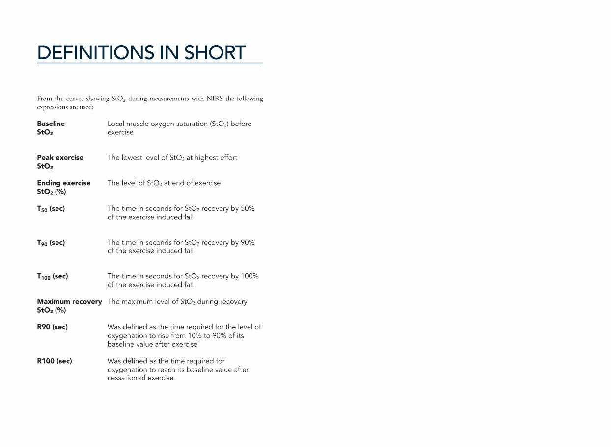
From the curves showing StO2 during measurements with NIRS the following expressions are used;
Baseline Local muscle oxygen saturation (StO²) before StO² exercise
Peak exercise The lowest level of StO² at highest effortStO²
Ending exercise The level of StO² at end of exercise StO² (%)
T50 (sec) The time in seconds for StO² recovery by 50% of the exercise induced fall
T90 (sec) The time in seconds for StO² recovery by 90% of the exercise induced fall
T100 (sec) The time in seconds for StO² recovery by 100% of the exercise induced fall
Maximum recovery The maximum level of StO² during recovery StO² (%)
R90 (sec)� � 7>Ã�`iw�i`�>Ã�Ì�i�Ì��i�ÀiµÕ�Ài`�v�À�Ì�i��iÛi���v�� oxygenation to rise from 10% to 90% of its baseline value after exercise
R100 (sec)�� � 7>Ã�`iw�i`�>Ã�Ì�i�Ì��i�ÀiµÕ�Ài`�v�À�� � oxygenation to reach its baseline value after cessation of exercise
DEFINITIONS IN SHORT
1514 Kajsa Rennerfelt | Methods in Diagnosing Chronic Anterior Compartment Syndrome.
Compartment syndrome is de%ned as a condition in which elevated intramus-cular pressure compromises local blood "ow and impairs function of the mus-cle tissue within a closed compartment (Matsen 3rd and Krugmire Jr 1978). Compartment syndromes are tradition-ally divided into an acute and a chronic form.
#e acute form occurs after a traumatic injury that induces a rapid irreversible pressure increase within a speci%c mus-cle compartment. #e acute compart-ment syndrome is a medical emergency and might require immediate surgical intervention. Chronic compartment syndrome is a recurrent, exercised-in-duced condition in which intramuscu-lar pressure increases to extreme levels during exercise and it impedes local muscle blood "ow and the neuromus-cular function in the a$ected tissue. Chronic compartment syndrome usu-ally occurs in athletes who participate in running or repetitive impact sports (Reneman 1975, Allen and Barnes 1986). #e chronic compartment syn-drome is a reversible form of abnor-mally increased intramuscular pressure during exercise. If the patient stops ex-ercising, the symptoms will reverse.
In 1956, Mavor wrote a case report in which he described a professional foot-ball player who experienced exercise-in-duced leg pain. #e football player was cured by fasciotomy (Mavor 1956). Patients with chronic compartment syndrome are free from symptoms during rest (Styf and Korner 1987, Styf 1989, Padhiar and King 1996, Ota et al. 1999, van den Brand et al. 2004). In patients with therapy-resistant leg pain, approximately 30% su$er from chronic compartment syndrome and it should be borne in mind that co-mor-bidity, such as medial tibial syndrome, peroneal tunnel syndrome and muscu-lar rupture, is common in this group of patients (Styf 1988).
#is thesis focuses on chronic anterior compartment syndrome (CACS) and how to establish the diagnosis. Howev-er, knowledge of the pathophysiology of acute compartment syndrome makes it easier to understand the development of elevated IMP in chronic compart-ment syndrome, the clinical presenta-tion and the evaluation of the di$erent diagnostic methods available.
INTRODUCTION
introduction
16 17Kajsa Rennerfelt | Methods in Diagnosing Chronic Anterior Compartment Syndrome.
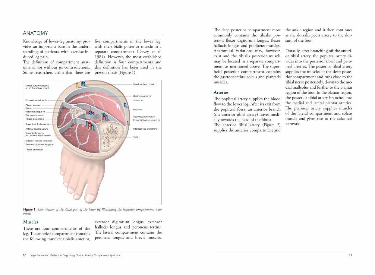
Muscles#ere are four compartments of the leg. #e anterior compartment contains the following muscles; tibialis anterior,
extensor digitorum longus, extensor hallucis longus and peroneus tertius. #e lateral compartment contains the peroneus longus and brevis muscles.
Figure 1. Cross-section of the distal part of the lower leg illustrating the muscular compartments with vessels.
Tibia
Interosseous membrane
Plantaris
Intermuscular septumFlexor digitorium longus m
Soleus m
Gastrocnemius m
Small saphenous vein
FibulaFibular vessels
Medial sural cutaneous nerve (from tibial nerve)
Posterior crural septum
Anterior crural septum
Super!cial !bular nerve
Tibialis anterior m
Extensor hallucis longus m
Extensor digitorum longus m
Deep !bular nerveand anterior tibial vessels
Tibialis posterior m
Peroneus brevis m
Peroneus longus m
Knowledge of lower-leg anatomy pro-vides an important base in the under-standing of patients with exercise-in-duced leg pain.#e de%nition of compartment anat-omy is not without its contradictions. Some researchers claim that there are
%ve compartments in the lower leg, with the tibialis posterior muscle in a separate compartment (Davey et al. 1984). However, the most established de%nition is four compartments and this de%nition has been used in the present thesis (Figure 1).
ANATOMY#e deep posterior compartment most commonly contains the tibialis pos-terior, "exor digitorum longus, "exor hallucis longus and popliteus muscles. Anatomical variations may, however, exist and the tibialis posterior muscle may be located in a separate compart-ment, as mentioned above. #e super-%cial posterior compartment contains the gastrocnemius, soleus and plantaris muscles.
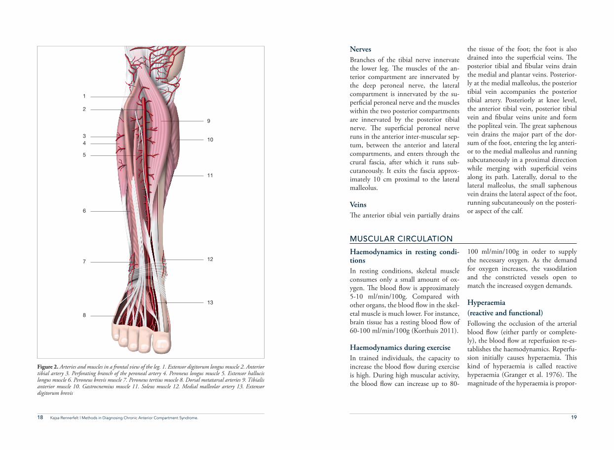
Arteries#e popliteal artery supplies the blood "ow to the lower leg. After its exit from the popliteal fossa, an anterior branch (the anterior tibial artery) leaves medi-ally towards the head of the %bula.#e anterior tibial artery (Figure 2) supplies the anterior compartment and
the ankle region and it then continues as the dorsalis pedis artery to the dor-sum of the foot.
Dorsally, after branching o$ the anteri-or tibial artery, the popliteal artery di-vides into the posterior tibial and pero-neal arteries. #e posterior tibial artery supplies the muscles of the deep poste-rior compartment and runs close to the tibial nerve posteriorly, down to the me-dial malleolus and further to the plantar region of the foot. In the plantar region, the posterior tibial artery branches into the medial and lateral plantar arteries. #e peroneal artery supplies muscles of the lateral compartment and soleus muscle and gives rise to the calcaneal network.
Kajsa Rennerfelt | Methods in Diagnosing Chronic Anterior Compartment Syndrome.
18 19Kajsa Rennerfelt | Methods in Diagnosing Chronic Anterior Compartment Syndrome.
1
2
10
9
11
13
12
43
5
6
7
8
Figure 2. Arteries and muscles in a frontal view of the leg. 1. Extensor digitorum longus muscle 2. Anterior tibial artery 3. Perforating branch of the peroneal artery 4. Peroneus longus muscle 5. Extensor hallucis longus muscle 6. Peroneus brevis muscle 7. Peroneus tertius muscle 8. Dorsal metatarsal arteries 9. Tibialis anterior muscle 10. Gastrocnemius muscle 11. Soleus muscle 12. Medial malleolar artery 13. Extensor digitorum brevis
NervesBranches of the tibial nerve innervate the lower leg. #e muscles of the an-terior compartment are innervated by the deep peroneal nerve, the lateral compartment is innervated by the su-per%cial peroneal nerve and the muscles within the two posterior compartments are innervated by the posterior tibial nerve. #e super%cial peroneal nerve runs in the anterior inter-muscular sep-tum, between the anterior and lateral compartments, and enters through the crural fascia, after which it runs sub-cutaneously. It exits the fascia approx-imately 10 cm proximal to the lateral malleolus.
Veins#e anterior tibial vein partially drains
the tissue of the foot; the foot is also drained into the super%cial veins. #e posterior tibial and %bular veins drain the medial and plantar veins. Posterior-ly at the medial malleolus, the posterior tibial vein accompanies the posterior tibial artery. Posteriorly at knee level, the anterior tibial vein, posterior tibial vein and %bular veins unite and form the popliteal vein. #e great saphenous vein drains the major part of the dor-sum of the foot, entering the leg anteri-or to the medial malleolus and running subcutaneously in a proximal direction while merging with super%cial veins along its path. Laterally, dorsal to the lateral malleolus, the small saphenous vein drains the lateral aspect of the foot, running subcutaneously on the posteri-or aspect of the calf.
MUSCULAR CIRCULATION
Haemodynamics in resting condi-tionsIn resting conditions, skeletal muscle consumes only a small amount of ox-ygen. #e blood "ow is approximately 5-10 ml/min/100g. Compared with other organs, the blood "ow in the skel-etal muscle is much lower. For instance, brain tissue has a resting blood "ow of 60-100 ml/min/100g (Korthuis 2011).
Haemodynamics during exerciseIn trained individuals, the capacity to increase the blood "ow during exercise is high. During high muscular activity, the blood "ow can increase up to 80-
100 ml/min/100g in order to supply the necessary oxygen. As the demand for oxygen increases, the vasodilation and the constricted vessels open to match the increased oxygen demands.
Hyperaemia (reactive and functional)Following the occlusion of the arterial blood "ow (either partly or complete-ly), the blood "ow at reperfusion re-es-tablishes the haemodynamics. Reperfu-sion initially causes hyperaemia. #is kind of hyperaemia is called reactive hyperaemia (Granger et al. 1976). #e magnitude of the hyperaemia is propor-
20 21Kajsa Rennerfelt | Methods in Diagnosing Chronic Anterior Compartment Syndrome.
tional to the extent of reduced blood "ow and the time of the reduction in blood "ow. After maximum vasodila-tion is reached, the return to a normal blood "ow is re-established.Functional hyperaemia occurs after
heavy muscle activity and can, at least partly, be explained by vasodilator me-tabolites that are released from the ac-tive muscle cells and contribute to the vasodilation (Björnberg et al. 1989, Korthuis 2011).
Pathophysiology Acute compartment syndrome is a con-dition in which increased pressure with-in a closed space contributes to a reduc-tion in oxygen perfusion and decreased blood "ow. It can be due to arterial ob-struction, after arterial clamping during surgery, a$ected venous "ow or external pressure, for example. When the arteri-
al blood "ow (Figure 3) is a$ected by occlusion or prolonged external com-pression, the endothelial cells in the capillary membranes become damaged, which results in increased permeability. Ischaemia leads to an in"ammatory re-action by triggering the immune system (Rodrigues and Granger 2010).
ACUTE COMPARTMENT SYNDROME
Figure 3. !e arterial "ow rate is explained by the Poiseuille’s Law. Volume "ow rate is given by the pres-sure di#erence divided by the resistance. !e impact on one aspect in"uences the other factors. F=Volume "ow rate, P=pressure, R=Resistance to "ow, =viscosity, L=length, $=constant 3.14, r=radius.
External pressure due to subcutaneous oedema or a tight dressing may con-tribute to an increase in intramuscu-lar pressure (IMP), initially caused by
compressed venous "ow. In the case of trauma, damaged tissue requires a per-fusion pressure exceeding 40 mmHg to meet the metabolic demands, whereas
healthy tissue requires less and needs perfusion pressure exceeding 30 mmHg (Heppenstall et al. 1988). #e rela-tionship between mean arterial pres-sure (MAP) and IMP is important to consider in the assessment in a situa-tion with increased IMP and suspected acute compartment syndrome.
Mechanisms of trauma to the lower legBlunt trauma to the lower leg can con-tribute to soft-tissue bleeding, swelling and subsequent compartment syn-drome (Matava et al. 1994). Fractures, especially closed tibial fractures, may contribute to acute compartment syn-drome. External pressure during an ex-tended period of unconsciousness, e.g. related to drug abuse, can induce crush syndrome. Of applied external pressure, 50-100% is transmitted into deep tis-sue (Brace and Guyton 1977). More-over, massive tissue damage can occur in relation to burn injuries and this can subsequently lead to acute compart-ment syndrome.
Crush syndromeOriginally, crush syndrome was de-scribed after the earthquake in Messi-na, Sicily, in 1909 (Better 1997). Crush syndrome occurs after a high-energy trauma that results in major injuries to soft tissue and bone.
When rabdomyolysis occurs, toxic me-tabolites leak into the systematic circu-lation and eventually contribute to a life-threatening condition. Acute renal
failure is caused by myoglobin precip-itating the distal convoluted tubuli of the kidneys (Vanholder et al. 2000). At reperfusion, metabolites are washed out into the circulation. Leukocytes migrate to the damaged tissue only after reper-fusion due to the need for oxygen. #e production of free radicals begins when oxygen is available. Myoglobin, potassi-um, calcium and phosphorus leak into the vascular system and can contribute to renal failure, cardiac arrhythmias and even seizures. #e hypoxic muscles pro-duce lactic acid that is eliminated by the liver. Failure of this function due to hypovolemia will give rise to meta-bolic acidosis (Vanholder et al. 2000). Traumatic rabdomyolysis may also be caused by hyperthermia and electrical insults (Vanholder et al. 2000).
Symptoms and signs#e patient experiences severe pain and swelling of the a$ected body part. #e most important patient-reported symp-tom is pain upon passive motion. If the lower leg is a$ected, passive motion of the ankle and increasing pain at rest are clinical signs. Paraesthesia and impaired circulation and, %nally, pulselessness are late signs. Establishing the diagnosis of acute compartment syndrome based on clinical %ndings alone is not advisable (Elliott and Johnstone 2003).
DiagnosisMonitoring IMP as a matter of rou-tine is suggested in young patients or in those who are unconscious (Elliott and Johnstone 2003). #e level of IMP
22 23Kajsa Rennerfelt | Methods in Diagnosing Chronic Anterior Compartment Syndrome.
should also be considered in conjunc-tion with the patient’s circulatory status and the extent of tissue damage (Elliott and Johnstone 2003). #e level of IMP is related to the MAP, as MAP minus IMP equals the perfusion pressure. It should be remembered that hypoten-sion could lower the critical level of perfusion. #e oxygen requirements of the tissue should also be considered, as damaged requires more oxygen. #e IMP needs to be monitored continu-ously to observe the pressure changes.Acute compartment syndrome in sedat-ed patients and in patients with cerebral damage and impaired consciousness is di(cult to diagnose, as communication between the patients and treating doc-tor is lacking. A delayed diagnosis can be devastating, as the sequelae are severe and young patients are often a$ected (McQueen 1996).
Treatment #e perfusion pressure must be con-sidered when fasciotomy is decided on (Whitesides Jr et al. 1975). It has been
demonstrated that fasciotomy within 12 hours after the onset of an acute compartment syndrome leaves 68% of all patients with no sequelae (Sheridan and Matsen 3rd 1976).
Fasciotomy is performed under tourni-quet control, if possible. Using a 20-30 cm long, para-%bular incision, all four compartments can be reached through a single incision (Matsen et al. 1980). Two incisions, one lateral and one me-dial, can also be used. From the lateral incision, the anterior and lateral com-partments can be reached and, from the medial incision, the super%cial and deep posterior compartment can be reached (Mubarak and Owen 1977). During decompression of the lateral compartment, the super%cial peroneal nerve should be decompressed as well, as it runs through the fascia tunnel (Styf and Morberg 1997). Care needs to be taken not to damage this nerve during fasciotomy.
CHRONIC ANTERIOR COMPARTMENT SYNDROME
(CACS)
Chronic compartment syndrome is an exercise-induced reversible elevation of the intramuscular pressure within one or more a$ected compartments. #e %rst reported case of chronic compart-ment syndrome was a self-experienced incident by Dr Edward Wilson in 1912, which took place during a race in which he was attempting to reach the South Pole before Roald Amundsen. In
his diary, Dr Wilson wrote “My left leg is exceedingly painful .... and hobbled alongside the sledge on foot. #e whole of the Tibialis anticus is swollen and tight …” (Freedman 1954).
Muscle volume changes that occur during exercise was described by Bar-croft and Kato 1916 (Barcroft and Kato 1916). Some 40 years later, in 1956,
Mavor reported on a young football player with exercise-induced leg pain that was relieved by fasciotomy (Mavor 1956). #e connection between ex-ercise-induced leg pain and increased intramuscular pressure was observed and demonstrated by intramuscular pressure measurements in a study by French and Price in 1962 (French and Price 1962).
Incidence In relation to the entire body, chronic compartment syndrome occurs in the lower leg in 95% of all cases (Barnes 1996). #e remaining 5% occur in the forearm, hand, thigh and occasion-ally the foot (Styf 2003). Among pa-tients with exercise-induced leg pain, the incidence of chronic compartment syndrome is high. #e patients report-ed in the literature are, however, from selected cohorts. In studies conducted on patients with chronic compartment syndrome, the patients are admitted to specialist clinics due to therapy-resis-tant, exercise-induced leg pain. Among these cohorts, the incidence is usually 20-30%, or even higher. Turnipseed et al. examined 400 patients and found 175 female patients and 74 male pa-tients (62%) with chronic compartment syndrome (Turnipseed 2002). Styf and Körner found 22 patients of 80 (28%) (Styf and Korner 1987) and 11 of 46 patients (24%) in a later study (Styf and Korner 1986). With increasing physical activity in society, it appears likely that men and women will be represented more equally.
Physiology and pathophysiology In exercising healthy muscle, the IMP increases as a physiological condition (Styf et al. 1989). #e patients su$er from exercise-induced leg pain caused by elevated IMP due to the increased transudation of "uid during exercise (Reneman 1975, Hargens et al. 1978, Rorabeck and Clarke 1978). However, the factors leading to the pathological increase in IMP in patients with CACS are not fully known. In resting muscle, the blood "ow rate is low and can in fact increase as much as 50 times during exercise in well-trained athletes (Sejerst-ed and Hargens 1986) and the muscle volume may increase up to 20% during exercise (Barcroft and Kato 1916). At the onset of exercise, during the %rst 15 minutes, the "uid %ltration increases from intravascular "uid to the intersti-tial space (Korthuis 2011). #e forces are explained by Starling’s equation (Figure 4). During the onset of exercise, the rate of "uid transported from the vascular space to the interstitial space exceeds the compensatory mechanism (Korthuis 2011). #e muscle circula-tion is enhanced by arteriolar vasodila-tion to match the increased oxygen de-mands (Kjellmer 1965). #e arteriolar surface is increased by recruiting closed capillaries (Kjellmer 1965). #e increase in interstitial "uid is a consequence of the enhanced osmolality by the metab-olites released from the exercising mus-cle into the interstitial space (Lundvall 1971). As the exercise progresses, the transcapillary %ltration slows down and a balance is reached. In physiological
24 25Kajsa Rennerfelt | Methods in Diagnosing Chronic Anterior Compartment Syndrome.
conditions, these mechanisms are bal-anced and increased interstitial pressure during exercise enhances the lymphatic drainage (Kjellmer 1964). #e muscu-lar contractile forces with their rhyth-mic contractions function as a muscular pump for the lymphatic system.
#e balance of net "ow with IMP and blood pressure within normal range fails temporary in patients with CACS. #ere is a pathological elevation of the IMP during and after exercise and the compliance in the muscular com-partment decreases in CACS patients in comparison to healthy individu-als (Reneman 1975, Allen and Barnes 1986, Styf et al. 1987). #e oxygen consumption during exercise in pa-tients with CACS has been studied and a lower level of local muscular oxygen saturation compared with patients with other causes of leg pain during exercise has been observed (Mohler et al. 1997, van den Brand et al. 2005). Studies of muscular blood "ow have shown a decreased level in patients with CACS compared with patients with other caus-es of leg pain (French and Price 1962, Styf et al. 1987, Abraham et al. 1998). During rhythmical muscular work, the muscular tissue contracts and relaxes. It is only during the relaxation phase that the musculature is perfused (Folkow et al. 1970). In patients with CACS, the IMP increases during exercise. As the IMP increases, the relaxation pressure also increases, which contributes to a decrease in muscle perfusion during ex-ercise (Styf et al. 1987).
Histological findings#e histological structure of the fas-cia plays an uncertain role in chronic compartment syndrome. In a study by Turnipseed et al., an increase in the thickness of the fascia in patients with chronic compartment syndrome compared with healthy subjects was demonstrated (Turnipseed et al. 1995). In a study by Styf et al. in 1986, eight patients had a biopsy taken in conjunc-tion with fasciotomy. #ese specimens were normal in terms of in"ammato-ry changes and no other abnormalities in the fascia muscular tissue were seen (Styf and Korner 1986). In muscular biopsies, an increase in water content (Wallensten and Karlsson 1984) and water and lactate content (Qvarfordt et al. 1983) has been shown.
Capillary density has been shown to be reduced in muscular tissue, in biopsies from patients with CACS, compared with healthy individuals. One year after fasciotomy, the capillary density did not recover (Edmundsson et al. 2010).
Symptoms and signs Patients with CACS typically experi-ence pain in the anterior aspect of the leg during exercise. #e average patient is 26-28 years of age and physically active (Detmer et al. 1985, Styf and Korner 1987, Turnipseed 2002). #e patients’ gender distribution varies in di$erent studies between equal, predominantly women and predominantly men (Det-mer et al. 1985, Turnipseed 2002, van den Brand et al. 2005, Van der Wal et
al. 2014). #e patients are commonly active in sport, usually running (Det-mer et al. 1985, Slimmon et al. 2002, Waterman et al. 2013). Pain occurs within minutes after the initiation of activity and subsides at rest. Strenuous activity can result in pain persisting for hours. Moreover, symptoms, such as experiencing stabbing pain and even-tually the inability to extend the ankle and sometimes numbness, can devel-op as well. #e pain increases during activity and %nally forces the athletes to cease the activity (Allen and Barnes 1986, Styf et al. 1987). Even if the main reported limitation relates to running, many patients also experience limita-tions while walking (Edmundsson et al. 2007). CACS is more compatible with activities such as cycling, swimming and gym training.
Patients with CACS have a higher re-ported physical activity level than that reported in the general population (Styf and Korner 1987). #e CACS can be at least partly explained by strenuous exer-cise that a$ects the soft tissues as a re-sult of micro-trauma and in"ammatory reaction. #e reason why the anterior compartment is most often exposed could be due to the anatomical restric-tions caused by the deep osteofascial membranes, tibia and %bula and the tight super%cial muscular fascia.
Diagnosis#e diagnostic methods and criteria are still the subject of debate (Barnes 1997, Aweid et al. 2012). For many years, the gold standard for diagnostics has been the measurement of intramuscu-lar pressure (IMP) (French and Price 1962, Reneman 1975, Hargens et al. 1977, Styf et al. 1987). At present, this is still the most accepted method, but it has the limitation of being invasive. Non-invasive methods have therefore been requested and several methods have been evaluated. Blood "ow during and after exercise has been investigated to study the pathophysiology, where the increase of blood "ow during exercise in healthy individuals is not seen in patients with CACS (French and Price 1962, Styf et al. 1987). Amendola et al. did not %nd indications of ischaemia (Amendola et al. 1990). Magnetic reso-nance imaging (MRI) after exercise has shown oedema in the a$ected muscle (Verleisdonk et al. 2001). Ultrasound after exercise has shown increased "uid in the interstitial space but no increase in muscular volume (Birtles et al. 2002, Birtles et al. 2003). However, none of these has been shown to be convincing in clinical practice.
26 27Kajsa Rennerfelt | Methods in Diagnosing Chronic Anterior Compartment Syndrome.
!e characteristics of patients with chronic compartment syndrome
• Normal clinical %ndings at examination• In some patients, fascial defects; usually more pronounced with muscular herniations after exercise• Exercise-induced leg pain with tender and hard muscles• In some cases, impaired muscular function and numbness after activity• Exercise-induced leg pain over the anterior compartment with reversed symptoms at rest• Swelling and tenderness over the anterior compartment immediately after exercise• IMP ≥ 30 mmHg, one minute after exercise• IMP ≥ 20 mmHg, %ve minutes after exercise
TREATMENT OF CACS
Non-surgical treatment#e non-surgical treatment of CACS has produced unsatisfactory results (Detmer et al. 1985, Styf et al. 1987, Van der Wal et al. 2014). In a study by Styf, the symptoms in CACS patients were persistent, despite various con-servative treatments, such as diuretics, anti-in"ammatory drugs, physiother-apy and stretching. Shoe modi%cation has also been tried, as well as di$erent training programmes. No bene%ts are reported after the conservative treat-ments (Styf 1988).
Surgical treatmentIn 1956, Mavor presented a case report in which a professional football player, 24 years of age, su$ered from what was clinically diagnosed as CACS. #e pa-tient was successfully operated on with fasciotomy of the anterior compart-ment. Fasciotomy of the compartments of the leg can be performed safely and satisfactorily according to reports from Detmer and Sharpe, who presented a report of fasciotomy in 100 patients and 233 compartments (Detmer et al. 1985).
Release of the anterior and lateral compartmentsUsing a vertical incision on the ven-tral part of the leg, both the anterior and the lateral compartments can be reached using the technique developed by Mubarak. However, it is important to make sure that the fascial split in the distal direction is long enough not to cause a muscular herniation in the distal “V” (Mubarak and Owen 1977, Detmer et al. 1985). Sensory nerves exit from the lateral portion of the anterior compartment and care should be taken (Detmer et al. 1985). #e fascia is split using scissors one centimetre lateral to the anterior margin of the tibia, begin-ning on the mid-portion of the tibia and continuing in the distal and prox-imal direction. #e intramuscular sep-tum must be identi%ed and the fascia of the lateral compartment should be split, in the proximal direction as well as distally, where care needs to be taken with the super%cial peroneal nerve.
Surgical outcome#e surgical outcome has been report-ed as good or excellent in several stud-ies (Wallenstein 1983, Styf and Korner 1986, Fronek et al. 1987, Rorabeck et al. 1988, Schepsis et al. 1993, van den Brand et al. 2004). In a study by Ed-mundsson et al., the results in 57 pa-tients were excellent in six, good in 35, fair in 15 and poor in one (Edmunds-son et al. 2007). A recent study com-prising 611 patients who had varying types of compartment syndrome (ante-rior, lateral, posterior) in the lower leg
indicated that 28% were not able to return to full activity after fasciotomy (Waterman et al. 2013). #ere were, however, limitations to this study, with a lack of information relating to IMP values and surgical methods.
Detmer et al. reported that as many as 90% improved partially or fully (mean follow-up 4.5 months) in their study of 100 patients (Detmer et al. 1985) and a similar result was reported by Styf and Körner in 19 patients (mean follow-up 25 months) (Styf and Korner 1986). Slimmon et al. performed a long-term follow-up (mean follow-up 51 months) to evaluate the results of fasciotomy, combined with partial fasciectomy. Of 50 patients who underwent surgery, 60% reported a good or excellent out-come, but 58% returned to activity at a lower level than before the injury. Surgery without a satisfactory outcome might be due to the insu(cient length of the fascial split (Puranen and Ala-vaikko 1981, Bell 1986).
Co-morbidity in patients with exer-cise-induced leg pain is not extensively reported in the literature. Co-existing diagnoses, such as medial tibial syn-drome periostitis, peroneal tunnel syn-drome, popliteal entrapment syndrome or muscular rupture, were reported in 42% of the patients with CACS in a study by Styf (Styf 1988). #e pain from elevated IMP during exercise might mask other sources of pain that can be revealed after fasciotomy.
28 29Kajsa Rennerfelt | Methods in Diagnosing Chronic Anterior Compartment Syndrome.
DIFFERENTIAL DIAGNOSES IN EXERCISE-INDUCED LEG
PAIN
Patients with CACS might have ad-ditional diagnoses. A discussion of di$erential diagnoses in patients with exercise-induced leg pain is important. In a study by Styf, as many as 42% of all patients with CACS su$ered from co-morbidities, such as periostitis, pe-roneal tunnel syndrome and medial tib-ial syndrome (Styf 1988). #e addition-al diagnoses might be expressed during rest and blur the clinical picture of CACS. #e a$ected patients are limited in physical activities, as the symptoms are not spontaneously relieved. #e fol-lowing diagnoses are commonly found to co-exist with CACS.
Medial tibial syndrome and perios-titisMedial tibial syndrome gives rise to symptoms in the form of exercise-in-duced pain over the distal third of the posteromedial aspect of the tibia. Devas reported this syndrome in the literature in 1958 (Devas 1958). #e pain often subsides at rest, but some patients have consistent pain for hours or even days after strenuous exercise. Physical exam-ination reveals local tenderness in the distal third of the medial border of the tibia.
Medial tibial syndrome may co-exist in patients with CACS and has been ob-served (Styf 1988). Previously, medial tibial syndrome was divided into three types, according to Detmer, Types I, II and III (Detmer 1986). Type I was related to radiological bony changes, sometimes with micro-fractures, at the osteofascial border at the origin of the soleus muscle (Holder and Michael 1984). #e radiological changes cor-relate with histological changes with the proliferation of osteoblasts. However, in the periosteum, no evidence of in"am-matory changes has been demonstrated (Johnell et al. 1982). Type II was de-%ned as periostalgia and traction peri-ostitis. Type III was de%ned as a chronic compartment syndrome in the deep dorsal compartment. In a study by Bar-bour et al., biopsies were taken from the patients during fasciotomy. #e study comprised 19 patients with deep pos-terior compartment syndrome and 11 controls. Fibroblastic activity, chronic in"ammatory cells and increased vas-cularity were seen in the biopsy mate-rial from the patients with deep dorsal compartment syndrome (Barbour et al. 2004).
In a study by Moen et al., 52 athletes with medial tibial syndrome were ex-amined with MRI. Periosteal and bone marrow oedema were found in more than 40% of the patients (Moen et al. 2014). However, no signi%cant di$er-
• Medial tibial syndrome• Periostitis• Muscle rupture
ences were found between symptomat-ic and non-symptomatic legs from the MRI examinations.
Medial tibial syndrome might be ex-plained by rapidly increasing activity. Histologically, no in"ammatory chang-es have been shown in the periosteum, but, in the crural fascia, 13 of 33 biop-sies revealed in"ammatory changes in a study by Johnell et al. (Johnell et al. 1982). #e patients with medial tibial syndrome experience pain, which in-tensi%es during activity.
Periostitis does not respond well to treatment with anti-in"ammatory medication, or physiotherapy. Reduced activity will contribute to less pain. #e clinical outcome after surgical treat-ment, i.e. fasciotomy, is not as success-ful as in patients with CACS.
Muscle rupture#e patients experience pain, which is usually located in the medial muscle belly of the gastrocnemius, close to the muscle-tendon junction. #is condition is common in recreational runners, but it is most typical in fast eccentric-action sports, such as tennis or "oorball. #e muscle %bres are strained due to over-stretching and the mechanism is load by "exion of the ankle joint with the knee extended. Clinical examination reveals pain at the muscle-tendon junc-tion. Ultrasonography may reveal scar tissue close to the muscle-tendon junc-tion. #e primary symptom is usually acute pain while running. #e patient often describes the pain in a similar
manner as in Achilles tendon rupture (Styf 2003). Treatment with physio-therapy to regain full muscle strength is successful. #e healing time is usually three to six weeks.
Tibial stress fractures comprise almost 50% of all stress fractures in athletes. Running is the most common activity leading to stress fracture of the tibia (Matheson et al. 1987). Usually stress fractures are seen in the push-o$/land-ing leg according to a study in 29 pa-tients by Ekenman et al. (Ekenman et al. 1996). In a study comprising 295 mil-itary recruits, 31% were found to have stress fractures and more than 50% of these fractures were located in the tibia. Of the total numbers of stress fractures, 35% were asymptomatic (Milgrom et al. 1985). During running, the external impact force must be absorbed by bone and soft tissue. Bone mainly absorbs the energy, while the soft tissue does so to a lesser extent (Burr and Milgrom 2001). #e remodelling of bone occurs when the forces are in harmony according to Wolf ’s Law (Chamay and Tschantz 1972). Bone remodelling occurs due to microtrauma in order to maintain its strength. During heavy training, as the muscles become fatigued, the bone must absorb more of the load from the external forces. #e load absorption function will be impaired and this can lead to stress fractures (Burr and Mil-grom 2001). Stress fractures are more common in women due to a higher rate of muscle fatigue, resulting in increased tension of the tibia (Burr and Milgrom 2001).
30 31Kajsa Rennerfelt | Methods in Diagnosing Chronic Anterior Compartment Syndrome.
In arterial entrapment, the symptoms are divided into anatomical and func-tional (Turnipseed 2002). Functional popliteal entrapment is regarded as an
overuse injury in a similar manner to CACS. Venous insu(ciency is uncom-mon in patients with exercise-induced leg pain.
INTRAMUSCULAR PRESSURE
DefinitionIntramuscular pressure (IMP) is mea-sured as the hydrostatic pressure in the muscular interstitial space. In resting muscle, this pressure is normally below 10 mmHg. IMP can be de%ned by the Starling equation. Starling de%ned tis-sue pressure in 1896 (Starling 1896). A change in IMP is due to a shift in "uids or external impact. #e net %ltration rate is described by the Starling equa-tion (Figure 4).
Four di"erent pressures act over the capillary membrane
• Pc = vascular hydrostatic pressure • Pt = interstitial hydrostatic pressure • πc = vascular oncotic pressure • πt = interstitial oncotic pressure
Figure 4. Starling’s Law explains the net "uid across the capillary membrane as the equation Jc=Kf((Pc-Pt)- ($c- $i)). Pressure in mmHg.
!e forces according to Starling’s Law
JV = transcapillary "uid transport (ml/min/100g tissue)Kf = capillary %ltration coe(cient (ml/min/100g tissue)Pc = capillary "uid hydrostatic pressure (mmHg)
Pt = tissue "uid hydrostatic pressure (mmHg) = capillary membrane re"ection coe(cient (0-1, no dimension)πC = capillary "uid oncotic pressureπt = tissue "uid oncotic pressure
Figure 5. !e interstitial space consists of a heterogeneous mixture of "uid and gel. Pressures from the interstitial space and from intravascular forces determine the shift in the "uids.
#e interstitial space is approximate-ly 10% of the skeletal muscle weight (Aukland and Reed 1993). It contains three quarters of the body "uid con-tent (Aukland and Reed 1993). #e interstitial space consists of a heteroge-neous mixture of "uid and gel. #e two phases of "uid and gel were described by Guiton et al. back in 1971 (Guyton et al. 1971). #e framework or matrix
is composed of collagen %bres. #e in-terstitial space is located in the space between the capillary network and the lymphatic system. #e change in interstitial-space volume is explained by the Starling equation and "uid shift is seen as changes in the tissue pressure (Figure 5). Lymphatic drainage exports the "uid back to the vascular system (Aukland and Reed 1993).
INTERSTITIAL SPACE (IMP)
32 Kajsa Rennerfelt | Methods in Diagnosing Chronic Anterior Compartment Syndrome. 33
In 1962, French and Price performed an analysis of how to diagnose CACS (French and Price 1962). #e study comprised two patients with exer-cise-induced leg pain and 18 healthy subjects. #e study was performed with tissue pressure measurements of the anterior compartment and clearance of radioactive sodium to monitor the blood "ow. #e tests were followed by an exercise test. An exercise test with external pressure produced by an in-"ated tourniquet, was also performed, to observe changes in pain and intra-muscular pressure during intramuscular pressure elevation. #ese tests are still up to date in modi%ed forms when it
comes to observing intramuscular pres-sure and changes in blood "ow (Styf et al. 1987, Breit et al. 1997, Mohler et al. 1997, Abraham et al. 1998). El-evated tissue pressures in the lower leg were found in patients with clinical signs of chronic anterior compartment syndrome (CACS), while elevation in blood "ow post-exercise was absent in the patients with CACS compared with the healthy individuals with elevated IMP (French and Price 1962). Normal arterial pulsations were observed in the a$ected compartment, as well as clini-cally observed oscillations of the a$ect-ed compartment explained by systolic pulsations.
HISTORICAL PERSPECTIVES OF IMP
IMP CHANGES
IMP BEFORE, DURING AND AFTER EXERCISE TEST
#e anatomical structure of the anteri-or compartment with rigid walls most probably makes it especially vulner-able to elevations of IMP. In a study by Allen and Barnes, there was a more pronounced elevation of IMP in the
anterior compartment compared with the deep posterior compartment after the infusion of a small amount of "u-id in amputated legs (Allen and Barnes 1986).
#ere is no universally accepted IMP level to establish the diagnosis of CACS (Aweid et al. 2012, Roberts and Frank-lyn‐Miller 2012). According to the literature, di$erent criteria have been used. #e intramuscular pressure in normal resting muscle tissue is 5-11 mmHg (Fronek et al. 1987, Nkele et al. 1988, Rorabeck et al. 1988). Intra-
muscular pressure during activity has been studied in detail (Styf et al. 1989, Barnes 1997) and pressure above 50 mmHg has been identi%ed as diagnos-tic for CACS (Puranen and Alavaikko 1981, McDermott et al. 1982, Allen and Barnes 1986). Today, the criteria formulated by Pedowitz et al. (Pedow-itz et al. 1990) are commonly used,
but, in actual fact, the criteria di$er in di$erent studies (Tzortziou et al. 2006, Edmundsson et al. 2010). Moreover, there is no general standard in terms of the levels of IMP and the most op-timal time point at which the pressure measurement should be performed. In a recent study by Aweid et al., overlap-ping values were found in the groups of patients with chronic compartment syndrome (mainly CACS, but all com-partments of the lower leg were includ-ed) and healthy individuals (Aweid et al. 2012).
IMP criteria by di"erent authors
However, overlapping values have not been found in reported mean IMP levels between patients and control subjects when IMP was measured one minute post-exercise.
#e criteria for CACS formulated by Pedovitz et al. are widely used, but they have been discussed and criticised due to the fact that they are based on a small number of patients (n=45) (Roberts and Franklyn‐Miller 2012).
One or more of the following were con-sidered to be present to ful%l the criteria for chronic compartment syndrome; 1) resting pressure of ≥ 15 mm Hg, 2) a one-minute post-exercise pressure of ≥ 30 mm Hg, or 3) a %ve-minute post-ex-ercise pressure of ≥ 20 mm Hg. #e pa-tients underwent clinical examination followed by an exercise test. #e clin-ical %ndings were normal, but several patients had muscular herniation of the lower leg.
IMP criteria during activity Puranen and Alavaikko 1981 (>50mmHg)Allen and Barnes 1986 (>50mmHg)McDermott et al. 1982(>85mmHg)
IMP criteria during rest before exerciseTurnipseed 2002 (>15mmHg)
IMP criteria during rest after exerciseStyf and Korner 1987 (>30mmHg)Pedowitz, Hargens et al. 1990 (>30mmHg)Fronek et al. 1987 (>10 mm Hg at rest and/or >25 mm Hg 5 min after exercise)
34 35Kajsa Rennerfelt | Methods in Diagnosing Chronic Anterior Compartment Syndrome.
without side-holes should not be used, due to the risk of false high measure-ments. Di$erent devices are available and a detailed knowledge of the exact technique and the potential pitfalls of each method is necessary (Figure 6).
Infusion techniques IMP can be measured using a contin-uous or non-continuous microcapillary infusion system. #e volume of saline solution used in the non-continuous microcapillary "uid system is signi%-cantly lower compared with that in the continuous infusion system (Styf 2003). To monitor an exercise test, a rapid response is necessary and the mi-crocapillary "uid-%lled system has been shown to be suitable (Styf and Korner 1986).
Non-infusion techniques #ere is a risk of blood or tissue clotting if no infusion is used. A wick catheter has been shown to possess low dynamic properties (Styf and Korner 1986) and a slit catheter requires "uid to be inter-mittently "ushed to prevent clotting (Allen and Barnes 1986). Both tech-niques need "uid to function.
Optical techniquesOptical transducers have been shown to have properties that are well suited to IMP (Crenshaw et al. 1990). #ese devices are suitable for measurements during exercise and during rest.
TECHNIQUES FOR MEASURING IMP
PERFORMING IMP MEASUREMENTS
#e emphasis is on an IMP-measure-ment post-exercise test. IMP measure-ment can be performed using needle or catheter techniques with infusion or non-infusion (Figure 6). Several mon-
itoring techniques, such as a needle manometer (Brace et al. 1975), wick catheter (Mubarak et al. 1976) and the microcapillary infusion system (Styf and Korner 1986), have been described.
#e position of the needle and possible clotting by blood or tissues at the tip of the needle are sources of uncertain or even false results. #e position of the needle is of the greatest importance. #e pressure has been shown to di$er according to the depth of the needle or catheter in the muscular tissue (Sejer-sted et al. 1984). #e position of the ankle helps to increase or reduce the IMP (Gershuni et al. 1984). #e time point of measurement is also of ma-jor importance. As an increase in IMP during exercise is a physiological pro-cess (Puranen and Alavaikko 1981, Styf et al. 1989), the pressure measurement performed one minute after exercise
enables the physiological process in pa-tients with other causes or exercise-in-duced leg pain to decline in pressure. #e IMP in patients with CACS does not normalise for %ve minutes (Pedow-itz et al. 1990). #e person making the IMP measurement must be aware of the possible pitfalls during the IMP measurement procedure to avoid false results.
Measurement positionTo insert the needle correctly, anatom-ical landmarks must be identi%ed to penetrate the correct muscle compart-ment. #e needle should be inserted parallel to the muscle %bres to reduce
Di!erent needles In a study by Boody and Wong-worawat, three di$erent IMP devices and three di$erent needles were com-pared with a reference model (Boody and Wongworawat 2005). #e study
revealed clinically signi%cant di$erenc-es between di$erent devices and nee-dles. It is important to be aware of the di$erences when making the measure-ments in day-by-day clinical work. One important result is that straight needles
Figure 6. Occlusion of the catheter tip is a problem that can be caused by blood clotting or tissue inside the tip of the needle. To prevent this, several methods have been developed with di#erent constructions at the tip of the needle. A. Wick catheter B. Catheter with multiple side-holes C. Slit catheter D. Catheter with a cover for a SLIT catheter or catheter for optical use.
IMP CRITERIA FOR CACS
#e criteria formulated by Pedowitz et al. (Pedowitz et al. 1990) are well doc-umented. #e diagnostic criteria for CACS used in this thesis are a modi%-cation of the criteria formulated by Pe-dovitz et al. Clinical signs of CACS, ex-ercise-induced leg pain and a minimum of 30mmHg one minute post-exercise
were used to establish the diagnosis of CACS in the present thesis. No exam-ination of IMP during rest before exer-cise was however performed. During he IMP measuring adequate investigation technique and correct use of the equip-ment must be controlled.
36 37Kajsa Rennerfelt | Methods in Diagnosing Chronic Anterior Compartment Syndrome.
Risks during IMP measurement#e risk of injury, such as bleeding and infection, is small, but the procedure
may create some discomfort and pain for the patient (Mohler et al. 1997, van den Brand et al. 2005).
the tissue trauma during insertion (Zhang et al. 2011). Occlusion of the needle sometimes occurs and this can be avoided by penetrating the skin with a separate needle which is withdrawn before placing the needle used for the IMP (Styf 2003). Local anaesthesia can be applied, but it is not necessary. To control the position of the needle, ex-ternal compression can be performed by the investigator running his/her %n-gertips over the needle tip. #e curve of the IMP will show a direct rise in ampli-tude if external compression is applied directly over the needle tip (Styf 2003).
Control of functioning needle/cath-eter
Anatomical landmarks of the mus-cular compartments Active extension and "exion of the ankle joint usually enables the mus-cle bellies to be well de%ned. A good
knowledge of anatomical landmarks is essential for correct intramuscular pres-sure measurements.
ReproducibilityDi$erent devices have been used and re-producibility is one important concern (Boody and Wongworawat 2005). Vari-ations in measurements can be due to di$erent devices or incorrect measure-ment techniques but also to biological variations in compartment pressure.
Factors a"ecting pressure measure-ments and the level of IMP
• Increased intramuscular pressure during active muscular contraction
• Needle positional control by ultrasound
• #e needle should be at heart level during measurement of the IMP in order to avoid an incorrect increase or
decrease in IMP.• External pressure over the
needle is followed by a quick rise in amplitude on the IMP curve.
• #e pressure transducer should be kept at the same level as the patient’s heart.
• Occlusion by tissue or blood clotting
• Timing of measurement in relation to the exercise test
• Muscular tension at rest after exercise
• External compression• Level of e$ort made by the
patient during the exercise test• Ability to reproduce pain by
an accurate exercise test• Correct muscular
compartment• Correctly placed needle in the
tissue in relation to muscle %bers and vessels
NEAR-INFRARED SPECTROSCOPY (NIRS)
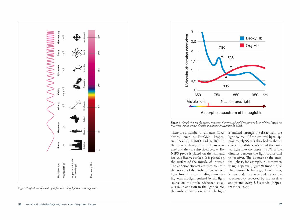
NIRS is a non-invasive method. Since the end of the 1980s, NIRS has been used to study local muscle oxygenation at rest and during exercise (Ferrari et al. 2011). #e blood changes in colour depending on the level of oxygenation. #is is due to the optical properties of haemoglobin, which are used by NIRS at wavelengths between 700 and 900 nm (Figure 7).
Oxygenated blood is bright red and de-oxygenated blood is deep red, dark or almost blue, depending on the optical spectra. When it comes to monitoring the systemic oxygenation, pulse oxime-try is an established method. However,
local muscle oxygenation can also be monitored. Haemoglobin changes its optical properties when binding to ox-ygen (Figure 8). #e chromophore, the part of the haemoglobin molecule that binds oxygen, absorbs and re"ects light (Scheeren et al. 2012). Oxygenated haemoglobin binds wavelengths at 830 nm and deoxygenated haemoglobin binds them at 780 nm. Myoglobin and haemoglobin are not possible to di$er-entiate, as they are covered in the same optical spectra (Mohler et al. 1997). #e di$erence between admitted and received light is de%ned as the change in muscle oxygen saturation.
38 39Kajsa Rennerfelt | Methods in Diagnosing Chronic Anterior Compartment Syndrome.
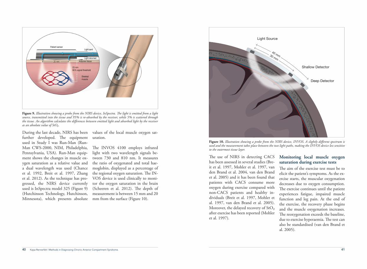
#ere are a number of di$erent NIRS devices, such as RunMan, InSpec-tra, INVOS, NIMO and NIRO. In the present thesis, three of them were used and they are described below. #e NIRS probe is placed on the skin and has an adhesive surface. It is placed on the surface of the muscle of interest. #e adhesive stickers are used to limit the motion of the probe and to restrict light from the surroundings interfer-ing with the light emitted by the light source on the probe (Scheeren et al. 2012). In addition to the light source, the probe contains a receiver. #e light
is emitted through the tissue from the light source. Of the emitted light, ap-proximately 95% is absorbed by the re-ceiver. #e distance/depth of the emit-ted light into the tissue is 95% of the distance between the light source and the receiver. #e distance of the emit-ted light is, for example, 23 mm when using InSpectra (Figure 9) (model 325, Hutchinson Technology, Hutchinson, Minnesota). #e recorded values are continuously collected by the receiver and printed every 3.5 seconds (InSpec-tra model 325).Figure 7. Spectrum of wavelengths found in daily life and medical practice.
Figure 8. Graph showing the optical properties of oxygenated and deoxygenated haemoglobin. Myoglobin is covered within the wavelengths and cannot be separated by NIRS.
0,5
1
1,5
2
2,5Deoxy Hb
780
830
805
Oxy Hb
3
0650
Visible light Near infrared light
Mol
ecul
ar a
bsor
ptio
n co
effic
ient
Absorption spectrum of hemoglobin
750 850 950 nm
40 41Kajsa Rennerfelt | Methods in Diagnosing Chronic Anterior Compartment Syndrome.
During the last decade, NIRS has been further developed. #e equipment used in Study I was Run-Man (Run-Man CWS-2000, NIM, Philadelphia, Pennsylvania, USA). Run-Man equip-ment shows the changes in muscle ox-ygen saturation as a relative value and a dual wavelength was used (Chance et al. 1992, Breit et al. 1997, Zhang et al. 2012). As the technique has pro-gressed, the NIRS device currently used is InSpectra model 325 (Figure 9) (Hutchinson Technology, Hutchinson, Minnesota), which presents absolute
values of the local muscle oxygen sat-uration.
#e INVOS 4100 employs infrared light with two wavelength signals be-tween 730 and 810 nm. It measures the ratio of oxygenated and total hae-moglobin, displayed as a percentage of the regional oxygen saturation. #e IN-VOS device is used clinically to moni-tor the oxygen saturation in the brain (Scheeren et al. 2012). #e depth of measurement is between 15 mm and 20 mm from the surface (Figure 10).
#e use of NIRS in detecting CACS has been assessed in several studies (Bre-it et al. 1997, Mohler et al. 1997, van den Brand et al. 2004, van den Brand et al. 2005) and it has been found that patients with CACS consume more oxygen during exercise compared with non-CACS patients and healthy in-dividuals (Breit et al. 1997, Mohler et al. 1997, van den Brand et al. 2005). Moreover, the delayed recovery of StO2 after exercise has been reported (Mohler et al. 1997).
Monitoring local muscle oxygen saturation during exercise tests#e aim of the exercise test must be to elicit the patient’s symptoms. As the ex-ercise starts, the muscular oxygenation decreases due to oxygen consumption. #e exercise continues until the patient experiences fatigue, impaired muscle function and leg pain. At the end of the exercise, the recovery phase begins and the muscle oxygenation increases. #e reoxygenation exceeds the baseline, due to exercise hyperaemia. #e test can also be standardised (van den Brand et al. 2005).
Figure 9. Illustration showing a probe from the NIRS device, InSpectra. !e light is emitted from a light source, transmitted into the tissue and 95% is re-absorbed by the receiver, while 5% is scattered through the tissue. An algorithm calculates the di#erences between emitted light and absorbed light by the receiver as an absolute value of StO%.
Figure 10. Illustration showing a probe from the NIRS device, INVOS. A slightly di#erent spectrum is used and the measurement takes place between the two light paths, making the INVOS device less sensitive to the outermost tissue layer.
Light Source
Shallow Detector
Deep Detector
40 mm30 mm
42 43Kajsa Rennerfelt | Methods in Diagnosing Chronic Anterior Compartment Syndrome.
When NIRS is used for diagnostic pur-poses in the examination of patients with suspected CACS, an exercise test must be performed, as it is the changes in oxygenation that are the parameter of interest. A probe is attached to the skin (Figure 7) and the muscular oxy-genation is expressed in per cent on the monitor. #e baseline value before the start of exercise functions as the base-
line reference for the changes in muscle oxygen saturation during exercise. #e model of NIRS that was previously in practical use was the RunMan device. It presented the local muscular oxygen-ation saturation as a relative value. #e NIRS device used today is the InSpectra device, which presents an absolute val-ue for the muscular oxygen saturation.
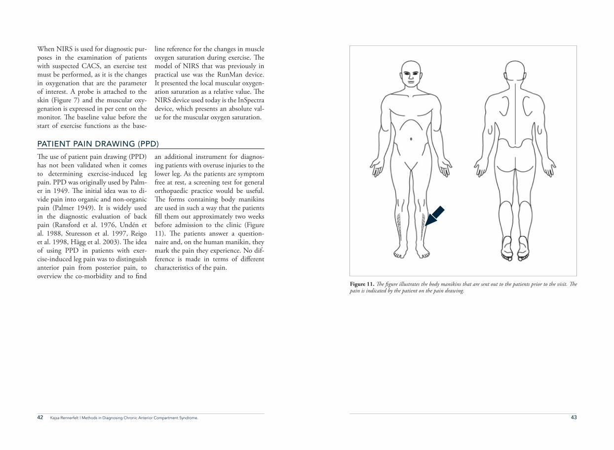
PATIENT PAIN DRAWING (PPD)
#e use of patient pain drawing (PPD) has not been validated when it comes to determining exercise-induced leg pain. PPD was originally used by Palm-er in 1949. #e initial idea was to di-vide pain into organic and non-organic pain (Palmer 1949). It is widely used in the diagnostic evaluation of back pain (Ransford et al. 1976, Undén et al. 1988, Sturesson et al. 1997, Reigo et al. 1998, Hägg et al. 2003). #e idea of using PPD in patients with exer-cise-induced leg pain was to distinguish anterior pain from posterior pain, to overview the co-morbidity and to %nd
an additional instrument for diagnos-ing patients with overuse injuries to the lower leg. As the patients are symptom free at rest, a screening test for general orthopaedic practice would be useful. #e forms containing body manikins are used in such a way that the patients %ll them out approximately two weeks before admission to the clinic (Figure 11). #e patients answer a question-naire and, on the human manikin, they mark the pain they experience. No dif-ference is made in terms of di$erent characteristics of the pain.
Figure 11. !e &gure illustrates the body manikins that are sent out to the patients prior to the visit. !e pain is indicated by the patient on the pain drawing.
44 45Kajsa Rennerfelt | Methods in Diagnosing Chronic Anterior Compartment Syndrome.
More specifically, the aims were to
• Investigate whether the magnitude of intramuscular oxygenation during exercise and the reoxygenation time at rest after exercise, following an exercise test, using NIRS, are reliable measurements for diagnosing CACS (Studies I and III),
• Compare two NIRS devices (INVOS and InSpectra) in healthy individuals with experimentally induced ischaemia in leg skeletal muscle (Study II),
• Investigate the e$ect of subcutaneous tissue thickness on measurements made with NIRS devices (Study II) and
• Validate the usefulness of PPD in identifying CACS in patients with exercise-in-duced leg pain (Study IV).
44 Kajsa Rennerfelt | Methods in Diagnosing Chronic Anterior Compartment Syndrome.
#e overall aim of this thesis was to investigate/evaluate near-infrared spectroscopy (NIRS) and patient pain drawing (PPD) in diagnosing chronic anterior compart-ment syndrome (CACS) in patients with exercise-induced leg-pain.
aimsAIMS
46 47Kajsa Rennerfelt | Methods in Diagnosing Chronic Anterior Compartment Syndrome.46 Kajsa Rennerfelt | Methods in Diagnosing Chronic Anterior Compartment Syndrome.
Intramuscular pressure (IMP) is regard-ed as the gold standard method for es-tablishing the diagnosis of chronic an-terior compartment syndrome (CACS). However, other methods for diagnos-ing CACS are the subject of debate, as are the exact cut-o$ levels for regard-ing an IMP as pathological (Barnes 1997, Aweid et al. 2012, Roberts and Franklyn‐Miller 2012). Variations in IMP can be caused by the examina-tion techniques and the placement of the needle or catheter (Gershuni et al. 1984, Nakhostine et al. 1993, Water-man et al. 2013). #is contributes to the uncertainty of relying on IMP mea-surements. New methods have been suggested and assessed, but none of these has as yet been found to be su-perior to IMP measurement. #e lack of symptoms at rest also suggests that the exercise test is of major importance in establishing the diagnosis of CACS.
Further research in this %eld was there-fore needed when this thesis was initiat-ed, to improve the diagnostic procedure and to validate the diagnostic methods in current use. One of the new diagnos-tic methods is near-infrared spectrosco-py (NIRS), which, in previous studies, has been suggested to be useful (Mohler et al. 1997, van den Brand et al. 2004). However, there was a need for larger studies to evaluate NIRS in more de-tail. Moreover, pain drawings produced by patients with exercise-induced leg pain could contribute to the di$eren-tiation of patients with CACS from patients with other causes of leg pain. Pain drawings have mostly been used in patients with lumbar pain (Hägg et al. 2003). #e clinical usefulness of PPD in diagnosing the causes of exercise-in-duced leg pain and its sensitivity and speci%city as a diagnostic instrument need to be studied.
why is thisthesisneeded?
WHY IS THIS THESIS NEEDED?
48 49Kajsa Rennerfelt | Methods in Diagnosing Chronic Anterior Compartment Syndrome.48 Kajsa Rennerfelt | Methods in Diagnosing Chronic Anterior Compartment Syndrome.
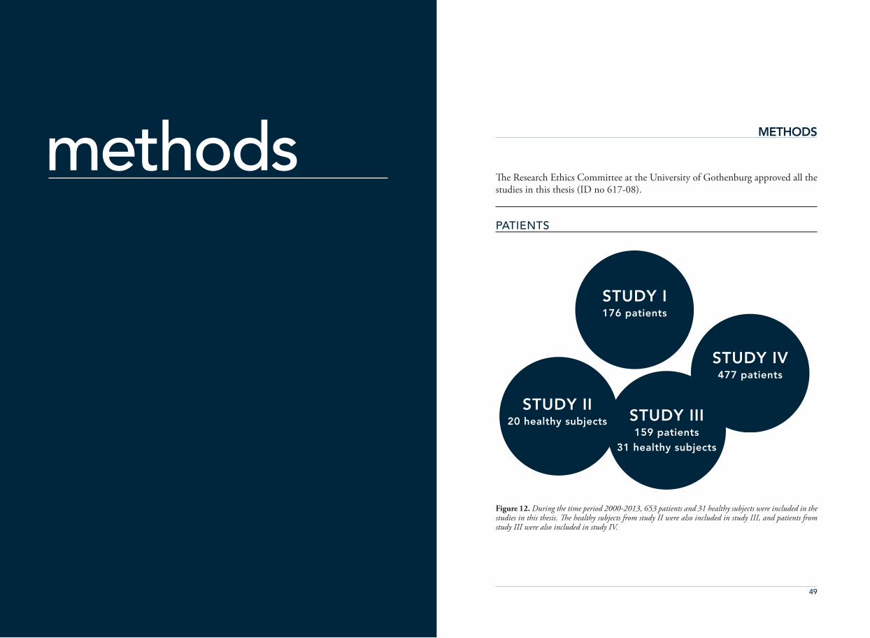
#e Research Ethics Committee at the University of Gothenburg approved all the studies in this thesis (ID no 617-08).
methodsPATIENTS
STUDY I176 patients
STUDY IV477 patients
STUDY II20 healthy subjects STUDY III
159 patients31 healthy subjects
Figure 12. During the time period 2000-2013, 653 patients and 31 healthy subjects were included in the studies in this thesis. !e healthy subjects from study II were also included in study III, and patients from study III were also included in study IV.
METHODS
50 51Kajsa Rennerfelt | Methods in Diagnosing Chronic Anterior Compartment Syndrome.
9000, Siemens, Gothenburg), (Zhang et al. 2011). #e skin was penetrated by a 1.2 mm diameter needle, with four side-holes at its tip. #e needle was inserted at a 30° angle to the long axis of the leg in the distal direction, into the belly of the tibialis anterior muscle (Zhang et al. 2011) parallel with the muscle %bres. To maintain the bulging of "uid at the tip of the needle at the beginning of the measurement, an infu-sion of 0.9% saline solution was given through the system and continuing to
the tip of the needle, with an infusion rate of 0.2 ml/h. #ere was no continu-ous infusion throughout the rest of the test time. #e tip of the catheter and the transducer were placed at heart level to minimize the hydrostatic artefacts and the position of the tip of the catheter was controlled using ultrasonography. #e patient lay supine in a relaxed posi-tion with the legs straight (Gershuni et al. 1984).
Study IOne hundred and seventy-six patients were included in the study during the time period 2000-2005, 73 men and 103 women; mean age was 34 (SD=15) years. #e duration of pain was 71 (range 3-360) months. #e patients were referred due to exercise-induced leg pain, with clinical signs of chron-ic anterior compartment syndrome (CACS) and all patients referred during this period were included provided the ability to perform an exercise test. #e study was non-randomized.
Study IIIOne hundred and %fty-nine patients were included in the study during the time period 2009-2012, 76 men and 83 women, with a median age of 29 years (range14-67) years. All patients were referred due to exercise-induced leg
pain and clinical signs of chronic ante-rior compartment syndrome (CACS). All patients referred during this period were included provided the ability to perform an exercise test.
Study IV#e study comprised 477 consecutive patients, 258 men and 219 women, with a median age of 31 (range 15-70) years, all with exercise-induced leg pain. #e patients were referred to the ortho-paedic department during 2009-2013.
HEALTHY SUBJECTS
Study IITwenty healthy subjects (10 women and 10 men) were recruited for this study. Median age was 43 (range 34-60) years. BMI mean 24 (SD 3). #e pa-tients had no history of vascular disease that necessitated medical intervention.
Study III#irty-one healthy subjects, 14 men and 17 women, with a median age of 36 (range 20-60) years were included. Twenty of them were also included in study II.
INTRAMUSCULAR PRESSURE (STUDIES I, III AND IV)
#e measurements were performed in the most symptomatic leg in all pa-tients. Intramuscular pressure (IMP) was measured 60 seconds after the exer-
cise test in all patients in the supine po-sition using a micro-capillary infusion system (Hemo 4, Siemens, Erlangen, Germany) and monitor (Siemens SC
NEAR-INFRARED SPECTROSCOPY (STUDIES I, II AND III)
StO2 was monitored by InSpectra (an (near-infrared spectroscopy) NIRS de-vice, tissue spectrometer model 325, Hutchinson Technology, Hutchinson, Minnesota). InSpectra employs infra-red light with wave-length signals be-tween 650 and 900 nm. #e distance between the light source and the detec-tor is 25 mm and approximately 95% of the detected optical signal is from a depth between zero and 23 mm. #e registration is continuous with data given every 3.5 seconds throughout the measurements. #e time for initiation of the exercise was indicated, as well as the %nish of the exercise and the end of the recovery time. Before every patient or healthy subjects calibration was per-formed by placing the probe in the cal-ibration-box.
InSpectra records the local oxygen satu-ration of the tibialis anterior muscle be-fore, during and after the exercise test.
#e NIRS probe was placed centrally over the tibialis anterior muscle in the most symptomatic leg and the measure-ments were randomised between right and left leg. #e measurement started with the patient/healthy subject resting in a supine position, followed by exer-cising in standing position, and there-after a recovery period with the patient resting supine. #e measurable range of StO2 is between 1 and 99%.
INVOS 4100 employs infrared light with two wavelength signals between 730 and 810 nm, it measures the ratio of oxygenated and total haemoglobin, displayed as a percentage of the region-al oxygen saturation. INVOS measures continuously every 4 seconds. INVOS uses one light source emitting light and two detectors. #e two detectors are located 30 and 40 mm from the light source. #e penetration depth of INVOS device is generally accepted to

52 53Kajsa Rennerfelt | Methods in Diagnosing Chronic Anterior Compartment Syndrome.
be half the distance between the light source and the sensor, 15 and 20 mm respectively. #e sensor was placed di-rectly on the skin, centrally over the tib-ialis anterior muscle randomly on either right or left leg and the local muscular oxygen saturation was analysed every 4 seconds.
#e measurable range of StO2 is be-tween 15 and 95%.Reoxygenation at rest after exercise was calculated as the time period required for the level of muscular StO2 to rise from the level of StO2 at the end of exercise to 50% (T50), 90% (T90) and 100% (T100) of the baseline value.
BLOOD PRESSURE (STUDIES I, II AND III)
Blood pressure was measured before and after the exercise test in all study objects using a pressure manometer (NAIS, Matsushita, Electronic Works,
Japan). Local perfusion pressure was calculated as the di$erence between the mean arterial blood pressure and IMP.
ULTRASONOGRAPHY IMAGING (STUDIES II AND III)
#e skin and subcutaneous tissue above the crural fascia and the dis-tance between the skin and the tip of the IMP needle were measured using ultrasonography imaging, with a linear
probe (L10-5, Acuson CV70, Siemens Medical Solutions Inc, USA). #is was performed with the subject in the su-pine position and with relaxed muscles.
SURFACE ELECTROMYOGRAPHY, EMG (STUDY III)
By using EMG the muscular activity can be controlled. During IMP mea-surement, the muscles must be relaxed in order not to in"uence the results of the IMP. Two bipolar surface electrodes were used (pre-gelled, Blue Sensor, Medicotest A/S, Denmark). #ey were placed 15 cm below the knee joint and a reference electrode was placed on the lateral malleolus. #e EMG signal was recorded before, during and after the exercise test. #e signals from the EMG
were ampli%ed in two steps – %rst by a custom-made pre-ampli%er (gain 100 pass second-order Butterworth %lter; fo = 10Hz) and then by the main am-pli%er (variable gain 1-196, low pass second-order Butterworth %lter; fo = 2 kHz) before recording with a data acquisition system (Pentium III PC, 12-bits DAQ board and Lab View soft-ware, National Instruments Corpora-tion, Austin, TX)(Zhang et al. 2011).
VISUAL ANALOGUE SCALE (STUDIES I, II AND III)
#e intensity of leg pain was evaluat-ed with a 10 cm visual analogue scale
(VAS) ranging from 0 cm (no pain) to 10 cm (worst imaginable pain).
STATISTICAL METHODS
Study I #e recorded intramuscular oxygen-ation data for each patient were ana-lyzed as follows:De%nitions of recovery time for intra-muscular oxygenation after exercise are shown on a typical curve obtained from near-infrared spectroscopy mea-surement before, during, and after an exercise test in one patient (Figure 16). R90 is de%ned as the time required for the level of oxygenation to rise from 10% to 90% of its baseline value after exercise. R100 is de%ned as the time re-quired for oxygenation to return to its baseline value after end of exercise.
1. Level of intramuscular oxygenation during exercise; the lowest value of oxy-genation recorded during exercise with a thigh tourniquet was de%ned as the physiological minimum, 0%. For analy-sis of the data, the levels of oxygenation that were recorded for each patient were normalized to a scale that ranged from 0% (the physiological minimum) to 100%.
2. Baseline value of muscular oxygen saturation was de%ned as the average level of intramuscular oxygenation re-corded during the 30-s rest period be-fore exercise (100%). R90 was de%ned
as the time required for the level of oxy-genation to rise from 10% to 90% of its baseline value after exercise. R100 was de%ned as the time required for oxy-genation to reach its baseline value after cessation of exercise.
#e time of intramuscular reoxygen-ation after exercise is presented as mean and standard deviation (SD). Di$erence between the patients with and without CACS was evaluated with analysis of variance. #e Mann–Whit-ney U-test was used for comparison be-tween groups (CACS and non-CACS). #e level of signi%cance was de%ned as p<0.05.
Study II #e measured values of muscle oxy-gen saturation (StO2) are presented as mean, median, standard deviation (SD) and range. Baseline StO2 values detect-ed by two NIRS devices were compared with values obtained during ischemia and post-ischemia recovery. VAS data are presented as median and range. Wilcoxon signed rank test was used to compare matched pairs. #e signif-icance of intergroup di$erences was determined using the Mann–Whitney U-test. #e level of signi%cance was de-%ned as p<0.05. Correlations between
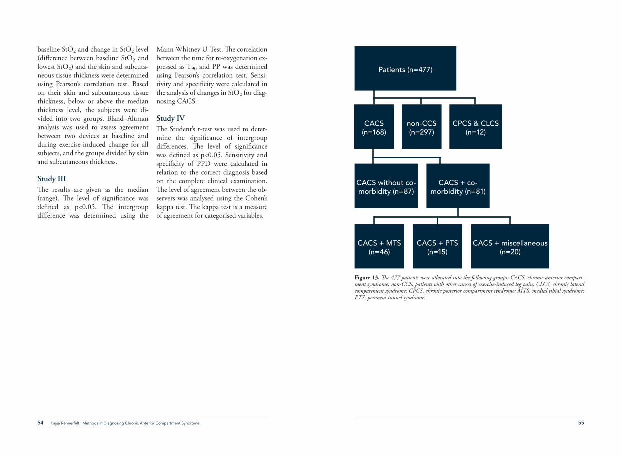
54 55Kajsa Rennerfelt | Methods in Diagnosing Chronic Anterior Compartment Syndrome.
baseline StO2 and change in StO2 level (di$erence between baseline StO2 and lowest StO2) and the skin and subcuta-neous tissue thickness were determined using Pearson’s correlation test. Based on their skin and subcutaneous tissue thickness, below or above the median thickness level, the subjects were di-vided into two groups. Bland–Altman analysis was used to assess agreement between two devices at baseline and during exercise-induced change for all subjects, and the groups divided by skin and subcutaneous thickness.
Study III#e results are given as the median (range). #e level of signi%cance was de%ned as p<0.05. #e intergroup di$erence was determined using the
Mann-Whitney U-Test. #e correlation between the time for re-oxygenation ex-pressed as T90 and PP was determined using Pearson’s correlation test. Sensi-tivity and speci%city were calculated in the analysis of changes in StO2 for diag-nosing CACS.
Study IV#e Student’s t-test was used to deter-mine the signi%cance of intergroup di$erences. #e level of signi%cance was de%ned as p<0.05. Sensitivity and speci%city of PPD were calculated in relation to the correct diagnosis based on the complete clinical examination. #e level of agreement between the ob-servers was analysed using the Cohen’s kappa test. #e kappa test is a measure of agreement for categorised variables.
Patients (n=477)
CACS
(n=168)
CACS + MTS
(n=46)
CACS without co-
morbidity (n=87)
CACS + co-
morbidity (n=81)
non-CCS
(n=297)
CACS + PTS
(n=15)
CPCS & CLCS
(n=12)
CACS + miscellaneous
(n=20)
Figure 13. !e 477 patients were allocated into the following groups: CACS, chronic anterior compart-ment syndrome; non-CCS, patients with other causes of exercise-induced leg pain; CLCS, chronic lateral compartment syndrome; CPCS, chronic posterior compartment syndrome; MTS, medial tibial syndrome; PTS, peroneus tunnel syndrome.
56 57Kajsa Rennerfelt | Methods in Diagnosing Chronic Anterior Compartment Syndrome.
STUDY I
Why was this study performed?#e study was performed to evaluate the use of near-infrared spectroscopy (NIRS) to diagnose chronic anteri-or compartment syndrome (CACS). During the study time period, the NIRS device RunMan was used (Breit et al. 1997, Mohler et al. 1997). Pre-vious studies have indicated di$erenc-es between CACS and other causes of leg pain during exercise when NIRS is used. A greater degree of tissue deoxy-genation during exercise has previously been observed in patients with CACS, as well as a delay in muscular oxygen saturation compared with patients who have other causes of leg pain. #e study by Breit et al. was performed on healthy individuals and the compartment syn-drome was induced by external pressure (Breit et al. 1997). As ischemia has been discussed as an important factor in the development of CACS, it is of great in-terest to monitor muscular oxygen sat-uration since other diagnostic methods have focused on di$erent parameters.
HypothesisExercise-induced leg pain in patients with CACS is related to muscle isch-emia, and NIRS is useful to detect pa-tients with CACS and compromised tissue perfusion.
MethodsOne hundred seventy-six consecutive patients (73 men, 103 women) with a mean age of 34 (range 14–76) years were studied between 2000 and 2005. #ey all had clinical signs of CACS.#e diagnosis of CACS was con%rmed by a thorough history and clinical ex-amination before and again after an exercise test that elicited the symptoms. Exercise test monitored by NIRS;A NIRS probe was placed centrally over the tibialis anterior muscle of the most symptomatic leg. #e patients per-formed two intensive indoor exercise tests to elicit symptoms – maximal con-centric dorsi"exion of both feet (60 cy-cles/min) in the standing position until they were unable to continue because of muscle fatigue, leg pain, impaired
56 Kajsa Rennerfelt | Methods in Diagnosing Chronic Anterior Compartment Syndrome.
summaryof papers
SUMMARY OF PAPERS
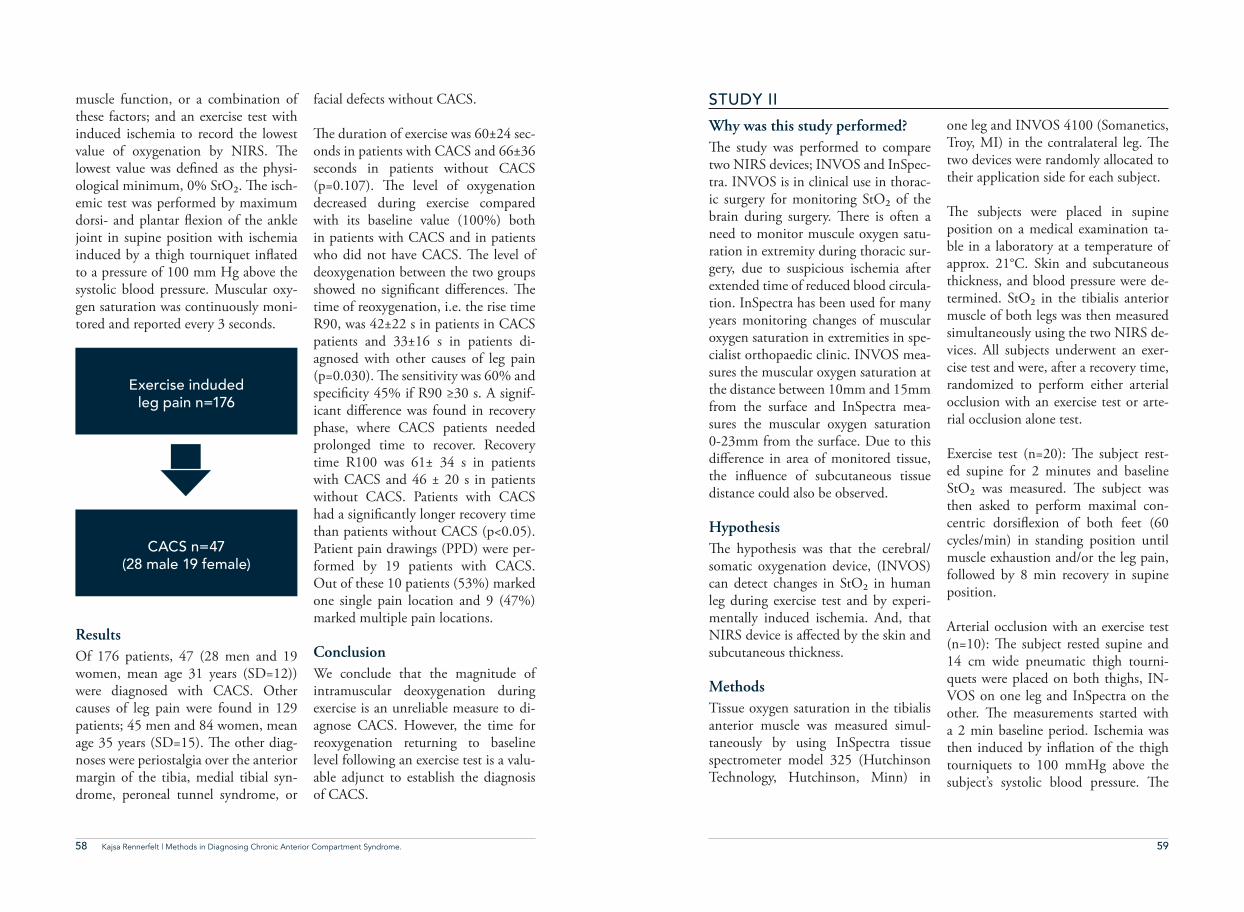
58 59Kajsa Rennerfelt | Methods in Diagnosing Chronic Anterior Compartment Syndrome.
muscle function, or a combination of these factors; and an exercise test with induced ischemia to record the lowest value of oxygenation by NIRS. #e lowest value was de%ned as the physi-ological minimum, 0% StO2. #e isch-emic test was performed by maximum dorsi- and plantar "exion of the ankle joint in supine position with ischemia induced by a thigh tourniquet in"ated to a pressure of 100 mm Hg above the systolic blood pressure. Muscular oxy-gen saturation was continuously moni-tored and reported every 3 seconds.
ResultsOf 176 patients, 47 (28 men and 19 women, mean age 31 years (SD=12)) were diagnosed with CACS. Other causes of leg pain were found in 129 patients; 45 men and 84 women, mean age 35 years (SD=15). #e other diag-noses were periostalgia over the anterior margin of the tibia, medial tibial syn-drome, peroneal tunnel syndrome, or
facial defects without CACS.
#e duration of exercise was 60±24 sec-onds in patients with CACS and 66±36 seconds in patients without CACS (p=0.107). #e level of oxygenation decreased during exercise compared with its baseline value (100%) both in patients with CACS and in patients who did not have CACS. #e level of deoxygenation between the two groups showed no signi%cant di$erences. #e time of reoxygenation, i.e. the rise time R90, was 42±22 s in patients in CACS patients and 33±16 s in patients di-agnosed with other causes of leg pain (p=0.030). #e sensitivity was 60% and speci%city 45% if R90 ≥30 s. A signif-icant di$erence was found in recovery phase, where CACS patients needed prolonged time to recover. Recovery time R100 was 61± 34 s in patients with CACS and 46 ± 20 s in patients without CACS. Patients with CACS had a signi%cantly longer recovery time than patients without CACS (p<0.05). Patient pain drawings (PPD) were per-formed by 19 patients with CACS. Out of these 10 patients (53%) marked one single pain location and 9 (47%) marked multiple pain locations.
ConclusionWe conclude that the magnitude of intramuscular deoxygenation during exercise is an unreliable measure to di-agnose CACS. However, the time for reoxygenation returning to baseline level following an exercise test is a valu-able adjunct to establish the diagnosis of CACS.
CACS n=47
(28 male 19 female)
Exercise induded
leg pain n=176
STUDY II
Why was this study performed?#e study was performed to compare two NIRS devices; INVOS and InSpec-tra. INVOS is in clinical use in thorac-ic surgery for monitoring StO2 of the brain during surgery. #ere is often a need to monitor muscule oxygen satu-ration in extremity during thoracic sur-gery, due to suspicious ischemia after extended time of reduced blood circula-tion. InSpectra has been used for many years monitoring changes of muscular oxygen saturation in extremities in spe-cialist orthopaedic clinic. INVOS mea-sures the muscular oxygen saturation at the distance between 10mm and 15mm from the surface and InSpectra mea-sures the muscular oxygen saturation 0-23mm from the surface. Due to this di$erence in area of monitored tissue, the in"uence of subcutaneous tissue distance could also be observed. Hypothesis#e hypothesis was that the cerebral/somatic oxygenation device, (INVOS) can detect changes in StO2 in human leg during exercise test and by experi-mentally induced ischemia. And, that NIRS device is a$ected by the skin and subcutaneous thickness.
MethodsTissue oxygen saturation in the tibialis anterior muscle was measured simul-taneously by using InSpectra tissue spectrometer model 325 (Hutchinson Technology, Hutchinson, Minn) in
one leg and INVOS 4100 (Somanetics, Troy, MI) in the contralateral leg. #e two devices were randomly allocated to their application side for each subject. #e subjects were placed in supine position on a medical examination ta-ble in a laboratory at a temperature of approx. 21°C. Skin and subcutaneous thickness, and blood pressure were de-termined. StO2 in the tibialis anterior muscle of both legs was then measured simultaneously using the two NIRS de-vices. All subjects underwent an exer-cise test and were, after a recovery time, randomized to perform either arterial occlusion with an exercise test or arte-rial occlusion alone test.
Exercise test (n=20): #e subject rest-ed supine for 2 minutes and baseline StO2 was measured. #e subject was then asked to perform maximal con-centric dorsi"exion of both feet (60 cycles/min) in standing position until muscle exhaustion and/or the leg pain, followed by 8 min recovery in supine position.
Arterial occlusion with an exercise test (n=10): #e subject rested supine and 14 cm wide pneumatic thigh tourni-quets were placed on both thighs, IN-VOS on one leg and InSpectra on the other. #e measurements started with a 2 min baseline period. Ischemia was then induced by in"ation of the thigh tourniquets to 100 mmHg above the subject’s systolic blood pressure. #e
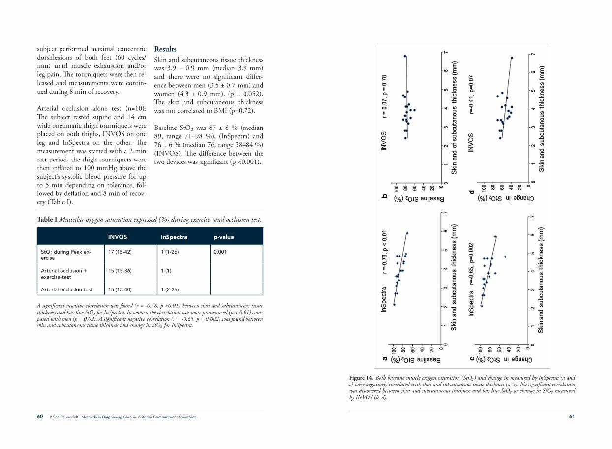
60 61Kajsa Rennerfelt | Methods in Diagnosing Chronic Anterior Compartment Syndrome.
subject performed maximal concentric dorsi"exions of both feet (60 cycles/min) until muscle exhaustion and/or leg pain. #e tourniquets were then re-leased and measurements were contin-ued during 8 min of recovery.
Arterial occlusion alone test (n=10): #e subject rested supine and 14 cm wide pneumatic thigh tourniquets were placed on both thighs, INVOS on one leg and InSpectra on the other. #e measurement was started with a 2 min rest period, the thigh tourniquets were then in"ated to 100 mmHg above the subject’s systolic blood pressure for up to 5 min depending on tolerance, fol-lowed by de"ation and 8 min of recov-ery (Table I).
ResultsSkin and subcutaneous tissue thickness was 3.9 ± 0.9 mm (median 3.9 mm) and there were no signi%cant di$er-ence between men (3.5 ± 0.7 mm) and women (4.3 ± 0.9 mm), (p = 0.052). #e skin and subcutaneous thickness was not correlated to BMI (p=0.72).
Baseline StO2 was 87 ± 8 % (median 89, range 71–98 %), (InSpectra) and 76 ± 6 % (median 76, range 58–84 %) (INVOS). #e di$erence between the two devices was signi%cant (p <0.001).
INVOS InSpectra p-value
StO2 during Peak ex-
ercise
17 (15-42) 1 (1-26) 0.001
Arterial occlusion +
exercise-test
15 (15-36) 1 (1)
Arterial occlusion test 15 (15-40) 1 (2-26)
Table I Muscular oxygen saturation expressed (%) during exercise- and occlusion test.
A signi&cant negative correlation was found (r = -0.78, p <0.01) between skin and subcutaneous tissue thickness and baseline StO% for InSpectra. In women the correlation was more pronounced (p < 0.01) com-pared with men (p = 0.02). A signi&cant negative correlation (r = -0.65, p = 0.002) was found between skin and subcutaneous tissue thickness and change in StO% for InSpectra.
andpo
stischem
ichy
peraem
iaweredetected
inallsubjects
even
thou
ghthebaseline
levels
ofox
ygenationas
wellas
thelowestox
ygenationleveldiffered
sign
ificantly
between
thedevices.
Inthisstud
y,muscleischem
iawas
indu
cedby
exercise,
exercise
incombination
witharterial
occlusion
andarterial
occlusion
alon
e.Both
InSpectra
and
INVOS
equipm
ent
were
able
todetect
adecrease
inStO
2du
ring
the
Fig.2
Bothbaseline
muscleox
ygen
saturation
(StO
2)andchange
inStO
2(differencebetweenbaseline
StO
2andlowestStO
2)measured
byInSpectra
werenegatively
correlated
withskin
andsubcutaneous
tissue
thickn
ess(a,c).No
sign
ificant
correlation
was
discov
ered
betweenskin
andsubcutaneous
thickn
essandbaseline
StO
2or
change
inStO
2measuredby
INVOS(b,d)
Fig.3
Bland
–Altman
plotsdisplaying
thedifference
invalueof
muscleox
ygen
saturation
(StO
2)draw
nagainstthemeanbetween
InSpectra
andIN
VOS,A
better
agreem
entis
show
nforthosewith
less
(\3.9mm)skin
andsubcutaneous
thickn
ess,
both
atbaseline
(a)and
atchange
inStO
2(c)compared
with
thosewith
deeper
([3.9mm)skin
andsubcutaneous
thickn
ess(b,d)
60JClinMon
itCom
put(201
4)28
:57–
62
123Figure 14. Both baseline muscle oxygen saturation (StO%) and change in measured by InSpectra (a and
c) were negatively correlated with skin and subcutaneous tissue thickness (a, c). No signi&cant correlation was discovered between skin and subcutaneous thickness and baseline StO% or change in StO% measured by INVOS (b, d).
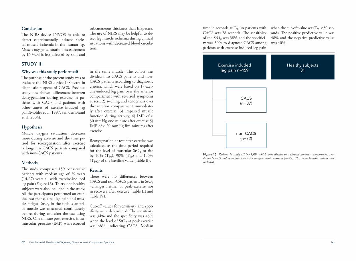
62 63Kajsa Rennerfelt | Methods in Diagnosing Chronic Anterior Compartment Syndrome.
time in seconds at T90 in patients with CACS was 28 seconds. #e sensitivity of the StO2 was 38% and the speci%ci-ty was 50% to diagnose CACS among patients with exercise-induced leg pain
when the cut-o$ value was T90 ≥30 sec-onds. #e positive predictive value was 48% and the negative predictive value was 40%.
Conclusion#e NIRS-device INVOS is able to detect experimentally induced skele-tal muscle ischemia in the human leg. Muscle oxygen saturation measurement by INVOS is less a$ected by skin and
subcutaneous thickness than InSpectra. #e use of NIRS may be helpful to de-tect leg muscle ischemia during clinical situations with decreased blood circula-tion.
STUDY III
Why was this study performed?#e purpose of the present study was to evaluate the NIRS-device InSpectra in diagnostic purpose of CACS. Previous study has shown di$erences between deoxygenation during exercise in pa-tients with CACS and patients with other causes of exercise induced leg pain(Mohler et al. 1997, van den Brand et al. 2004).
HypothesisMuscle oxygen saturation decreases more during exercise and the time pe-riod for reoxygenation after exercise is longer in CACS patients compared with non-CACS patients.
Methods#e study comprised 159 consecutive patients with median age of 29 years (14-67) years all with exercise-induced leg pain (Figure 15). #irty-one healthy subjects were also included in the study. All the participants performed an exer-cise test that elicited leg pain and mus-cle fatigue. StO2 in the tibialis anteri-or muscle was measured continuously before, during and after the test using NIRS. One minute post-exercise, intra-muscular pressure (IMP) was recorded
in the same muscle. #e cohort was divided into CACS patients and non-CACS patients according to diagnostic criteria, which were based on 1) exer-cise-induced leg pain over the anterior compartment with reversed symptoms at rest, 2) swelling and tenderness over the anterior compartment immediate-ly after exercise, 3) impaired muscle function during activity, 4) IMP of ≥ 30 mmHg one minute after exercise 5) IMP of ≥ 20 mmHg %ve minutes after exercise.
Reoxygenation at rest after exercise was calculated as the time period required for the level of muscular StO2 to rise by 50% (T50), 90% (T90) and 100% (T100) of the baseline value (Table II).
Results#ere were no di$erences between CACS and non-CACS patients in StO2 –changes neither at peak-exercise nor in recovery after exercise (Table III and Table IV).
Cut-o$ values for sensitivity and spec-i%city were determined. #e sensitivity was 34% and the speci%city was 43% when the level of StO2 at peak exercise was ≤8%, indicating CACS. Median
Healthy subjects
31
Exercise induded
leg pain n=159
CACS
(n=87)
non-CACS
(n=72)
Figure 15. Patients in study III (n=159), which were divides into chronic anterior compartment syn-drome (n=87) and non-chronic anterior compartment syndrome (n=72). !irty-one healthy subjects were included.
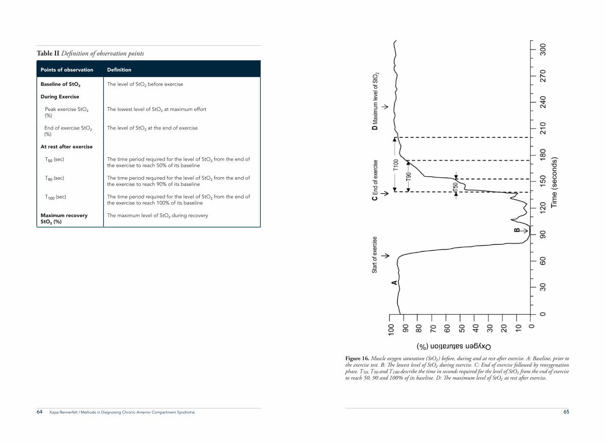
64 65Kajsa Rennerfelt | Methods in Diagnosing Chronic Anterior Compartment Syndrome.
Table II De&nition of observation points
Points of observation &GƂPKVKQP
Baseline of StO² The level of StO² before exercise
During Exercise
Peak exercise StO²
(%)
The lowest level of StO² at maximum effort
End of exercise StO²
(%)The level of StO
² at the end of exercise
At rest after exercise
T50 (sec) The time period required for the level of StO² from the end of
the exercise to reach 50% of its baseline
T90 (sec) The time period required for the level of StO² from the end of
the exercise to reach 90% of its baseline
T100 (sec) The time period required for the level of StO² from the end of
the exercise to reach 100% of its baseline
Maximum recovery StO² (%)
The maximum level of StO² during recovery
Figure 16. Muscle oxygen saturation (StO%) before, during and at rest after exercise. A: Baseline, prior to the exercise test. B: !e lowest level of StO% during exercise. C: End of exercise followed by reoxygenation phase. T50, T90 and T100 describe the time in seconds required for the level of StO% from the end of exercise to reach 50, 90 and 100% of its baseline. D: !e maximum level of StO% at rest after exercise.
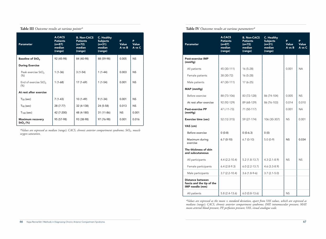
66 67Kajsa Rennerfelt | Methods in Diagnosing Chronic Anterior Compartment Syndrome.
Parameter
A.CACSPatients (n=87) median (range)
B. Non-CACSPatients (n=72)median (range)
C. Healthy Subjects (n=31)median (range)
P ValueA vs B
P ValueA vs C
Baseline of StO² 92 (45-98) 84 (40-98) 88 (59-98) 0.005 NS
During Exercise
Peak exercise StO²
(%)
1 (1-36) 3 (1-54) 1 (1-44) 0.003 NS
End of exercise StO²
(%)1 (1-68) 17 (1-69) 1 (1-54) 0.001 NS
At rest after exercise
T50 (sec) 7 (1-43) 10 (1-49) 9 (1-34) 0.001 NS
T90 (sec) 28 (7-77) 32 (4-138) 24 (5-58) 0.013 NS
T100 (sec) 42 (7-200) 48 (4-180) 31 (11-86) NS 0.001
Maximum recoveryStO² (%)
95 (57-98) 93 (38-98) 97 (76-98) 0.001 0.016
Table III Outcome results at various points'
'Values are expressed as median (range); CACS, chronic anterior compartment syndrome; StO%, muscle ocygen saturation.
Table IV Outcome results at various parameters'
'Values are expressed as the mean ± standard deviation, apart from VAS values, which are expressed as medians (range); CACS, chronic anterior compartment syndrome; IMP, intramuscular pressure; MAP, mean arterial blood pressure; PP, perfusion pressure; VAS, visual analogue scale.
Parameter
A.CACSPatients (n=87) median (range)
B. Non-CACSPatients (n=72)median (range)
C. Healthy Subjects (n=31)median (range)
P ValueA vs B
P ValueA vs C
Post-exercise IMP (mmHg)
All patients 45 (30-111) 16 (5-28) 0.001 NA
Female patients 38 (30-72) 16 (5-28)
Male patients 47 (30-111) 17 (6-25)
MAP (mmHg)
Before exercise 88 (73-106) 83 (72-128) 86 (74-104) 0.005 NS
At rest after exercise 92 (92-129) 89 (68-129) 86 (76-103) 0.014 0.010
Post-exercise PP (mmHg)
47 (-11-73) 71 (50-117) 0.001 NA
Exercise time (sec) 52 (12-315) 59 (27-174) 106 (30-307) NS 0.001
VAS (cm)
Before exercise 0 (0-8) 0 (0-6.3) 0 (0)
Maximum during
exercise
6.7 (0-10) 6.7 (0-10) 5.0 (0-9) NS 0.034
The thickness of skin and subcutaneous
All participants 4.4 (2.2-10.4) 5.2 (1.8-13.7) 4.3 (2.1-8.9) NS NS
Female participants 6.4 (2.8-9.3) 6.0 (2.2-13.7) 4.6 (3.3-8.9)
Male participants 3.7 (2.2-10.4) 3.6 (1.8-9-6) 3.7 (2.1-5.0)
Distance between fascia and the tip of the IMP needle (mm)
All patients 5.8 (2.4-13.6) 6.0 (0.8-13.6) NS
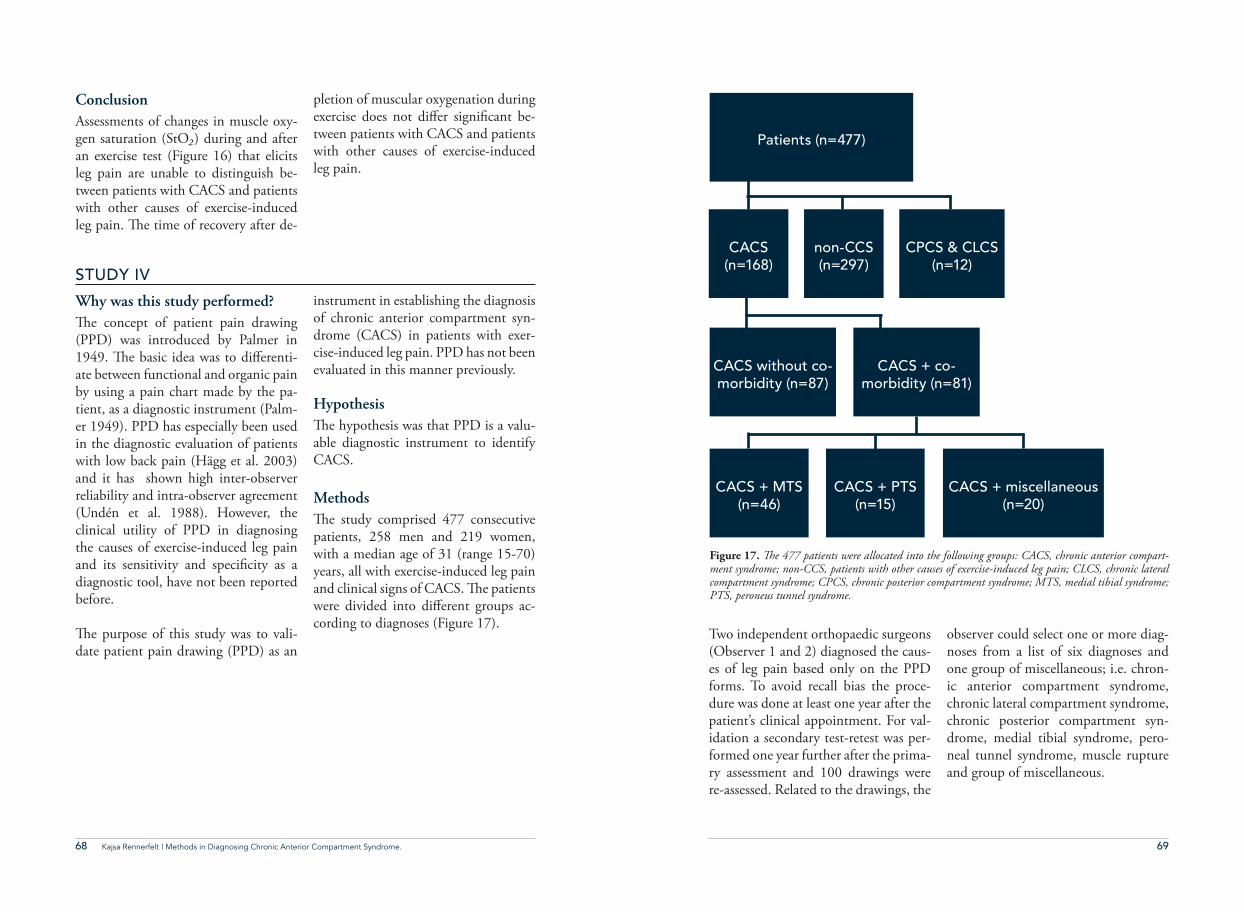
68 69Kajsa Rennerfelt | Methods in Diagnosing Chronic Anterior Compartment Syndrome.
Two independent orthopaedic surgeons (Observer 1 and 2) diagnosed the caus-es of leg pain based only on the PPD forms. To avoid recall bias the proce-dure was done at least one year after the patient’s clinical appointment. For val-idation a secondary test-retest was per-formed one year further after the prima-ry assessment and 100 drawings were re-assessed. Related to the drawings, the
observer could select one or more diag-noses from a list of six diagnoses and one group of miscellaneous; i.e. chron-ic anterior compartment syndrome, chronic lateral compartment syndrome, chronic posterior compartment syn-drome, medial tibial syndrome, pero-neal tunnel syndrome, muscle rupture and group of miscellaneous.
ConclusionAssessments of changes in muscle oxy-gen saturation (StO%) during and after an exercise test (Figure 16) that elicits leg pain are unable to distinguish be-tween patients with CACS and patients with other causes of exercise-induced leg pain. #e time of recovery after de-
pletion of muscular oxygenation during exercise does not di$er signi%cant be-tween patients with CACS and patients with other causes of exercise-induced leg pain.
STUDY IV
Why was this study performed?#e concept of patient pain drawing (PPD) was introduced by Palmer in 1949. #e basic idea was to di$erenti-ate between functional and organic pain by using a pain chart made by the pa-tient, as a diagnostic instrument (Palm-er 1949). PPD has especially been used in the diagnostic evaluation of patients with low back pain (Hägg et al. 2003) and it has shown high inter-observer reliability and intra-observer agreement (Undén et al. 1988). However, the clinical utility of PPD in diagnosing the causes of exercise-induced leg pain and its sensitivity and speci%city as a diagnostic tool, have not been reported before.
#e purpose of this study was to vali-date patient pain drawing (PPD) as an
instrument in establishing the diagnosis of chronic anterior compartment syn-drome (CACS) in patients with exer-cise-induced leg pain. PPD has not been evaluated in this manner previously. Hypothesis#e hypothesis was that PPD is a valu-able diagnostic instrument to identify CACS.
Methods#e study comprised 477 consecutive patients, 258 men and 219 women, with a median age of 31 (range 15-70) years, all with exercise-induced leg pain and clinical signs of CACS. #e patients were divided into di$erent groups ac-cording to diagnoses (Figure 17).
Patients (n=477)
CACS
(n=168)
CACS + MTS
(n=46)
CACS without co-
morbidity (n=87)
CACS + co-
morbidity (n=81)
non-CCS
(n=297)
CACS + PTS
(n=15)
CPCS & CLCS
(n=12)
CACS + miscellaneous
(n=20)
Figure 17. !e 477 patients were allocated into the following groups: CACS, chronic anterior compart-ment syndrome; non-CCS, patients with other causes of exercise-induced leg pain; CLCS, chronic lateral compartment syndrome; CPCS, chronic posterior compartment syndrome; MTS, medial tibial syndrome; PTS, peroneus tunnel syndrome.
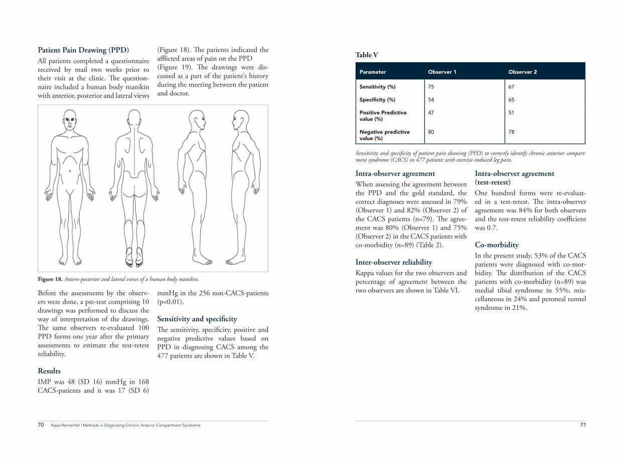
70 71Kajsa Rennerfelt | Methods in Diagnosing Chronic Anterior Compartment Syndrome.
Intra-observer agreementWhen assessing the agreement between the PPD and the gold standard, the correct diagnoses were assessed in 79% (Observer 1) and 82% (Observer 2) of the CACS patients (n=79). #e agree-ment was 80% (Observer 1) and 75% (Observer 2) in the CACS patients with co-morbidity (n=89) (Table 2).
Inter-observer reliabilityKappa values for the two observers and percentage of agreement between the two observers are shown in Table VI.
Intra-observer agreement (test-retest)One hundred forms were re-evaluat-ed in a test-retest. #e intra-observer agreement was 84% for both observers and the test-retest reliability coe(cient was 0.7.
Co-morbidityIn the present study, 53% of the CACS patients were diagnosed with co-mor-bidity. #e distribution of the CACS patients with co-morbidity (n=89) was medial tibial syndrome in 55%, mis-cellaneous in 24% and peroneal tunnel syndrome in 21%.
Before the assessments by the observ-ers were done, a pre-test comprising 10 drawings was performed to discuss the way of interpretation of the drawings. #e same observers re-evaluated 100 PPD forms one year after the primary assessments to estimate the test-retest reliability.
ResultsIMP was 48 (SD 16) mmHg in 168 CACS-patients and it was 17 (SD 6)
mmHg in the 256 non-CACS-patients (p<0.01).
Sensitivity and specificity#e sensitivity, speci%city, positive and negative predictive values based on PPD in diagnosing CACS among the 477 patients are shown in Table V.
Patient Pain Drawing (PPD) All patients completed a questionnaire received by mail two weeks prior to their visit at the clinic. #e question-naire included a human body manikin with anterior, posterior and lateral views
(Figure 18). #e patients indicated the a-icted areas of pain on the PPD (Figure 19). #e drawings were dis-cussed as a part of the patient’s history during the meeting between the patient and doctor.
Figure 18. Antero-posterior and lateral views of a human body manikin.
Parameter Observer 1 Observer 2
Sensitivity (%) 75 67
5RGEKƂEKV[��� 54 65
Positive Predictivevalue (%)
47 51
Negative predictivevalue (%)
80 78
Table V
Sensitivity and speci&city of patient pain drawing (PPD) to correctly identify chronic anterior compart-ment syndrome (CACS) in 477 patients with exercise-induced leg pain.
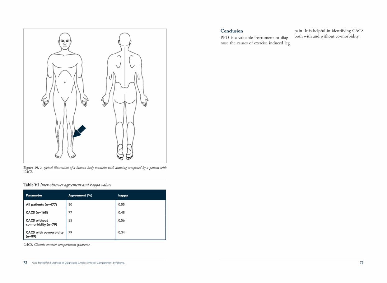
72 73Kajsa Rennerfelt | Methods in Diagnosing Chronic Anterior Compartment Syndrome.
ConclusionPPD is a valuable instrument to diag-nose the causes of exercise induced leg
pain. It is helpful in identifying CACS both with and without co-morbidity.
Figure 19. A typical illustration of a human body-manikin with drawing completed by a patient with CACS.
Parameter Agreement (%) kappa
All patients (n=477) 80 0.55
CACS (n=168) 77 0.48
CACS without co-morbidity (n=79)
85 0.56
CACS with co-morbidity (n=89)
79 0.34
Table VI Inter-observer agreement and kappa values
CACS, Chronic anterior compartment syndrome.
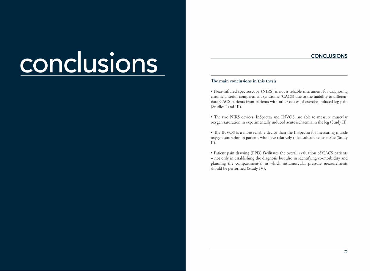
74 75Kajsa Rennerfelt | Methods in Diagnosing Chronic Anterior Compartment Syndrome.
"e main conclusions in this thesis
• Near-infrared spectroscopy (NIRS) is not a reliable instrument for diagnosing chronic anterior compartment syndrome (CACS) due to the inability to di$eren-tiate CACS patients from patients with other causes of exercise-induced leg pain (Studies I and III).
• #e two NIRS devices, InSpectra and INVOS, are able to measure muscular oxygen saturation in experimentally induced acute ischaemia in the leg (Study II). • #e INVOS is a more reliable device than the InSpectra for measuring muscle oxygen saturation in patients who have relatively thick subcutaneous tissue (Study II).
• Patient pain drawing (PPD) facilitates the overall evaluation of CACS patients – not only in establishing the diagnosis but also in identifying co-morbidity and planning the compartment(s) in which intramuscular pressure measurements should be performed (Study IV).
74 Kajsa Rennerfelt | Methods in Diagnosing Chronic Anterior Compartment Syndrome.
conclusionsCONCLUSIONS
76 77Kajsa Rennerfelt | Methods in Diagnosing Chronic Anterior Compartment Syndrome.
discussionElevated intramuscular pressure has been known as a possible cause of ex-ercise-induced leg pain in recreational runners and athletes since the 1960-70s. It is usually the anterior muscu-lar compartment in the lower leg that is involved in the syndrome known as chronic anterior compartment syn-drome (CACS). Exercise elicits the pain and the patients are free from symp-toms at rest. To diagnose CACS, the measurement of intramuscular pressure by inserting a needle has long been the method regarded as the gold standard (Reneman 1975, Wallensten and Eriks-son 1984, Styf et al. 1987). However, it would be desirable if a reliable, non-in-vasive diagnostic method for this pa-tient group was available.
#e main focus of this thesis was to investigate the potential for using near-infrared spectroscopy (NIRS) as an alternative method to IMP mea-surement to diagnose CACS. NIRS measures the local muscular oxygen saturation. #ree di$erent devices based on NIRS methodology were in-vestigated in three of the four studies included in this thesis. In addition, the opportunity to acquire information
from patient pain drawings (PPD) in order to establish the CACS diagnosis was investigated. #e %ndings in CACS patients were compared with those in patients with other established diagno-ses, causing lower leg pain. Moreover, in Studies II and III, healthy individu-als were included in the comparison of the two modern NIRS devices.
NIRS as a diagnostic method for CACS#e %rst generation of NIRS, called RunMan, was used in Study I. #e measurement of muscle oxygenation is given as a relative value by the Run-Man. #e further development of the methodology resulted in the InSpectra device, which delivers an absolute value for the local muscle oxygen saturation. Another NIRS device called INVOS was developed to monitor the local oxygen saturation of the brain during cardiothoracic surgery, where it is cur-rently used in daily practice (Scheeren et al. 2012). Due to the need sometimes to monitor skeletal muscle in the leg when suspicions of hypoxia might arise, Study II was designed.
DISCUSSION
78 79Kajsa Rennerfelt | Methods in Diagnosing Chronic Anterior Compartment Syndrome.
#e use of NIRS to diagnose CACS was suggested, based on the %nding that patients with CACS di$er in mus-cular oxygen saturation during exercise compared with patients who have other causes of leg pain, as well as when com-pared with healthy individuals (Breit et al. 1997, Mohler et al. 1997, van den Brand et al. 2005). #e a$ected mus-cles in patients with CACS have been observed to deoxygenate to a lower level and to reoxygenate after exercise during a prolonged period, compared with the muscles in patients with other causes of leg pain (Breit et al. 1997, Mohler et al. 1997, van den Brand et al. 2004, van den Brand et al. 2005, Zhang et al. 2012).
Findings using the first generation of NIRS device (RunMan) in CACS patientsStudy I consists of a relatively large cohort, comprising 47 CACS patients and 129 patients with other causes of leg pain. In the two sub-cohorts, i.e. patients with and without CACS, the muscles in the lower leg were found to deoxygenate to the same extent during exercise and no signi%cant di$erences between these two groups could be ob-served. #ese results are, however, not in line with the results presented by Mohler et al. (Mohler et al. 1997). #e divergent results were surprising and no explanation was found when compar-ing the design of these two studies. #e exercise test used in Study I appears to be comparable to the exercise test per-formed by Mohler et al. and the tests
were performed in a similar manner. So the di$erences in the results in Study I and in the study performed by Mohler et al. are di(cult to explain.
When comparing with the study by Mohler et al., it has to be remembered that all their study objects were contin-uously monitored with IMP. #is is a strength of that study, con%rming the diagnosis by using the gold standard method. However, the study by Mohler et al. had limitations, as it only com-prised 10 patients with CACS and eight patients with other causes of leg pain. #e 10 healthy volunteers in their study can be regarded as a strength.
Findings using the second gen-eration of NIRS device in CACS patientsIn order to further investigate the op-portunity to use NIRS as a diagnostic instrument in patients with CACS, Study III was designed, using a more modern NIRS device than that used in Study I; i.e. the InSpectra device. More-over, a larger patient cohort was includ-ed in this study; a total of 159 patients with lower leg pain, plus 31 healthy individuals. As far as we know, this is the largest patient cohort with CACS (n=87) investigated using the NIRS technique in the literature.
In Study III, %ndings similar to those in Study I were observed with regard to the deoxygenation phase. It was ob-served that the magnitude of deoxy-genation during exercise did not di$er
in clinical terms between patients with CACS and patients with other causes of leg pain. #e magnitude of deoxygen-ation during exercise re"ects a variation in work intensity and might be in"u-enced by leg pain among the patients.
#e current results also di$er from the results presented by Mohler et al. in terms of the increased magnitude of deoxygenation in patients with CACS compared with patients with other causes of leg pain (Mohler et al. 1997). Moreover, in the study by van den Brand et al., patients with CACS (n=42) were reported to have more pro-nounced deoxygenation compared with patients with other causes of leg pain (van den Brand et al. 2005). However, both these studies included a limited number of patients with other causes of leg pain. #ere were eight control patients in the study by Mohler et al. and only three in the study by van den Brand et al.
Moreover, no di$erences during de-oxygenation were recorded between CACS patients and healthy individu-als in Study III. #is %nding also dif-fered from the %ndings of Mohler et al., where healthy individuals (n=10) were included as well. #e reasons for the di$erent %ndings in the present study and other previous studies are not com-pletely clear, but they may be related to the relatively large inter-individual vari-ation, which can easily cause large dif-ferences when the sample size is small, and to the performance of the exercise test.
In Study I, and in the study by Mohler et al., the recovery time for muscular oxygenation (time for the oxygenation to return to the baseline value, T100) was found to be prolonged in patients with CACS when compared with pa-tients with other causes of leg pain. In the study by van den Brand et al., there were no indications of a prolonged re-oxygenation phase (van den Brand et al. 2004). #e impact of the reoxygenation period is therefore not really clear con-sidering the results in Study I.
However, as the results from Study III showed no di$erences in the recovery phase between patients with CACS and patients with other causes of leg pain, the impact of the recovery period could be regarded as not being especially use-ful. However, when CACS patients, as well as patients with lower leg pain caused by other conditions, were com-pared with healthy individuals in Study III, a prolonged recovery time was ob-served for both patient groups com-pared with the controls. As both patient groups showed similar changes, the re-covery phase does not appear to be a valuable instrument in distinguishing patients with CACS from patients with other causes of lower leg pain. Further-more, the poor sensitivity (38%) and speci%city (50%) when analysing the level of muscle oxygen saturation at T90 indicate several limitations in diagnos-ing CACS.
80 81Kajsa Rennerfelt | Methods in Diagnosing Chronic Anterior Compartment Syndrome.
Assessing agreement between INVOS and In Spectra in healthy individualsIn Study II, the two NIRS devices, In-Spectra and INVOS, both representing the second generation of NIRS devices, were compared. #e INVOS is used in daily practice during cardiac surgery to monitor StO2 of the brain. Both devic-es show absolute oxygen saturation val-ues. #e study investigated the changes in StO2 in a muscular compartment in the lower extremity of 20 healthy indi-viduals during experimentally induced muscle ischaemia, using both methods. #e most important %nding was that the INVOS, which was developed to monitor the oxygenation of brain tis-sue, can also be used to detect skeletal muscle ischaemia.
#ere are some di$erences between the two methods and they may play a role in some conditions. #e InSpectra measures the spectrum of light between 650-900 nm. #e wide range of chang-es in haemoglobin saturation that can be detected by the InSpectra is there-fore between 1-99%. #e INVOS, on the other hand, detects the spectrum of light between 730-810 nm and this corresponds to changes in oxygenation between 15-95% saturation. In clinical terms, this means that the InSpectra is able to detect changes within a wider range of light compared with the IN-VOS. If the INVOS is used clinically during suspected severe hypoxia in skeletal muscle, values below 15% of oxygen saturation must be considered
as somewhere between 1-15% of satu-ration.
One known advantage of the INVOS is that it does not monitor the most super%cial tissue layers. It measures the oxygen saturation of the muscle at a tissue depth of approximately 15-20 mm (Figure 10). #is means that the most super%cial layer is not within the range of the light. It was also clearly demonstrated in Study II that the IN-VOS was less sensitive to a thick layer of subcutaneous tissue than the InSpectra, when the two methods were compared in individuals with thick subcutaneous tissue (Figure 14).
Evaluation of pain drawings as a diagnostic instrument in patients with CACSEstablishing the diagnosis of CACS is a complex process, as it cannot be per-formed by observing only one param-eter or by using a single test available today. As a result, one of the aims of the present thesis was to investigate wheth-er pain drawings (PPD) produced by patients with exercise-induced leg pain were able to help to di$erentiate the patients with CACS from a group of patients with other diagnoses of lower leg pain. In Study IV, PPDs from pa-tients, all diagnosed with CACS using clinical %ndings and IMP measure-ments (n=168), were compared with those produced by a group of patients with lower leg pain with other causes (n=309). In this study, the sensitivity of two independent observers in diagnos-
ing CACS in a cohort of 477 patients with exercise-induced leg pain was 67-75%. #e speci%city was 54-65%, the positive predictive value 47-51% and the negative predictive value 78-80%. #e results might appear to be fair, but, as an additional diagnostic instrument, PPD should be considered to be valu-able. When it comes to pain drawings, the method has previously been used to dis-criminate between organic and non-or-ganic pain (Palmer 1949). Pain draw-ings have mostly been used in patients with lumbar pain (Hägg et al. 2003). #e patient simply indicates the a$ect-ed area on a body manikin. #e clinical usefulness of PPD in diagnosing the causes of exercise-induced leg pain, to-gether with its sensitivity and speci%city as a diagnostic instrument, has not been reported before.
Even if the predictive value of selecting the CACS patients from a non-speci%c group of patients with lower leg pain was relatively low, PPD might be valu-able as an additional instrument in the diagnostic evaluation of CACS. Using PPD to select patients at primary care level for referral to a specialist is, howev-er, not advisable, based on these results.
Limitations and strengths of the studiesStudy I#e CACS diagnosis in Study I was determined clinically. Only in cases of uncertainty were IMP measurements
performed, which is a limitation of this study. A further limitation in this study was that no healthy volunteers were included. On the other hand, Study I consisted of a large cohort of 47 CACS patients and 129 patients with other causes of leg pain.
Study IIIA limitation of Study III is that the 31 healthy controls did not undergo IMP measurement. A major strength of Study III was the large number of pa-tients included, 87 patients with CACS, 72 patients with other diagnoses result-ing in lower leg pain. All the patients in this study were examined with IMP measurements and the position of the measurement needle was controlled us-ing ultrasound. #e diagnosis of CACS was based on IMP measurements, to-gether with the patient history and clin-ical examination.
Study IIOne of the limitations of Study II was that the comparison of the two meth-ods was performed in healthy individ-uals, in which an ischaemic condition of the muscle was induced by exercise and arterial occlusion with and without exercise to the leg. However, the lim-itation of the NIRS method is that, in patients with relatively thick subcutane-ous tissue, it will cause the same inaccu-racy in a cohort of patients during tho-racic surgery as in healthy individuals. #e strength of this study was that it was carried out in a controlled manner in a healthy group of patients.
82 83Kajsa Rennerfelt | Methods in Diagnosing Chronic Anterior Compartment Syndrome.
Study IVOne of the limitations is that the eval-uated instrument was not created to discriminate between di$erent organic pain diagnoses. A strength was that a large number of patients with lower leg pain were evaluated (477 patients).
"e value of the exercise test in diagnosing CACS using di!erent methods measuring changes in muscle parametersNo solitary test measuring muscular changes, such as intramuscular pres-sure or oxygen saturation, can indicate changes speci%c to CACS and the clue to the diagnosis might be the exercise test. As the patients are free of symp-toms during rest, it is important that the test really does elicit the symptoms.
#e pain that is elicited by exercise is usually severe, but only for a very short time. Pain in patients with CACS can be partly explained by traction of the periosteum, as the IMP is elevated and the pacinian corpuscles of the fascia are stimulated (Balduini et al. 1993). In all patients, the physiological increase in metabolites, such as potassium and hydrogen ions and lactate, develops as oxygen decreases and pain will thereby increase. Patients who su$er from ad-ditional diagnoses, i.e. co-morbidity, such as medial tibial syndrome, perone-al tunnel syndrome or muscle rupture, might experience di$erent components of pain that may be di(cult to distin-guish from CACS.
However, based on the results in the present thesis, the exercise test appears to be dependent on the intensity of the performance at exercise. It can be spec-ulated that an athlete, who is used to ex-ercising in painful conditions, can focus on the exercise test as a challenge and thereby reach low levels of muscular oxygen saturation despite pain, whereas other patients might give up much ear-lier during the test. #is could explain the %ndings in some previous studies, where a lower level of muscular oxygen-ation during exercise in patients with CACS compared with other patients has been observed.
A confounding factor when it comes to the exercise test is the co-morbidity of lower leg pain. Co-morbidity was ob-served in Study IV in more than every second patient and might in"uence the performance ability during the ex-ercise test. #ese patients can be mis-diagnosed, as the pain during the test may limit the performance and force the patient to stop before the IMP has increased. Increased knowledge of addi-tional diagnoses can be acquired from PPD and this is an advantage when ad-justing the exercise test. Most probably, the exercise test should be individual-ised.
Present knowledge of aetiology and diagnosis of CACS #e aetiology of CACS is still un-known. Ischaemia has been a major issue in the discussion of chronic com-partment syndrome and reduced blood
"ow has been observed in patients with CACS (Qvarfordt et al. 1983, Styf et al. 1987). Opposite %ndings, such as no indication of ischaemia, have also been reported (Amendola et al. 1990). An additional and important %nding in Study III was that, post-exercise, 86% of the patients with CACS had a per-fusion pressure equal to or above 30 mmHg. In previous studies, local per-fusion pressure exceeding 30 mmHg has been indicated to be satisfactory for healthy tissue (Heppenstall et al. 1988). It might therefore be correct to conclude that, in patients with CACS, who have elevated IMP after exercise for only a very short time if at all, the perfusion pressure is below the tissue
demands. Moreover, the knowledge that a large proportion of patients ful-%lling the criteria of CACS also su$er from other lower leg conditions (lower leg co-morbidity) might blur the clini-cal picture.
In conclusion, based on current knowl-edge, the diagnosis of CACS should be made in a multifaceted procedure, including a detailed patient history, pa-tient pain drawing and an exercise test adjusted to elicit the symptoms, fol-lowed by IMP measurement. Non-in-vasive tests, such as NIRS, have not been shown to be able to replace the invasive IMP measurement.
84 85Kajsa Rennerfelt | Methods in Diagnosing Chronic Anterior Compartment Syndrome.84 Kajsa Rennerfelt | Methods in Diagnosing Chronic Anterior Compartment Syndrome.
future per-spectives Even if our understanding of chronic
anterior compartment syndrome (CACS) has improved dramatically since the %rst reports on this condition in the 1960s, there are still knowledge gaps to be %lled. Why does this happen? Are some patients more prone to develop this condition than others? Do genetics or speci%c exercise patterns predispose people to developing CACS? For diagnostic evaluation and treatment, these questions may not have the highest priority, but an increased knowledge of aetiology and pathophysiology may be valuable when it comes to possible prevention.
To diagnose CACS, the development of non-invasive techniques measuring intramuscular pressure, preferably by continuous monitoring during muscle activation in real life and not just in a laboratory setting, would be of bene%t. #is type of monitoring would then also be able to detect increased intramuscu-
lar pressure. As the treatment of CACS is a relatively simple surgical procedure, but still a surgical intervention with a possible risk of post-operative morbid-ity, diagnostic instruments of this kind would be highly desirable.
Considering the diagnostic aspect, larger studies, especially with men and women evaluated in di$erent cohorts, would be desirable. It is probable that the diagnostic criteria would be di$er-ent in these two groups.
Studies with long-term follow-up after conservative (non-surgical) treatment, especially in young girls, would be valu-able, since the surgical outcome often reveals co-morbidity.
Finally, the exercise test might need to be standardised for study purposes. Or, on the other hand, con%rmation that the exercise test should be individual-ised is required.
FUTURE PERSPECTIVES
86 87Kajsa Rennerfelt | Methods in Diagnosing Chronic Anterior Compartment Syndrome.86 Kajsa Rennerfelt | Methods in Diagnosing Chronic Anterior Compartment Syndrome.
acknow- ledgement Jorma Styf, MD, PhD, Adjunct Pro-
fessor, main supervisor and co-author. #ank you for introducing me into the research, and for sharing your knowl-edge. We have had many interesting discussions.
Jón Karlsson, MD, PhD, Professor, Head of Section, Institute of Clinical Sciences, supervisor and co-author, #ank you for your endless support and motivation to keep the project moving forward. You are always positive and there is never a problem for the end-less time you spend reviewing papers. I am very proud that you gave me this chance!
Qiuxia Zhang, PhD. Supervisor and co-author, #ank you for your endless patience and your fantastic pedagogic ability. It has been a lot of fun to work with you. Your great knowledge in the %eld is fantastic. I am happy to be your friend.
Helena Brisby, MD, PhD, Professor, Head of Department of Orthopaedics, and college on the Spine Team, You are the super-woman! #ank you for taking me under your wings. Your knowledge and guidance is invaluable. I admire you!
Andreas Nygren, MD, PhD and co-author, #ank you for sharing your knowledge. To work with you was in-teresting and fun.
Bengt Eriksson, MD, PhD, Adjunct professor. #ank you for your interest in my work and for the interesting dis-cussions.
Colleagues on the spine team, #ank you for your support and for the nice feeling of friendship!
Linda Johansson Research Adminis-trator, #ank you for your endless help in all kinds of questions and situations. Your help is truly invaluable, and you
ACKNOWLEDGEMENT
88 89Kajsa Rennerfelt | Methods in Diagnosing Chronic Anterior Compartment Syndrome.
always take your time and make sure everything is going the right direction. Nothing is ever a problem for you even if you have so many of us to keep on the right track! You are the compass in the jungle of academic papers!
Christer Johansson, Statistical Con-sultant, #ank you for your help and support. It is interesting and fun to dis-cuss the statistical challenges with you.
Eva Fermen, Secretary on the Ortho-paedic Department, #ank you for in-terest in the process and encouragement and support.
Camilla Eriksson-Svensson, Secre-tary on the Orthopaedic Department, #ank you for your help and for your support.
Friends and Colleagues in the Gran Canaria Group, For interesting discus-sions and friendship in the process of research.
Department of Occupational Ortho-paedics, #ank you for enthusiastic support thorough the process.
Pontus Andersson, Pontus Art Pro-duction, thank you for fantastic illus-trations. You are a real pro!
Sara Brisby Jeppsson, #ank you for helping me with the layout of Paper III and IV.
Gu#ni Ólafsson IT-designer, #ank you so much for all help with the layout of the book!
Jeanette Kliger for all help with the English language in the thesis.
My Dear Friends, #ank you for al-ways supporting and believing in me. All encouraging words, small funny SMSes, understanding and listening in di(cult and in good times. I sincerely hope I am as good friend for you!
Birgitta, Carina, Marika, My Aunts, You are a smile in my heart.
Mona and Lasse Andersson, My par-ents in law, #ank you for always being there.
Kerstin and Ulf Rennerfelt, My par-ents, for bringing me up in believe that everything is possible. #ank you for always supporting my projects, and for believing in me!
Patrik, Clara, Ebba, Axel and Tuva-Lisa, My beloved family, #ank you for being so supportive and encour-aging through out this time! You are all that matters to me!
90 91Kajsa Rennerfelt | Methods in Diagnosing Chronic Anterior Compartment Syndrome.
Abraham, P., Leftheriotis, G. and Sau-met, J. (1998). Laser Doppler "owme-try in the diagnosis of chronic compart-ment syndrome. J Bone Joint Surg Br 80(2): 365-369.
Allen, M. and Barnes, M. (1986). Ex-ercise pain in the lower leg. Chronic compartment syndrome and medial tibial syndrome. J Bone Joint Surg Br 68(5): 818-823.
Amendola, A., Rorabeck, C., Vellett, D., Vezina, W., Rutt, B. and Nott, L. (1990). #e use of magnetic resonance imaging in exertional compartment syndromes. Am J Sports Med 18(1): 29-34.
Aukland, K. and Reed, R. (1993). In-terstitial-lymphatic mechanisms in the control of extracellular "uid volume. Physiol Rev 73(1): 1-78.
Aweid, O., Del Buono, A., Malliaras, P., Iqbal, H., Morrissey, D., Ma$ulli, N. and Padhiar, N. (2012). Systematic review and recommendations for intra-compartmental pressure monitoring in diagnosing chronic exertional compart-ment syndrome of the leg. Clin J Sport
Med 22(4): 356-370.
Balduini, F., Shenton, D., O’Con-nor, K. and Heppenstall, R. (1993). Chronic exertional compartment syn-drome: correlation of compartment pressure and muscle ischemia utilizing 31P-NMR spectroscopy. Clin Sports Med 12(1): 151-165.
Barbour, T., Briggs, C., Bell, S., Brad-shaw, C., Venter, D. and Brukner, P. (2004). Histology of the fascial-peri-osteal interface in lower limb chronic deep posterior compartment syndrome. Br J Sports Med 38(6): 709-717.
Barcroft, J. and Kato, T. (1916). E$ects of functional activity in striated mus-cle and the submaxillary gland. Philos Trans R Soc Lond B Biol Sci: 149-182.
Barnes, M. (1997). Diagnosis and man-agement of chronic compartment syn-dromes: a review of the literature. Br J Sports Med 31(1): 21-27.
Bell, S. (1986). Repeat compartment decompression with partial fasciectomy. J Bone Joint Surg Br 68(5): 815-817.
90 Kajsa Rennerfelt | Methods in Diagnosing Chronic Anterior Compartment Syndrome.
referencesREFERENCES
92 93Kajsa Rennerfelt | Methods in Diagnosing Chronic Anterior Compartment Syndrome.
Detmer, D. (1986). Chronic shin splints. Sports Med 3(6): 436-446.
Detmer, D., Sharpe, K., Su%t, R. and Girdley, F. (1985). Chronic compart-ment syndrome: diagnosis, manage-ment, and outcomes. Am J Sports Med 13(3): 162-170.
Devas, M. (1958). Stress fractures of the tibia in athletes. J Bone Joint Surg 2(20): 22.
Edmundsson, D., Toolanen, G. and Sojka, P. (2007). Chronic compartment syndrome also a$ects nonathletic sub-jects: a prospective study of 63 cases with exercise-induced lower leg pain. Acta Orthop 78(1): 136-142.
Edmundsson, D., Toolanen, G., #or-nell, L. and Stål, P. (2010). Evidence for low muscle capillary supply as a patho-genic factor in chronic compartment syndrome. Scand J Med Sci Sports 20(6): 805-813.
Ekenman, I., Tsai-Felländer, L., West-blad, P., Turan, I. and Rolf, C. (1996). A study of intrinsic factors in patients with stress fractures of the tibia. Foot Ankle Int 17(8): 477-482.
Elliott, K. and Johnstone, A. (2003). Diagnosing acute compartment syn-drome. J Bone Joint Surg Br 85(5): 625-632.
Ferrari, M., Muthalib, M. and Quaresi-ma, V. (2011). #e use of near-infrared spectroscopy in understanding skeletal
muscle physiology: recent develop-ments. Philos T Roy Soc A 369(1955): 4577-4590.
Folkow, B., Gaskell, P. and Waaler, B. (1970). Blood "ow through limb mus-cles during heavy rhythmic exercise. Acta Physiol Scand 80(1): 61-72.
Freedman, B. (1954). Dr. Edward Wil-son of the Antarctic. P Roy Soc Med 47(3): 183.
French, E. and Price, W. (1962). An-terior tibial pain. Brit Med J 2(5315): 1290.
Fronek, J., Mubarak, S., Hargens, A., Lee, Y., Gershuni, D., Gar%n, S. and Akeson, W. (1987). Management of chronic exertional anterior compart-ment syndrome of the lower extremity. Clin Ort Relat Res 220: 217-227.
Gershuni, D., Yaru, N., Hargens, A., Lieber, R., O’Hara, R. and Akeson, W. (1984). Ankle and knee position as a factor modifying intracompartmental pressure in the human leg. J Bone Joint Surg Am 66(9): 1415-1420.
Granger, H., Goodman, A. and Grang-er, D. (1976). Role of resistance and exchange vessels in local microvascular control of skeletal muscle oxygenation in the dog. Circ Res 38(5): 379-385.
Guyton, A., Granger, H. and Taylor, A. (1971). Interstitial "uid pressure. Physiol Rev 51(3): 527-563.
Better, O. (1997). History of the crush syndrome: from the earthquakes of Messina, Sicily 1909 to Spitak, Arme-nia 1988. Am J Nephrol 17(3-4): 392-394.
Birtles, D., Rayson, M., Jones, D., Padhiar, N., Casey, A. and Newham, D. (2003). E$ect of eccentric exercise on patients with chronic exertional com-partment syndrome. Eur J Appl Physiol 88(6): 565-571.
Birtles, D., Minden, D., Wickes, S. J., Casey, A., Rayson, M., Jones, D. and Newham, D. (2002). Chronic exer-tional compartment syndrome: muscle changes with isometric exercise. Med Sci Sports Exerc 34(12): 1900-1906.
Björnberg, J., Maspers, M. and Mel-lander, S. (1989). Metabolic control of large‐bore arterial resistance vessels, arterioles, and veins in cat skeletal mus-cle during exercise. Acta Physi Scand 135(2): 83-94.
Boody, A. and Wongworawat, M. (2005). Accuracy in the measurement of compartment pressures: a compar-ison of three commonly used devices. J Bone Joint Surg Am 87(11): 2415-2422.
Brace, R. and Guyton, A. (1977). Trans-mission of applied pressure through tissues: interstitial "uid pressure, solid tissue pressure, and total tissue pressure. Exp Biol Med 154(2): 164-167.
Brace, R., Guyton, A. and Taylor, A.
(1975). Reevaluation of the needle method for measuring interstitial "uid pressure. Am J Physiol 229(3): 603-607.
Breit, G., Gross, J., Watenpaugh, D., Chance, B. and Hargens, A. (1997). Near-infrared spectroscopy for moni-toring of tissue oxygenation of exercis-ing skeletal muscle in a chronic com-partment syndrome model. J Bone Joint Surg Am 79(6): 838-843.
Burr, D. and Milgrom, C. (2001). Mus-culoskeletal fatigue and stress fractures, CRC Press.
Chamay, A. and Tschantz, P. (1972). Mechanical in"uences in bone remod-eling. Experimental research on Wol$’s law. J Biomech 5(2): 173-180.
Chance, B., Dait, M., Zhang, C., Hamaoka, T. and Hagerman, F. (1992). Recovery from exercise-induced desatu-ration in the quadriceps muscles of elite competitive rowers. Am J Physiol 262(3 Pt 1): 766-775.
Crenshaw, A., Styf, J., Mubarak, S. and Hargens, A. (1990). A new “transducer‐tipped” %ber optic catheter for measur-ing intramuscular pressures. J Orthop Res 8(3): 464-468.
Davey, J., Rorabeck, C. and Fowler, P. (1984). #e tibialis posterior muscle compartment An unrecognized cause of exertional compartment syndrome. Am J Sports Med 12(5): 391-397.
94 95Kajsa Rennerfelt | Methods in Diagnosing Chronic Anterior Compartment Syndrome.
Matsen 3rd, F., Winquist, R. and Krug-mire Jr, R. (1980). Diagnosis and man-agement of compartmental syndromes. J Bone Joint Surg Am 62(2): 286-291.
Mavor, G. (1956). #e anterior tibial syndrome. J Bone Joint Surg 38: 513-517.
McDermott, A., Marble, A., Yabsley, R. and Phillips, M. (1982). Monitoring dynamic anterior compartment pres-sures during exercise A new technique using the STIC catheter. Am J Sports Med 10(2): 83-89.
McQueen, M. (1996). Compartment monitoring in tibilal fractures. #e pressure threshold for decompression. J Bone Joint Surg Br 78(1): 99-104.
Milgrom, C., Giladi, M., Stein, M., Kashtan, H., Margulies, J., Chisin, R., Steinberg, R. and Aharonson, Z. (1985). Stress fractures in military re-cruits. A prospective study showing an unusually high incidence. J Bone Joint Surg Br 67(5): 732-735.
Moen, M., Schmikli, S., Weir, A., Steeneken, V., Stapper, G., Slegte, R., Tol, J. and Backx, F. (2014). A prospec-tive study on MRI %ndings and prog-nostic factors in athletes with MTSS. Scand J Med Sci Sports 24(1): 204-210.
Mohler, L., Styf, J., Pedowitz, R., Har-gens, A. and Gershuni, D. (1997). Intramuscular deoxygenation during exercise in patients who have chronic anterior compartment syndrome of the
leg. J Bone Joint Surg Am 79(6): 844-849.
Mubarak, S., Hargens, A., Owen, C., Garetto, L. and Akeson, W. (1976). #e wick catheter technique for mea-surement of intramuscular pressure. A new research and clinical tool. J Bone Joint Surg 58(7): 1016-1020.
Mubarak, S. and Owen, C. (1977). Double-incision fasciotomy of the leg for decompression in compartment syndromes. J Bone Joint Surg Am 59(2): 184-187.
Nakhostine, M., Styf, J., van Leu-ven, S., Hargens, A. and Gershuni, D. (1993). Intramuscular pressure varies with depth: the tibialis anterior muscle studied in 12 volunteers. Acta Orthop 64(3): 377-381.
Nkele, C., Aindow, J. and Grant, L. (1988). Study of pressure of the normal anterior tibial compartment in di$erent age groups using the slit-catheter meth-od. J Bone Joint Surg 70(1): 98-101.
Ota, Y., Senda, M., Hashizume, H. and Inoue, H. (1999). Chronic compart-ment syndrome of the lower leg: a new diagnostic method using near-infrared spectroscopy and a new technique of endoscopic fasciotomy. Arthroscopy 15(4): 439-443.
Padhiar, N. and King, J. B. (1996). Ex-ercise induced leg pain-chronic com-partment syndrome. Is the increase in intra-compartment pressure exercise
Hargens, A., Akeson, W., Mubarak, S., Owen, C., Evans, K., Garetto, L., Gon-salves, M. and Schmidt, D. (1978). Fluid balance within the canine an-terolateral compartment and its rela-tionship to compartment syndromes. J Bone Joint Surg 60(4): 499-505.
Hargens, A., Mubarak, S., Owen, C., Garetto, L. and Akeson, W. (1977). Interstitial "uid pressure in muscle and compartment syndromes in man. Mi-crovasc Res 14(1): 1-10.
Heppenstall, R., Sapega, A., Scott, R., Shenton, D., Park, Y., Maris, J. and Chance, B. (1988). #e compartment syndrome. An experimental and clinical study of muscular energy metabolism using phosphorus nuclear magnetic resonance spectroscopy. Clin Orthop Relat Res(226): 138-155.
Holder, L. and Michael, R. (1984). #e speci%c scintigraphic pattern of” shin splints in the lower leg”: concise com-munication. J Nucl Med 25(8): 865-869.
Hägg, O., Fritzell, P., Hedlund, R., Möller, H., Ekselius, L. and Nordwall, A. (2003). Pain-drawing does not pre-dict the outcome of fusion surgery for chronic low-back pain: a report from the Swedish Lumbar Spine Study. Eur Spine J 12(1): 2-11.
Johnell, O., Rausing, A., Wendeberg, B. and Westlin, N. (1982). Morpholog-ical bone changes in shin splints. Clin Orthop Relat R 167: 180-184.
Kjellmer, I. (1964). #e e$ect of exer-cise on the vascular bed of skeletal mus-cle. Acta Physiol Scand 62(1‐2): 18-30.
Kjellmer, I. (1965). On the Competi-tion between Metabolic Vasodilatation and Neurogenic Vasoconstriction in Skeletal Muscle1. Acta Physiol Scand 63(4): 450-459.
Kjellmer, I. (1965). Studies on exercise hyperemia. Acta Physiol Scand Supple-ment Volume 244
Korthuis, R. (2011). Skeletal muscle circulation, Morgan & Claypool Life Sciences.
Lundvall, J. (1971). Tissue hyperosmo-lality as a mediator of vasodilatation and transcapillary "uid "ux in exercis-ing skeletal muscle. Acta physiol Scand 379: 1-142.
Matava, M., Whitesides Jr, T., Seiler III, J., Hewan-Lowe, K. and Hutton, W. (1994). Determination of the com-partment pressure threshold of muscle ischemia in a canine model. J Trauma Acute Care Surg 37(1): 50-58.
Matheson, G., Clement, D., McKen-zie, D., Taunton, J., Lloyd-Smith, D. and MacIntyre, J. (1987). Stress frac-tures in athletes A study of 320 cases. Am J Sports Med 15(1): 46-58.
Matsen 3rd, F. and Krugmire Jr, R. (1978). Compartmental syndromes. Surg Gynecol Obstet 147(6): 943-949.
96 97Kajsa Rennerfelt | Methods in Diagnosing Chronic Anterior Compartment Syndrome.
ation by near infrared spectroscopy (NIRS): background and current appli-cations. J Clin Monitor Comp 26(4): 279-287.
Schepsis, A., Martini, D. and Corbett, M. (1993). Surgical management of ex-ertional compartment syndrome of the lower leg Long-term followup. Am J Sports Med 21(6): 811-817.
Sejersted, O., Hargens, A., Kardel, K., Blom, P., Jensen, O. and Hermansen, L. (1984). Intramuscular "uid pressure during isometric contraction of human skeletal muscle. J Appl Physiol 56(2): 287-295.
Sejersted, O. and Hargens, A. (1986). Regional pressure and nutrition of skel-etal muscle during isometric contrac-tion. Tissue Nutrition and Viability, Springer: 263-283.
Sheridan, G. and Matsen 3rd, F. (1976). Fasciotomy in the treatment of the acute compartment syndrome. J Bone Joint Surg 58(1): 112-115.
Slimmon, D., Bennell, K., Brukner, P., Crossley, K. and Bell, S. (2002). Long-term outcome of fasciotomy with par-tial fasciectomy for chronic exertional compartment syndrome of the lower leg. Am J Sports Med 30(4): 581-588.
Starling, E. (1896). On the absorption of "uids from the connective tissue spaces. J Physiol 19(4): 312-326.
Sturesson, B., Udén, G. and Udén, A.
(1997). Pain pattern in pregnancy and” catching” of the leg in pregnant wom-en with posterior pelvic pain. Spine 22(16): 1880-1883.
Styf, J. (1988). Diagnosis of exercise-in-duced pain in the anterior aspect of the lower leg. Am J Sports Med 16(2): 165-169.
Styf, J. (1989). Chronic exercise-in-duced pain in the anterior aspect of the lower leg. An overview of diagnosis. Sports Med 7(5): 331-339.
Styf, J. (2003). Compartment syn-dromes: diagnosis, treatment, and com-plications, CRC press.
Styf, J., Korner, L. and Suurkula, M. (1987). Intramuscular pressure and muscle blood "ow during exercise in chronic compartment syndrome. J Bone Joint Surg Br 69(2): 301-305.
Styf, J. and Morberg, P. (1997). #e super%cial peroneal tunnel syndrome. Results of treatment by decompression. J Bone Joint Surg Br 79(5): 801-803.
Styf, J., Crenshaw, A. and Hargens, A. (1989). Intramuscular pressures during exercise Comparison of measurements with and without infusion. Acta Or-thop 60(5): 593-596.
Styf, J. and Korner, L. M. (1986). Chronic anterior-compartment syn-drome of the leg. Results of treatment by fasciotomy. J Bone Joint Surg Am 68(9): 1338-1347.
speci%c? Brit J Sport Med 30(4): 360-362.
Palmer, H. (1949). Pain charts; a de-scription of a technique whereby func-tional pain may be diagnosed from or-ganic pain. New Zeal Med J 48(264): 187.
Pedowitz, R., Hargens, A., Mubarak, S. and Gershuni, D. (1990). Modi%ed criteria for the objective diagnosis of chronic compartment syndrome of the leg. Am J Sports Med 18(1): 35-40.
Puranen, J. and Alavaikko, A. (1981). Intracompartmental pressure increase on exertion in patients with chronic compartment syndrome in the leg. J Bone Joint Surg 63(8): 1304-1309.
Qvarfordt, P., Christenson, J., Eklöf, B., Ohlin, P. and Saltin, B. (1983). Intramuscular pressure, muscle blood "ow, and skeletal muscle metabolism in chronic anterior tibial compartment syndrome. Clin Orthop Relat R 179: 284-290.
Ransford, A., Cairns, D. and Mooney, V. (1976). #e pain drawing as an aid to the psychologic evaluation of pa-tients with low-back pain. Spine 1(2): 127-134.
Reigo, T., Tropp, H. and Timpka, T. (1998). Pain drawing evaluation-the problem with the clinically biased sur-geon: Intra-and interobserver agree-ment in 50 cases related to clinical bias.
Acta Orthop 69(4): 408-411.
Reneman, R. (1975). #e Anterior and the Lateral Compartmental Syndrome of the Leg Due to Intensive Use of Mus-cles. Clin Orthop Relat R 113: 69-80.
Roberts, A. and Franklyn‐Miller, A. (2012). #e validity of the diagnos-tic criteria used in chronic exertional compartment syndrome: a systematic review. Scand J Med Sci Sports 22(5): 585-595.
Rodrigues, S. and Granger, D. (2010). Role of blood cells in ischaemia–reper-fusion induced endothelial barrier fail-ure. Cardiovasc Res 87(2): 291-299.
Rorabeck, C., Bourne, R., Fowler, P., Finlay, J. and Nott, L. (1988). #e role of tissue pressure measurement in diag-nosing chronic anterior compartment syndrome. Am J Sports Med 16(2): 143-146.
Rorabeck, C., Fowler, P. and Nott, L. (1988). #e results of fasciotomy in the management of chronic exertional compartment syndrome. Am J Sports Med 16(3): 224-227.
Rorabeck, C. and Clarke, K. (1978). #e pathophysiology of the anteri-or tibial compartment syndrome: an experimental investigation. J Trauma 18(5): 299-304.
Scheeren, T., Schober, P. and Schwarte, L. (2012). Monitoring tissue oxygen-
98 99Kajsa Rennerfelt | Methods in Diagnosing Chronic Anterior Compartment Syndrome.
syndrome or chronic anterior-com-partment syndrome. J Bone Joint Surg 65(9): 1252-1255.
Wallensten, R. and Eriksson, E. (1984). Intramuscular Pressures in Exercise-In-duced Lower Leg Pain. Int J Sports Med 5(01): 31-35.
Wallensten, R. and Karlsson, J. (1984). Histochemical and metabolic changes in lower leg muscles in exercise-induced pain. Int J Sports Med 5(4): 202-208.
Waterman, C., Laughlin, C., Kil-coyne, C., Cameron, K. and Owens, L. (2013). Surgical Treatment of Chronic Exertional Compartment Syndrome of the LegFailure Rates and Postoperative Disability in an Active Patient Popula-tion. J Bone Joint Surg 95(7): 592-596.
Whitesides Jr, T., Haney, T., Morimoto, K. and Harada, H. (1975). Tissue pres-sure measurements as a determinant for the need of fasciotomy. Clin Orthop Relat R 113: 43-51.
Zhang, Q., Jonasson, C. and Styf, J. (2011). Simultaneous intramuscular pressure and surface electromyography measurement in diagnosing the chronic compartment syndrome. Scand J Med Sci Sports 21(2): 190-195.
Zhang, Q., Rennerfelt, K. and Styf, J. (2012). #e magnitude of intramus-cular deoxygenation during exercise is an unreliable measure to diagnose the cause of leg pain. Scand J Med Sci Sports 22(5): 690-694.
Styf, J. and Korner, L. M. (1986). Mi-crocapillary infusion technique for measurement of intramuscular pres-sure during exercise. Clin Orthop Relat Res(207): 253-262.
Styf, J. and Korner, L. M. (1987). Diag-nosis of chronic anterior compartment syndrome in the lower leg. Acta Orthop Scand 58(2): 139-144.
Turnipseed, W. (2002). Diagnosis and management of chronic compartment syndrome. Surgery 132(4): 613-619.
Turnipseed, W. (2002). Popliteal en-trapment syndrome. J Vasc Surg 35(5): 910-915.
Turnipseed, W., Hurschler, C. and Van-derby Jr, R. (1995). #e e$ects of el-evated compartment pressure on tibial arteriovenous "ow and relationship of mechanical and biochemical charac-teristics of fascia to genesis of chronic anterior compartment syndrome. J Vasc Surg 21(5): 810-817.
Tzortziou, V., Ma$ulli, N. and Padhiar, N. (2006). Diagnosis and management of chronic exertional compartment syndrome (CECS) in the United King-dom. Clin J Sport Med 16(3): 209-213.
Undén, A., Åström, M. and Bergenudd, H. (1988). Pain drawings in chronic back pain. Spine 13(4): 389-392.
van den Brand, J., Nelson, T., Ver-leisdonk, E. and van der Werken, C.
(2005). #e diagnostic value of intra-compartmental pressure measurement, magnetic resonance imaging, and near-infrared spectroscopy in chronic exertional compartment syndrome: a prospective study in 50 patients. Am J Sports Med 33(5): 699-704.
van den Brand, J., Verleisdonk, E. and van der Werken, C. (2004). Near in-frared spectroscopy in the diagnosis of chronic exertional compartment syn-drome. Am J Sports Med 32(2): 452-456.
Van der Wal, W., Heesterbeek, P., Van den Brand, J. and Verleisdonk, E. (2014). #e natural course of chronic exertional compartment syndrome of the lower leg. Knee Surg Sport Tr A: 1-6.
Vanholder, R., Sever, M., Erek, E. and Lameire, N. (2000). Rhabdomyolysis. J Am Soc Nephrol 11(8): 1553-1561.
Verleisdonk, E., Van Gils, A. and Van der Werken, C. (2001). #e diagnostic value of MRI scans for the diagnosis of chronic exertional compartment syn-drome of the lower leg. Skeletal radiol 30(6): 321-325.
von Volkmann, R. (1872). Verletzun-genund Krannkheiten der Bewegung-sorgane. Handbuch der Allgemeinen und Speziellen Chirugie: 234.
Wallensten, R. (1983). Results of fas-ciotomy in patients with medial tibial

paper IZhang Q, Rennerfelt K, Styf J
The magnitude of intramuscular deoxygenation during exercise is an unreliable method to diagnose the cause of leg pain
Scand J Med Sci Sports. 2012;22(5):690-694.

paper IINygren A, Rennerfelt K, Zhang Q
Detection of changes in muscle oxygen saturation in human leg: a comparison of two near-infrared spectroscopy devices
J Clin Monit Comput. 2014:28 (1):57-62

paper IIIRennerfelt K, Zhang Q, Karlsson J, Styf J
Changes in muscle oxygen saturation have low sensitivity in diagnosing chronic anterior compartment syndrome of the leg
Conditionally accepted, J Bone Joint Surg

paper IVRennerfelt K, Zhang Q, Karlsson J, Styf J
Patient Pain Drawing is a valuable instrument to assess the causes of exercise-induced leg pain
Manuscript