Embed Size (px)
Citation preview
Vol. 57, No. 1APPLIED AND ENVIRONMENTAL MICROBIOLOGY, Jan. 1991, p. 219-2220099-2240/91/010219-04$02.O0/0Copyright © 1991, American Society for Microbiology
Immobilization of Bacteria and Saccharomyces cerevisiae inPoly(Tetrafluoroethylene) Membranes
F. W. HYDE,* G. R. HUNT, AND L. A. ERREDECorporate Research Laboratories, Building 201-BW-II,
3M Company, St. Paul, Minnesota 55144-1000Received 10 September 1990/Accepted 26 October 1990
A novel method for immobilization of bacteria and Saccharomyces cerevisiae cells is described. Microorgan-isms may be entrapped in a matrix of poly(tetrafluoroethylene) (PTFE) fibrils. Cells are blended with an
aqueous emulsion of PTFE stabilized with Triton X-100 surfactant to form a thick paste. The paste iscalendered biaxially in a standard rubber mill. This process causes fibrillation of the PTFE and formation ofthe fibril matrix, which serves only to impart physical integrity to the composite microporous membrane. Thecells trapped in the membrane were shown to be viable by incubation of the membrane on solid media and inbroth culture. This bioactive membrane represents a new means of immobilization of cells for bioprocessing.
Immobilization of living microorganisms has been de-scribed by several investigators (1) as being useful in theproduction of specialty chemicals for industrial use. Fermen-tations which are currently performed in large vessels haveproblems with complete mixing of nutrients and biomass.Problems exist also with the purification of chemicals gen-erated by microorganisms in fermentation vessels.
Immobilization of bacteria, yeast cells, and fungi has beendone in a variety of ways. Matrices for entrapment includecalcium alginate, carageenan, agar, cellulose, polyacrylate,and polyamide (1). These methods have their own problemsassociated with them, such as dispersion of cells, flow ofnutrients into and wastes away from the cells (largelyinhibited by the viscosity of the immobilization preparation),and purification of the desired cell product from the immo-bilization matrix.
In this report, we describe a novel means of immobilizingmicrobial cells in a fibril matrix of poly(tetrafluoroethylene)(PTFE) (Teflon 30B; Dupont, Wilmington, Del.). The pro-cess involves working a mixture of wet cells and PTFE on astandard rubber mill to generate a tough, leathery membranewhich is permeable to growth substrates but not to foreigncells or colloidal materials which have diameters equal to orgreater than those of the cells trapped in the membrane (4).Despite the mechanically harsh procedure of preparing themicroporous membrane, there is little, if any, detrimentaleffect on the viability of the cells entrapped therein. We havedemonstrated the viability of cells entrapped in the matrix bychemical means, such as production of ethanol by yeastcells, and by cultivation, such as growth of a membranecomposed of 85% Serratia marcescens cells.
MATERIALS AND METHODSConstruction of biomembranes. PTFE membranes contain-
ing microorganisms were constructed by mixing viable cellswith a PTFE emulsion and calendering the mixture on astandard rubber mill. Bacterial cells (S. marcescens orPseudomonas aeruginosa) were grown in tryptic soy broth(Difco Laboratories, Detroit, Mich.) to mid-log phase in a6-liter Erlenmeyer flask. The cells were harvested after 24 hof growth at 37°C by centrifugation in a Sorvall RC5C
* Corresponding author.
high-speed centrifuge. The yeast was commercially availablebaker's yeast (Saccharomyces cerevisiae) purchased from alocal grocery outlet. Bacterial cell pellets were washed oncewith phosphate-buffered saline (PBS) (10 mM sodium phos-phate [pH 7.3] with 15 mM NaCl). The wet weight of thecells was approximately 12 g. A slurry of PTFE particles inemulsion form (Teflon 30B [59.3% solids by weight], Du-pont) was added dropwise to the slurry bf cells to a finalconcentration of Teflon of 15% by weight for the S. marces-cens mixture and 10% for the P. aeruginosa mixture. Eachmixture was centrifuged at 5,000 rpm for 10 min to concen-trate the PTFE and cells. The supernatant was removed, andthe pellet was resuspended in PBS to remove the surfactant.After a second centrifugation, the mixture was removedfrom the centrifuge bottle, leaving a tough dough which wasmilled in multiple directions on a standard rubber mill. Theresulting membrane was washed overnight in nutrient broth
FIG. 1. S. marcescens membrane. The dark area, bacterialgrowth, appears red (not evident in this black and white photo-graph). When incubated for several more days at 30°C, the redgrowth spread to the entire surface of the membrane.
219
Dow
nloa
ded
from
http
s://j
ourn
als.
asm
.org
/jour
nal/a
em o
n 03
Jan
uary
202
2 by
128
.22.
175.
1.
220 HYDE ET AL.
A0.ff
-jtS
ffE:. .;_S -C; 000000000:S0000000000000Aa000002ooox; .0 f:0000000000 0s:Af 00 X 00 a:D 00 :X;; ft f010,0005Xt 0:_
:-
00il0. ,000-
0f1
-
_
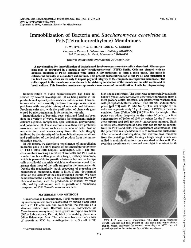
00:X; 0 000 50000:d :0:00000:j0000000;0 7:0; 0100yH 0 2pH FFIG. 2. Scanning electron micrographs of freshly constructed microporous yeast membranes. (A) Cut yeast membrane (original
magnification, x2,000). (B) Oblique view of yeast membrane (original magnification, x2,000). (C) Micrograph of edge of yeast membraneshowing Teflon fibrils (original magnification, x2,000). (D) Fivefold magnification of panel C.
which had been diluted 1:10 with water. The membrane wascut into the appropriate size and shape for the experimentson cell viability.
Cell viability in PTFE membranes. To determine thatmicroorganisms survived the milling-calendering process,experiments were performed to evaluate growth and bio-chemical activity in membranes. To determine that microor-ganisms were indeed viable, a small piece of the bacterial oryeast membrane was excised from the master sheet andplaced onto the surface of tryptic soy agar (Difco) plates.The plates were placed into a 30°C incubator for 72 h toevaluate growth of the organisms. The cut membrane wouldallow any viable microorganisms to escape from the PTFEmatrix and grow out onto the solid medium. The bacterialmembranes were placed at 30°C because Serratia spp.produce a bright red pigment at this temperature.
Biological activity of microorganism-ifiled PTFE mem-branes. Experiments were performed to determine the bio-chemical characteristics of the yeast membrane on the basisof conversion of glucose to ethanol by the entrapped yeastcells. Membranes were cut into a circular shape of 44.2 cm2
and placed into the flow cell of an Amicon (Danvers, Mass.)402 model 3155 ultrafiltration device. A glucose solution (1%[wt/vol] in water) was passed through the yeast membrane atvarious flow rates. The ethanol present in the filtrate wasdetected by gas chromatography with a Hewlett Packard5880 gas chromatograph. The membranes evaluated forbiological functionality were all of the same size.
P. aeruginosa was evaluated for its ability to remove apesticide, 2,4,5-trichlorophenoxyacetic acid (2,4,5-T), fromwater. The membrane was permeated with tryptic soy brothfor 72 h and then with 420 ml of distilled water. The reservoirabove the membrane was charged with a solution of 2,4,5-T(21 ,ug/ml in water). The pesticide was passed through themembrane at a flow rate of 8 ml/h. The amount of 2,4,5-Tremaining in the water sample was determined by gaschromatography.
Visualization of microorganisms in PTFE membranes.Electron microscopy of the PTFE membrane containing S.cerevisiae was performed with a freeze-fractured membrane.The membrane was frozen in liquid nitrogen and broken.The membrane was sputter coated with uranyl acetate and
APPL. ENVIRON. MICROBIOL.
Dow
nloa
ded
from
http
s://j
ourn
als.
asm
.org
/jour
nal/a
em o
n 03
Jan
uary
202
2 by
128
.22.
175.
1.
BACTERIUM AND YEAST IMMOBILIZATION 221
A B
C
/
D
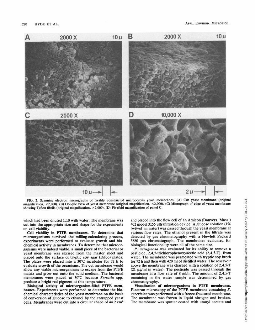
FIG. 3. Scanning electron micrographs of the yeast-filled microporous membrane after 1 month of use. The membrane was prepared asdescribed in the text for scanning electron microscopy. (A) Yeast cells enmeshed in the membrane. (B) Cross section of freeze-fracturedmembrane. The large particles are 20-pum-diameter polystyrene beads picked up from the mill, where they had been deposited by previousexperiments. (C) x 10 magnification of panel B. (D) x 3.3 magnification of panel C. Scale is denoted on each plate.
placed into the electron path of a Jeol 840 scanning electronmicroscope. Images at a magnification of x500 were ex-
posed on Polaroid type 55 film.
RESULTS
The PTFE-microorganism matrix formed easily on a stan-dard rubber mill when a work-intensive calendering proce-
dure was used. The permeability of the membrane was
determined by the expression log F = 1.52 - 0.280V, whereF is the flow rate in milliliters per minute and V is the totalvolume of substrate solution that has passed through themembrane (2, 3). This relationship was established by usingwater to permeate the membrane.When the water was replaced by a nutrient solution such
as tryptic soy broth, the flow rate decreased logarithmicallyaccording to the expression log F = 0.176 - 0.213 (V - 1.2),because of the faster rate of accumulation of particulatematerial (now in gel form) on the proximal side of themembrane. When this gel was removed from the membrane,the flow rate returned to that exhibited at V = 1.2 liters (i.e.,just prior to the change from water to nutrient broth).
Membranes containing 85% (wt/wt) S. marcescens placed
on the surface of a tryptic soy agar plate were incubated at30°C. No color change was noted after 36 h at this temper-ature. A spreading red area was observed on the membraneafter 48 h at 30°C (Fig. 1). The color spread until the entiremembrane was red (approximately 6 days). The slow growthof the organism is likely attributable to the recovery periodnecessary for the organisms after the milling process and tothe slow diffusion of nutrients from the agar into the mem-brane. Several red colonies were visible on the surface of themembrane after 48 h, the progeny of organisms situated atthe surface of the membrane.Membranes containing 85% (wt/wt) S. cerevisiae cells
were placed onto tryptic soy agar plates and incubated at30°C. Series of electron micrographs of the membrane im-mediately after construction (Fig. 2) and after 1 month of use(Fig. 3) are shown. The fibrils noted in the photographs arethe PTFE fibrils, and intact yeast cells are abundant in thearea. After 24 h at this temperature, yeast cells weredetected spreading away from the cut edge of the membrane,and colonylike growth (small areas of butter-texturedgrowth) was observed on the surface of the membrane. Thisindicated that the yeast cells had survived the milling pro-cess, as had the bacteria.
VOL. 57, 1991
Dow
nloa
ded
from
http
s://j
ourn
als.
asm
.org
/jour
nal/a
em o
n 03
Jan
uary
202
2 by
128
.22.
175.
1.
APPL. ENVIRON. MICROBIOL.
gooi
700
X 6Q0500
ti 4002 300j 200
0 10 20 30 40 50 60Residence fime (min.)
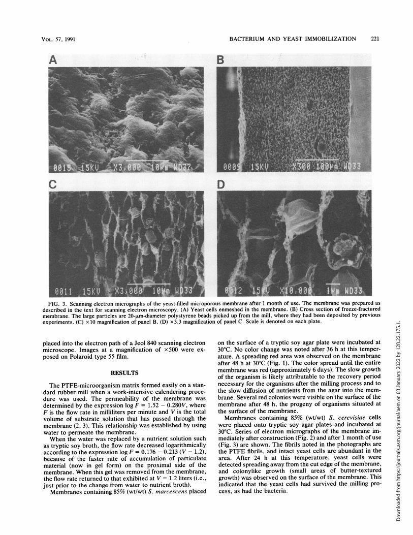
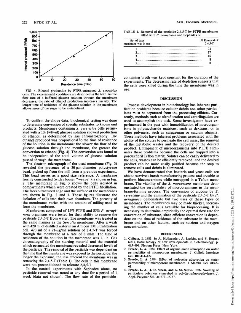
FIG. 4. Ethanol production by PTFE-entrapped S. cerevisiaecells. The experimental conditions are described in the text. As theflow rate of a buffered glucose solution through the membranedecreases, the rate of ethanol production increases linearly. Thelonger time of residence of the glucose solution in the membraneallows more of the sugar to be metabolized.
To confirm the above data, biochemical testing was doneto determine conversion of specific substrates to known endproducts. Membranes containing S. cerevisiae cells perme-ated with a 1% (wt/vol) glucose solution showed productionof ethanol, as determined by gas chromatography. Theethanpl produced was proportional to the time of residenceof the solution in the membrane: the slower the flow of theglucose solution through the membrane, the greater theconversion to ethanol (Fig. 4). The conversion was found tobe independent of the total volume of glucose solutionpassed through the membrane.The electron micrograph of the used membrane (Fig. 3)
revealed the presence of a 20-,um-diameter polystyrenebead, picked up from the mill from a previous experiment.This bead serves as a good size reference. A membranefreshly constructed (with a looser "weave") is shown in Fig.2. The membrane in Fig. 3 shows the cells isolated incompartments which were created by the PTFE fibrillation.The freeze-fractured edge and the surface of the membranesare shown in Fig. 2 and 3. These figures illustrate theisolation of cells into their own chambers. The porosity ofthe membranes varies with the amount of milling used toform the membrane.Membranes composed of 15% PTFE and 85% P. aerugi-
nosa organisms were tested for their ability to remove thepesticide 2,4,5-T from water. The membrane was treated inthe same manner as the Serratia membrane. After a washwith 420 ml of distilled water in an Amicon TM ultrafiltrationcell, 420 ml of a 21-,uglml solution of 2,4,5-T was forcedthrough the membranie at a rate of 8 ml/h. The time ofresidence of the solution in the membrane was 1.1 h. Gaschromatography of the starting material and the materialwhich permeated the membrane revealed decreased levels ofthe pesticide. The removal of the pesticide was dependent onthe time that the membrane was exposed to the pesticide: thelonger the exposure, the less efficient the membrane was inremoving the 2,4,5-T (Table 1). The cells in this membranewere not preconditioned to tolerate 2,4,5-T.
In the control experiments with Sephadex alone, nopesticide removal was noted at any time for a period of 1week (data not shown). The flow rate of the pesticide-
TABLE 1. Removal of the pesticide 2,4,5-T by PTFE membranesfilled with P. aeruginosa and Sephadex R
No. of days % Removal ofmembrane was in use 2,4,5-T
1....................................... 232.................................... 17
3....................................... 16
4 .. . . . . .. . . . . ...................................... 8
5 ..................................... . . .. . . . .. . . .
containing broth was kept constant for the duration of theexperiments. The decreasing rate of depletion suggests thatthe cells were killed during the time the membrane was inuse.
DISCUSSION
Process development in biotechnology has inherent puri-fication problems because cellular debris and other particu-lates must be separated from the processing effluent. Cur-rently, methods such as ultrafiltration and centrifugation areused to accomplish this task. Some investigators have ex-perimented in the past with immobilization of microorgan-isms in polysaccharide matrices, such as dextrans, or inother polymers, such as carageenan or calcium alginate.These methods have inherent problems associated with theability of the solutes to permeate the cell mass, the removalof the metabolic wastes and the recovery of the desiredproduct. Entrapment of microorganisms into PTFE elimi-nates these problems because the cells are trapped into aporous fibril Teflon matrix. Solutes can be easily delivered tothe cells, wastes can be efficiently removed, and the desiredproduct can be more easily purified because the step toremove cells and debris is eliminated.We have demonstrated that bacteria and yeast cells are
able to survive a harsh manufacturing process and are able toperform bioconversions while entrapped in a Teflon mem-brane. The viability of the S. marcescens membrane dem-onstrated the survivability of microorganisms in the mem-brane-forming process. The conversion of glucose by S.cerevisiae and the elimination of the pesticide 2,4,5-T by P.aeruginosa demonstrate but two uses of these types ofmembranes. The membranes may be made thicker, increas-ing the number of cells available for bioprocessing. It isnecessary to determine empirically the optimal flow rate forconversion of substrate, since efficient conversion is depen-dent on the time of residence of the substrate in the mem-brane and on other factors, such as nutrient and oxygenconcentrations.
REFERENCES1. Chibata, I. 1983. In A. Hollaender, A. Laskin, and P. Rogers
(ed.), Basic biology of new developments in biotechnology, p.465-496. Plenum Press, New York.
2. Errede, L. A. 1984. Effect of organic anion adsorption on waterpermeability of microporous membranes. J. Colloid InterfaceSci. 100:414-422.
3. Errede, L. A. 1984. Effect of molecular adsorption on waterpermeability of microporous membranes. J. Membr. Sci. 20:45-61.
4. Errede, L. A., J. D. Stoesz, and L. M. Sirvio. 1986. Swelling ofparticulate polymers enmeshed in poly(tetrafluoroethylene). J.Appl. Polymer Sci. 31:2721-2737.
222 HYDE ET AL.
Dow
nloa
ded
from
http
s://j
ourn
als.
asm
.org
/jour
nal/a
em o
n 03
Jan
uary
202
2 by
128
.22.
175.
1.