Embed Size (px)
Citation preview
Lecture 2
Dr. Dalia Mohsen Associate prof. of microbiology
Dr. Dalia Mohsen Associate prof. of microbiology
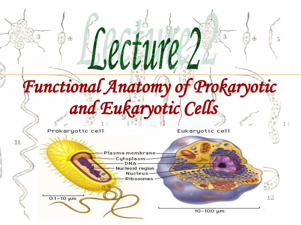
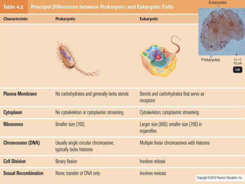
Functional Anatomy of Prokaryotic and Eukaryotic Cells

Dr. Dalia Mohsen Associate prof. of microbiology
All living cells are classified into Prokaryotic and Eukaryotic Cells, based on their structural and functional characteristics.
Prokaryote comes from the Greek words for pre-nucleus.
Eukaryote comes from the Greek words for true nucleus.

Dr. Dalia Mohsen Associate prof. of microbiology
• Archaea
• BacteriaPROKARYOTIC
CELLS
• Fungi
• protozoa,
• algae,
• plants
• animals
EUKARYOTIC CELLS

Important
• Viruses -Non-cellular elements that do not fit
into any organizational scheme of living cells.
(will be discussed later)
Dr. Dalia Mohsen Associate prof. of microbiology

Dr. Dalia Mohsen Associate prof. of microbiology
Dr. Dalia Mohsen Associate prof. of microbiology
Dr. Dalia Mohsen Associate prof. of microbiology
Dr. Dalia Mohsen Associate prof. of microbiology
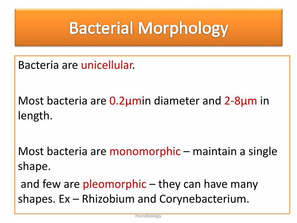
Bacteria are unicellular.
Most bacteria are 0.2µmin diameter and 2-8µm in length.
Most bacteria are monomorphic – maintain a single shape.
and few are pleomorphic – they can have many shapes. Ex – Rhizobium and Corynebacterium.
Dr. Dalia Mohsen Associate prof. of microbiology
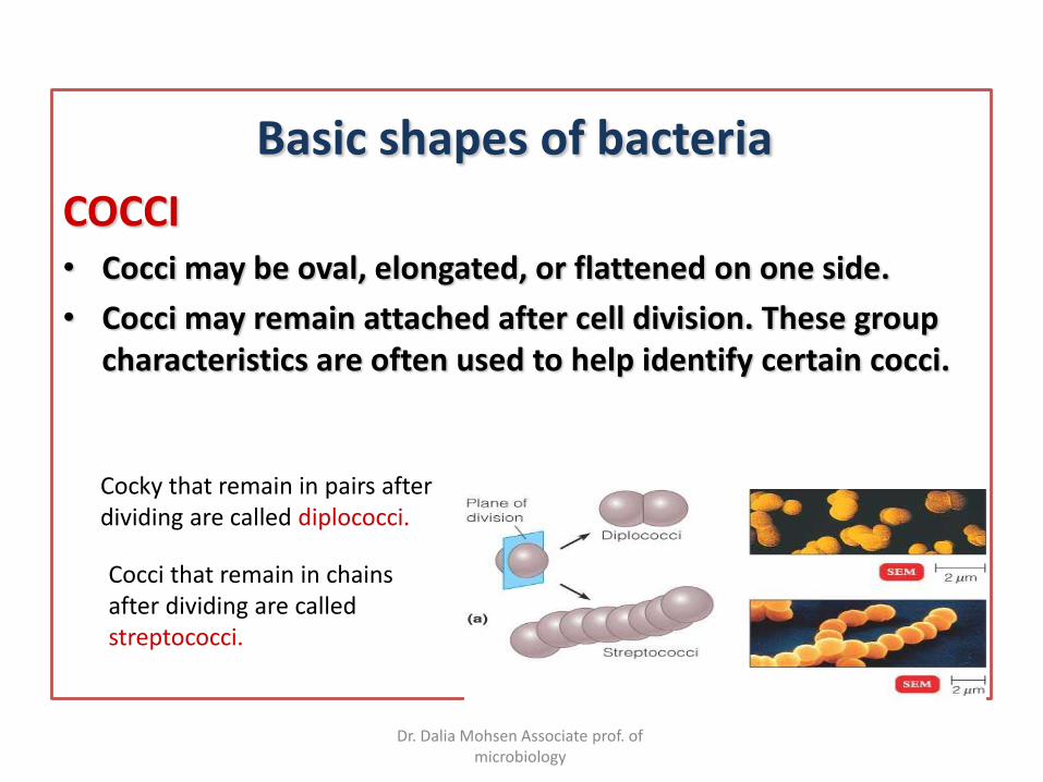
Basic shapes of bacteria
COCCI• Cocci may be oval, elongated, or flattened on one side.
• Cocci may remain attached after cell division. These group characteristics are often used to help identify certain cocci.
Cocky that remain in pairs after dividing are called diplococci.
Cocci that remain in chains after dividing are called streptococci.
Dr. Dalia Mohsen Associate prof. of microbiology
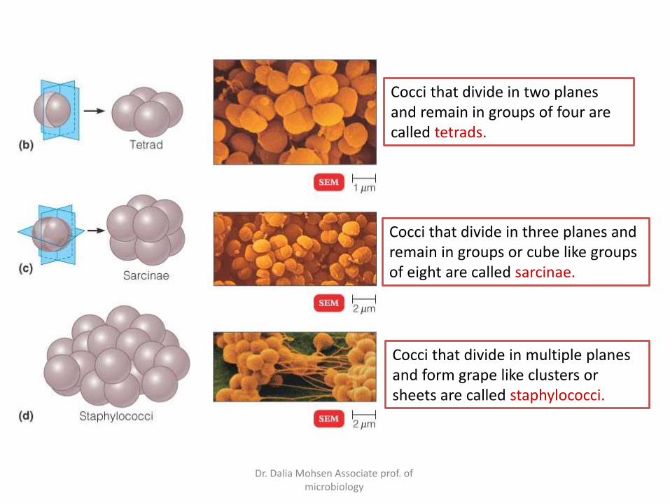
Cocci that divide in two planes and remain in groups of four are called tetrads.
Cocci that divide in three planes and remain in groups or cube like groups of eight are called sarcinae.
Cocci that divide in multiple planes and form grape like clusters or sheets are called staphylococci.
Dr. Dalia Mohsen Associate prof. of microbiology
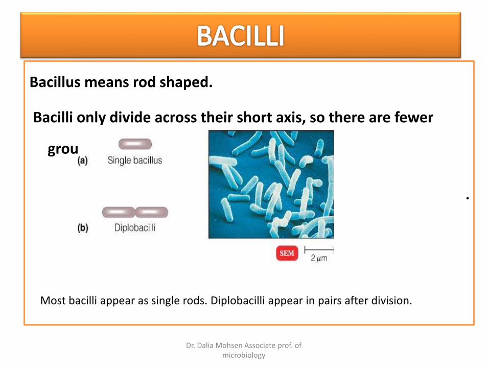
Bacillus means rod shaped.
Bacilli only divide across their short axis, so there are fewer
groups.
•
Most bacilli appear as single rods. Diplobacilli appear in pairs after division.
Dr. Dalia Mohsen Associate prof. of microbiology
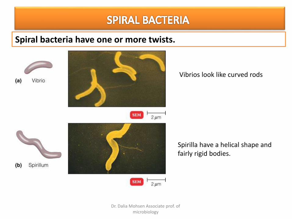
Spiral bacteria have one or more twists.
Vibrios look like curved rods
Spirilla have a helical shape and fairly rigid bodies.
Dr. Dalia Mohsen Associate prof. of microbiology
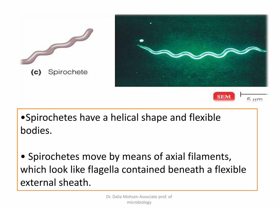
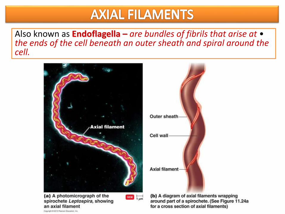
•Spirochetes have a helical shape and flexible bodies.
• Spirochetes move by means of axial filaments, which look like flagella contained beneath a flexible external sheath.
Dr. Dalia Mohsen Associate prof. of microbiology
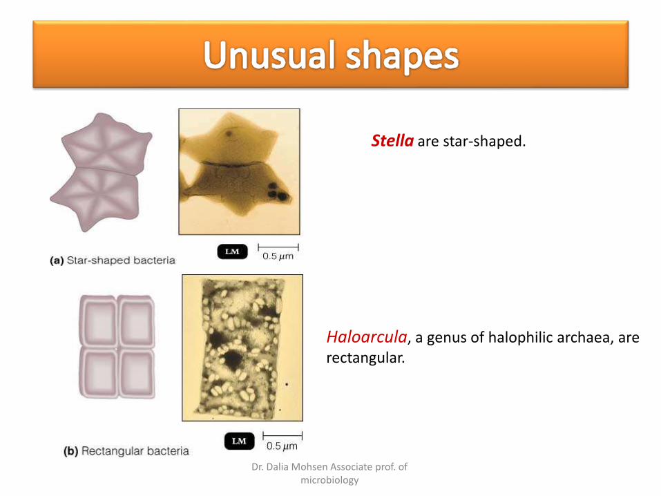
Stella are star-shaped.
Haloarcula, a genus of halophilic archaea, are rectangular.
Dr. Dalia Mohsen Associate prof. of microbiology
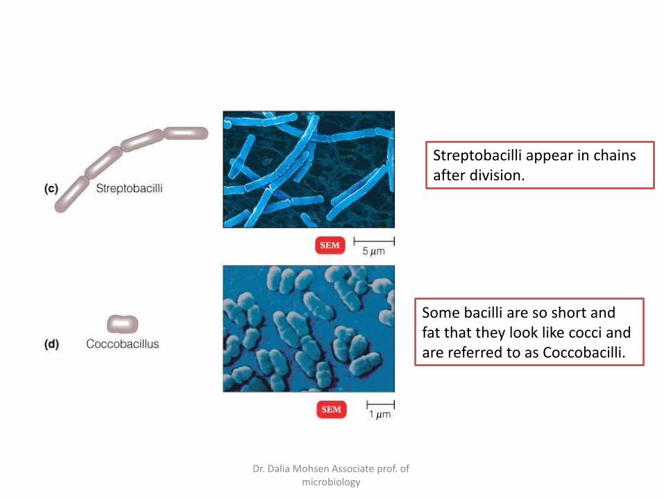
Streptobacilli appear in chains after division.
Some bacilli are so short and fat that they look like cocci and are referred to as Coccobacilli.

Dr. Dalia Mohsen Associate prof. of microbiology
movement
attachment
virulent factor
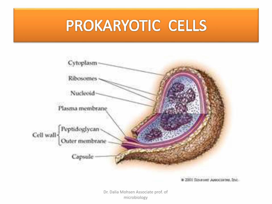
STRUCTURE OF A PROKARYOTIC CELL
Dr. Dalia Mohsen Associate prof. of microbiology
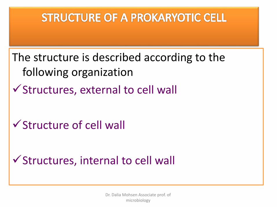
The structure is described according to the following organization
Structures, external to cell wall
Structure of cell wall
Structures, internal to cell wall
Dr. Dalia Mohsen Associate prof. of microbiology

Dr. Dalia Mohsen Associate prof. of microbiology
Glycocalyx
Flagella
Axial filaments
Fimbriae
Pili
Dr. Dalia Mohsen Associate prof. of microbiology
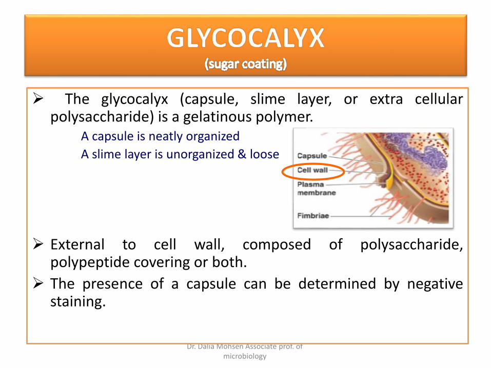
The glycocalyx (capsule, slime layer, or extra cellularpolysaccharide) is a gelatinous polymer.
A capsule is neatly organized
A slime layer is unorganized & loose
External to cell wall, composed of polysaccharide,polypeptide covering or both.
The presence of a capsule can be determined by negativestaining.
Dr. Dalia Mohsen Associate prof. of microbiology
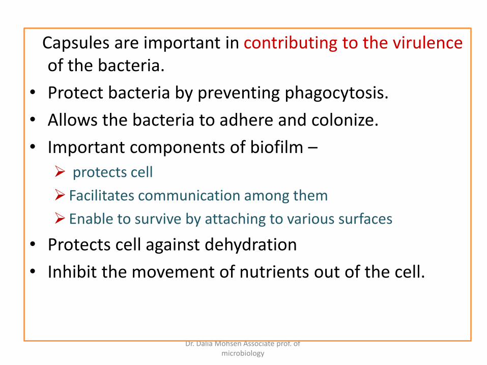
Capsules are important in contributing to the virulenceof the bacteria.
• Protect bacteria by preventing phagocytosis.
• Allows the bacteria to adhere and colonize.
• Important components of biofilm –
protects cell
Facilitates communication among them
Enable to survive by attaching to various surfaces
• Protects cell against dehydration
• Inhibit the movement of nutrients out of the cell.

Dr. Dalia Mohsen Associate prof. of microbiology
Capsulated bacteria –
• Streptococcus pneumoniae
• Klebsiella pneumoniae
• Haemophilus influenzae
• Bacillus anthracis
• Streptococcus mutans
• Yersinia pestis

Dr. Dalia Mohsen Associate prof. of microbiology
Streptococcus pneumoniae (in vivo)

Dr. Dalia Mohsen Associate prof. of microbiology
K. pneumoniae Haemophilus influenzae
Dr. Dalia Mohsen Associate prof. of microbiology
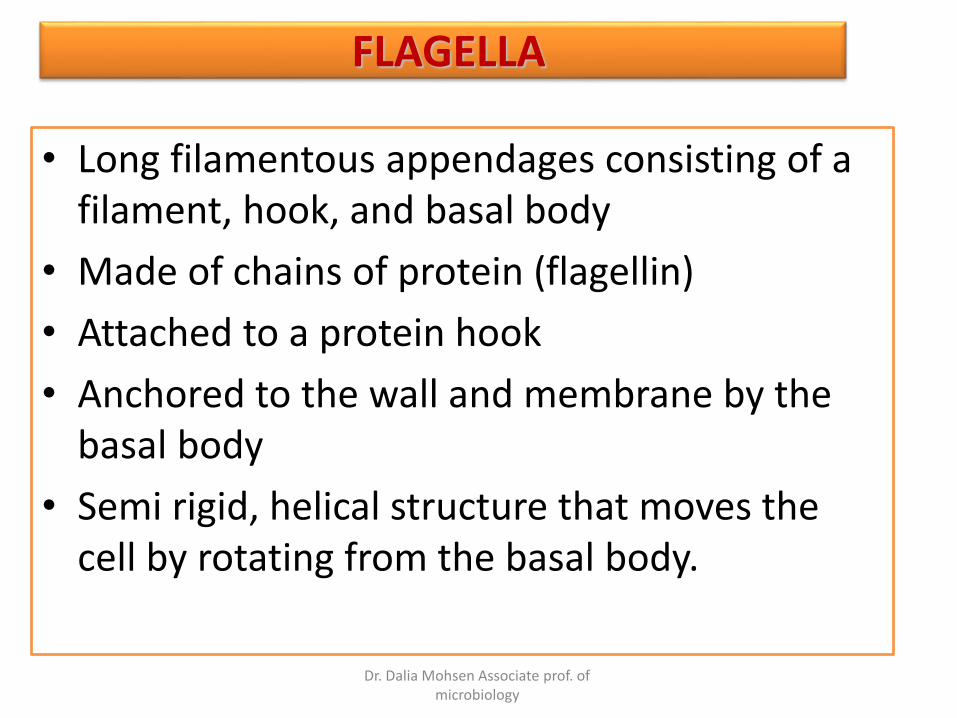
• Long filamentous appendages consisting of a filament, hook, and basal body
• Made of chains of protein (flagellin)
• Attached to a protein hook
• Anchored to the wall and membrane by the basal body
• Semi rigid, helical structure that moves the cell by rotating from the basal body.
FLAGELLA

Dr. Dalia Mohsen Associate prof. of microbiology
Flagella are anchored by pairs of rings associated with the plasma membrane and cell wall. Gram positive bacteria have only the inner pair of rings

Dr. Dalia Mohsen Associate prof. of microbiology
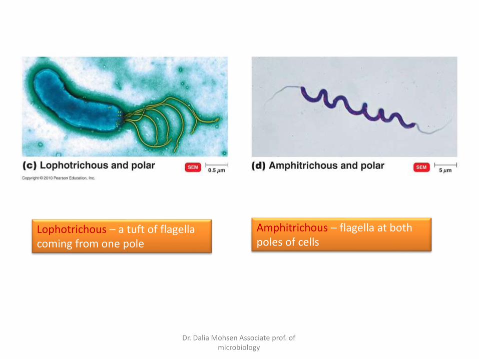
Flagella Arrangement
Peritrichous – distributed over the entire cell
Monotrichous – single flagellum at one pole
Dr. Dalia Mohsen Associate prof. of microbiology
Lophotrichous – a tuft of flagella coming from one pole
Amphitrichous – flagella at both poles of cells
Dr. Dalia Mohsen Associate prof. of microbiology
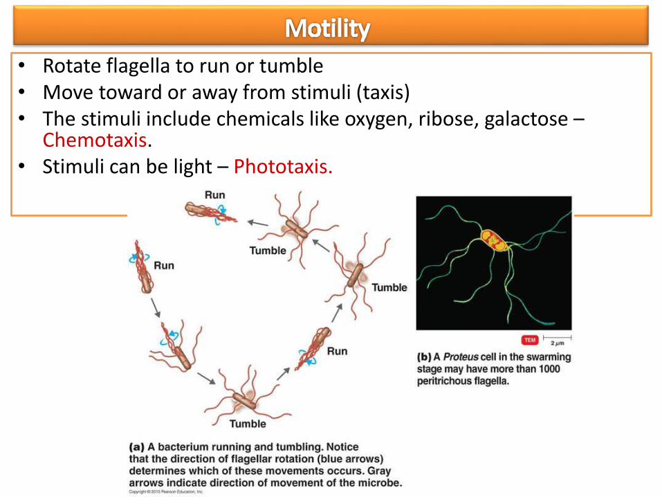
• Rotate flagella to run or tumble• Move toward or away from stimuli (taxis)• The stimuli include chemicals like oxygen, ribose, galactose –
Chemotaxis.• Stimuli can be light – Phototaxis.
Dr. Dalia Mohsen Associate prof. of microbiology
• Flagellar (H) protein functions as an antigen.
• Flagella proteins are H - antigens - useful in distinguishing the variants within the species of gram-negative bacteria.
• Example – 50 different H antigen for E. coli are identified.
• E. coli O157:H7 – associated with food borne epidemics.
Dr. Dalia Mohsen Associate prof. of microbiology
•Also known as Endoflagella – are bundles of fibrils that arise at the ends of the cell beneath an outer sheath and spiral around the cell.
Dr. Dalia Mohsen Associate prof. of microbiology
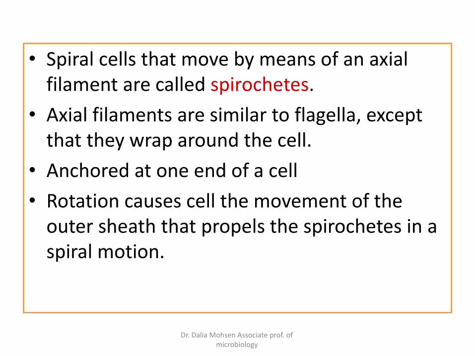
• Spiral cells that move by means of an axial filament are called spirochetes.
• Axial filaments are similar to flagella, except that they wrap around the cell.
• Anchored at one end of a cell
• Rotation causes cell the movement of the outer sheath that propels the spirochetes in a spiral motion.
Dr. Dalia Mohsen Associate prof. of microbiology
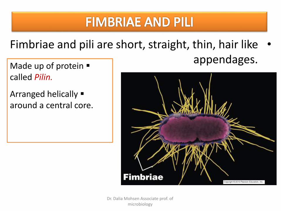
•Fimbriae and pili are short, straight, thin, hair like appendages.
Made up of protein called Pilin.
Arranged helically around a central core.

Dr. Dalia Mohsen Associate prof. of microbiology
• Fimbriae –
– Occur at poles or evenly distributed.
– Number can vary from few to several hundreds
– Allow attachment to surfaces and adhere to each other
• Pili –
– Longer than Fimbriae
– Only one or two per cell
– are used to transfer DNA from one cell to another byConjugation – (sex Pili).
– Involved in motility called twitching motility – short jerkyintermittent movements, seen in Neisseria gonorrhoeae.
– Other type of motility is gliding motility – smooth glidingmovement of mycobacterium.

Dr. Dalia Mohsen Associate prof. of microbiology
Is a complex, semi rigid structure responsible for the shape of the cell.
Surrounds the underlying, fragile plasma membrane.
Dr. Dalia Mohsen Associate prof. of microbiology
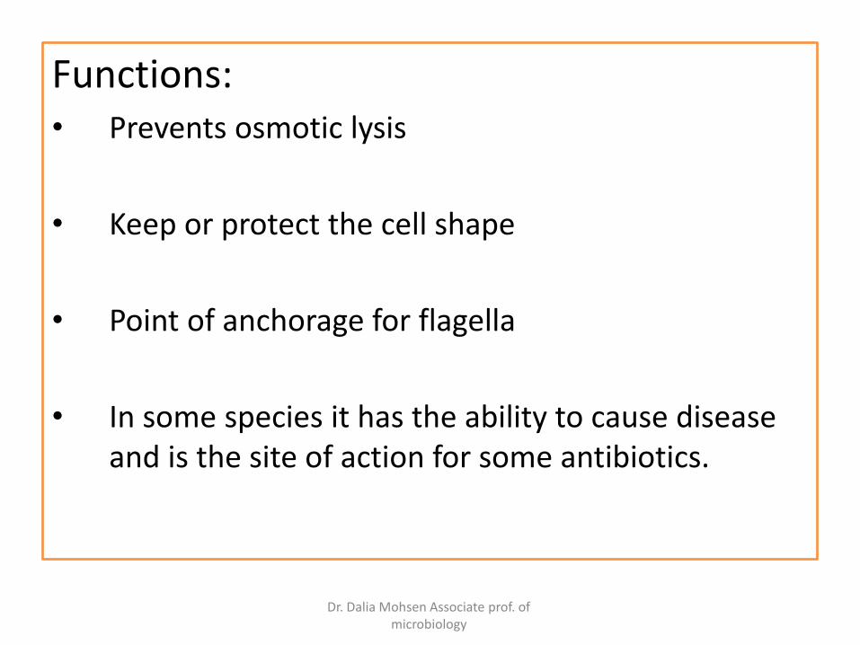
Functions:• Prevents osmotic lysis
• Keep or protect the cell shape
• Point of anchorage for flagella
• In some species it has the ability to cause disease and is the site of action for some antibiotics.

Dr. Dalia Mohsen Associate prof. of microbiology
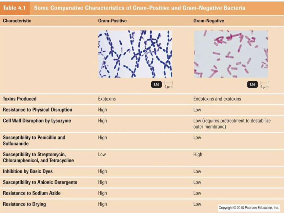
Composition and Characteristics
• PEPTIDOGLYCAN - Main component of bacterial cell wall (also known as murein) - a polymer consisting of disaccharideN-acetyl glucoseamine (NAG) & N-acetyl muramic acid (NAM)linked by polypeptides chains.
N-acetyl glucosamine (NAG) and N-acetyl muramic acid (NAM) joined as in peptidoglycan

Dr. Dalia Mohsen Associate prof. of microbiology
• Alternating NAM and NAG molecules form a carbohydrate backbone (the glycanportion).
• Rows of NAG and NAM are linked by polypeptides (the peptido - portion).
•The structure of the polypeptide cross-bridges may vary but they always have atetra peptide side chain, which consists of 4 amino acids attached to NAMs. Theamino acids occur in alternating D and L forms.
Dr. Dalia Mohsen Associate prof. of microbiology
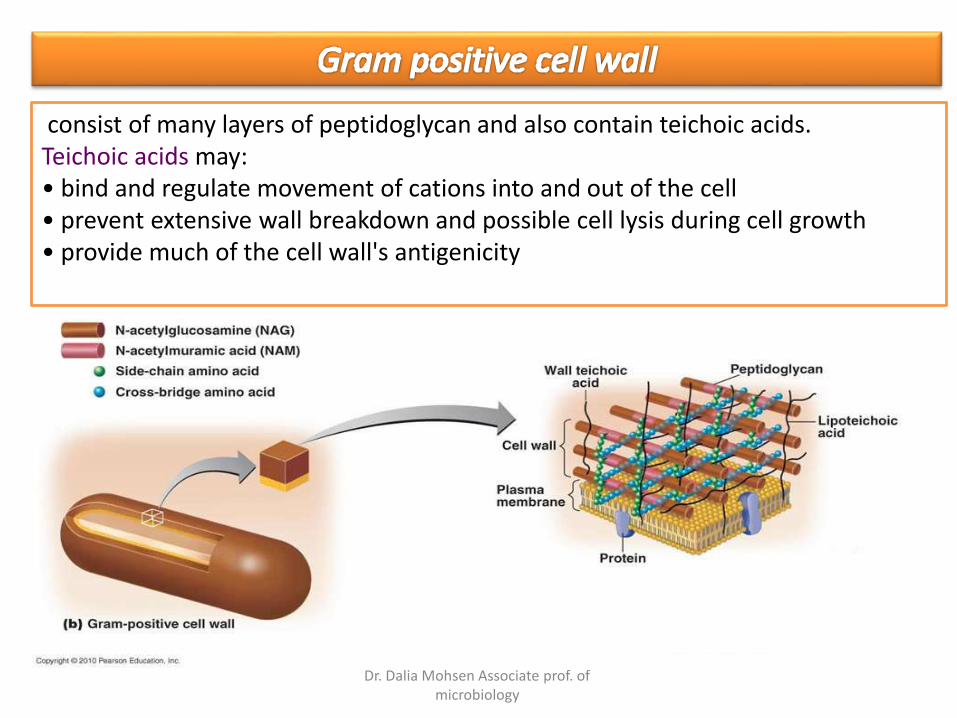
consist of many layers of peptidoglycan and also contain teichoic acids.Teichoic acids may:• bind and regulate movement of cations into and out of the cell• prevent extensive wall breakdown and possible cell lysis during cell growth• provide much of the cell wall's antigenicity
Dr. Dalia Mohsen Associate prof. of microbiology
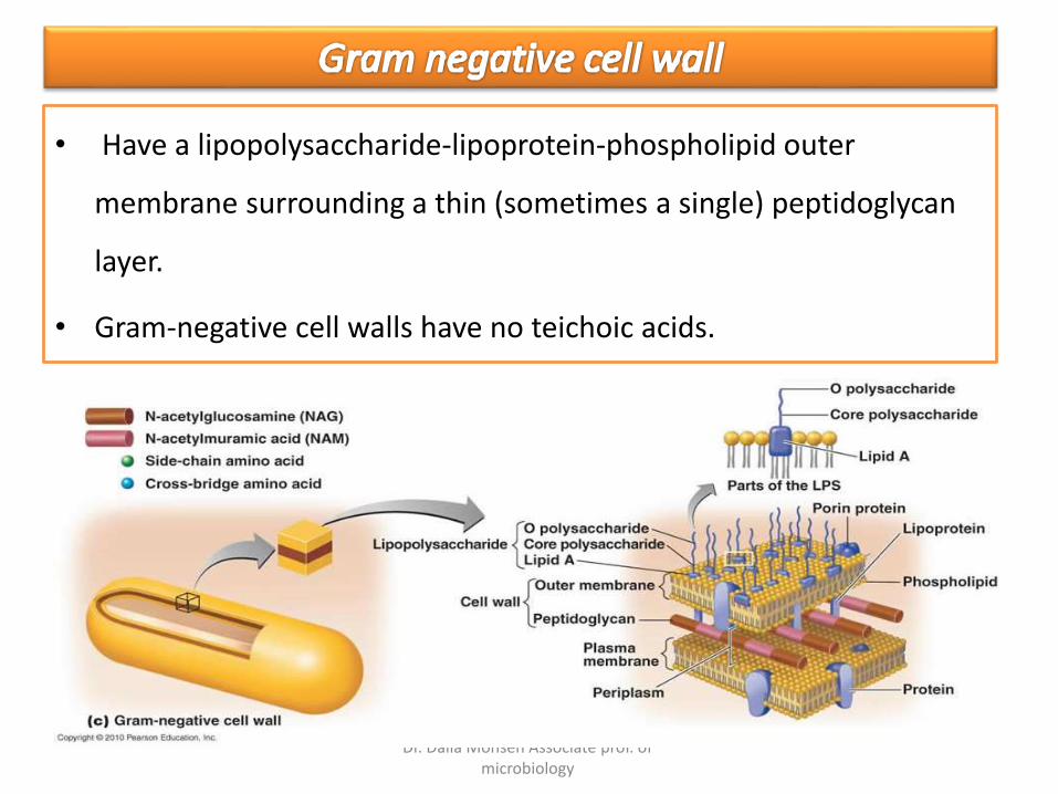
• Have a lipopolysaccharide-lipoprotein-phospholipid outer
membrane surrounding a thin (sometimes a single) peptidoglycan
layer.
• Gram-negative cell walls have no teichoic acids.
Dr. Dalia Mohsen Associate prof. of microbiology
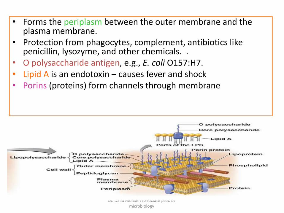
• Forms the periplasm between the outer membrane and the plasma membrane.
• Protection from phagocytes, complement, antibiotics like penicillin, lysozyme, and other chemicals. .
• O polysaccharide antigen, e.g., E. coli O157:H7.• Lipid A is an endotoxin – causes fever and shock• Porins (proteins) form channels through membrane

Dr. Dalia Mohsen Associate prof. of microbiology
Dr. Dalia Mohsen Associate prof. of microbiology
Dr. Dalia Mohsen Associate prof. of microbiology
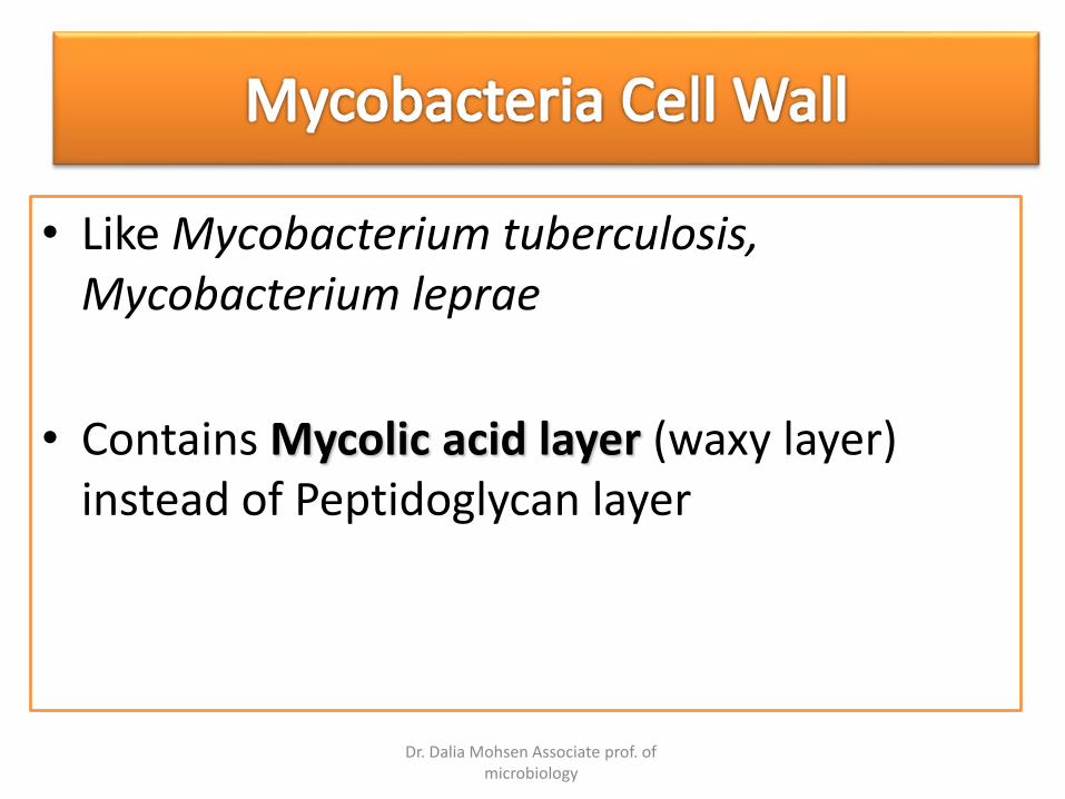
• Like Mycobacterium tuberculosis, Mycobacterium leprae
• Contains Mycolic acid layer (waxy layer) instead of Peptidoglycan layer
Dr. Dalia Mohsen Associate prof. of microbiology
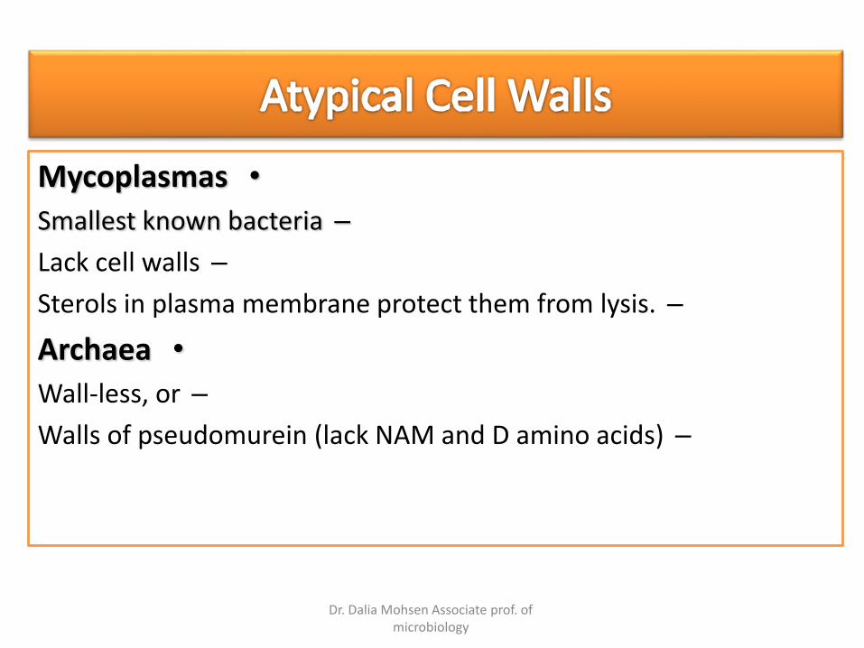
•Mycoplasmas
–Smallest known bacteria
–Lack cell walls
–Sterols in plasma membrane protect them from lysis.
•Archaea
–Wall-less, or
–Walls of pseudomurein (lack NAM and D amino acids)
Dr. Dalia Mohsen Associate prof. of microbiology
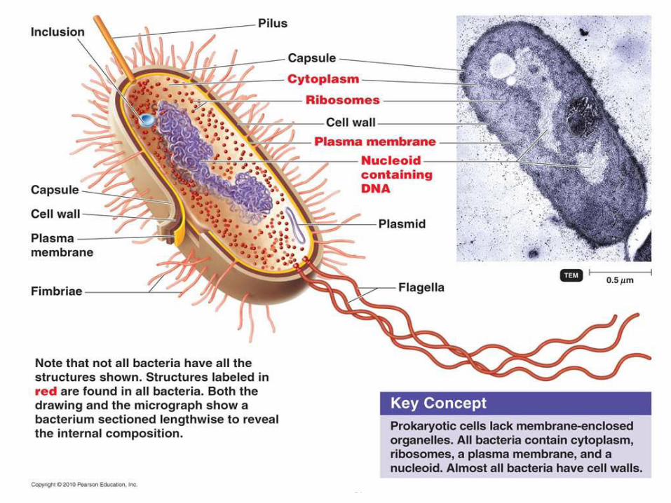
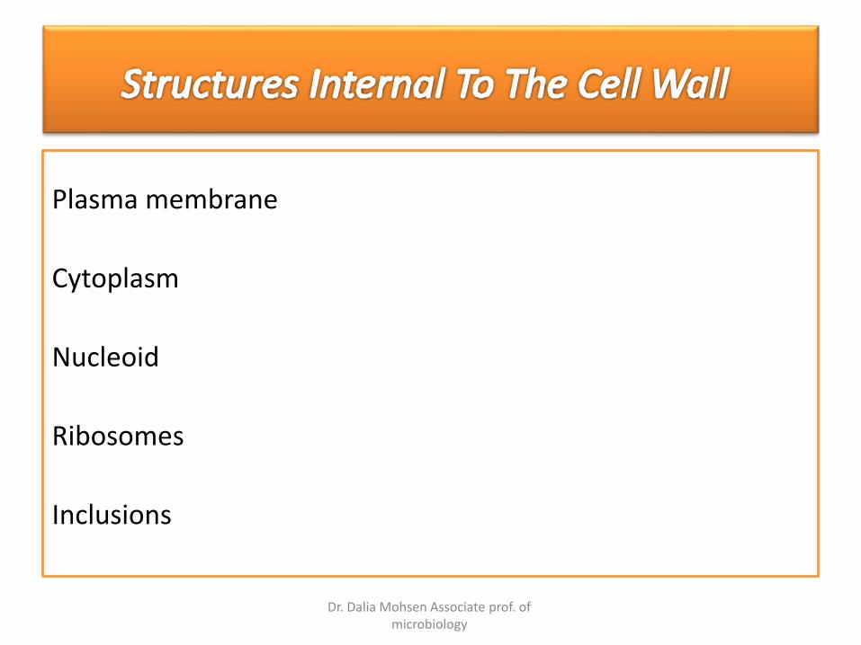
Plasma membrane
Cytoplasm
Nucleoid
Ribosomes
Inclusions
Dr. Dalia Mohsen Associate prof. of microbiology
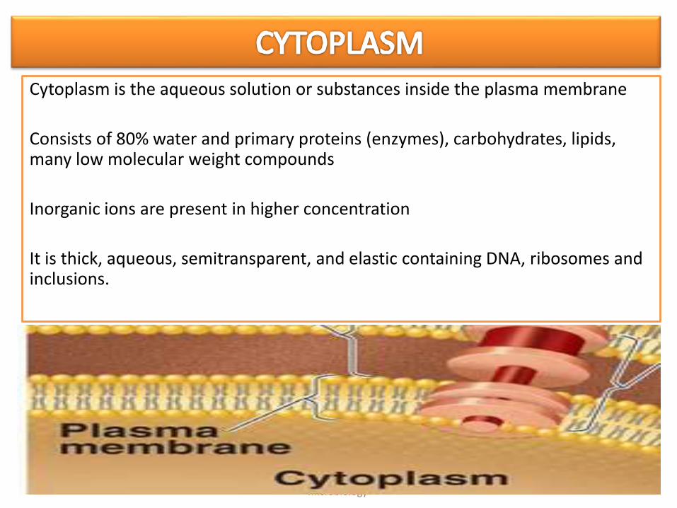
Cytoplasm is the aqueous solution or substances inside the plasma membrane
Consists of 80% water and primary proteins (enzymes), carbohydrates, lipids, many low molecular weight compounds
Inorganic ions are present in higher concentration
It is thick, aqueous, semitransparent, and elastic containing DNA, ribosomes and inclusions.

Dr. Dalia Mohsen Associate prof. of microbiology
Bacteria contains negative supercoiled single covalently closed circular chromosome (cccc) – single , long, continuous, and frequently circularly arranged thread of double stranded DNA called Bacterial Chromosome
Nuclear area (nucleoid), there is no nucleus
Bacteria can also contain plasmids, which are circular, extra-chromosomal
DNA molecules.

Dr. Dalia Mohsen Associate prof. of microbiology
The cytoplasm of a prokaryote contains numerous 70s ribosomes; ribosomes consist of rRNA and protein.
Protein synthesis occurs at ribosomes; it can be inhibited by certain antibiotics.
The difference between prokaryotic (70s) and eukaryotic (80s) ribosomes allows antibiotics to selectively target the prokaryotic ribosomes while sparing eukaryotic ribosomes.
Dr. Dalia Mohsen Associate prof. of microbiology
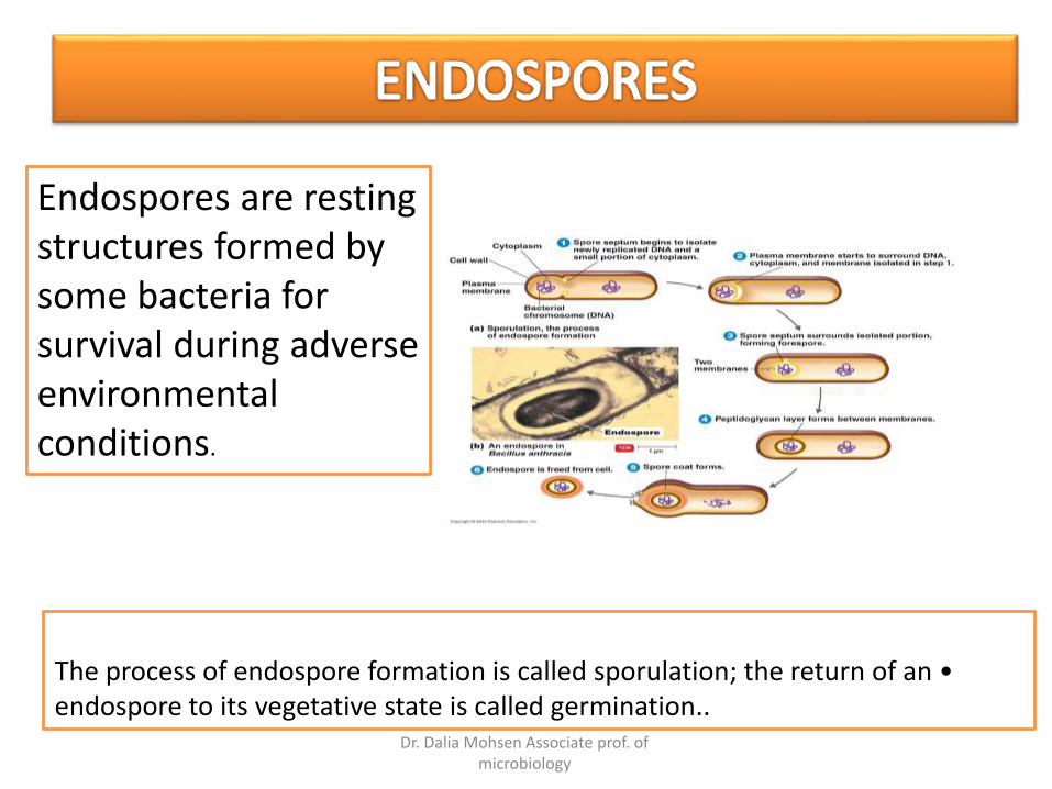
•The process of endospore formation is called sporulation; the return of an endospore to its vegetative state is called germination..
Endospores are resting structures formed by some bacteria for survival during adverse environmental conditions.
Dr. Dalia Mohsen Associate prof. of microbiology
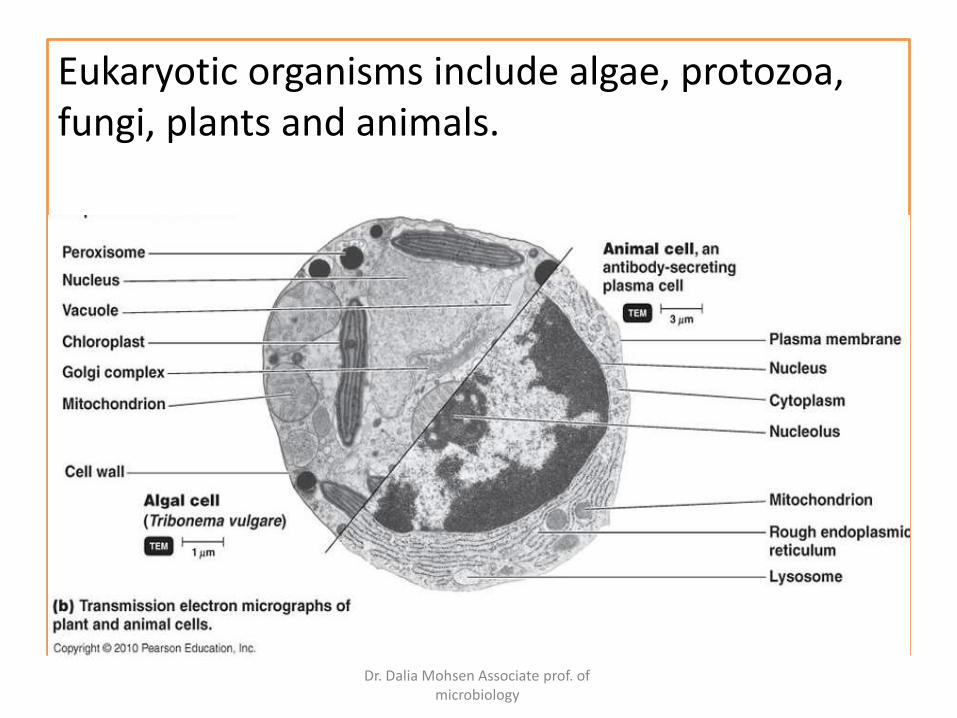
Eukaryotic organisms include algae, protozoa, fungi, plants and animals.
Dr. Dalia Mohsen Associate prof. of microbiology Figure 4.23a, b
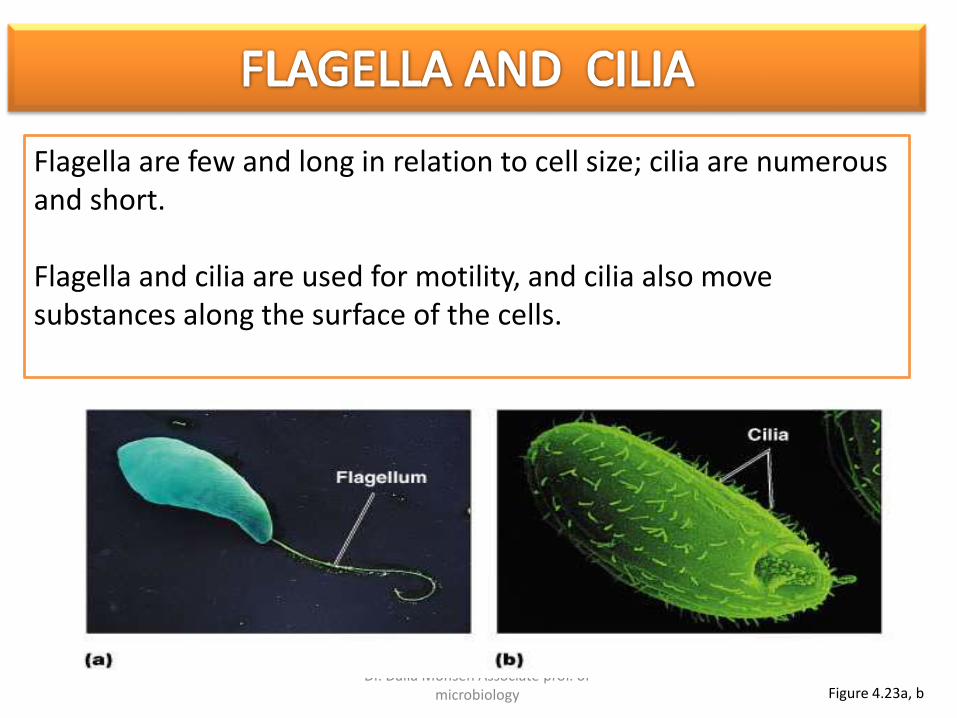
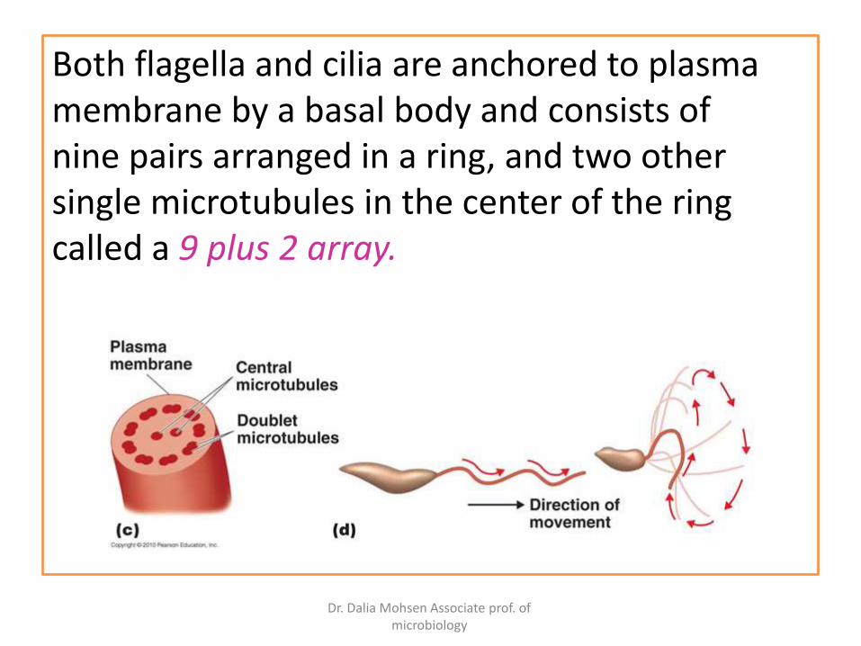
Flagella are few and long in relation to cell size; cilia are numerous and short.
Flagella and cilia are used for motility, and cilia also move substances along the surface of the cells.
Dr. Dalia Mohsen Associate prof. of microbiology
Both flagella and cilia are anchored to plasma membrane by a basal body and consists of nine pairs arranged in a ring, and two other single microtubules in the center of the ring called a 9 plus 2 array.
Dr. Dalia Mohsen Associate prof. of microbiology
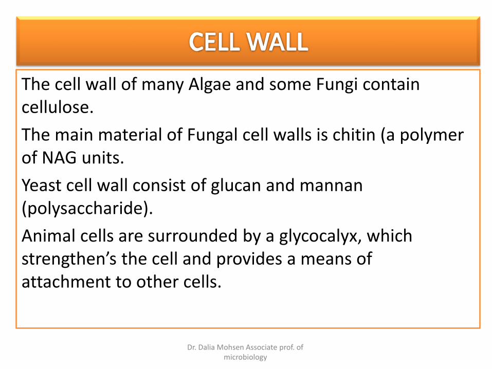
The cell wall of many Algae and some Fungi contain cellulose.
The main material of Fungal cell walls is chitin (a polymer of NAG units.
Yeast cell wall consist of glucan and mannan(polysaccharide).
Animal cells are surrounded by a glycocalyx, which strengthen’s the cell and provides a means of attachment to other cells.
Dr. Dalia Mohsen Associate prof. of microbiology
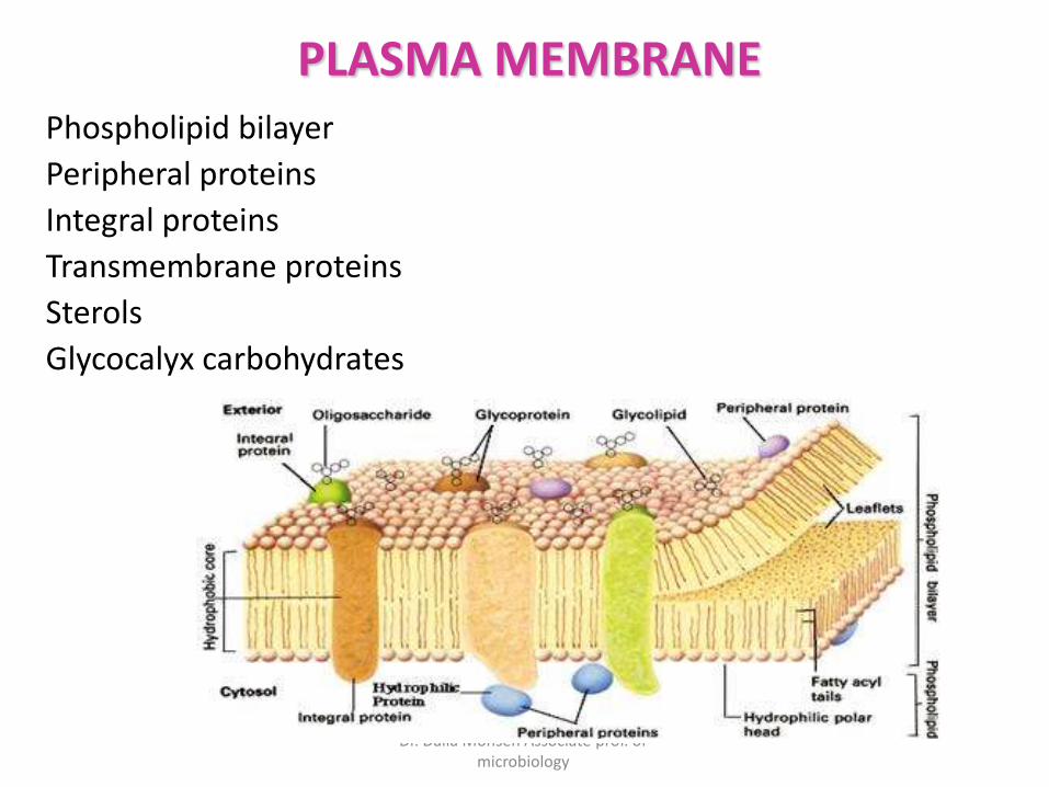
Phospholipid bilayer
Peripheral proteins
Integral proteins
Transmembrane proteins
Sterols
Glycocalyx carbohydrates
PLASMA MEMBRANE
Dr. Dalia Mohsen Associate prof. of microbiology
Cytoplasm – encompasses substance inside the plasma membrane and outside the cell
Cytosol - Fluid portion of cytoplasm
Cytoskeleton – provides support and shape and assists in transporting substances through the cell
Cytoplasmic streaming - Movement of cytoplasm throughout cells helps distribute nutrients and move
the cell over the surface