Embed Size (px)
Citation preview

Marine Microbes

Key Concepts
• Microbial life in the sea is extremely diverse, including members of all three domains of life as well as viruses.
• Marine virology is an emerging field of study, due to recognition of the critical role that viruses may play in population control of other microbes, in nutrient cycling, and in marine pathology.

Key Concepts
• Photosynthetic and chemosynthetic bacteria and archaeons are important primary producers in marine ecosystems.
• Heterotrophic bacteria, archaeons, and fungi play essential roles in recycling nutrients in the marine environment.
Key Concepts• Marine eukaryotic microbes are primary
producers, decomposers, and consumers, and some contribute significantly to the accumulation of deep-sea sediments.
• Populations of several kinds of photosynthetic marine microbes may form harmful blooms that affect other marine and maritime organisms directly and indirectly.


Marine Viruses
• Virology—the study of viruses
• Viruses are diverse and are more abundant than any other organism in the sea
• Have significance for marine food webs, population biology and diseases of marine organisms
• Viruses of marine eukaryotic hosts first reported in the 1970s
• Reliable counts of marine viruses made in the 1980s

Viral Characteristics• Most authorities do not consider them to be alive• Viruses consist of bits of DNA or RNA surrounded
by protein• Have no metabolism, and rely entirely on host
organism for energy, material and organelles to reproduce themselves
• Viral replication must occur within a host cell• Origin of viruses: two hypotheses
– highly reduced prokaryotic parasites– renegade genes
• Viruses infect all groups of living organisms, but may be specialized
Viral Characteristics
• Viral structure– virus particle is called a virion when outside
the host cell– virion composed of a nucleic acid core
surrounded by a coat of protein called a capsid (together called a nucleocapsid)
– may have a protective envelope, a membrane derived from the host’s nuclear or cell membrane

http://www.ncbi.nlm.nih.gov/ICTVdb/Images/Viroscoop2005_07minPoster.jpg

Viral Characteristics
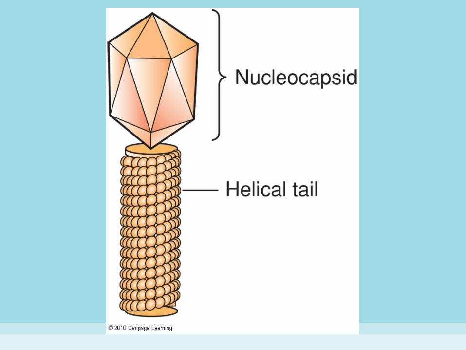
• Viral structure (con’t)– viral shapes:
• icosahedral viruses—capsid with 20 triangular faces composed of protein subunits
• helical viruses—protein subunits of the capsid spiral around the central core of nucleic acid
• binal viruses—those with icosahedral heads and helical tails
– some virions have filaments and other parts used to attach to and infect the host cell

Viral Characteristics
• Viral life cycles– lytic cycle—a rapid cycle of infection,
replication of viral nucleic acids and proteins, assembly of virions, and release of virions by lysis (rupture) of the cell
– lysogenic cycle—the viral nucleic acid is inserted into the host genome and may reside there through multiple cell divisions before becoming lytic


Lytic Cycle
Lysogenic Cycle
LysisInfection Replication
Stepped Art
Fig. 6-3, p. 128

Biodiversity and Distribution of Marine Viruses
• 10 times more abundant than marine prokaryotes, may reach 1010 virons per liter of seawater, 1013 per kilogram of sediment
• Estimated 100 to 10,000 genotypes• Most planktonic viruses are icosahdral or
binal bacteriophages (“bacteria eaters”) with lytic life cycles
• Sediment viruses are typically helical and lysogenic
Ecology of Marine Viruses• Viruses kill host cells, and thus control populations
of bacteria and other microbes in plankton communities
• Viruses also responsible for chronic infection and mass mortality of populations of marine animals
• Bacterial lysis can alter biogeochemical cycles and planktonic food webs
• Viral populations are probably controlled by several biotic and abiotic factors– e.g. alteration by light, adsorption onto suspended
particles, ingestion by microbes, failure to attach to appropriate host cell
Marine Bacteria
• General characteristics– simple, prokaryotic organization: no nuclei or
membrane-bound organelles, few genes, nonliving cell wall
– reproduce asexually by binary fission– many shapes and sizes
• bacillus—rod shape• coccus—spherical shape• Spirillum – cork screw shape
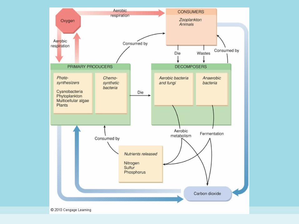
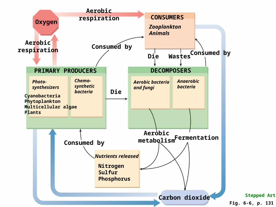
Carbon dioxide
Oxygen
Aerobicrespiration
Aerobicrespiration
PRIMARY PRODUCERS
Photo-synthesizers
CyanobacteriaPhytoplanktonMulticellular algaePlants
Chemo-syntheticbacteria
Consumed byConsumed by
CONSUMERS
ZooplanktonAnimals
Consumed by
Nutrients released
NitrogenSulfurPhosphorus
Aerobicmetabolism Fermentation
WastesDie
Die
Anaerobicbacteria
DECOMPOSERS
Aerobic bacteriaand fungi
Stepped Art
Fig. 6-6, p. 131
Nutritional Types
• Cyanobacteria (blue-green bacteria)– photosynthetic bacteria which are found in
environments high in dissolved oxygen, and produce free oxygen
– store excess photosynthetic products as cyanophycean starch and oils
– primary photosynthetic pigments are chlorophyll a and chlorophyll b
– accessory pigments include carotenoids and phycobilins


Light energy
Sulfate(SO4
2–)Carbohydrates
(CH2O)x
(a) Cyanobacteria – Free oxygen produced
Carbon dioxide(CO2)
Water(H2O)
Hydrogen sulfide(H2S)
Carbon dioxide(CO2)
Light energy
(b) Purple and green bacteria – No free oxygen produced
Oxygen(O2)
Carbohydrates(CH2O)x
Stepped Art
Fig. 6-8, p. 132

Nutritional Types (Cyanobacteria)
• Cyanobacteria (con’t)– chromatic adaptation—response of pigment
composition to the quality of light in the sea– may exist as single cells or form dense mats
held together by mucilage• form associates called stromatolites—a coral-like
mound of microbes that trap sediment and precipitate minerals in shallow tropical seas
Nutritional Types
• Other photosynthetic bacteria– anaerobic green and purple sulfur and non-
sulfur bacteria do not produce oxygen– the primary photosynthetic pigments are
bacteriochlorophylls – sulfur bacteria are obligate anaerobes
(tolerating no oxygen)– non-sulfur bacteria are facultative anaerobes
(respiring when in low oxygen or in the dark and photosynthesizing anaerobically when in the presence of light)

Nutritional Types• Chemosynthetic bacteria
– use energy derived from chemical reactions that involve substances such as ammonium ion, sulfides and elemental sulfur, nitrites, hydrogen, and ferrous ion
– chemosynthesis is less efficient than photosynthesis, so rates of cell growth and division are slower
– found around hydrothermal vents and some shallower habitats where needed materials are available in abundance


Carbondioxide (CO2)
Hydrogensulfide (H2S)
Magma (molten rock)
Water(H2O)
Carbohydrates
Produce
Elementalsulfur (S)
Carbondioxide (CO2)
Animalcommunity
Carbondioxide (CO2)
Hydrogensulfide (H2S)
Chemosynthetic bacteria (in animal tissues, in water, and on rocks)
Stepped Art
Fig. 6-10, p. 134
Nutritional Types
• Heterotrophic bacteria– decomposers that obtain energy and
materials from organic matter in their surroundings
– return many chemicals to the marine environment through respiration and fermentation
– populate the surface of organic particles suspended in the water by secreting mucilage (glue-like substance)

Nutritional Types (Heterotrophic Bacteria)
• Heterotrophic bacteria– association of heterotrophic bacteria with
particles in the water column aids with:• consolidation: adjacent particles adhere• lithification: formation of mineral cement between
particles• sedimentation: settling of particles
– marine snow: large, cobweb-like drifting structures formed by mucus secreted by many kinds of plankton, where particles may accumulate
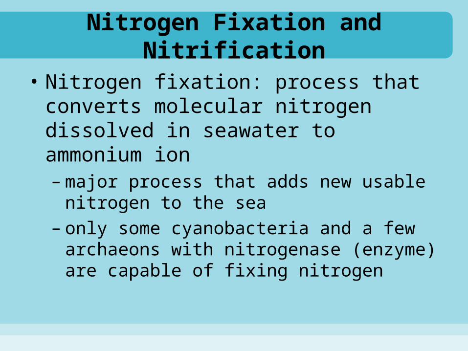
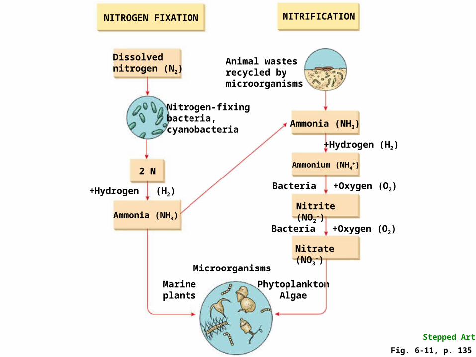
Nitrogen Fixation and Nitrification
• Nitrogen fixation: process that converts molecular nitrogen dissolved in seawater to ammonium ion– major process that adds new usable nitrogen
to the sea– only some cyanobacteria and a few
archaeons with nitrogenase (enzyme) are capable of fixing nitrogen
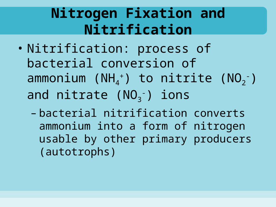
Nitrogen Fixation and Nitrification
• Nitrification: process of bacterial conversion of ammonium (NH4
+) to nitrite (NO2
-) and nitrate (NO3-) ions
– bacterial nitrification converts ammonium into a form of nitrogen usable by other primary producers (autotrophs)

Nitrogen-fixingbacteria,cyanobacteria
2 N
+Hydrogen (H2)
Ammonia (NH3)
Dissolvednitrogen (N2)
NITROGEN FIXATION
Marineplants
Microorganisms
PhytoplanktonAlgae
NITRIFICATION
Animal wastesrecycled bymicroorganisms
Ammonium (NH4+)
Bacteria +Oxygen (O2)
Nitrite (NO2–)
Nitrate (NO3–)
Ammonia (NH3)
+Hydrogen (H2)
Bacteria +Oxygen (O2)
Stepped Art
Fig. 6-11, p. 135
Symbiotic Bacteria• Many bacteria have evolved symbiotic
relationships with a variety of marine organisms• Endosymbiotic theory
– mitochondria, plastids & hydrogenosomes evolved as symbionts within other cells
• Chemosynthetic bacteria live within tube worms and clams
• Some deep-sea or nocturnal animals host helpful bioluminescent bacteria– photophores– embedded in the ink sacs of squid

Archaea• General characteristics
– small (0.1 to 15 micrometers)– prokaryotic– adapted to extreme environmental conditions:
high and low temperatures, high salinities, low pH, and high pressure
– formerly considered bacteria– differences from bacteria
• cell walls lack special sugar-amino acid compounds in bacterial cell walls
• cell membranes contain different lipids, which help stabilize them under extreme conditions

• Nutritional Types– archaea includes photosynthesizers,
chemosynthesizers and heterotrophs– most are methanogens: anaerobic organisms
that metabolize organic matter for energy, producing methane as a waste product
– halobacteria (photosynthetic), thrive at high salinities, trap light using bacteriorhodopsins, purple proteins
Archaea

• Hyperthermophiles– organisms that can survive at temperatures
exceeding 100o C, such as near deep-sea vents– Potential for biomedical and industrial
application
Archaea
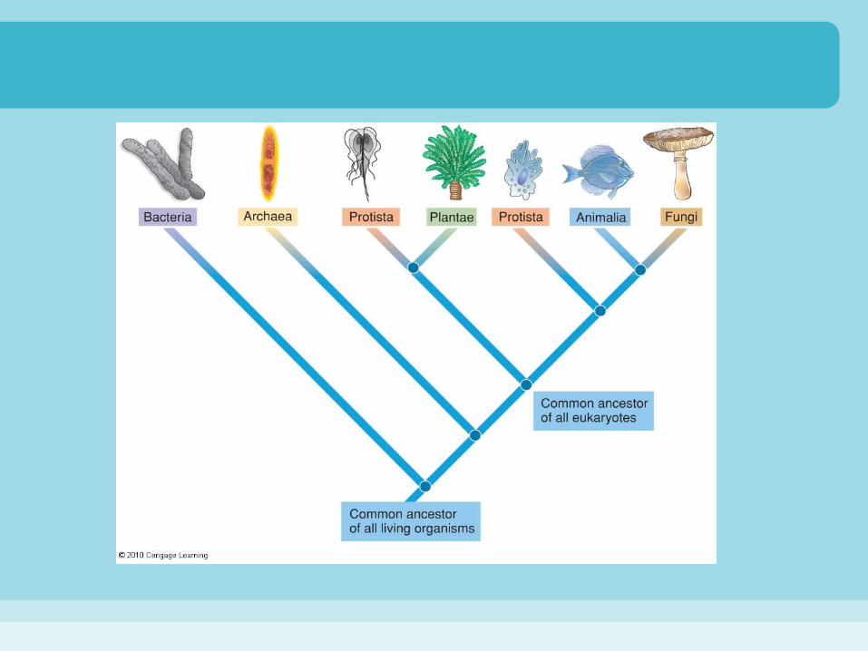
Eukarya
• Eukarya includes all organisms with eukaryotic cells
• Examples:– plants– animals– fungi– algae– single-celled animal-like protozoa

Fungi
• History of marine mycology– marine fungi first discovered in 1849– marine fungi’s ecological role is difficult to
evaluate; biomass needs to be quantified– important in marine ecosystems as
decomposers, prey, pathogens and symbionts

Fungi
• General features of fungi– eukaryotes with cell walls of chitin– many are unicellular yeasts– filamentous fungi grow into long, multi-cellular
filaments called hyphae that can branch to produce a tangled mass called a mycelium
– heterotrohic decomposers that recycle organic material
• can digest lignin (major component of wood)
Fungi
• General features of fungi (con’t)– store energy as glycogen– kingdom Fungi is divided into 4 phyla:
• Chytridiomycota (motile cells)• Zygomycota (e.g. black bread mold)• Basidiomycota (club fungi, e.g. mushrooms)• Ascomycota (sac fungi)
– in the sea, ascomycotes are the most diverse and abundant fungi
Fungi• Ecology and physiology of marine fungi
– can be either obligately marine, requiring ocean or brakish water or facultatively marine (primarily of terrestrial or fresh water origin)
– salinity is toxic to fungi, so they must devote energy to removing sodium
– most marine fungi live on wood from land– some live on grass in salt marshes– others live on algae, mangroves or sand– fungi decompose the chitinous remains of dead
crustaceans in open sea plankton communities
Reproduction of Marine Fungi• Marine yeasts reproduce asexually by
budding—mitosis that produces daughter cells of unequal size
• Filamentous marine fungi reproduce asexually by production of conidiospores on the tips of hyphae
• Filamentous marine ascomycotes can reproduce sexually by forming a fruiting body called an ascocarp, a structure which produces ascospores


Maritime Lichens
• Lichens: mutualistic associations between a fungus and an alga– fungi are usually ascomycotes– algae are usually green or blue-green bacteria
• The fungus provides attachment, general structure, minerals, moisture
• The alga produces organic matter through photosynthesis

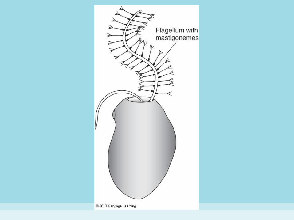
Stramenophiles• A diverse group of eukaryotic organisms
unified by the nature of their cells’ 2 flagella
• The special flagella– 1 flagellum is a simple form, usually with a
light-sensing body at the base; senses light– 2nd bears many mastigonemes (hair-like
filaments) with a thickened base and a branching tip along the shaft; used for swimming

Stramenophiles
• Heterokont: refers to the different form of the 2 flagella
• Ochrophytes: photosynthetic type that are usually golden brown– e.g. diatoms, silicoflagellates and brown algae

Diatoms
• Extremely diverse and distinct members of marine phytoplankton
• Diatom structure– frustule—a two-part, box-shaped organic cell
wall impregnated with silica– valve—one half of a frustule; 1 valve is larger
and fits over the other like a box lid– 2 basic diatom shapes:
• radially symmetrical valves (generally planktonic)• bilaterally symmetrical valves (generally benthic)

Diatoms
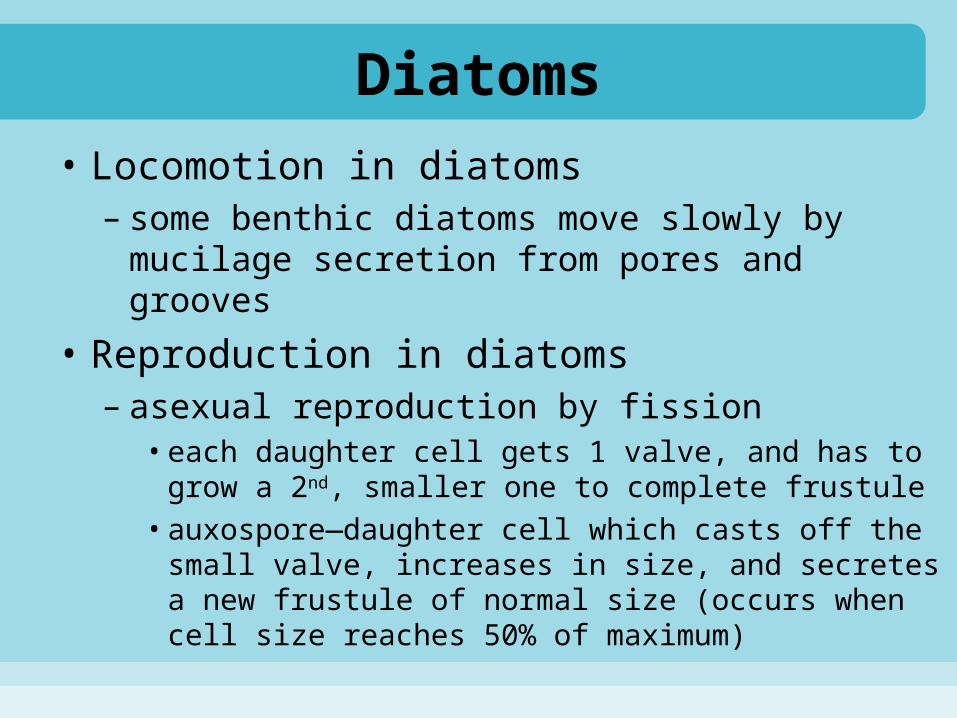
• Locomotion in diatoms– some benthic diatoms move slowly by mucilage
secretion from pores and grooves
• Reproduction in diatoms– asexual reproduction by fission
• each daughter cell gets 1 valve, and has to grow a 2nd, smaller one to complete frustule
• auxospore—daughter cell which casts off the small valve, increases in size, and secretes a new frustule of normal size (occurs when cell size reaches 50% of maximum)


New cell Frustule formation
Zygote
Growth ofthe cell
(auxospore)
Gametefrom another
Gametesreleased
Gametesformed
Cells’ divisioncontinues untilcells become toosmall to divide
Asexual Reproduction Sexual Reproduction
Mitosis
Mitosis
MitosisMitosis
Stepped Art
Fig. 6-19, p. 144
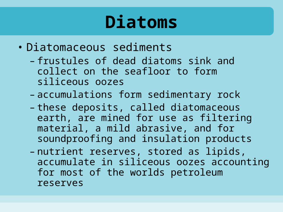
Diatoms• Diatomaceous sediments
– frustules of dead diatoms sink and collect on the seafloor to form siliceous oozes
– accumulations form sedimentary rock– these deposits, called diatomaceous earth, are
mined for use as filtering material, a mild abrasive, and for soundproofing and insulation products
– nutrient reserves, stored as lipids, accumulate in siliceous oozes accounting for most of the worlds petroleum reserves
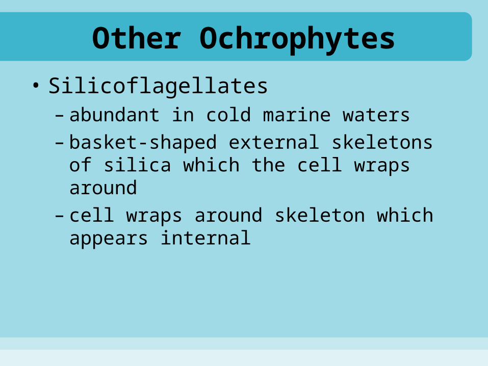
Other Ochrophytes
• Silicoflagellates– abundant in cold marine waters– basket-shaped external skeletons of silica
which the cell wraps around– cell wraps around skeleton which appears
internal

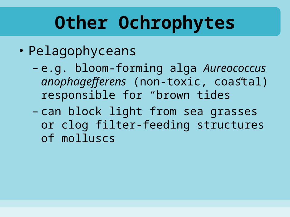
Other Ochrophytes
• Pelagophyceans– e.g. bloom-forming alga Aureococcus
anophagefferens (non-toxic, coastal) responsible for “brown tides”
– can block light from sea grasses or clog filter-feeding structures of molluscs

Labyrinthomorphs• Spindle-shaped, mucous secreting osmotrophic
cells• Labyrinthulids
– e.g. Labyrinthula zosterae, causes devastating eelgrass wasting disease
• Thraustochytrids– planktonic and benthic decomposers– some are pathogens of shellfish– used to produce dietary supplements: oils extracted
from some species are high in polyunsaturated omega-3 fatty acid docosahexaenoic acid (DHA)

Haptophytes• Photosynthetic organisms with 2 simple
flagella both used for locomotion• Have haptonema: a unique structure
arising from the cell surface between the 2 flagella, captures food
• Most are coccolithophores with a surface coating of disc-shaped scales (coliths) of calcium carbonate– remains form calcereous oozes

Haptophytes
• Account for up to 40% of carbonate production in modern seas
• High reflectance of chalky coccolithophores and their production of dimethyl sulfide may have impact on global climate change


Alveolates• Recent re-grouping of several kinds of
marine microbes• Have membranous sacs (alveoli) beneath
their cell membranes– pellicle: term for the cell surface if the
combination of cell membrane and alveoli is complex (distinct from cell wall)
• Examples:– dinoflagellates– ciliates– apicomplexans (strictly parasitic)

Alveolates
• Dinoflagellates– globular, unicellular (sometimes colonial) with
2 flagella– dinosporin: a unique chemical associated with
the cellulose plates within the alveoli of dinoflagellates
– Most are planktonic, some are benthic and others parasitic, also can be bioluminescent – Bioluminescent Bay, Puerto Rico
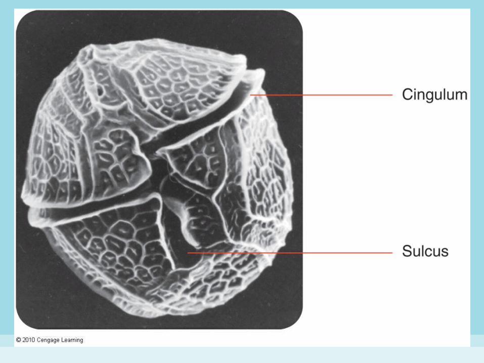

Alveolates (Dinoflagellates)• Dinoflagellate structure
– heterokont flagella– simple flagellum encircles the cell in the cingulum (a
horizontal groove) and produces a spinning motion– longer flagellum with hair-like filaments trails down the
sulcus (a longitudinal groove) and imparts most of the forward motion to the cell
– unarmored dinoflagellates have few or no cellulose plates in the pellicle; armored dinoflagellates have multiple layers of them
– number, size and shapes of plates are used to identify different species
Alveolates (Dinoflagellates)• Dinoflagellate nutrition
– photosynthetic ones have chlorophylls a and c, beta-carotene and peridinin (a xanthophyll which imparts a golden-brown color)
– mixotrophic photosynthetic ones supplement photosynthesis by osmotrophy (absorbing nutrients) or phagotrophy (engulfing nutrients)
• Reproduction in dinoflagellates– asexual reproduction by fission– sexual reproduction by fusion and meiosis– often have dormant stages (cyst formation)

Alveolates (Dinoflagellates)
• Ecological roles of dinoflagellates– major component of phytoplankton– some are parasites of copepods (crustaceans)– zooxanthellae: species lacking flagella which
are symbionts of jellyfish, corals and molluscs– photosynthetic zooxanthellae provide food for
hosts– hosts provide carbon dioxide, other nutrients,
and shelter

Alveolates (Dinoflagellates)
• Harmful Algal Blooms (HABs)– occur when photosynthetic dinoflagellates
undergo a population explosion– colors the water red, orange or brown– dinoflagellates that cause HABs produce toxins
• paralytic shellfish poisoning (PSP) occurs in humans who consume shellfish contaminated with these toxins
• toxins cannot be destroyed by cooking
– oxygen content of the water may be reduced to deadly levels as bacteria decompose animals killed by dinoflagellate toxins

Alveolates• Ciliates
– protozoans that bear cilia for locomotion and for gathering food
• membranelles—tufts or long rows of fused adjacent cilia
• cytostome—an organelle serving as a permanent site for phagocytosis of food
– planktonic and benthic– major links in marine food chains– form symbiotic and parasitic relationships– reproduce asexually by binary fission and
sexually by conjugation (nuclei transfer)


Alveolates (Ciliates)
• Types of marine ciliates– scuticociliates (have a dense and uniform
distribution of cilia on their body)– oligotrichs (have few cilia)– tintinnids (usually lack body cilia and secrete an
organic, loosely fitting shell, the lorica)
• Ecological roles of marine ciliates– most are heterotrophs; some harbor autotrophic
symbionts or chloroplasts– link hetero- and autotrophic blue-green bacteria
to higher levels in the food chain

Choanoflagellates
• Phylum of marine and freshwater flagellated cells that are more closely related to animals than any other group of one-celled microbes
• Unicellular or colonial– colonies may be stalked or embedded in a
gelatinous mass– cell often surrounded by a lorica of siliceous
rods; flagellum is surrounded by a funnel-shaped collar of microvilli
• Highly efficient consumers of bacteria

Amoeboid Protozoans• All have an organelle called a pseudopod—an
extension of the cell surface that can change shape and is used for locomotion (benthic species) and food capture (benthic and pelagic)
• Are hererotrophs consuming bacteria and other small organisms
• Most have a test—an externally secreted organic membrane often covered with foreign particles or strengthened by mineral secretions
Amoeboid Protozoans
• Two major phyla: – foraminiferans (abundant, diverse)– actinopods, which include:
• radiolarians (predominant type)• acantharians• heliozoans

Amoeboid Protozoans
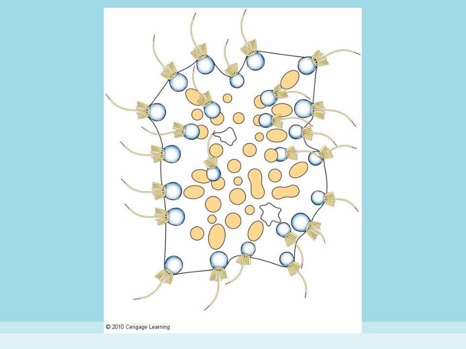
• Foraminiferans (forams)– have branched pseudopods that form
reticulopods (elaborate, net-like structures) used to:
• snare prey• crawl (benthic)• reduce sinking rate (pelagic)
– consume bacteria and diatoms– some harbor symbiotic green and red algae
and zooxanthellae
Amoeboid Protozoans (Foraminiferans)
• Foraminiferan test– elaborate, multi-chambered tests of calcium
carbonate– globigerina ooze: sediments of dead planktonic
forams, largely Globigerina
• Foraminiferans and zooxanthellae– zooxanthellae live symbiotically within the
cytoplasm of many forams from nutrient-poor waters
– photosynthetic zooxanthellae use foram waste products (e.g. CO2, ammonia) as nutrients
Amoeboid Protozoans• Radiolarians
– named for long, needle-like pseudopods• central nuclear region is surrounded by a capsule—
an external organic membrane • pseudopods pass through pores in the capsule and
form a region called the calymma• pseudopods capture food and slow sinking
– radiolarian oozes form from the internal siliceous skeleton of dead radiolarians
– live in the photic zone and capture phyto- and zooplankton, sometimes copepods
– larger radiolarians prey on copepods and other planktonic crustaceans