Embed Size (px)
Citation preview
Synthesis of methylphosphonic acid by marine microbes: asource for methane in the aerobic ocean
William W. Metcalf1,3,*, Benjamin M. Griffin1,†, Robert M. Cicchillo1,2,#, Jiangtao Gao1,2,Sarath Chandra Janga1, Heather A. Cooke1,2,‡, Benjamin T. Circello1,3, Bradley S. Evans1,Willm Martens-Habbena4, David A. Stahl4, and Wilfred A. van der Donk1,2,*
1Institute for Genomic Biology, University of Illinois, 1206 W. Gregory, Urbana, IL 61801. USA.2Department of Chemistry and Howard Hughes Medical Institute, University of Illinois at Urbana-Champaign, 600 S. Matthews Ave., Urbana, IL 61801. USA.3Department of Microbiology, University of Illinois, 601 S. Goodwin Ave, Urbana, IL 61801. USA.4Department of Civil and Environmental Engineering, University of Washington, 302 More Hall,Box 352700, Seattle, WA 98195-2700. USA.
AbstractRelative to the atmosphere, much of the aerobic ocean is supersaturated with methane; however,the source of this important greenhouse gas remains enigmatic. Catabolism of methylphosphonicacid by phosphorus-starved marine microbes, with concomitant release of methane, has beensuggested to explain this phenomenon, yet methylphosphonate is not a known natural product, norhas it been detected in natural systems. Further, its synthesis from known natural products wouldrequire unknown biochemistry. Here we show that the marine archaeon Nitrosopumilus maritimusencodes a pathway for methylphosphonate biosynthesis and that it produces cell-associatedmethylphosphonate esters. The abundance of a key gene in this pathway in metagenomic datasetssuggests that methylphosphonate biosynthesis is relatively common in marine microbes, providinga plausible explanation for the methane paradox.
Methane plays a key role in the global carbon cycle and is a potent greenhouse gas. As such,knowledge of its sources and sinks is essential to climate change models and to understandthe flow of carbon within the biosphere. An unsolved problem in this area is the observationthat vast sections of the aerobic ocean are supersaturated with this gas, despite the fact thatstrictly anaerobic archaea are the only significant biological source of methane known (1).The amount of methane produced in these aerobic environments is substantial, comprisingas much as 4% of the global methane budget (2). It has been suggested that anaerobicmicroenvironments within the aerobic ecosystem could allow production of methane byknown methanogens; however, this is contested on a variety of grounds (for a discussion see(1, 3)). Recently, Karl et al. suggested a new model in which methane would be producedwhen aerobic marine microorganisms use methylphosphonic acid (MPn) as a source ofphosphorus (2). The model is based on several observations: (i) a well-studied bacterialenzyme, carbon-phosphorus (C-P) lyase, produces methane from MPn (4), (ii), C-P lyasegenes are abundant in marine microbes (5, 6), (iii) phosphonates comprise a significantfraction of the available phosphorus pool in marine systems (7, 8), and (iv) incubation of
*To whom correspondence should be addressed. [email protected], [email protected].†Synthetic Genomics, San Diego, CA#Dow AgroSciences, Indianapolis, IN‡Alkermes, Waltham, MA
NIH Public AccessAuthor ManuscriptScience. Author manuscript; available in PMC 2013 February 28.
Published in final edited form as:Science. 2012 August 31; 337(6098): 1104–1107. doi:10.1126/science.1219875.
NIH
-PA Author Manuscript
NIH
-PA Author Manuscript
NIH
-PA Author Manuscript
seawater microcosms with MPn leads to methane production (2). While this model isconceptually appealing, it has a significant missing link: MPn has never been detected inmarine ecosystems, nor is it a known natural product. Moreover, based on knownphosphonate biosynthetic pathways (9), it is difficult to see how MPn could be made withoutinvoking unusual biochemistry.
With one exception, all known phosphonate biosynthetic pathways begin with formation ofthe C-P bond by the enzyme phosphoenolpyruvate mutase (Ppm) (9). We have used the ppmgene as a molecular marker to identify the genes responsible for synthesis of phosphonicacid antibiotics in numerous microorganisms (10-13). During the course of this work, weidentified a putative phosphonate biosynthetic gene cluster in Nitrosopumilus maritimus, amember of the ubiquitous Group I marine Thaumarchaeota whose members are among themost abundant organisms in marine surface waters (14, 15). Based on the experimentallyvalidated functions of homologous enzymes (10, 16, 17), it is very likely that N. maritimushas the capacity to synthesize 2-hydroxyethylphosphonate (HEP), which is a commonintermediate in phosphonate biosynthetic pathways (Fig. S1A, Table S1). Immediatelyadjacent to the putative HEP biosynthetic genes is an operon encoding a putativeoxidoreductase, two putative sulfatases and a protein of the cupin superfamily that wedesignated MpnS.
MpnS has weak homology to hydroxypropylphosphonate epoxidase (HppE) andhydroxyethylphosphonate dioxygenase (HepD), two enzymes that catalyze Fe(II)- andoxygen-dependent transformations of similar phosphonate substrates (Figs S1B & S2).Thus, we suspected that MpnS might be a similar phosphonate biosynthetic enzyme. To testthis, we cloned and overexpressed the mpnS gene in Escherichia coli (18). Cell extractscontaining MpnS stoichiometrically convert 13C-labelled HEP to a product whose retentiontime and molecular mass are identical to MPn in liquid chromatography-mass spectrometry(LC-MS) experiments (Figs. 1 & S3). Using purified MpnS protein and HEP labeledwith 13C at either the 1- or 2- position, we conclusively showed that the products of theMpnS reaction are MPn and HCO3
- (Fig. 1B & 1C). The MpnS-catalyzed reaction requiresboth Fe(II) and molecular oxygen, but does not require an exogenous electron donor. Thus,like HepD, MpnS is an Fe(II)-dependent oxygenase that cleaves the unactivated carbon-carbon bond of HEP. However, the two enzymes catalyze distinct reactions. In the HepDreaction, the reducing equivalents needed for incorporation of oxygen into the cleavageproducts are derived equally from the C-1 and C-2 carbons of HEP, whereas MpnS catalyzesthe asymmetric oxidation of HEP, with all four electrons being derived from the C-2 carbon,affording the more reduced phosphonate product MPn.
Having shown that MpnS catalyzes the synthesis of MPn in vitro, we asked whether N.maritimus synthesizes phosphonic acids using 31P NMR spectroscopy (Fig. 2A). The 1H-decoupled 31P spectrum of the soluble cell extract displayed two peaks in the 10-30 ppmrange characteristic of phosphonic acids (19). The relative abundance of the two peaksvaried with sample preparation and could be seen in both the soluble and cell debrisfractions after sonication (Fig S4). Based on spiking of the sample with an authenticstandard neither peak can be attributed to free methylphosphonate; however, because thephosphorus compounds are cell associated we expected them to be covalently linked to alarger, more complex molecule, thus changing the chemical shift in the 31P NMR spectrum.Accordingly, we conducted a series of 31P-1H Heteronuclear Multiple Bond Correlation(HMBC) experiments to identify the atoms linked to the P nuclei seen in the NMR spectra(Fig 2B). Because the behavior of phosphonate esters in such experiments is not welldocumented, we also synthesized and characterized a series of phosphonate esters to supportour assignments (Figs S5-S7). Based on these experiments the 31P NMR peak at 28.7 ppmcan be confidently assigned as an ester of methylphosphonate. Further support for this
Metcalf et al. Page 2
Science. Author manuscript; available in PMC 2013 February 28.
NIH
-PA Author Manuscript
NIH
-PA Author Manuscript
NIH
-PA Author Manuscript
conclusion was provided by high-resolution mass spectrometry, which revealed the presenceof free methylphosphonate after strong acid hydrolysis of N. maritimus cell debris (Fig 2Cand S8). Based on these results and the gene context of the MpnS locus (Table S1), wesuspect that N. maritimus synthesizes an exopolysaccharide decorated withmethylphosphonate, similar to the HEP- and aminoethylphosphonate-modified polymersfound in many bacteria and lower eukaryotes (20).
The data presented above suggest that N. maritimus produces a cell-associatedmethylphosphonate ester via an MpnS-dependent biosynthetic pathway. To link this findingto the larger marine environment we screened the Global Oceanic Survey (GOS)metagenomic dataset (21) for the presence of MpnS homologs. We also searched forhomologs of the related HepD and HppE proteins. Initially we screened the assembled GOSscaffolds, finding forty-six MpnS and twenty HepD homologs using a BLASTP cutoff valueof 10-10 (Table S2). No HppE homologs were observed. Importantly, none of the HepDhomologs were identified using N. maritimus MpnS as the query sequence; likewise none ofthe MpnS homologs were identified using HepD as a query. Thus, BLASTP clearlydifferentiates between the two homologous groups, supporting the assignment of therecovered sequences as MpnS and HepD proteins, respectively. To independently supportthese functional assignments we constructed maximum likelihood phylogenetic treesincluding biochemically validated MpnS, HepD and HppE proteins (Fig 3A & S9). We alsoused a hierarchical clustering method to examine all putative and validated MpnS, HepDand HppE proteins (Fig S10). In both cases robust support for the functional assignmentswas obtained. Thus, we conclude that the recovered GOS MpnS homologs are likely to bemethylphosphonate synthases.
Additional support for the function of the MpnS homologs was revealed by analysis ofneighboring genes found in GOS DNA scaffolds (Fig 3B, Table S3). Many of the nearbyORFs are homologous to those found in the N. maritimus gene cluster, including thephosphonate biosynthetic genes ppm, ppd, and pdh, as well as homologs of the sulfatasesand nucleotidyl transferase genes, suggesting that the GOS scaffolds encode genes for thesynthesis of similar MPn esters. Several other genes found on the scaffolds provide evidencefor the identity of the organisms in which they are found. One of the scaffolds includes a23S rRNA gene that can be confidently placed within the SAR11 clade betweenPelagibacter species (Fig S11), while two of the manC genes are nearly identical to onesfound in Pelagibacter sp. HTCC7211. Although the mpnS gene is absent in sequencedPelagibacter genomes, these data strongly support the conclusion that some members of thisgenus have the capacity to synthesize MPn.
Relatives of Nitrosopumilus and Pelagibacter are among the most abundant organisms in thesea, with global populations estimated at 1028 for both ammonia-oxidizing Thaumarchaeota(14) and members of the SAR11 clade (22). Thus, the observation of mpnS in somemembers of these genera is consistent with the idea that MPn synthesis is prevalent inmarine systems. To provide direct support for this notion, we measured the abundance of thempnS gene relative to the abundance of typical single-copy genes as previously described(23). We also quantified the occurrence of the ppm gene to provide an estimate the relativeoccurrence of phosphonate synthesis in general (Table S4). Based on these data, we estimatethat ca. 16% of marine microbes are capable of phosphonate biosynthesis, while 0.6% havethe capacity to synthesize MPn. Because the GOS samples are confined to the upper fewmeters of the ocean, extrapolation of this analysis to the deeper ocean should be viewed withsome skepticism. Nevertheless, the upper 200 m of the world's oceans are thought to containca. 3.6 ×1028 microbial cells with an average generation time of ca. two weeks (24). Thus,even with the relatively modest abundance of MPn biosynthesis suggested by our data, itseems quite possible that these cells could provide sufficient MPn precursor to account for
Metcalf et al. Page 3
Science. Author manuscript; available in PMC 2013 February 28.
NIH
-PA Author Manuscript
NIH
-PA Author Manuscript
NIH
-PA Author Manuscript
the observed methane production in the aerobic ocean via the C-P lyase dependent scenariosuggested by Karl et al (2).
Supplementary MaterialRefer to Web version on PubMed Central for supplementary material.
AcknowledgmentsThis work was supported by the National Institutes of Health (GM PO1 GM077596 and F32 GM095024), theHoward Hughes Medical Institute and the National Science Foundation NSF (MCB-0604448, OCE-1046017 andMCB-0920741). Its contents are solely the responsibility of the authors and do not necessarily represent the officialviews of the NIGMS, NIH, NSF or HHMI.
References and Notes1. Rogers, JE.; Whitman, WB., editors. Microbial production and consumption of greenhouse gases :
methane, nitrogen oxides, and halomethanes. American Society for Microbiology; Washington,D.C.: 1991.
2. Karl DM, et al. Nat. Geosci. 2008; 1:473.
3. Reeburgh WS. Chem. Rev. 2007; 107:486. [PubMed: 17261072]
4. Daughton CG, Cook AM, Alexander M. FEMS Microbiol. Lett. 1979; 5:91.
5. Martinez A, Tyson GW, DeLong EF. Environ. Microbiol. 2010; 12:222. [PubMed: 19788654]
6. Ilikchyan IN, McKay RML, Zehr JP, Dyhrman ST, Bullerjahn GS. Environ. Microbiol. 2009;11:1314. [PubMed: 19220397]
7. Clark LL, Ingall ED, Benner R. Am. J. Sci. 1999; 299:724.
8. Clark LL, Ingall ED, Benner R. Nature. 1998; 393:426.
9. Metcalf WW, van der Donk WA. Annu. Rev. Biochem. 2009; 78:65. [PubMed: 19489722]
10. Borisova SA, Circello BT, Zhang JK, van der Donk WA, Metcalf WW. Chem. Biol. Jan 29.201017:28. [PubMed: 20142038]
11. Blodgett JA, Zhang JK, Metcalf WW. Antimicrob. Agents and Ch. 2005; 49:230.
12. Eliot AC, et al. Chem Biol. 2008; 15:765. [PubMed: 18721747]
13. Circello BT, Eliot AC, Lee JH, van der Donk WA, Metcalf WW. Chem Biol. 2010; 17:402.[PubMed: 20416511]
14. Karner MB, DeLong EF, Karl DM. Nature. 2001; 409:507. [PubMed: 11206545]
15. Konneke M, et al. Nature. 2005; 437:543. [PubMed: 16177789]
16. Shao Z, et al. J. Biol. Chem. 2008; 283:23161. [PubMed: 18544530]
17. Seidel HM, Freeman S, Seto H, Knowles JR. Nature. 1988; 335:457. [PubMed: 3138545]
18. Materials and methods are available as supplementary material on Science Online.
19. Tebby, JC., editor. CRC handbook of phosphorus-31 nuclear magnetic resonance data. CRC Press;Boca Raton: 1991.
20. Hilderbrand, RL., editor. The Role of Phosphonates in Living Systems. CRC Press; Boca Raton:1983.
21. Yooseph S, et al. PLoS. Biol. 2007; 5:432.
22. Morris RM, et al. Nature. 2002; 420:806. [PubMed: 12490947]
23. Howard EC, Sun S, Biers EJ, Moran MA. Environ. Microbiol. 2008; 10:2397. [PubMed:18510552]
24. Whitman WB, Coleman DC, Wiebe WJ. Proc. Natl. Acad. Sci. USA. 1998; 95:6578. [PubMed:9618454]
25. Dunwell JM, Culham A, Carter CE, Sosa-Aguirre CR, Goodenough PW. Trends Biochem. Sci.2001; 26:740. [PubMed: 11738598]
26. Liu PH, et al. J. Am. Chem. Soc. 2001; 123:4619. [PubMed: 11457256]
Metcalf et al. Page 4
Science. Author manuscript; available in PMC 2013 February 28.
NIH
-PA Author Manuscript
NIH
-PA Author Manuscript
NIH
-PA Author Manuscript
27. Cicchillo RM, et al. Nature. 2009; 459:871. [PubMed: 19516340]
28. Higgins LJ, Yan F, Liu PH, Liu HW, Drennan CL. Nature. 2005; 437:838. [PubMed: 16015285]
29. Nair SK, van der Donk WA. Arch. Biochem. Biophys. 2011; 505:13. [PubMed: 20854789]
30. Kuemin M, van der Donk WA. Chem. Commun. 2010; 46:7694.
31. Whitteck JT, et al. Angew. Chem., Int. Ed. Engl. 2007; 46:9089. [PubMed: 17990255]
32. Martens-Habbena W, Berube PM, Urakawa H, de la Torre JR, Stahl DA. Nature. 2009; 461:976.[PubMed: 19794413]
33. Papadopoulos JS, Agarwala R. Bioinformatics. 2007; 23:1073. [PubMed: 17332019]
34. Stamatakis A. Bioinformatics. 2006; 22:2688. [PubMed: 16928733]
35. Olson SA. Brief Bioinform. 2002; 3:87. [PubMed: 12002227]
36. de Hoon MJ, Imoto S, Nolan J, Miyano S. Bioinformatics. 2004; 20:1453. [PubMed: 14871861]
37. Saldanha AJ. Bioinformatics. 2004; 20:3246. [PubMed: 15180930]
38. Desper R, Gascuel O. Mol. Biol. Evol. 2004; 21:587. [PubMed: 14694080]
39. Markowitz VM, et al. Nuc Acids Res. 2010; 38:D382.
Metcalf et al. Page 5
Science. Author manuscript; available in PMC 2013 February 28.
NIH
-PA Author Manuscript
NIH
-PA Author Manuscript
NIH
-PA Author Manuscript
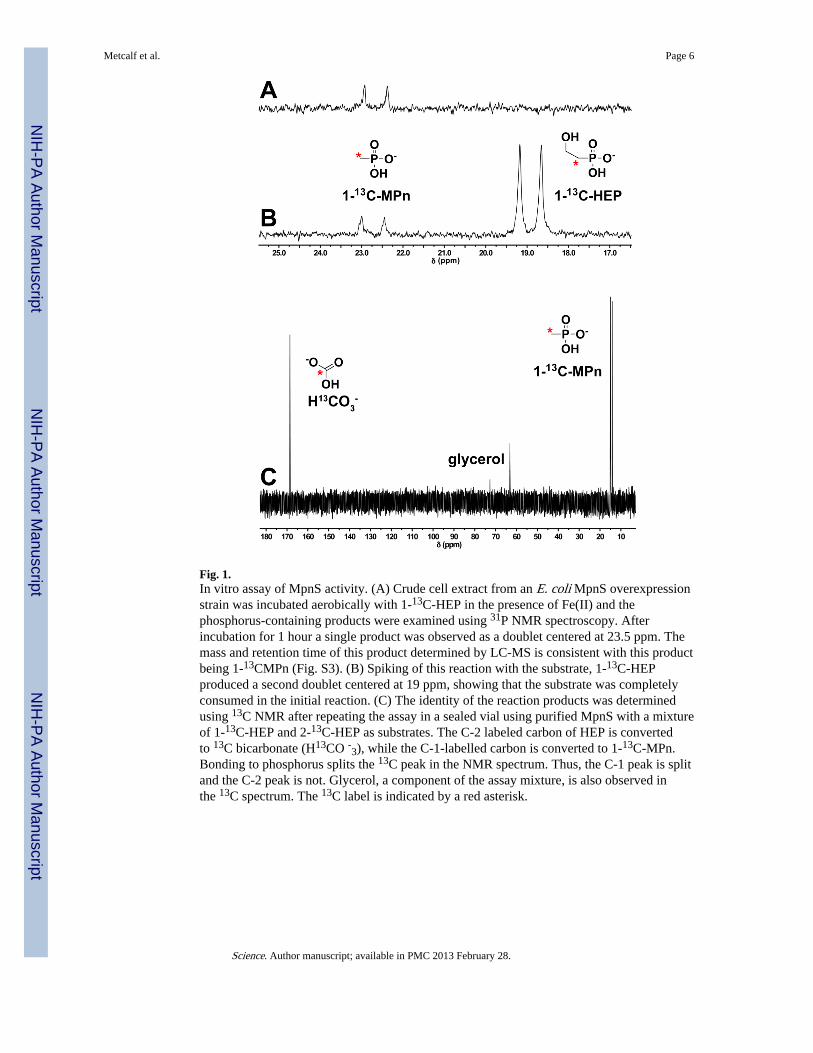
Fig. 1.In vitro assay of MpnS activity. (A) Crude cell extract from an E. coli MpnS overexpressionstrain was incubated aerobically with 1-13C-HEP in the presence of Fe(II) and thephosphorus-containing products were examined using 31P NMR spectroscopy. Afterincubation for 1 hour a single product was observed as a doublet centered at 23.5 ppm. Themass and retention time of this product determined by LC-MS is consistent with this productbeing 1-13CMPn (Fig. S3). (B) Spiking of this reaction with the substrate, 1-13C-HEPproduced a second doublet centered at 19 ppm, showing that the substrate was completelyconsumed in the initial reaction. (C) The identity of the reaction products was determinedusing 13C NMR after repeating the assay in a sealed vial using purified MpnS with a mixtureof 1-13C-HEP and 2-13C-HEP as substrates. The C-2 labeled carbon of HEP is convertedto 13C bicarbonate (H13CO -3), while the C-1-labelled carbon is converted to 1-13C-MPn.Bonding to phosphorus splits the 13C peak in the NMR spectrum. Thus, the C-1 peak is splitand the C-2 peak is not. Glycerol, a component of the assay mixture, is also observed inthe 13C spectrum. The 13C label is indicated by a red asterisk.
Metcalf et al. Page 6
Science. Author manuscript; available in PMC 2013 February 28.
NIH
-PA Author Manuscript
NIH
-PA Author Manuscript
NIH
-PA Author Manuscript
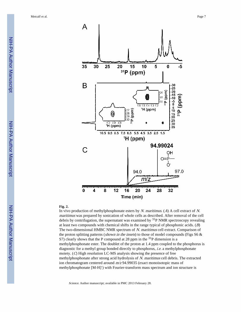
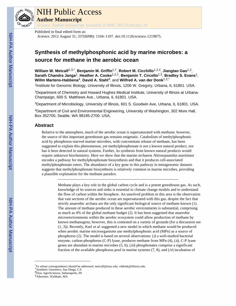
Fig. 2.In vivo production of methylphosphonate esters by N. maritimus. (A) A cell extract of N.maritimus was prepared by sonication of whole cells as described. After removal of the celldebris by centrifugation, the supernatant was examined by 31P NMR spectroscopy revealingat least two compounds with chemical shifts in the range typical of phosphonic acids. (B)The two-dimensional HMBC NMR spectrum of N. maritimus cell extract. Comparison ofthe proton splitting patterns (shown in the insets) to those of model compounds (Figs S6 &S7) clearly shows that the P compound at 28 ppm in the 31P dimension is amethylphosphonate ester. The doublet of the proton at 1.4 ppm coupled to the phosphorus isdiagnostic for a methyl group bonded directly to phosphorus, i.e. a methylphosphonatemoiety. (C) High resolution LC-MS analysis showing the presence of freemethylphosphonate after strong acid hydrolysis of N. maritimus cell debris. The extractedion chromatogram centered around m/z 94.99035 (exact monoisotopic mass ofmethylphosphonate [M-H]-) with Fourier-transform mass spectrum and ion structure is
Metcalf et al. Page 7
Science. Author manuscript; available in PMC 2013 February 28.
NIH
-PA Author Manuscript
NIH
-PA Author Manuscript
NIH
-PA Author Manuscript
shown in the inset. The chromatographic and MS fragmentation pattern is identical to anauthentic MPn standard (Fig S8).
Metcalf et al. Page 8
Science. Author manuscript; available in PMC 2013 February 28.
NIH
-PA Author Manuscript
NIH
-PA Author Manuscript
NIH
-PA Author Manuscript
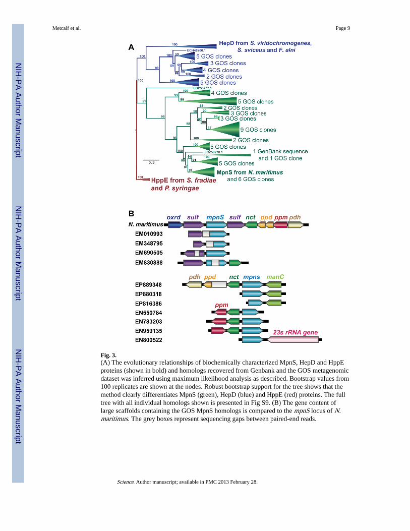
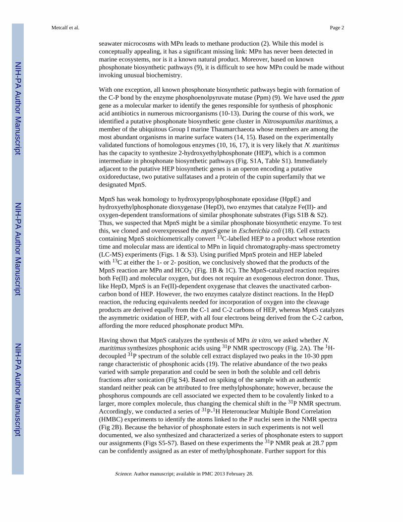
Fig. 3.(A) The evolutionary relationships of biochemically characterized MpnS, HepD and HppEproteins (shown in bold) and homologs recovered from Genbank and the GOS metagenomicdataset was inferred using maximum likelihood analysis as described. Bootstrap values from100 replicates are shown at the nodes. Robust bootstrap support for the tree shows that themethod clearly differentiates MpnS (green), HepD (blue) and HppE (red) proteins. The fulltree with all individual homologs shown is presented in Fig S9. (B) The gene content oflarge scaffolds containing the GOS MpnS homologs is compared to the mpnS locus of N.maritimus. The grey boxes represent sequencing gaps between paired-end reads.
Metcalf et al. Page 9
Science. Author manuscript; available in PMC 2013 February 28.
NIH
-PA Author Manuscript
NIH
-PA Author Manuscript
NIH
-PA Author Manuscript