Embed Size (px)
Citation preview
Comparative Characterization of Two Marine Alginate Lyasesfrom Zobellia galactanivorans Reveals Distinct Modes ofAction and Exquisite Adaptation to Their Natural Substrate*
Received for publication, March 6, 2013, and in revised form, June 17, 2013 Published, JBC Papers in Press, June 19, 2013, DOI 10.1074/jbc.M113.467217
François Thomas‡§1, Lena C. E. Lundqvist¶, Murielle Jam‡§, Alexandra Jeudy�**, Tristan Barbeyron‡§,Corine Sandström¶, Gurvan Michel‡§, and Mirjam Czjzek‡§2
From the ‡University of Marie and Pierre Curie Paris 6, UMR 7139, Marine Plants and Biomolecules, Station Biologique de Roscoff,F-29682 Roscoff, Brittany, France, §CNRS, UMR 7139, Marine Plants and Biomolecules, Station Biologique de Roscoff, F-29682Roscoff, Brittany, France, the ¶Department of Chemistry, Swedish University of Agricultural Sciences, SE-750 07 Uppsala, Sweden,the �University of Marie and Pierre Curie Paris 6, FR2424, Station Biologique de Roscoff, F-29682 Roscoff, Brittany, France, and**CNRS, FR2424, Station Biologique de Roscoff, F-29682 Roscoff, Brittany, France
Background: Alginolytic systems frommarine bacteria are crucial for algal biomass conversion, yet their molecular mech-anisms remain poorly understood.Results: Structural and biochemical characterization of two paralogous marine alginate lyases highlights details on comple-mentary roles and differences with terrestrial enzymes.Conclusion: Bacterial alginolytic enzymes are specifically adapted to the unique characteristics of the natural substrate.Significance:Marine microbes evolved complex degradation systems targeting habitat-specific polysaccharides.
Cell walls of brown algae are complex supramolecular assem-blies containing various original, sulfated, and carboxylatedpolysaccharides. Among these, the major marine polysaccha-ride component, alginate, represents an important biomass thatis successfully turned over by the heterotrophicmarine bacteria.In the marine flavobacterium Zobellia galactanivorans, thecatabolism and uptake of alginate are encoded by operon struc-tures that resemble the typical Bacteroidetes polysaccharideuti-lization locus. The genome ofZ. galactanivorans contains sevenputative alginate lyase genes, five of which are localized withintwo clusters comprising additional carbohydrate-related genes.This study reports on the detailed biochemical and structuralcharacterization of two of these. We demonstrate here thatAlyA1PL7 is an endolytic guluronate lyase, and AlyA5 cleavesunsaturated units, �-L-guluronate or �-D-manuronate residues,at the nonreducing end of oligo-alginates in an exolytic fashion.Despite a common jelly roll-fold, these striking differences ofthe mode of action are explained by a distinct active site topol-ogy, an open cleft in AlyA1PL7, whereas AlyA5 displays a pockettopology due to the presence of additional loops partiallyobstructing the catalytic groove. Finally, in contrast to PL7 algi-nate lyases from terrestrial bacteria, both enzymes proceedaccording to a calcium-dependent mechanism suggesting anexquisite adaptation to their natural substrate in the context ofbrown algal cell walls.
Brown algae dominate the primary production in temperateand polar rocky shores and represent a huge marine biomass.Indeed, coastal regions are considered as carbon sinks, retain-ing about 200�1012 g of carbon/year (1). Their cell walls includea minor fraction of crystalline cellulose and a majority of ani-onic polysaccharides, alginates, and sulfated fucoidans (2).Phlorotannins, which consist of halogenated and/or sulfatedphenolic compounds (3, 4), and 5% of proteins (5) completethis complex supramolecular assemblage. Among these com-pounds, alginate can account for up to 40% of the dry weight ofthe algal biomass (6). This linear polysaccharide is composed of�-D-mannuronate (M)3 and its C5 epimer �-L-guluronate (G),which are arranged in three types of repeating structures asfollows: poly-G stretches, poly-M stretches, and heteropoly-meric random sequences (poly-MG) (7). The gelling propertiesstrongly depend on the poly-G content of the algal polysaccha-ride, because these G blocks form highly viscous solutions andgels through interconnectedmetal ion chelation (mainly Ca2�)(8). Within the algal cell wall, the functional properties of algi-nate aremodulated through changes inGandMcontent. Thesevariations can be species-specific or depend on season andenvironmental conditions; however, to date only very few stud-ies exploring such relationships are available (9–12). Phylo-genomic analysis of the carbohydrate metabolism of the modelbrown alga Ectocarpus siliculosus has revealed that brown algaehorizontally acquired the alginate biosynthetic pathway froman ancestral Actinobacterium (13). Indeed, alginates are alsoproduced as exopolysaccharides by some bacteria, such asthose belonging to the genera Azotobacter and Pseudomonas(14, 15). The main differences at the molecular level between
* This work was supported by European Community’s Seventh FrameworkProgramme FP7/2007-2013 under Grant Agreement 222628 and by theRegion Bretagne and the CNRS.
The atomic coordinates and structure factors (codes 3zpy and 4be3) have beendeposited in the Protein Data Bank (http://wwpdb.org/).
1 Present address: Biology Dept., Watson Laboratory, Woods Hole Oceano-graphic Institution, Woods Hole, MA 02543.
2 To whom correspondence should be addressed. Tel.: 33-298-29-23-75; Fax:33-298-29-23-24; E-mail: [email protected].
3 The abbreviations used are: M, �-D-mannuronate; G, �-L-guluronate; PDB,Protein Data Bank; r.m.s.d., root mean square deviation; C-PAGE, carbohy-drate electrophoresis; DEH, 4-deoxy-L-erythro-5-hexoseulose uronic acid;PL, polysaccharide lyase; DP, degrees of polymerization.
THE JOURNAL OF BIOLOGICAL CHEMISTRY VOL. 288, NO. 32, pp. 23021–23037, August 9, 2013© 2013 by The American Society for Biochemistry and Molecular Biology, Inc. Published in the U.S.A.
AUGUST 9, 2013 • VOLUME 288 • NUMBER 32 JOURNAL OF BIOLOGICAL CHEMISTRY 23021
by guest on May 16, 2020
http://ww
w.jbc.org/
Dow
nloaded from
algal and bacterial alginates are the presence of O-acetylgroups at C2 and/or C3 in the bacterial alginates (7, 16) andtheir higher proportion of M units. From an ancestral bacte-rial exopolymer, alginate further evolved in brown algae intoa pivotal cell wall polysaccharide, which is constrained by thenecessity to interact with other cell wall components (2, 13).These evolutionary constraints likely explain the loss ofacetyl groups in algal alginates together with the increasedimportance of the G units and of their interactions with cal-cium ions to control gel properties. This hypothesis is con-sistent with the expansion of the mannuronan C5-epimerasegene families observed in the brown algae Laminaria digi-tata (17–19) and E. siliculosus (13).Alginate constitutes an abundant nutrient resource for het-
erotrophicmarine bacteria and thus in coastal ecosystems playsan ecological role similar to that of cellulosic and hemicellulosicbiomass in terrestrial environments. However, the comprehen-sion of themolecular bases for the assimilation of algal alginatesby marine bacteria remains at best fragmentary. We haverecently reported the first operons specific for alginate assimi-lation in a marine bacterium, Zobellia galactanivorans (20).This flavobacterium, which is well known to degrade sulfatedgalactans from red seaweeds (21–23), is also able to use alg-inates from brown algae as the sole carbon source (24). Thealginolytic system of Z. galactanivorans encompasses sevenalginate lyases (AlyA1 toAlyA7) belonging to four distinct fam-ilies of polysaccharide lyases (PL (25)), families PL6, -7, -14, and-17. Although alyA1 and alyA7 are isolated genes, alyA4, alyA5,and alyA6 are transcribed on the samemRNA. alyA2 and alyA3are localized in a large operon comprising other carbohydrate-related genes, notably a TonB-dependent receptor and its asso-ciated SusD-like protein likely involved in oligo-alginate uptake(20). Such gene organization is typically found in Bacteroidetesand is referred to as a polysaccharide utilization locus (26, 27).With the exception of alyA7, all these genes are up-regulated inthe presence of alginate (20). The complexity of this degrada-tion system questions the exact role of the different alginatelyases. Notably, is there a functional redundancy providing arobustness to this alginolytic system?Do these enzymes displaydistinct substrate specificities? Do they proceed by differentmodes of action, which could have synergistic effects? To dis-criminate between these hypotheses, which are not necessarilymutually exclusive, the in-depth biochemical study of theseenzymes is a prerequisite.AlyA1 is a secreted modular protein with an N-terminal car-
bohydrate-bindingmodule of the family 32 (CBM32) appendedto a C-terminal PL7 module, and AlyA5 is predicted to be anouter membrane lipoprotein consisting of a lone PL7 catalyticdomain. Moreover, these PL7modules are extremely divergentwith only 16% sequence identity, and several large insertionsare present in AlyA5 in comparison with AlyA1 (20). Wedescribe here the detailed study of structure-function relation-ships of the catalytic modules of two paralogous alginate lyasesAlyA1 and AlyA5 from Z. galactanivorans, by a combination ofcrystallographic and biochemical approaches, including reac-tion product analysis by NMR spectroscopy.
EXPERIMENTAL PROCEDURES
Substrate Materials—Sodium alginate samples with threedifferent M/G ratios (0.5, 0.9, and 2.0) were provided by Dan-isco. Fractions of oligoguluronates, oligomannuronates, andmixed MG oligosaccharides were prepared according to Hauget al. (9). This method has been reported to yield fragmentswith DP ranging from 4 to �30 (28). Their respective M/Gcomposition was controlled by NMR (data not shown).Reduction of Oligoguluronates—Oligosaccharides were re-
duced following a protocol adapted from Abdel-Akher andSandstrom (29). Briefly, 3 mg of sodium borohydride wereadded to 500 �l of oligoguluronates (1 mg�ml�1), and the mix-ture was incubated overnight at room temperature. The reac-tion was acidified by adding drops of acetic acid until no moreH2 production was observed. The sample was then dried undervacuum and resuspended in 1 drop of 17.5 M acetic acid and 1ml of methanol. Solvent was evaporated under a nitrogen flux.The amount of reducing ends was estimated by the ferricyanideassay (30) using a calibration curve with 20–400 �g�ml�1 glu-cose. Before the treatment, the ferricyanide assay measured29.4 �g�ml�1 eq�glucose in the oligoguluronate solution. Noreducing ends could be detected by the same assay after thereduction.Phylogenetic Analyses—Members of the PL7 family were
selected in the CAZY database (31), and their sequences werealigned usingMAFFTwith the L-INS-i algorithm and the scor-ing matrix Blosum62 (32). The alignment was manually editedin MEGA 4.1 (33). 180 sites were used to derive a phylogenetictree using a maximum likelihood method conducted with thePhyMLprogram (34) implemented online on Phylogeny.fr (35).Bootstrap values were calculated from 100 resamplings of thedataset.Cloning of TwoGenes Coding for PL7 Alginate Lyases—Prim-
ers were designed to amplify the coding region correspondingto the mature protein for AlyA5 and for the catalytic module ofAlyA1 only, hereafter called AlyA1PL7. The genes were ampli-fied by PCR from Z. galactanivorans genomic DNA using theoligonucleotide primers as follows: forward ggggggGGATCC-tgtaaagacaaacctaaggccactacg and reverse ccccccGAATTCtca-ctgggcttctggggcgcttc for AlyA5; forward ggggggAGATCTtccg-gtggttcgtccacccttc and reverse ccccccGAATTCttaattgtgggtta-cgcttaggtttttg for the catalytic domain of AlyA1PL7. Using therestriction sites EcoRI and BamHI for AlyA5, and EcoRI andBglII for AlyA1PL7, the PCR products were then ligated into theexpression vector pFO4, resulting in a recombinant proteinwith an N-terminal hexa-histidine tag. The plasmids weretransformed into Escherichia coli BL21(DE3) expressionstrains. All cloning procedures were performed as described inGroisillier et al. (36).Protein Expression and Purification Procedures—For both
enzymes, the same procedure was applied. Two-milliliters pre-cultures of the recombinant E. coliBL21(DE3) cells were grownovernight at 37 °C in LB medium containing ampicillin (100�g.ml�1). 200ml of autoinducible ZYPmediumwith ampicillin(37) were inoculated with 200 �l of pre-culture and incubatedfor 3 days at 20 °C and 200 rpm. Cells were centrifuged for 20min at 5000 rpmat 4 °C, and pellets were stored at�20 °C. Cells
In-depth Characterization of Marine Alginate Lyases
23022 JOURNAL OF BIOLOGICAL CHEMISTRY VOLUME 288 • NUMBER 32 • AUGUST 9, 2013
by guest on May 16, 2020
http://ww
w.jbc.org/
Dow
nloaded from
were resuspended in 20ml of BufferA (25mMTris-HCl, pH7.5,200 mM NaCl, 5 mM imidazole) containing a mixture of anti-proteases (Complete EDTA-free, Roche Applied Science) andDNase. Cells were lysedwith a French press. Samples were thencentrifuged for 2 h at 20,000 � g. The supernatant was loadedon a Hyper Cell PAL column charged with 0.1 M NiSO4 andequilibrated with Buffer A. Proteins were eluted with a lineargradient between Buffer A and Buffer B (25 mM Tris-HCl, pH7.5, 200 mM NaCl, 1 M imidazole) in 60 ml at a flow rate of 1ml�min�1 and collected in fractions of 1 ml. Fractions showingthe presence of the recombinant proteins by SDS-PAGE werepooled. Aliquots of 5 ml were further purified on a size exclu-sion chromatography column (Superdex 75 16/60 pg, GEHealthcare) using Buffer C (25 mM Tris-HCl, pH 7.5, 200 mM
NaCl) at a flow rate of 1 ml�min�1. Fractions showing the pres-ence of the pure proteins were pooled for AlyA1PL7 (data notshown), although two samples, separating the fractions ofmonomeric and dimeric forms of AlyA5 (data not shown), wereprepared. Dynamic light scattering measurements were per-formed on 40 �l of concentrated protein solutions (�1mg�ml�1) in a quartz cuvette on a ZetaSizerNano-S instrument(Malverne Instruments).Enzymatic Assays—Protein concentration was determined
by measuring the absorbance at 280 nm. Theoretical molarextinction coefficients �280 were computed from the proteinsequences on the ProtParam server. Alginate lyase activity wasassayed by measuring the increase in absorbance at 235 nm(A235) of the reaction products (unsaturated uronates) for 5minin a 1-cm quartz cuvette containing 0.5 ml of reaction mixturein a thermostated spectrophotometer. One unit of activity wasdefined as an increase of 1 unit in A235 per min.The effects of temperature, NaCl, and chelating agents on
AlyA1PL7 activity were tested in 100 mM Tris-HCl buffer, pH7.5, using sodium alginate 0.05% as substrate and 5 �l of puri-fied enzyme at 1.2 �M. The chelating agents EDTA or EGTAwere either incubated at 1 mM final concentration withAlyA1PL7 (overnight, 4 °C) or added directly in the reactionmixture. The effect of pH on AlyA1PL7 activity was tested usingvarious buffers at 100 mM: citrate, pH 3.0–6.0, MOPS/NaOH(MOPS, pH 6.0–7.9), HEPES, pH 7.0–8.0, Tris-HCl, pH 7.0–8.5, and glycine-NaOH, pH 8.5–10.0. Kinetic parameters weredetermined at 30 °C in 100mMTris-HCl, pH 7.5, 200mMNaCl,with 10 alginate concentrations ranging from 0.075 to 2.5mg�ml�1. Substrate concentrations were derived from themolecular mass of an M or G monosaccharide (194 g�mol�1).Reactions were performed in triplicate. A molar extinctioncoefficient � � 8500 liter�mol�1�cm�1 (38) was used for thereaction products (unsaturated uronic acids) The kineticparameters were estimated by a hyperbolic regression algo-rithm with Hyper32.AlyA5 enzymatic assays on alginate polysaccharides were
conducted as for AlyA1PL7, using 5 �l of the purified enzyme at2.7�M. Reactions on saturated or unsaturated oligosaccharideswere performed at 30 °C in 100 mM Tris-HCl buffer, pH 7.5, 4mM CaCl2, 50 �g of substrate, and 5 �l of enzyme at 5.6 �M.Carbohydrate Electrophoresis (C-PAGE)—After enzymatic
degradation, aliquots were boiled for 10 min before analysis.Oligosaccharides were analyzed by C-PAGE. Aliquots (5 �l)
weremixedwith 15�l of loading buffer (10% sucrose and 0.01%phenol red). 5 �l were electrophoresed through a 6% (w/v)stacking and a 28% running, 0.75-mm thick, polyacrylamide gelin 50 mM Tris-HCl, 1 mM EDTA buffer, pH 8.7. Gels werestained with Alcian blue 0.5% (w/v) followed by silver nitrate0.4% (w/v) (73, 74). The coloration was developed with 7%sodium carbonate (w/v) containing formaldehyde, and thereaction was stopped by adding concentrated acetic acid.Monitoring of Alginate Lyases Reaction by 1H NMR—The
action of alginate lyases was directly followed at 30 °C in theNMR tube on crude alginates, G-, M-, and alternating MGblocks. The substrates were dissolved at a concentration of 5mg�ml�1 in a 100mMdeuterated phosphate buffer, pH 7.6, with200 mM NaCl and 5 mM CaCl2. The NMR experiments wereperformed on a Bruker DRX-400 MHz spectrometer using a5.0-mm 1H/13C/15N/31P QNP probe or on a Bruker Avance III600MHz spectrometer using a 5.0-mm1H/13C/15N/31P inversedetection QXI probe, and a 2.5-mm 1H/13C inverse detectionprobe, all equipped with z-gradients and controlled by Topspin1.3 and 2.1 software, respectively. The 1H NMR experimentswere recorded at 30 and 70 °Cusing a spectral width of 3000Hz,an acquisition time of 5.62 s, a pulsewidth of 8.5�s, a relaxationdelay of 4 s, and 128 scans. The residual HOD signal was sup-pressed using the NOESY pre-saturation pulse sequence inwhich saturation was applied during the relaxation delay of 4 sand the mixing time of 50 ms.Prior to an enzymatic reaction, a reference spectrum of the
substrate was acquired. The samplewas then removed from theprobe; the enzyme was added at the proper concentration, andthe tube was put back into the probe. The reaction was moni-tored as a function of time. To monitor the progress of degra-dation of the sample, series of 1H NMR spectra were recordedat 30 °C using the multizg or multizgvd programs from theBruker library.Analysis of End Products by NMR and Mass Spectrometry—
The enzymatic degradation was performed on 10 ml of 5mg�ml�1 alginate samples with addition of alginate lyases fromZ. galactanivorans at 30 °C for 18 h. The samples were thencentrifuged; the supernatant was lyophilized and dissolved in0.1 M ammonium acetate.
The oligouronates were fractionated according to their sizeby preparative gel filtration chromatography on a ÄKTA sys-temwith a 16�60 Superdex 30 column (AmershamBiosciences).Oligosaccharides were eluted with 0.1 M ammonium acetate ata flow rate of 0.8 ml�min�1 at room temperature. Unsaturatedoligo-alginates were detected byA235, and the relative amountsof the different end products were determined as the peak area.The fractions belonging to the same peak were pooled, lyophi-lized, and purified further by repeating the size exclusion chro-matography procedure on the Superdex 30 column. The purefractions were lyophilized and analyzed by ESI-MS using aBruker Esquire-LC mass spectrometer (Bruker Daltonics Inc.,Billerica, MA). The samples were dissolved in 1:1 MeOH-H2Oand were directly injected into the electrospray with a flow of 2�l�min�1. The mass range scanned was from 100 to 1100atomic mass units. All acquisitions were recorded in both neg-ative and positive mode and treated by Bruker DaltonicsEsquire LC 4.5, data analysis version 3.0. To determine the
In-depth Characterization of Marine Alginate Lyases
AUGUST 9, 2013 • VOLUME 288 • NUMBER 32 JOURNAL OF BIOLOGICAL CHEMISTRY 23023
by guest on May 16, 2020
http://ww
w.jbc.org/
Dow
nloaded from
structure of the end products, the oligosaccharide signals wereassigned from one-dimensional proton and selective CSSFTOCSY with Keeler filter (selcssfdizs.2) from Bruker libraryNMR spectra and from two-dimensional 1H-1H TOCSY NMRexperiments using a mixing time of 90 ms. The NMR spectrawere referenced at 30 °C using acetone (�H 2.225, �C 31.05) asinternal reference.Protein Crystallization and Three-dimensional Crystal Struc-
ture Determination—The first crystallization screening experi-ments for both the catalytic domain of AlyA1PL7 and the full-length dimeric sample of AlyA5 were carried out at 292 K withthe sitting-drop vapor diffusion method in 96-well Corningplates (catalog no. 3551). The crystallization screening was per-formed with JCSG� and PACT screens from Qiagen, whichmakes a total of 192 conditions in two 96-well plates. A crystal-lization robot (Proteomics Solutions, Honeybee) was used fordispensing the drops that contained 300 nl of protein solutionthat were mixed with 150 nl of reservoir solution. After visualidentification of initial crystallization conditions, these werefurther optimized in 24-well Linbro plates by the hanging-dropvapor diffusionmethod. ForAlyA1PL7, optimal conditionswereobtained for a protein solution of 11.3 mg�ml�1 concentrationsupplemented with oligoguluronate (0.1 mg�ml�1). Drops of 2�l volume of this protein solution were mixed with 1 �l ofcrystallization solution that contained 0.2 M KSCN and 28%PEG-MME 2000 and equilibrated against a reservoir contain-ing 500 �l. For AlyA5 (dimer) the optimized conditions wereobtained for a protein solution of 7.7 mg�ml�1. Drops of 2 �lvolume of this protein solution were mixed with 1 �l of crystal-lization solution that contained 22% PEG 3350 and 0.2 M sodi-um/potassium tartrate and equilibrated against a reservoir con-taining 200 �l. The native crystals of both alginate lyases wereflash-frozen at 100 K in a crystallization solution supplementedwith 10% glycerol. Data were collected to 1.43 Å resolution forAlyA1PL7 and 1.75 Å for AlyA5 on beamline PROXIMA-1equipped with an ADSC Quantum Q315r detector (SOLEIL, StAubin, France) and a wavelength of 0.98 Å. All data sets weretreated, scaled, and converted with the program suite XDS (39).The three-dimensional structures were solved by the molecularreplacementmethodwithALY-1 fromCorynebacterium sp. (PDB1UAI) for the crystals of AlyA1PL7 andA1-II� from Sphingomonassp. A1 (PDB 2Z42) as initial model, in the case of AlyA5. Thestarting phases were optimized using the programs ARP/wARP(40) andREFMAC5(41) aspartof theCCP4suite (42).The refine-mentwascarriedoutwithREFMAC5andthe finalmodelbuildingwith Coot (43). Water molecules were added automatically withREFMAC-ARP/wARP and visually verified. In all cases, the stere-ochemistry of the final structurewas evaluatedusingPROCHECK(44). The atomic coordinates and structure factors (codes 3zpyand 4be3) have beendeposited in the ProteinDataBank, ResearchCollaboratory for Structural Bioinformatics, Rutgers University,New Brunswick, NJ.
RESULTS
Phylogenetic Analysis
The PL7 family has been recently subdivided in subfamiliesbased on sequence similarities (25). To identify the subfamily
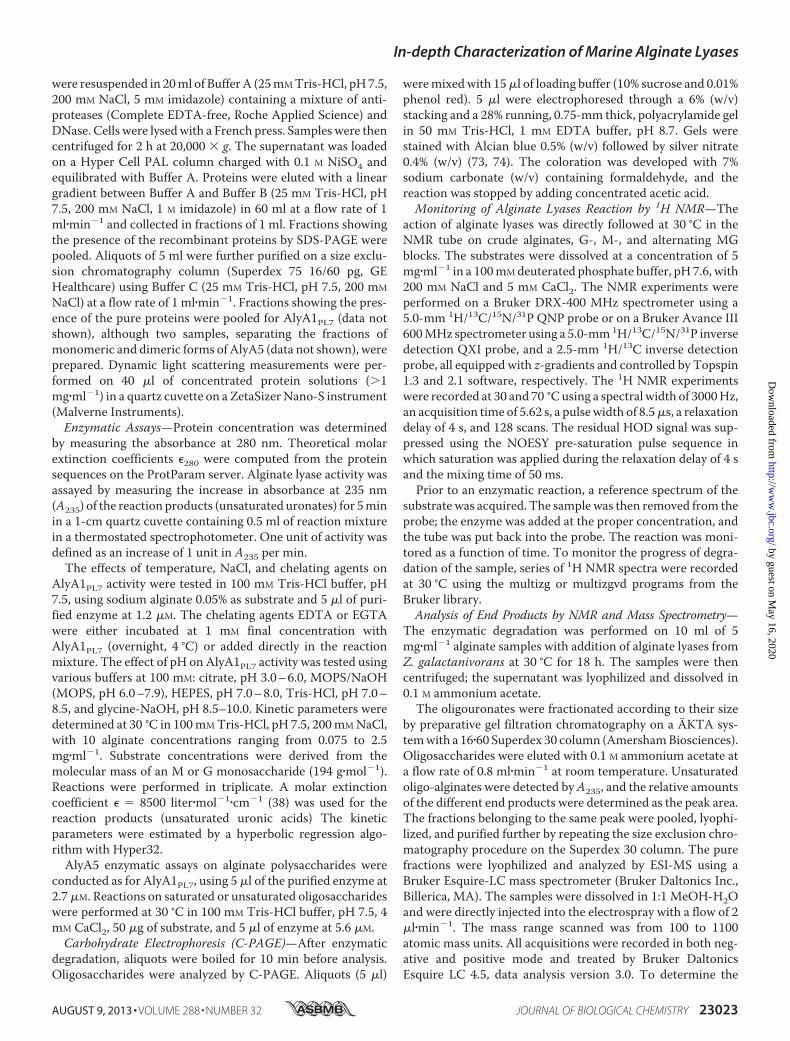
for the three PL7 enzymes AlyA1, AlyA2, and AlyA5 from Z.galactanivorans, 35 additional selected sequences from thesame family were used to derive a phylogenetic tree (Fig. 1).This approach clearly distinguished the five existing subfami-lies within the PL7 family (SF1–SF5). It reveals that the threePL7 enzymes encoded in theZ. galactanivorans genome belongto different subfamilies. AlyA1 is distantly related to enzymesclassified in SF3 and thus is a new member of this subfamily.AlyA5 can also be confidently assigned to SF5. AlyA2was foundto belong to a well supported group composed of four otherenzymes from marine Flavobacteriaceae. A condition imposedby Lombard et al. (25) when creating subfamilies was that thegroup contained at least five members. Thus, we propose thatAlyA2 and the four other enzymes in the same group belong toa new subfamily of PL7. This new subfamily, which we herebydefine as SF6, appears to be conserved only inmarine represen-tatives of the Flavobacteriaceae, possibly representing a niche-specific evolution of PL7 enzymes. As subfamilies correlate ingeneral with substrate specificities, they can help predict thepreferred substrate of new enzymes (25). To date, two alginatelyases have been characterized in SF3 and seven in SF5 (CAZYdatabase, December 2012). They all are classified as poly(�-L-guluronate) lyases (EC 4.2.2.11). Thus, we hypothesize thatAlyA1 and AlyA5 should preferentially cleave Gmotifs (Fig. 2).No prediction can be made on AlyA2 specificity, because noneof the proteins belonging to this new subfamily SF6 has yet beencharacterized.
Overexpression and Purification of AlyA1PL7 and AlyA5
To characterize the enzymatic activities and structural prop-erties of the new PL7 enzymes from Z. galactanivorans, thecoding sequences of the mature proteins (full-length for AlyA2and AlyA5 and only the catalytic module for AlyA1) werecloned into pFO4 expression vector. Expression tests in E. coliBL21(DE3) grown in ZYP medium (37) were successful for therecombinant proteins AlyA1PL7 andAlyA5 but failed for AlyA2(data not shown). We thus focused on the study of AlyA1PL7and AlyA5. High amounts of soluble recombinant proteinswere obtained and purified to homogeneity using a nickel affin-ity chromatography followed by gel filtration. The two steps ofpurification yielded 30 mg of recombinant AlyA1PL7 from 200ml of ZYPmedium. The apparent molecular mass was between26.5 and 29 kDa (estimated from the calibration of the sizeexclusion column and the SDS-PAGE, respectively). This isconcordant with the theoretical molecular mass of 27.5 kDacalculated for the protein sequence of the catalytic domainusing ProtParam implemented in the ExPASy ProteomicsServer (45). The recombinant AlyA5 eluted from the final sizeexclusion chromatography as two distinct peaks (data notshown), corresponding to apparent molecular masses of 69.5and 38.3 kDa. SDS-PAGE analyses showed that these two pop-ulations contained only one protein form under denaturingconditions, with an apparent molecular mass of 42 kDa. Com-pared with the theoretical molecular mass of 38.3 kDa, the pro-teins eluted in the first and second peaks correspond roughly toa dimer and a monomer of AlyA5, respectively. This was con-firmed by the difference in radius of gyration thatwasmeasuredby dynamic light scattering as 2.5 and 3.2 nm for the mono-
In-depth Characterization of Marine Alginate Lyases
23024 JOURNAL OF BIOLOGICAL CHEMISTRY VOLUME 288 • NUMBER 32 • AUGUST 9, 2013
by guest on May 16, 2020
http://ww
w.jbc.org/
Dow
nloaded from
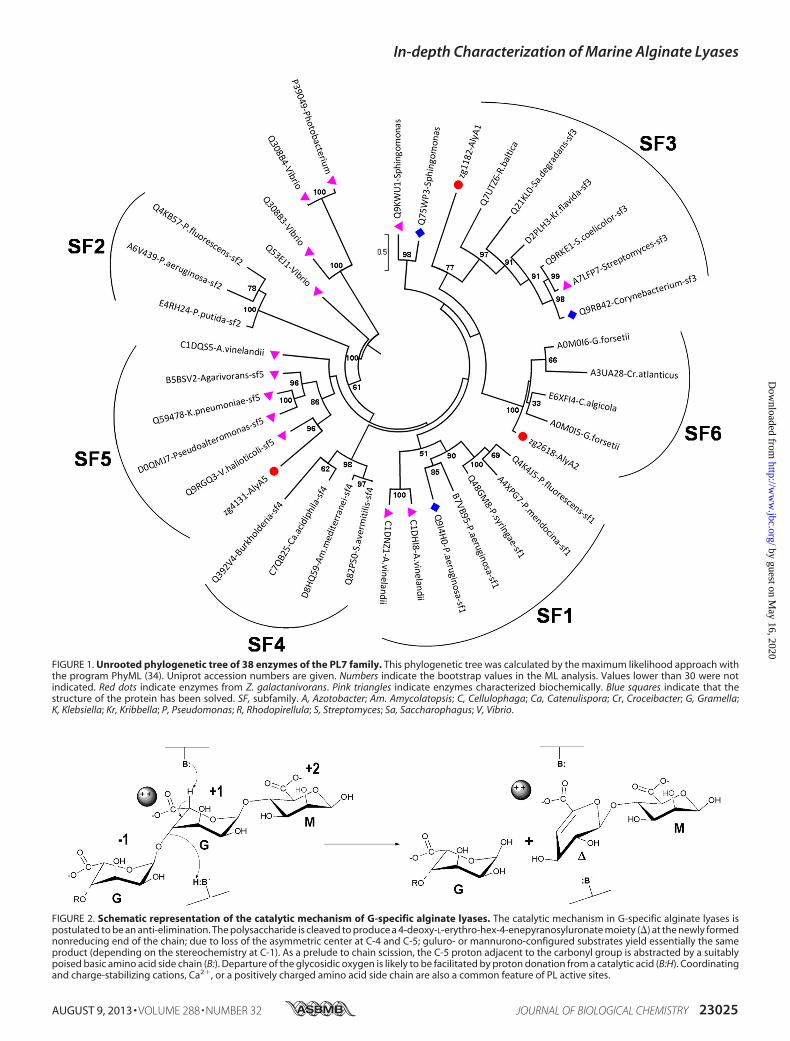
FIGURE 1. Unrooted phylogenetic tree of 38 enzymes of the PL7 family. This phylogenetic tree was calculated by the maximum likelihood approach withthe program PhyML (34). Uniprot accession numbers are given. Numbers indicate the bootstrap values in the ML analysis. Values lower than 30 were notindicated. Red dots indicate enzymes from Z. galactanivorans. Pink triangles indicate enzymes characterized biochemically. Blue squares indicate that thestructure of the protein has been solved. SF, subfamily. A, Azotobacter; Am. Amycolatopsis; C, Cellulophaga; Ca, Catenulispora; Cr, Croceibacter; G, Gramella;K, Klebsiella; Kr, Kribbella; P, Pseudomonas; R, Rhodopirellula; S, Streptomyces; Sa, Saccharophagus; V, Vibrio.
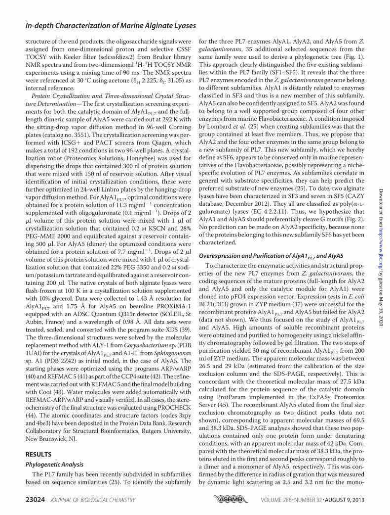
FIGURE 2. Schematic representation of the catalytic mechanism of G-specific alginate lyases. The catalytic mechanism in G-specific alginate lyases ispostulated to be an anti-elimination. The polysaccharide is cleaved to produce a 4-deoxy-L-erythro-hex-4-enepyranosyluronate moiety (�) at the newly formednonreducing end of the chain; due to loss of the asymmetric center at C-4 and C-5; guluro- or mannurono-configured substrates yield essentially the sameproduct (depending on the stereochemistry at C-1). As a prelude to chain scission, the C-5 proton adjacent to the carbonyl group is abstracted by a suitablypoised basic amino acid side chain (B:). Departure of the glycosidic oxygen is likely to be facilitated by proton donation from a catalytic acid (B:H). Coordinatingand charge-stabilizing cations, Ca2�, or a positively charged amino acid side chain are also a common feature of PL active sites.
In-depth Characterization of Marine Alginate Lyases
AUGUST 9, 2013 • VOLUME 288 • NUMBER 32 JOURNAL OF BIOLOGICAL CHEMISTRY 23025
by guest on May 16, 2020
http://ww
w.jbc.org/
Dow
nloaded from
disperse samples of purified monomeric and dimeric forms ofAlyA5, respectively. The purification of the recombinantAlyA5yielded 2.2 mg of monomeric form and 10 mg of dimeric formfrom cultures of 200 ml. The dimeric form was stable in solu-tion, as it eluted as a single peak after a second size exclusionchromatography, corresponding to a molecular mass of 69.5kDa (data not shown).
Characterization of the Recombinant AlyA1PL7
Biochemical Properties of AlyA1PL7—The action of therecombinant catalytic domain of AlyA1PL7 on alginate induceda strong increase in the absorbance at 235 nm, confirming thatit is an alginate lyase acting via the �-elimination mechanism,producing the 4,5-unsaturated 4-deoxy-L-erythro-hex-4-enepyranosyluronate (�) on the nonreducing end (Fig. 2). Theoptimal temperature and salinity were 30 °C and 200mMNaCl,respectively (Fig. 3, A and B). AlyA1PL7 showed the highestactivity at pH 7.0 in Tris-HCl buffer (Fig. 3C). This is similar tothe optimal pH determined for other alginate lyases frommarine bacteria (46). No activity was detected outside the rangeof pH 4.0–8.5. The choice of the buffering molecule appearedto be critical, as exemplified by the 40% decrease in activity in
MOPS buffer at pH 7.0 compared with Tris-HCl. Addition ofthe chelating agents, EDTA or EGTA, in the reaction mixturereduced the enzyme activity by 90 and 95%, respectively (datanot shown). This inhibition was only incomplete when thechelating agents were incubatedwith the enzyme overnight butnot added to the reaction mixture (43 and 58%, respectively).This suggests that the interaction between the enzyme anddivalent cations takes place during substrate fixation and notbefore. To determine themode of action of AlyA1PL7, the prod-ucts resulting from the degradation of alginate were sampledperiodically and analyzed by C-PAGE. After 2min, the polysac-charide fraction was already largely degraded (Fig. 3E). Follow-ing the reaction from 2 min to 14 h, the size of the detectedoligosaccharides decreased with time, rapidly reaching thesmallest DP of 2. The appearance of intermediate larger oligo-saccharides, DP4 to DP20, at early stages demonstrates thatAlyA1PL7 was acting with an endolytic mode on alginate.Substrate Specificity of AlyA1PL7—The enzyme activity was
measured on three polymeric alginate substrates, chosen fortheir difference in the M/G ratio. The kinetic parameters wereclearly correlated with the guluronate content of the substrate.
FIGURE 3. Effect of temperature (A), concentration of NaCl (B) and pH (C) on AlyA1PL7 and pH (D) on AlyA5 activity, C-PAGE analysis of AlyA1PL7degradation products (E) and degradation kinetics of alginate by AlyA5 (F). A and B, experiments were conducted in Tris-HCl buffer, pH 7.5, using sodiumalginate 0.05% as substrate and 12 nM of the purified enzyme. Values are mean S.D. (n � 3). A, effect of temperature. Activity at 30 °C was taken as 100%.B, effect of [NaCl]. Reactions were conducted at 30 °C. Activity with 200 mM NaCl was taken as 100%. C, effect of pH on AlyA1PL7 activity. Experiments wereundertaken at 30 °C in 100 mM buffer using sodium alginate 0.05% as substrate and 27 nM of the purified enzyme. Sodium citrate (open rhombuses), MOPS (opencircles), Tris-HCl (open triangles), HEPES (closed squares), and glycine-NaOH (closed circles) buffers were used in the assay. Activity in Tris-HCl, pH 7.0, was takenas 100%. D, effect of pH on AlyA5 activity. Experiments were done as above except that the protein concentration of purified AlyA5 was 56 nM. Activity in MOPS,pH 7.0, was taken as 100%. E, reaction was conducted at 30 °C in 100 mM Tris-HCl buffer, pH 7.5, using sodium alginate 0.05% as substrate and 12 nM of thepurified enzyme. Aliquots were sampled before adding the enzyme (lane 1) and after 2 min (lane 2), 5 min (lane 3), 10 min (lane 4), 30 min (lane 5), 1 h (lane 6),2 h (lane 7), and 14 h (lane 8). F, degradation kinetics of G (black), MG (dark gray), and M blocks (light gray) by AlyA5. A235 was measured at 30 °C for reactionmixtures (500 �l) composed of 100 mM Tris-HCl buffer, pH 7.5, 4 mM CaCl2, 50 �g of substrate, and 27 nM of enzyme.
In-depth Characterization of Marine Alginate Lyases
23026 JOURNAL OF BIOLOGICAL CHEMISTRY VOLUME 288 • NUMBER 32 • AUGUST 9, 2013
by guest on May 16, 2020
http://ww
w.jbc.org/
Dow
nloaded from
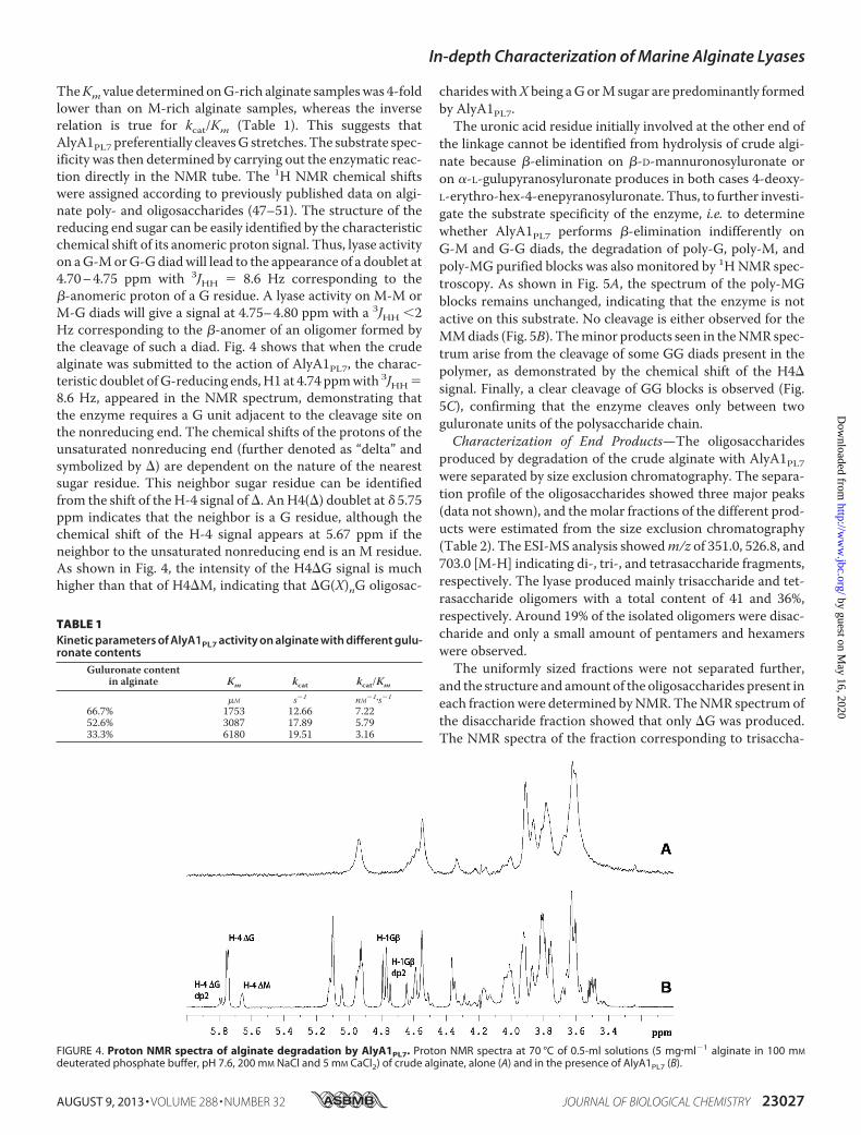
TheKm value determined onG-rich alginate sampleswas 4-foldlower than on M-rich alginate samples, whereas the inverserelation is true for kcat/Km (Table 1). This suggests thatAlyA1PL7 preferentially cleavesG stretches. The substrate spec-ificity was then determined by carrying out the enzymatic reac-tion directly in the NMR tube. The 1H NMR chemical shiftswere assigned according to previously published data on algi-nate poly- and oligosaccharides (47–51). The structure of thereducing end sugar can be easily identified by the characteristicchemical shift of its anomeric proton signal. Thus, lyase activityon aG-MorG-Gdiadwill lead to the appearance of a doublet at4.70–4.75 ppm with 3JHH � 8.6 Hz corresponding to the�-anomeric proton of a G residue. A lyase activity on M-M orM-G diads will give a signal at 4.75–4.80 ppm with a 3JHH 2Hz corresponding to the �-anomer of an oligomer formed bythe cleavage of such a diad. Fig. 4 shows that when the crudealginate was submitted to the action of AlyA1PL7, the charac-teristic doublet ofG-reducing ends,H1 at 4.74 ppmwith 3JHH�8.6 Hz, appeared in the NMR spectrum, demonstrating thatthe enzyme requires a G unit adjacent to the cleavage site onthe nonreducing end. The chemical shifts of the protons of theunsaturated nonreducing end (further denoted as “delta” andsymbolized by �) are dependent on the nature of the nearestsugar residue. This neighbor sugar residue can be identifiedfrom the shift of the H-4 signal of�. AnH4(�) doublet at � 5.75ppm indicates that the neighbor is a G residue, although thechemical shift of the H-4 signal appears at 5.67 ppm if theneighbor to the unsaturated nonreducing end is an M residue.As shown in Fig. 4, the intensity of the H4�G signal is muchhigher than that of H4�M, indicating that �G(X)nG oligosac-
charideswithX being aGorMsugar are predominantly formedby AlyA1PL7.The uronic acid residue initially involved at the other end of
the linkage cannot be identified from hydrolysis of crude algi-nate because �-elimination on �-D-mannuronosyluronate oron �-L-gulupyranosyluronate produces in both cases 4-deoxy-L-erythro-hex-4-enepyranosyluronate. Thus, to further investi-gate the substrate specificity of the enzyme, i.e. to determinewhether AlyA1PL7 performs �-elimination indifferently onG-M and G-G diads, the degradation of poly-G, poly-M, andpoly-MG purified blocks was also monitored by 1HNMR spec-troscopy. As shown in Fig. 5A, the spectrum of the poly-MGblocks remains unchanged, indicating that the enzyme is notactive on this substrate. No cleavage is either observed for theMMdiads (Fig. 5B). Theminor products seen in theNMR spec-trum arise from the cleavage of some GG diads present in thepolymer, as demonstrated by the chemical shift of the H4�signal. Finally, a clear cleavage of GG blocks is observed (Fig.5C), confirming that the enzyme cleaves only between twoguluronate units of the polysaccharide chain.Characterization of End Products—The oligosaccharides
produced by degradation of the crude alginate with AlyA1PL7were separated by size exclusion chromatography. The separa-tion profile of the oligosaccharides showed three major peaks(data not shown), and themolar fractions of the different prod-ucts were estimated from the size exclusion chromatography(Table 2). The ESI-MS analysis showedm/z of 351.0, 526.8, and703.0 [M-H] indicating di-, tri-, and tetrasaccharide fragments,respectively. The lyase produced mainly trisaccharide and tet-rasaccharide oligomers with a total content of 41 and 36%,respectively. Around 19% of the isolated oligomers were disac-charide and only a small amount of pentamers and hexamerswere observed.The uniformly sized fractions were not separated further,
and the structure and amount of the oligosaccharides present ineach fractionwere determined byNMR. TheNMR spectrumofthe disaccharide fraction showed that only �G was produced.The NMR spectra of the fraction corresponding to trisaccha-
FIGURE 4. Proton NMR spectra of alginate degradation by AlyA1PL7. Proton NMR spectra at 70 °C of 0.5-ml solutions (5 mg�ml�1 alginate in 100 mM
deuterated phosphate buffer, pH 7.6, 200 mM NaCl and 5 mM CaCl2) of crude alginate, alone (A) and in the presence of AlyA1PL7 (B).
TABLE 1Kinetic parameters of AlyA1PL7 activity on alginate with different gulu-ronate contents
Guluronate contentin alginate Km kcat kcat/Km
�M s�1 nM�1�s�1
66.7% 1753 12.66 7.2252.6% 3087 17.89 5.7933.3% 6180 19.51 3.16
In-depth Characterization of Marine Alginate Lyases
AUGUST 9, 2013 • VOLUME 288 • NUMBER 32 JOURNAL OF BIOLOGICAL CHEMISTRY 23027
by guest on May 16, 2020
http://ww
w.jbc.org/
Dow
nloaded from
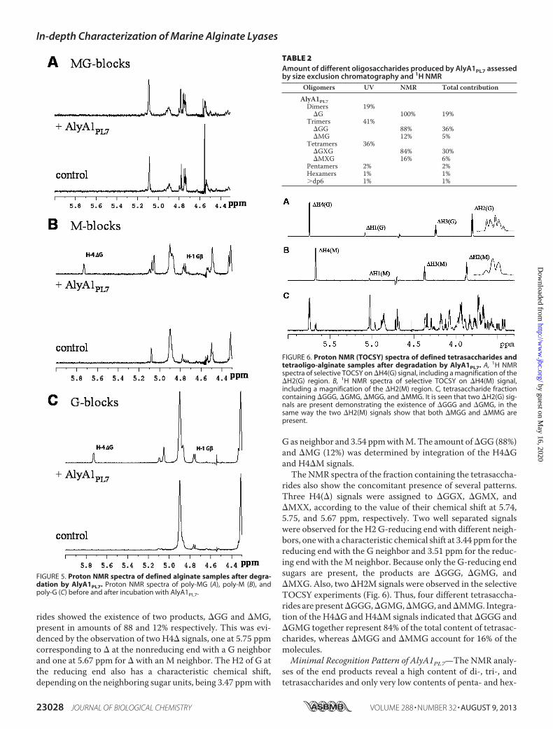
rides showed the existence of two products, �GG and �MG,present in amounts of 88 and 12% respectively. This was evi-denced by the observation of two H4� signals, one at 5.75 ppmcorresponding to � at the nonreducing end with a G neighborand one at 5.67 ppm for � with an M neighbor. The H2 of G atthe reducing end also has a characteristic chemical shift,depending on the neighboring sugar units, being 3.47 ppmwith
G as neighbor and 3.54 ppmwithM.The amount of�GG (88%)and �MG (12%) was determined by integration of the H4�Gand H4�M signals.The NMR spectra of the fraction containing the tetrasaccha-
rides also show the concomitant presence of several patterns.Three H4(�) signals were assigned to �GGX, �GMX, and�MXX, according to the value of their chemical shift at 5.74,5.75, and 5.67 ppm, respectively. Two well separated signalswere observed for the H2 G-reducing end with different neigh-bors, onewith a characteristic chemical shift at 3.44 ppm for thereducing end with the G neighbor and 3.51 ppm for the reduc-ing end with theM neighbor. Because only the G-reducing endsugars are present, the products are �GGG, �GMG, and�MXG. Also, two �H2M signals were observed in the selectiveTOCSY experiments (Fig. 6). Thus, four different tetrasaccha-rides are present�GGG,�GMG,�MGG, and�MMG. Integra-tion of the H4�G and H4�M signals indicated that �GGG and�GMG together represent 84% of the total content of tetrasac-charides, whereas �MGG and �MMG account for 16% of themolecules.Minimal Recognition Pattern of AlyA1PL7—The NMR analy-
ses of the end products reveal a high content of di-, tri-, andtetrasaccharides and only very low contents of penta- and hex-
FIGURE 6. Proton NMR (TOCSY) spectra of defined tetrasaccharides andtetraoligo-alginate samples after degradation by AlyA1PL7. A, 1H NMRspectra of selective TOCSY on �H4(G) signal, including a magnification of the�H2(G) region. B, 1H NMR spectra of selective TOCSY on �H4(M) signal,including a magnification of the �H2(M) region. C, tetrasaccharide fractioncontaining �GGG, �GMG, �MGG, and �MMG. It is seen that two �H2(G) sig-nals are present demonstrating the existence of �GGG and �GMG, in thesame way the two �H2(M) signals show that both �MGG and �MMG arepresent.
TABLE 2Amount of different oligosaccharides produced by AlyA1PL7 assessedby size exclusion chromatography and 1H NMR
Oligomers UV NMR Total contribution
AlyA1PL7Dimers 19%
�G 100% 19%Trimers 41%
�GG 88% 36%�MG 12% 5%
Tetramers 36%�GXG 84% 30%�MXG 16% 6%
Pentamers 2% 2%Hexamers 1% 1%�dp6 1% 1%
FIGURE 5. Proton NMR spectra of defined alginate samples after degra-dation by AlyA1PL7. Proton NMR spectra of poly-MG (A), poly-M (B), andpoly-G (C) before and after incubation with AlyA1PL7.
In-depth Characterization of Marine Alginate Lyases
23028 JOURNAL OF BIOLOGICAL CHEMISTRY VOLUME 288 • NUMBER 32 • AUGUST 9, 2013
by guest on May 16, 2020
http://ww
w.jbc.org/
Dow
nloaded from
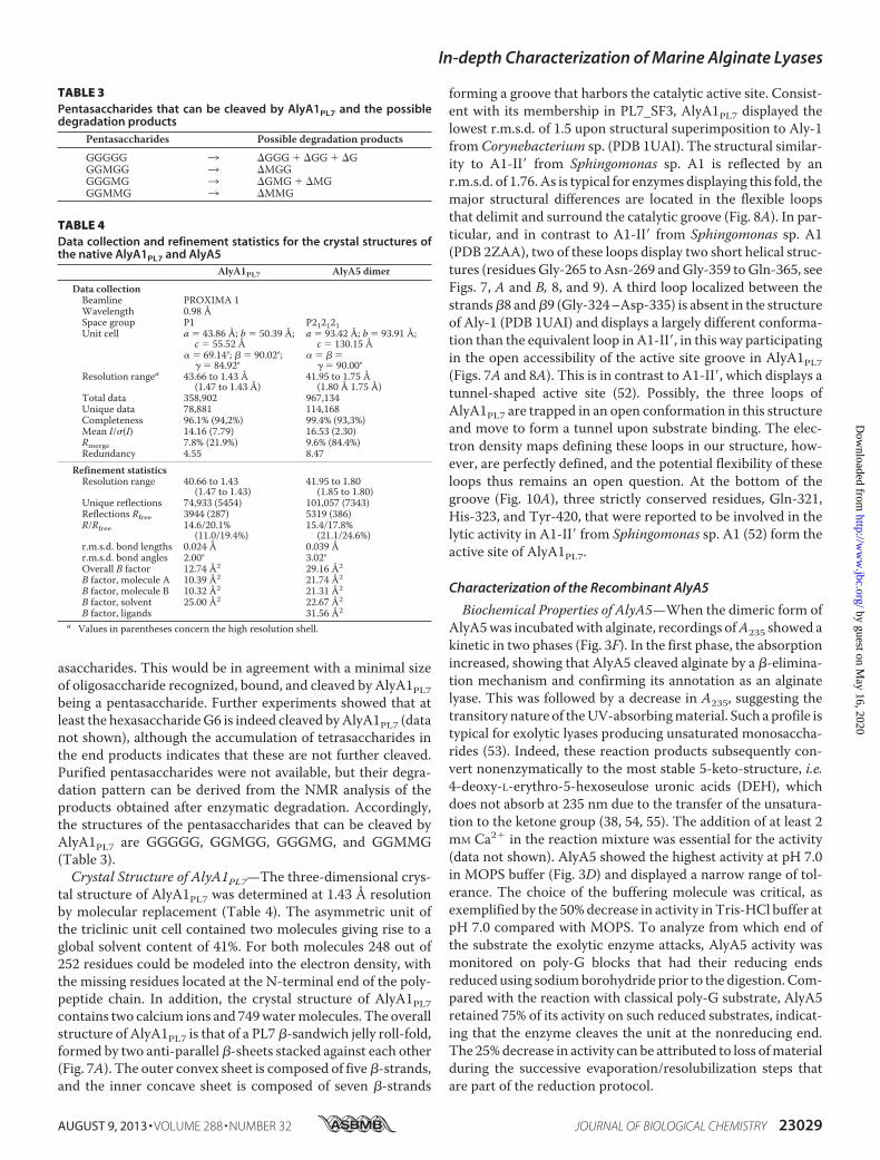
asaccharides. This would be in agreement with a minimal sizeof oligosaccharide recognized, bound, and cleaved by AlyA1PL7being a pentasaccharide. Further experiments showed that atleast the hexasaccharideG6 is indeed cleaved byAlyA1PL7 (datanot shown), although the accumulation of tetrasaccharides inthe end products indicates that these are not further cleaved.Purified pentasaccharides were not available, but their degra-dation pattern can be derived from the NMR analysis of theproducts obtained after enzymatic degradation. Accordingly,the structures of the pentasaccharides that can be cleaved byAlyA1PL7 are GGGGG, GGMGG, GGGMG, and GGMMG(Table 3).Crystal Structure of AlyA1PL7—The three-dimensional crys-
tal structure of AlyA1PL7 was determined at 1.43 Å resolutionby molecular replacement (Table 4). The asymmetric unit ofthe triclinic unit cell contained two molecules giving rise to aglobal solvent content of 41%. For both molecules 248 out of252 residues could be modeled into the electron density, withthe missing residues located at the N-terminal end of the poly-peptide chain. In addition, the crystal structure of AlyA1PL7contains two calcium ions and 749watermolecules. The overallstructure of AlyA1PL7 is that of a PL7 �-sandwich jelly roll-fold,formed by two anti-parallel�-sheets stacked against each other(Fig. 7A). The outer convex sheet is composed of five�-strands,and the inner concave sheet is composed of seven �-strands
forming a groove that harbors the catalytic active site. Consist-ent with its membership in PL7_SF3, AlyA1PL7 displayed thelowest r.m.s.d. of 1.5 upon structural superimposition to Aly-1from Corynebacterium sp. (PDB 1UAI). The structural similar-ity to A1-II� from Sphingomonas sp. A1 is reflected by anr.m.s.d. of 1.76.As is typical for enzymes displaying this fold, themajor structural differences are located in the flexible loopsthat delimit and surround the catalytic groove (Fig. 8A). In par-ticular, and in contrast to A1-II� from Sphingomonas sp. A1(PDB 2ZAA), two of these loops display two short helical struc-tures (residues Gly-265 toAsn-269 andGly-359 toGln-365, seeFigs. 7, A and B, 8, and 9). A third loop localized between thestrands�8 and�9 (Gly-324–Asp-335) is absent in the structureof Aly-1 (PDB 1UAI) and displays a largely different conforma-tion than the equivalent loop in A1-II�, in this way participatingin the open accessibility of the active site groove in AlyA1PL7(Figs. 7A and 8A). This is in contrast to A1-II�, which displays atunnel-shaped active site (52). Possibly, the three loops ofAlyA1PL7 are trapped in an open conformation in this structureand move to form a tunnel upon substrate binding. The elec-tron density maps defining these loops in our structure, how-ever, are perfectly defined, and the potential flexibility of theseloops thus remains an open question. At the bottom of thegroove (Fig. 10A), three strictly conserved residues, Gln-321,His-323, and Tyr-420, that were reported to be involved in thelytic activity in A1-II� from Sphingomonas sp. A1 (52) form theactive site of AlyA1PL7.
Characterization of the Recombinant AlyA5
Biochemical Properties of AlyA5—When the dimeric form ofAlyA5was incubatedwith alginate, recordings ofA235 showed akinetic in two phases (Fig. 3F). In the first phase, the absorptionincreased, showing that AlyA5 cleaved alginate by a �-elimina-tion mechanism and confirming its annotation as an alginatelyase. This was followed by a decrease in A235, suggesting thetransitory nature of theUV-absorbingmaterial. Such a profile istypical for exolytic lyases producing unsaturated monosaccha-rides (53). Indeed, these reaction products subsequently con-vert nonenzymatically to the most stable 5-keto-structure, i.e.4-deoxy-L-erythro-5-hexoseulose uronic acids (DEH), whichdoes not absorb at 235 nm due to the transfer of the unsatura-tion to the ketone group (38, 54, 55). The addition of at least 2mM Ca2� in the reaction mixture was essential for the activity(data not shown). AlyA5 showed the highest activity at pH 7.0in MOPS buffer (Fig. 3D) and displayed a narrow range of tol-erance. The choice of the buffering molecule was critical, asexemplified by the 50%decrease in activity inTris-HCl buffer atpH 7.0 compared with MOPS. To analyze from which end ofthe substrate the exolytic enzyme attacks, AlyA5 activity wasmonitored on poly-G blocks that had their reducing endsreducedusing sodiumborohydride prior to the digestion.Com-pared with the reaction with classical poly-G substrate, AlyA5retained 75% of its activity on such reduced substrates, indicat-ing that the enzyme cleaves the unit at the nonreducing end.The 25%decrease in activity can be attributed to loss ofmaterialduring the successive evaporation/resolubilization steps thatare part of the reduction protocol.
TABLE 3Pentasaccharides that can be cleaved by AlyA1PL7 and the possibledegradation products
Pentasaccharides Possible degradation products
GGGGG 3 �GGG � �GG � �GGGMGG 3 �MGGGGGMG 3 �GMG � �MGGGMMG 3 �MMG
TABLE 4Data collection and refinement statistics for the crystal structures ofthe native AlyA1PL7 and AlyA5
AlyA1PL7 AlyA5 dimer
Data collectionBeamline PROXIMA 1Wavelength 0.98 ÅSpace group P1 P212121Unit cell a � 43.86 Å; b � 50.39 Å;
c � 55.52 Åa � 93.42 Å; b � 93.91 Å;
c � 130.15 Å� � 69.14°; � � 90.02°;
� � 84.92°� � � �
� � 90.00°Resolution rangea 43.66 to 1.43 Å
(1.47 to 1.43 Å)41.95 to 1.75 Å
(1.80 Å 1.75 Å)Total data 358,902 967,134Unique data 78,881 114,168Completeness 96.1% (94,2%) 99.4% (93,3%)Mean I/�(I) 14.16 (7.79) 16.53 (2.30)Rmerge 7.8% (21.9%) 9.6% (84.4%)Redundancy 4.55 8.47
Refinement statisticsResolution range 40.66 to 1.43
(1.47 to 1.43)41.95 to 1.80
(1.85 to 1.80)Unique reflections 74,933 (5454) 101,057 (7343)Reflections Rfree 3944 (287) 5319 (386)R/Rfree 14.6/20.1%
(11.0/19.4%)15.4/17.8%
(21.1/24.6%)r.m.s.d. bond lengths 0.024 Å 0.039 År.m.s.d. bond angles 2.00° 3.02°Overall B factor 12.74 Å2 29.16 Å2
B factor, molecule A 10.39 Å2 21.74 Å2
B factor, molecule B 10.32 Å2 21.31 Å2
B factor, solvent 25.00 Å2 22.67 Å2
B factor, ligands 31.56 Å2
a Values in parentheses concern the high resolution shell.
In-depth Characterization of Marine Alginate Lyases
AUGUST 9, 2013 • VOLUME 288 • NUMBER 32 JOURNAL OF BIOLOGICAL CHEMISTRY 23029
by guest on May 16, 2020
http://ww
w.jbc.org/
Dow
nloaded from
Substrate Specificity of AlyA5—To assess the substrate spec-ificity of AlyA5, the activity was tested onMG,M, andGblocks.The reactions were followed by the absorbance at 235 nm andby 1H NMR (Figs. 3F and 11). The three substrates were
degraded, indicating that the enzyme has a broad substrate tol-erance and can cleave M-M, M-G, and G-G linkages at thenonreducing end. The activity was depending on the blockstructure. Specific activities were calculated in the first phase of
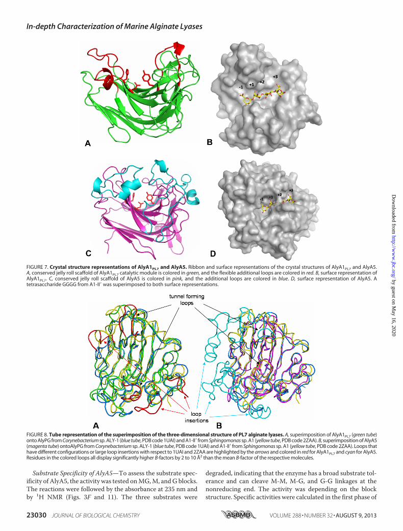
FIGURE 7. Crystal structure representations of AlyA1PL7 and AlyA5. Ribbon and surface representations of the crystal structures of AlyA1PL7 and AlyA5.A, conserved jelly roll scaffold of AlyA1PL7 catalytic module is colored in green, and the flexible additional loops are colored in red. B, surface representation ofAlyA1PL7. C, conserved jelly roll scaffold of AlyA5 is colored in pink, and the additional loops are colored in blue. D, surface representation of AlyA5. Atetrasaccharide GGGG from A1-II� was superimposed to both surface representations.
FIGURE 8. Tube representation of the superimposition of the three-dimensional structure of PL7 alginate lyases. A, superimposition of AlyA1PL7 (green tube)onto AlyPG from Corynebacterium sp. ALY-1 (blue tube, PDB code 1UAI) and A1-II� from Sphingomonas sp. A1 (yellow tube, PDB code 2ZAA). B, superimposition of AlyA5(magenta tube) ontoAlyPG from Corynebacterium sp. ALY-1 (blue tube, PDB code 1UAI) and A1-II� from Sphingomonas sp. A1 (yellow tube, PDB code 2ZAA). Loops thathave different configurations or large loop insertions with respect to 1UAI and 2ZAA are highlighted by the arrows and colored in red for AlyA1PL7 and cyan for AlyA5.Residues in the colored loops all display significantly higher B-factors by 2 to 10 Å2 than the mean B-factor of the respective molecules.
In-depth Characterization of Marine Alginate Lyases
23030 JOURNAL OF BIOLOGICAL CHEMISTRY VOLUME 288 • NUMBER 32 • AUGUST 9, 2013
by guest on May 16, 2020
http://ww
w.jbc.org/
Dow
nloaded from
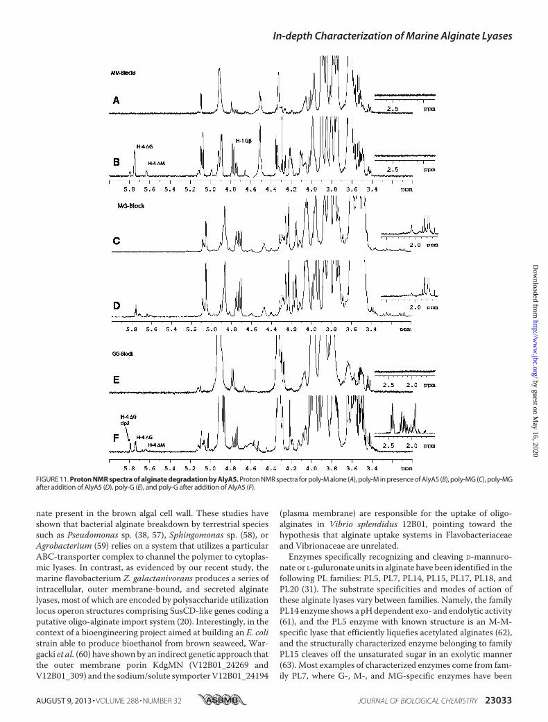
the reaction, corresponding to the increase in A235 (Fig. 3F).AlyA5 was much more active on G blocks (449.3 units�mg�1)than on MG blocks (13.5 units�mg�1) or M blocks (2.0units�mg�1). On the NMR spectra (Fig. 11), in addition to thecharacteristic H4� signals indicating formation of unsaturatedoligosaccharides, other signals were observed in the region1.6–2.6 and 3–4 ppm (see below).Characterization of End Products—The degradation of
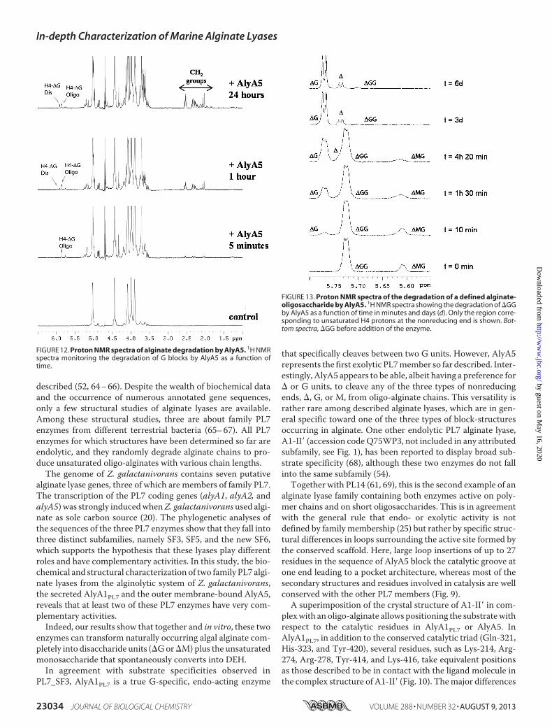
poly-G was followed by NMR to characterize the end productsresulting from AlyA5 activity. Fig. 12 shows that during thecourse of the reaction, the concentration of the disaccharide�G increased although the concentration of the longer unsat-urated oligosaccharides decreased. A small amount of theunstable �-(4,5)-unsaturated pyranose monomer was alsoidentified in the NMR spectra. As the reaction progressed, sig-nals corresponding to protons from CH2 groups appeared in
the NMR spectra (Fig. 12). These proton signals have chemicalshifts typical for CH2 groups close to a keto group or hemiketal(56). Thus, the NMR data confirm that one of the reactionproducts is DEH resulting from the spontaneous conversion ofthe unsaturated monosaccharide �. The concentration of theunsaturated disaccharide always stays low during the course ofthe reaction, compared with the signal intensities from DEH.This indicates that �G is a minor product of the degradationreaction.Minimal Recognition—Incubation of the unsaturated trisac-
charide�GGwithAlyA5 resulted in the formation of� and�Gdetected by 1H NMR (Fig. 13). The amount of �G was muchhigher than that of � confirming that the unsaturated mono-saccharide is not stable in aqueous solution. Concordantly, sig-nals corresponding to deoxy protons appeared in the 1.5–2.6ppm region of the 1H NMR spectra. As seen in Fig. 13, some
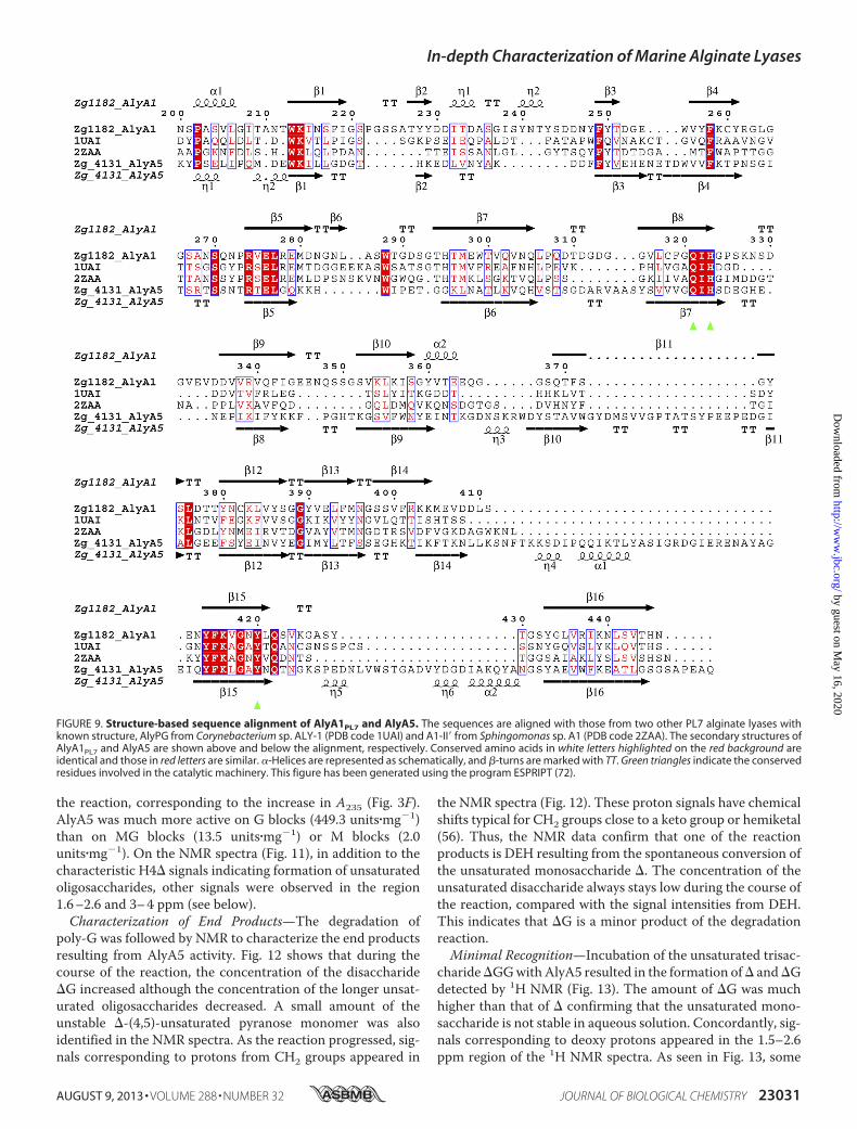
FIGURE 9. Structure-based sequence alignment of AlyA1PL7 and AlyA5. The sequences are aligned with those from two other PL7 alginate lyases withknown structure, AlyPG from Corynebacterium sp. ALY-1 (PDB code 1UAI) and A1-II� from Sphingomonas sp. A1 (PDB code 2ZAA). The secondary structures ofAlyA1PL7 and AlyA5 are shown above and below the alignment, respectively. Conserved amino acids in white letters highlighted on the red background areidentical and those in red letters are similar. �-Helices are represented as schematically, and �-turns are marked with TT. Green triangles indicate the conservedresidues involved in the catalytic machinery. This figure has been generated using the program ESPRIPT (72).
In-depth Characterization of Marine Alginate Lyases
AUGUST 9, 2013 • VOLUME 288 • NUMBER 32 JOURNAL OF BIOLOGICAL CHEMISTRY 23031
by guest on May 16, 2020
http://ww
w.jbc.org/
Dow
nloaded from
�MG was present as traces in the solution containing �GG.�MGwas degraded during the course of the reaction into� and�G indicating that AlyA5 is also able to cleave the �M glyco-sidic linkage. The fact that �M was not observed strengthens
the conclusion that AlyA5 attacks its substrate from the nonre-ducing end. The enzyme was also active on �MM producing� � �M (data not shown). The activity on �GMwas not stud-ied, but the combined data above suggest that this compoundshould also be a substrate for AlyA5. The saturated oligosac-charides MMM and GGG were also found to be substrates ofAlyA5, producing �M and �G, respectively. Disaccharideswere not substrate for the enzyme. Thus, the minimal recogni-tion oligosaccharides are saturated and unsaturated trisaccha-rides, and the enzyme is able to cleave aG,M, or� unit from thenonreducing end.Crystal Structure of AlyA5—AlyA5 represents the first three-
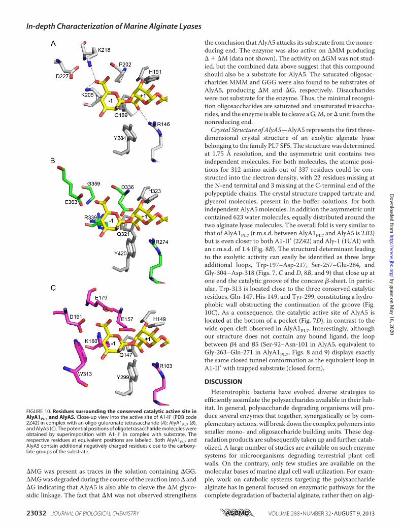
dimensional crystal structure of an exolytic alginate lyasebelonging to the family PL7 SF5. The structure was determinedat 1.75 Å resolution, and the asymmetric unit contains twoindependent molecules. For both molecules, the atomic posi-tions for 312 amino acids out of 337 residues could be con-structed into the electron density, with 22 residues missing atthe N-end terminal and 3 missing at the C-terminal end of thepolypeptide chains. The crystal structure trapped tartrate andglycerol molecules, present in the buffer solutions, for bothindependent AlyA5molecules. In addition the asymmetric unitcontained 623 water molecules, equally distributed around thetwo alginate lyase molecules. The overall fold is very similar tothat of AlyA1PL7 (r.m.s.d. between AlyA1PL7 and AlyA5 is 2.02)but is even closer to both A1-II� (2Z42) and Aly-1 (1UAI) withan r.m.s.d. of 1.4 (Fig. 8B). The structural determinant leadingto the exolytic activity can easily be identified as three largeadditional loops, Trp-197–Asp-217, Ser-257–Glu-284, andGly-304–Asp-318 (Figs. 7, C and D, 8B, and 9) that close up atone end the catalytic groove of the concave �-sheet. In partic-ular, Trp-313 is located close to the three conserved catalyticresidues, Gln-147, His-149, and Tyr-299, constituting a hydro-phobic wall obstructing the continuation of the groove (Fig.10C). As a consequence, the catalytic active site of AlyA5 islocated at the bottom of a pocket (Fig. 7D), in contrast to thewide-open cleft observed in AlyA1PL7. Interestingly, althoughour structure does not contain any bound ligand, the loopbetween �4 and �5 (Ser-92–Asn-101 in AlyA5, equivalent toGly-263–Gln-271 in AlyA1PL7, Figs. 8 and 9) displays exactlythe same closed tunnel conformation as the equivalent loop inA1-II� with trapped substrate (closed form).
DISCUSSION
Heterotrophic bacteria have evolved diverse strategies toefficiently assimilate the polysaccharides available in their hab-itat. In general, polysaccharide degrading organisms will pro-duce several enzymes that together, synergistically or by com-plementary actions, will break down the complex polymers intosmaller mono- and oligosaccharide building units. These deg-radation products are subsequently taken up and further catab-olized. A large number of studies are available on such enzymesystems for microorganisms degrading terrestrial plant cellwalls. On the contrary, only few studies are available on themolecular bases of marine algal cell wall utilization. For exam-ple, work on catabolic systems targeting the polysaccharidealginate has in general focused on enzymatic pathways for thecomplete degradation of bacterial alginate, rather then on algi-
FIGURE 10. Residues surrounding the conserved catalytic active site inAlyA1PL7 and AlyA5. Close-up view into the active site of A1-II� (PDB code2Z42) in complex with an oligo-guluronate tetrasaccharide (A); AlyA1PL7 (B),and AlyA5 (C). The potential positions of oligotetrasaccharide molecules wereobtained by superimposition with A1-II� in complex with substrate. Therespective residues at equivalent positions are labeled. Both AlyA1PL7 andAlyA5 contain additional negatively charged residues close to the carboxy-late groups of the substrate.
In-depth Characterization of Marine Alginate Lyases
23032 JOURNAL OF BIOLOGICAL CHEMISTRY VOLUME 288 • NUMBER 32 • AUGUST 9, 2013
by guest on May 16, 2020
http://ww
w.jbc.org/
Dow
nloaded from
nate present in the brown algal cell wall. These studies haveshown that bacterial alginate breakdown by terrestrial speciessuch as Pseudomonas sp. (38, 57), Sphingomonas sp. (58), orAgrobacterium (59) relies on a system that utilizes a particularABC-transporter complex to channel the polymer to cytoplas-mic lyases. In contrast, as evidenced by our recent study, themarine flavobacterium Z. galactanivorans produces a series ofintracellular, outer membrane-bound, and secreted alginatelyases, most of which are encoded by polysaccharide utilizationlocus operon structures comprising SusCD-like genes coding aputative oligo-alginate import system (20). Interestingly, in thecontext of a bioengineering project aimed at building an E. colistrain able to produce bioethanol from brown seaweed, War-gacki et al. (60) have shownby an indirect genetic approach thatthe outer membrane porin KdgMN (V12B01_24269 andV12B01_309) and the sodium/solute symporterV12B01_24194
(plasma membrane) are responsible for the uptake of oligo-alginates in Vibrio splendidus 12B01, pointing toward thehypothesis that alginate uptake systems in Flavobacteriaceaeand Vibrionaceae are unrelated.Enzymes specifically recognizing and cleaving D-mannuro-
nate or L-guluronate units in alginate have been identified in thefollowing PL families: PL5, PL7, PL14, PL15, PL17, PL18, andPL20 (31). The substrate specificities and modes of action ofthese alginate lyases vary between families. Namely, the familyPL14 enzyme shows a pHdependent exo- and endolytic activity(61), and the PL5 enzyme with known structure is an M-M-specific lyase that efficiently liquefies acetylated alginates (62),and the structurally characterized enzyme belonging to familyPL15 cleaves off the unsaturated sugar in an exolytic manner(63). Most examples of characterized enzymes come from fam-ily PL7, where G-, M-, and MG-specific enzymes have been
FIGURE 11. Proton NMR spectra of alginate degradation by AlyA5. Proton NMR spectra for poly-M alone (A), poly-M in presence of AlyA5 (B), poly-MG (C), poly-MGafter addition of AlyA5 (D), poly-G (E), and poly-G after addition of AlyA5 (F).
In-depth Characterization of Marine Alginate Lyases
AUGUST 9, 2013 • VOLUME 288 • NUMBER 32 JOURNAL OF BIOLOGICAL CHEMISTRY 23033
by guest on May 16, 2020
http://ww
w.jbc.org/
Dow
nloaded from
described (52, 64–66). Despite the wealth of biochemical dataand the occurrence of numerous annotated gene sequences,only a few structural studies of alginate lyases are available.Among these structural studies, three are about family PL7enzymes from different terrestrial bacteria (65–67). All PL7enzymes for which structures have been determined so far areendolytic, and they randomly degrade alginate chains to pro-duce unsaturated oligo-alginates with various chain lengths.The genome of Z. galactanivorans contains seven putative
alginate lyase genes, three of which are members of family PL7.The transcription of the PL7 coding genes (alyA1, alyA2, andalyA5) was strongly inducedwhenZ. galactanivorans used algi-nate as sole carbon source (20). The phylogenetic analyses ofthe sequences of the three PL7 enzymes show that they fall intothree distinct subfamilies, namely SF3, SF5, and the new SF6,which supports the hypothesis that these lyases play differentroles and have complementary activities. In this study, the bio-chemical and structural characterization of two family PL7 algi-nate lyases from the alginolytic system of Z. galactanivorans,the secreted AlyA1PL7 and the outer membrane-bound AlyA5,reveals that at least two of these PL7 enzymes have very com-plementary activities.Indeed, our results show that together and in vitro, these two
enzymes can transform naturally occurring algal alginate com-pletely into disaccharide units (�Gor�M)plus the unsaturatedmonosaccharide that spontaneously converts into DEH.In agreement with substrate specificities observed in
PL7_SF3, AlyA1PL7 is a true G-specific, endo-acting enzyme
that specifically cleaves between two G units. However, AlyA5represents the first exolytic PL7member so far described. Inter-estingly, AlyA5 appears to be able, albeit having a preference for� or G units, to cleave any of the three types of nonreducingends, �, G, or M, from oligo-alginate chains. This versatility israther rare among described alginate lyases, which are in gen-eral specific toward one of the three types of block-structuresoccurring in alginate. One other endolytic PL7 alginate lyase,A1-II� (accession codeQ75WP3, not included in any attributedsubfamily, see Fig. 1), has been reported to display broad sub-strate specificity (68), although these two enzymes do not fallinto the same subfamily (54).Together with PL14 (61, 69), this is the second example of an
alginate lyase family containing both enzymes active on poly-mer chains and on short oligosaccharides. This is in agreementwith the general rule that endo- or exolytic activity is notdefined by family membership (25) but rather by specific struc-tural differences in loops surrounding the active site formed bythe conserved scaffold. Here, large loop insertions of up to 27residues in the sequence of AlyA5 block the catalytic groove atone end leading to a pocket architecture, whereas most of thesecondary structures and residues involved in catalysis are wellconserved with the other PL7 members (Fig. 9).A superimposition of the crystal structure of A1-II� in com-
plexwith an oligo-alginate allows positioning the substratewithrespect to the catalytic residues in AlyA1PL7 or AlyA5. InAlyA1PL7, in addition to the conserved catalytic triad (Gln-321,His-323, and Tyr-420), several residues, such as Lys-214, Arg-274, Arg-278, Tyr-414, and Lys-416, take equivalent positionsas those described to be in contact with the ligand molecule inthe complex structure of A1-II� (Fig. 10). Themajor differences
FIGURE 13. Proton NMR spectra of the degradation of a defined alginate-oligosaccharide by AlyA5. 1H NMR spectra showing the degradation of �GGby AlyA5 as a function of time in minutes and days (d). Only the region corre-sponding to unsaturated H4 protons at the nonreducing end is shown. Bot-tom spectra, �GG before addition of the enzyme.
FIGURE 12. Proton NMR spectra of alginate degradation by AlyA5. 1H NMRspectra monitoring the degradation of G blocks by AlyA5 as a function oftime.
In-depth Characterization of Marine Alginate Lyases
23034 JOURNAL OF BIOLOGICAL CHEMISTRY VOLUME 288 • NUMBER 32 • AUGUST 9, 2013
by guest on May 16, 2020
http://ww
w.jbc.org/
Dow
nloaded from
between AlyA1PL7 and the A1-II�-substrate complex are as fol-lows. First, there is no equivalent to Asn-141 (A1-II� number-ing) that is part of the first loop (residues Cys-260 to Pro-273 inAlyA1PL7; Fig. 9). Second, Gln-97 is replaced by a shorter resi-due, Asn-216 in AlyA1PL7, making the distance to interact withthe substrate much longer. Third, in AlyA1PL7, an aspartateresidue (Asp-336, absent in A1-II�) is positioned close to thecatalytic active site.But themost striking feature that differentiatesAlyA1PL7 and
AlyA5 from other PL7 alginate lyases is the presence of severalmore negatively charged aspartate and glutamate residues inthe vicinity of the�1,�1 cleavage site (Fig. 10). InAlyA1PL7, theequivalent position of Pro-202 of A1-II� (Fig. 10A) is occupiedbyAsp-336 (Fig. 10B), whichwould be both in sterical clash andelectrostatic repulsion with the uronate group if the �1 G unitwould bind exactly in the same position as deduced from thesuperimposition with A1-II�. In A1-II�, Lys-218 interacts withthe G unit bound to the �1-binding site. The equivalent regionin AlyA1PL7 includes strand �9 and helix �2. The correspond-ing strand in A1-II� is badly superimposed with �9, and there isno equivalent of helix �2. As a consequence, the C� of Lys-218is roughly substituted by a glycine (Gly-359), and its side chainis spatially replaced by the acidic group of Glu-363. Again, thisconformation appears incompatible with a similar binding of aG unit in �1.In AlyA5 there are even three acidic residues that would
come into sterical clash with alginate G units if bound as inA1-II� (Fig. 10C); here, Pro-202 is replaced by Glu-157, andagain the region, including Lys-218 in A1-II�, is poorly con-served, with strands �9 and �10 of AlyA5 significantly dis-placed in comparison with the corresponding strands in A1-II�.Thus, Lys-218 is spatially substituted in AlyA5 by a cluster oftwo acidic residues, Glu-179 and Asp-191, with their sidechains pointing toward the carboxylic group of the modeled Gunit in subsite �1. Remarkably, these residues are situated inthe extended loops that are found in variable conformations inthe different PL7 alginate lyase structures. In our crystal struc-ture of AlyA5 the loops are closed above the active site groove,although closed forms in other PL7 enzymes are only observedin presence of substrate. Overall, the different loop conforma-tions observed in the various crystal structures are indicative offlexible loops, and most probably loop movements assist sub-strate binding and release. Together with the finding thatchelating agents inhibited the lyase activities, these structuralfeatures lead us to hypothesize that substrate binding and rec-ognition inAlyA1PL7 andAlyA5 includes interactionsmediatedby calcium ions. Loop movements displacing the acidic resi-dues together with calcium binding could bring the residuesfrom their actual conformations into positions for productiveinteractions of the enzyme with its natural calcium-chelatedalginate substrate. A similar substrate-binding mode, involvingacidic amino acids and calcium ions to a negatively chargedsubstrate, is observed in other polysaccharide lyases, such aschondroitin B lyase (70) andpectate lyases (71). The rationale ofsuch a variation in the binding and recognition mode of thehere studied PL7 alginate lyases, as compared with that of thepreviously described enzymes, would be found in the differentnature of alginate originating from bacteria or from brown sea-
weeds. Indeed, the negative charges of algal alginates are notmasked by acetyl group as in bacterial alginates. Moreover,alginate chains have multiple interactions with other cellwall compounds, polysaccharides, phlorotannins, and pro-teins but also inorganic ligands such as calcium or iodineions (2, 13). In conclusion, our biochemical and structuralanalyses of these first marine alginate lyases demonstratethat they have indeed complementary roles in the degradation ofalgal alginate. Moreover, these results shed light on the molec-ular basis for adapted specificity and binding mode to theirnatural substrate. Further work is under way to confirm thehypothesis described here and to proceed with the character-ization of all the other enzymatic players of the complete algi-nolytic system of Z. galactanivorans.
Acknowledgments—We are indebted to Andrew Thompson andPierre Legrand for help and support during data collection and treat-ment at beamline PROXIMA 1, SOLEIL (French Synchrotron at St.Auban).
REFERENCES1. Duarte, C. C.,Middelburg, J. J., andCaraco,N. (2005)Major role ofmarine
vegetation on the oceanic carbon cycle. Biogeosciences 2, 1–82. Popper, Z. A., Michel, G., Hervé, C., Domozych, D. S., Willats, W. G.,
Tuohy,M.G., Kloareg, B., and Stengel, D. B. (2011) Evolution and diversityof plant cell walls: from algae to flowering plants.Annu. Rev. Plant Biol. 62,567–590
3. Vreeland, V.,Waite, J. H., and Epstein, L. (1998) Polyphenols and oxidasesin substratum adhesion by marine algae and mussels. J. Phycol. 34, 1–18
4. Schoenwaelder,M. E., andWiencke, C. (2000) Phenolic compounds in theembryo development of several NorthernHemisphere fucoids. Plant Biol.2, 24–33
5. Quatrano, R. S., and Stevens, P. T. (1976) Cell wall assembly in Fucuszygotes: I. Characterization of the polysaccharide components. PlantPhysiol. 58, 224–231
6. Smidsrod, O., and Draget, K. I. (1996) Chemistry and physical propertiesof alginates. Carbohydr. Eur. 14, 6–13
7. Gacesa, P. (1988) Alginates. Carbohydr. Polym. 8, 161–1828. Grant, G. T., Morris, E. R., Rees, D. A., Smith, P. J., and Thom, D. (1973)
Biological interactions between polysaccharides and divalent cations: theegg-box model. FEBS Lett. 32, 195–198
9. Haug, A., Larsen, B., and Smidsrød, O. (1974) Uronic acid sequence inalginate from different sources. Carbohydr. Res. 32, 217–225
10. Kloareg, B., and Quatrano, R. (1988) Structure of the cell walls of marinealgae and ecophysiological functions of the matrix polysaccharides.Oceanogr. Mar. Biol. Ann. Rev. 26, 259–315
11. Skriptsova, A., Khomenko, V., and Isakov, V. V. (2004) Seasonal changesin growth rate,morphology and alginate content inUndaria pinnatifida atthe northern limit in the Sea of Japan. J. Appl. Phycol. 16, 17–21
12. Craigie, J. S.,Morris, E. R., Rees, D. A., and Thom,D. (1984) Alginate blockstructure in Phaeophyceae from Nova-Scotia-Variation with species, en-vironment and tissue-type. Carbohydr. Polym. 4, 237–252
13. Michel, G., Tonon, T., Scornet, D., Cock, J. M., and Kloareg, B. (2010) Thecell wall polysaccharide metabolism of the brown alga Ectocarpus silicu-losus. Insights into the evolution of extracellularmatrix polysaccharides inEukaryotes. New Phytologist. 188, 82–97
14. Ramsey, D. M., and Wozniak, D. J. (2005) Understanding the control ofPseudomonas aeruginosa alginate synthesis and the prospects formanage-ment of chronic infections in cystic fibrosis.Mol. Microbiol. 56, 309–322
15. Remminghorst, U., and Rehm, B. H. (2006) Alg44, a unique protein re-quired for alginate biosynthesis in Pseudomonas aeruginosa. FEBS Lett.580, 3883–3888
16. Skjåk-Braek, G., Grasdalen, H., and Larsen, B. (1986) Monomer sequenceand acetylation pattern in some bacterial alginates. Carbohydr. Res. 154,
In-depth Characterization of Marine Alginate Lyases
AUGUST 9, 2013 • VOLUME 288 • NUMBER 32 JOURNAL OF BIOLOGICAL CHEMISTRY 23035
by guest on May 16, 2020
http://ww
w.jbc.org/
Dow
nloaded from
239–25017. Charrier, B., Coelho, S. M., Le Bail, A., Tonon, T., Michel, G., Potin, P.,
Kloareg, B., Boyen, C., Peters, A. F., and Cock, J. M. (2008) Developmentand physiology of the brown alga Ectocarpus siliculosus: two centuries ofresearch. New Phytologist. 177, 319–332
18. Nyvall, P., Corre, E., Boisset, C., Barbeyron, T., Rousvoal, S., Scornet, D.,Kloareg, B., and Boyen, C. (2003) Characterization of mannuronan C-5-epimerase genes from the brown alga Laminaria digitata. Plant Physiol.133, 726–735
19. Roeder, V., Collén, J., Rousvoal, S., Corre, E., Leblanc, C., and Boyen, C.(2005) Identification of stress gene transcripts in Laminaria digitata (pha-eophyceae) protoplast cultures by expressed sequence tag analysis. J. Phy-col. 41, 1227–1235
20. Thomas, F., Barbeyron, T., Tonon, T., Génicot, S., Czjzek,M., andMichel,G. (2012) Characterization of the first alginolytic operons in a marinebacterium: from their emergence in marine Flavobacteria to their inde-pendent transfers to marine Proteobacteria and human gut Bacteroides.Environ. Microbiol. 14, 2379–2394
21. Jam, M., Flament, D., Allouch, J., Potin, P., Thion, L., Kloareg, B., Czjzek,M., Helbert, W., Michel, G., and Barbeyron, T. (2005) The endo-�-aga-rases AgaA and AgaB from the marine bacterium Zobellia galactaniv-orans: two paralogue enzymes with different molecular organizations andcatalytic behaviours. Biochem. J. 385, 703–713
22. Rebuffet, E., Groisillier, A., Thompson, A., Jeudy, A., Barbeyron, T.,Czjzek, M., and Michel, G. (2011) Discovery and structural characteriza-tion of a novel glycosidase family of marine origin. Environ. Microbiol. 13,1253–1270
23. Hehemann, J. H., Correc, G., Thomas, F., Bernard, T., Barbeyron, T., Jam,M., Helbert,W.,Michel, G., and Czjzek,M. (2012) Biochemical and struc-tural characterization of the complex agarolytic enzyme system fromthe marine bacterium Zobellia galactanivorans. J. Biol. Chem. 287,30571–30584
24. Thomas, F., Barbeyron, T., and Michel, G. (2011) Evaluation of referencegenes for real time quantitative PCR in the marine flavobacterium Zobel-lia galactanivorans. J. Microbiol. Methods 84, 61–66
25. Lombard, V., Bernard, T., Rancurel, C., Brumer, H., Coutinho, P. M., andHenrissat, B. (2010) A hierarchical classification of polysaccharide lyasesfor glycogenomics. Biochem. J. 432, 437–444
26. Koropatkin, N. M., Martens, E. C., Gordon, J. I., and Smith, T. J. (2008)Starch catabolism by a prominent human gut symbiont is directed by therecognition of amylose helices. Structure 16, 1105–1115
27. Koropatkin, N.M., Cameron, E. A., andMartens, E. C. (2012) How glycanmetabolism shapes the human gut microbiota. Nat. Rev. Microbiol. 10,323–335
28. Shimokawa, T., Yoshida, S., Kusakabe, I., Takeuchi, T., Murata, K., andKobayashi, H. (1997) Some properties and action mode of (134)-�-L-guluronan lyase from Enterobacter cloacae M-1. Carbohydr. Res. 304,125–132
29. Abdel-Akher,M.A., and Sandstrom,W.M. (1951) The abnormal reactionof glycine and related compounds with nitrous acid. Arch Biochem. 30,407–413
30. Kidby, D. K., and Davidson, D. J. (1973) A convenient ferricyanide estima-tion of reducing sugars in the nanomole range. Anal. Biochem. 55,321–325
31. Cantarel, B. L., Coutinho, P. M., Rancurel, C., Bernard, T., Lombard, V.,and Henrissat, B. (2009) The Carbohydrate-Active EnZymes database(CAZy): an expert resource for glycogenomics. Nucleic Acids Res. 37,D233–D238
32. Katoh, K., and Toh, H. (2008) Improved accuracy of multiple ncRNAalignment by incorporating structural information into a MAFFT-basedframework. BMC Bioinformatics 9, 212
33. Tamura, K., Dudley, J., Nei, M., and Kumar, S. (2007) MEGA4: Molecularevolutionary genetics analysis (MEGA) software version 4.0. Mol. Biol.Evol. 24, 1596–1599
34. Guindon, S., andGascuel, O. (2003) A simple, fast, and accurate algorithmto estimate large phylogenies by maximum likelihood. Syst. Biol. 52,696–704
35. Dereeper, A., Guignon, V., Blanc, G., Audic, S., Buffet, S., Chevenet, F.,
Dufayard, J. F., Guindon, S., Lefort, V., Lescot, M., Claverie, J. M., andGascuel, O. (2008) Phylogeny.fr: robust phylogenetic analysis for the non-specialist. Nucleic Acids Res. 36,W465–W469
36. Groisillier, A., Hervé, C., Jeudy, A., Rebuffet, E., Pluchon, P. F., Chevolot,Y., Flament, D., Geslin, C.,Morgado, I.M., Power, D., Branno,M.,Moreau,H., Michel, G., Boyen, C., and Czjzek, M. (2010)MARINE-EXPRESS: tak-ing advantage of high throughput cloning and expression strategies for thepost-genomic analysis of marine organisms.Microb. Cell Fact. 9, 45
37. Studier, F.W. (2005) Protein production by auto-induction in high densityshaking cultures. Protein Expr. Purif. 41, 207–234
38. Preiss, J., and Ashwell, G. (1962) Alginic acid metabolism in bacteria. I.Enzymatic formation of unsaturated oligosaccharides and 4-deoxy-L-erythro-5-hexoseulose uronic acid. J. Biol. Chem. 237, 309–316
39. Kabsch,W. (2010) XDS.Acta Crystallogr. D Biol. Crystallogr. 66, 125–13240. Perrakis, A., Harkiolaki, M., Wilson, K. S., and Lamzin, V. S. (2001) ARP/
wARP and molecular replacement. Acta Crystallogr. D Biol. Crystallogr.57, 1445–1450
41. Murshudov, G. N., Skubák, P., Lebedev, A. A., Pannu, N. S., Steiner, R. A.,Nicholls, R. A., Winn, M. D., Long, F., and Vagin, A. A. (2011) REFMAC5for the refinement of macromolecular crystal structures.Acta Crystallogr.D Biol. Crystallogr. 67, 355–367
42. Collaborative Computational Project No. 4 (1994) The CCP4 suite: pro-grams for protein crystallography. Acta Crystallogr. D Biol. Crystallogr.50, 760–763
43. Emsley, P., and Cowtan, K. (2004) Coot: model-building tools for molec-ular graphics. Acta Crystallogr. D Biol. Crystallogr. 60, 2126–2132
44. Laskowski, R. A., MacArthur, M. W., Moss, D. S., and Thornton, J. M.(1993) PROCHECK: a program to check the stereochemical quality ofprotein structures. J. Appl. Cryst. 26, 283–291
45. Gasteiger, E., Hoogland, C., Gattiker, A., Duvaud, S.,Wilkins,M. R., Appel,R. D., and Bairoch, A. (2005) in The Proteomics Protocols Handbook(Walker, J. M., ed) pp. 571–607, Humana Press Inc., Totowa, NJ
46. Wong, T. Y., Preston, L. A., and Schiller, N. L. (2000) ALGINATE LYASE:Review of major sources and enzyme characteristics, structure-functionanalysis, biological roles, and applications. Annu. Rev. Microbiol. 54,289–340
47. Chavagnat, F., Heyraud, A., Colin-Morel, P., Guinand, M., andWallach, J.(1998) Catalytic properties and specificity of a recombinant, overex-pressed D-mannuronate lyase. Carbohydr. Res. 308, 409–415
48. Ertesvåg, H., Erlien, F., Skjåk-Braek, G., Rehm, B. H., and Valla, S. (1998)Biochemical properties and substrate specificities of a recombinantly pro-duced Azotobacter vinelandii alginate lyase. J. Bacteriol. 180, 3779–3784
49. Gimmestad, M., Ertesvåg, H., Heggeset, T. M., Aarstad, O., Svanem, B. I.,and Valla, S. (2009) Characterization of three new Azotobacter vinelandiialginate lyases, one of which is involved in cyst germination. J. Bacteriol.191, 4845–4853
50. Heyraud, A., Gey, C., Leonard, C., Rochas, C., Girond, S., and Kloareg, B.(1996) NMR spectroscopy analysis of oligoguluronates and oligomannu-ronates prepared by acid or enzymatic hydrolysis of homopolymericblocks of alginic acid. Application to the determination of the substratespecificity of Haliotis tuberculata alginate lyase. Carbohydr. Res. 289,11–23
51. Zhang, Z., Yu, G., Guan, H., Zhao, X., Du, Y., and Jiang, X. (2004) Prepa-ration and structure elucidation of alginate oligosaccharides degraded byalginate lyase from Vibro sp. 510. Carbohydr. Res. 339, 1475–1481
52. Ogura, K., Yamasaki, M., Mikami, B., Hashimoto, W., and Murata, K.(2008) Substrate recognition by family 7 alginate lyase from Sphingomonassp. A1. J. Mol. Biol. 380, 373–385
53. Preiss, J., and Ashwell, G. (1963) Polygalacturonic acid metabolism inbacteria. I. Enzymatic formation of 4-deoxy-L-threo-5-hexoseulose uronicacid. J. Biol. Chem. 238, 1571–1583
54. Hashimoto,W., Miyake, O., Momma, K., Kawai, S., andMurata, K. (2000)Molecular identification of oligoalginate lyase of Sphingomonas sp. strainA1 as one of the enzymes required for complete depolymerization of algi-nate. J. Bacteriol. 182, 4572–4577
55. Ochiai, A., Hashimoto,W., andMurata, K. (2006) A biosystem for alginatemetabolism in Agrobacterium tumefaciens strain C58: molecular identifi-cation of Atu3025 as an exotype family PL-15 alginate lyase. Res. Micro-
In-depth Characterization of Marine Alginate Lyases
23036 JOURNAL OF BIOLOGICAL CHEMISTRY VOLUME 288 • NUMBER 32 • AUGUST 9, 2013
by guest on May 16, 2020
http://ww
w.jbc.org/
Dow
nloaded from
biol. 157, 642–64956. Kuorelahti, S., Jouhten, P., Maaheimo, H., Penttilä, M., and Richard, P.
(2006) L-Galactonate dehydratase is part of the fungal path for D-galac-turonic acid catabolism.Mol. Microbiol. 61, 1060–1068
57. Preiss, J., and Ashwell, G. (1962) Alginic acid metabolism in bacteria. II.The enzymatic reduction of 4-deoxy-L-erythro-5-hexoseulose uronic acidto 2-keto-3-deoxy-D-gluconic acid. J. Biol. Chem. 237, 317–321
58. Momma, K., Okamoto, M., Mishima, Y., Mori, S., Hashimoto, W., andMurata, K. (2000) A novel bacterial ATP-binding cassette transportersystem that allows uptake of macromolecules. J. Bacteriol. 182,3998–4004
59. Hashimoto,W., Kawai, S., andMurata, K. (2010) Bacterial supersystem foralginate import/metabolism and its environmental and bioenergy appli-cations. Bioeng. Bugs 1, 97–109
60. Wargacki, A. J., Leonard, E., Win, M. N., Regitsky, D. D., Santos, C. N.,Kim, P. B., Cooper, S. R., Raisner, R. M., Herman, A., Sivitz, A. B., Laksh-manaswamy, A., Kashiyama, Y., Baker, D., and Yoshikuni, Y. (2012) Anengineered microbial platform for direct biofuel production from brownmacroalgae. Science 335, 308–313
61. Ogura, K., Yamasaki, M., Yamada, T., Mikami, B., Hashimoto, W., andMurata, K. (2009) Crystal structure of family 14 polysaccharide lyase withpH-dependent modes of action. J. Biol. Chem. 284, 35572–35579
62. Yoon, H. J., Hashimoto,W.,Miyake, O.,Murata, K., andMikami, B. (2001)Crystal structure of alginate lyase A1-III complexed with trisaccharideproduct at 2.0 Å resolution. J. Mol. Biol. 307, 9–16
63. Ochiai, A., Yamasaki, M., Mikami, B., Hashimoto, W., and Murata, K.(2010) Crystal structure of exotype alginate lyase Atu3025 from Agrobac-terium tumefaciens. J. Biol. Chem. 285, 24519–24528
64. Murata, K., Inose, T., Hisano, T., Abe, S., Yonemoto, Y., Yamashita, T.,Takagi, M., Sakaguchi, K., Kimura, A., and Imanaka, T. (1993) Bacterialalginate lyase: enzymology, genetics, and application. J. Ferment. Bioeng.76, 427–437
65. Osawa, T., Matsubara, Y., Muramatsu, T., Kimura, M., and Kakuta, Y.
(2005) Crystal structure of the alginate (poly �-L-guluronate) lyase fromCorynebacterium sp. at 1.2 Å resolution. J. Mol. Biol. 345, 1111–1118
66. Yamasaki, M., Moriwaki, S., Miyake, O., Hashimoto, W., Murata, K., andMikami, B. (2004) Structure and function of a hypothetical Pseudomonasaeruginosa protein PA1167 classified into family PL-7: a novel alginatelyase with a �-sandwich fold. J. Biol. Chem. 279, 31863–31872
67. Yamasaki, M., Ogura, K., Hashimoto, W., Mikami, B., and Murata, K.(2005) A structural basis for depolymerization of alginate by polysaccha-ride lyase family-7. J. Mol. Biol. 352, 11–21
68. Miyake, O., Ochiai, A., Hashimoto, W., andMurata, K. (2004) Origin anddiversity of alginate lyases of families PL-5 and �7 in Sphingomonas sp.strain A1. J. Bacteriol. 186, 2891–2896
69. Suzuki, H., Suzuki, K., Inoue, A., and Ojima, T. (2006) A novel oligoalg-inate lyase from abalone,Haliotis discus hannai, that releases disaccharidefrom alginate polymer in an exolytic manner. Carbohydr. Res. 341,1809–1819
70. Michel, G., Pojasek, K., Li, Y., Sulea, T., Linhardt, R. J., Raman, R., Prabha-kar, V., Sasisekharan, R., and Cygler,M. (2004) The structure of chondroi-tin B lyase complexedwith glycosaminoglycan oligosaccharides unravels acalcium-dependent catalytic machinery. J. Biol. Chem. 279, 32882–32896
71. Scavetta, R. D., Herron, S. R., Hotchkiss, A. T., Kita, N., Keen, N. T., Benen,J. A., Kester, H. C., Visser, J., and Jurnak, F. (1999) Structure of a plant cellwall fragment complexed to pectate lyase C. Plant Cell 11, 1081–1092
72. Gouet, P., Robert, X., and Courcelle, E. (2003) ESPript/ENDscript: Ex-tracting and rendering sequence and 3D information from atomic struc-tures of proteins. Nucleic Acids Res. 31, 3320–3323
73. Cowman, M. K., Slahetka, M. F., Hittner, D. M., Kim, J., Forino, M., andGadelrab, G. (1984) Polyacrylamide gel electrophoresis and Alcian Bluestaining of sulphated glycosaminoglycan oligosaccharides. Biochem. J.221, 707–716
74. Zablackis, E., and Perez, J. (1990) A partially pyruvated carrageenan fromHawaiian Grateloupia filicina (Cryptonemiales, Rhodophyta). BotanicaMarina 33, 273–274
In-depth Characterization of Marine Alginate Lyases
AUGUST 9, 2013 • VOLUME 288 • NUMBER 32 JOURNAL OF BIOLOGICAL CHEMISTRY 23037
by guest on May 16, 2020
http://ww
w.jbc.org/
Dow
nloaded from
Barbeyron, Corine Sandström, Gurvan Michel and Mirjam CzjzekFrançois Thomas, Lena C. E. Lundqvist, Murielle Jam, Alexandra Jeudy, Tristan
Their Natural Substrate Reveals Distinct Modes of Action and Exquisite Adaptation togalactanivorans
ZobelliaComparative Characterization of Two Marine Alginate Lyases from
doi: 10.1074/jbc.M113.467217 originally published online June 19, 20132013, 288:23021-23037.J. Biol. Chem.
10.1074/jbc.M113.467217Access the most updated version of this article at doi:
Alerts:
When a correction for this article is posted•
When this article is cited•
to choose from all of JBC's e-mail alertsClick here
http://www.jbc.org/content/288/32/23021.full.html#ref-list-1
This article cites 73 references, 19 of which can be accessed free at
by guest on May 16, 2020
http://ww
w.jbc.org/
Dow
nloaded from