Embed Size (px)
Citation preview
Microbes in the Marine Environment
Chapter 1
Microbes in the Marine Environment
Viewed from space, it is clear why our planet would be better named “Ocean” than “Earth.” More than 70% of the planet’s surface is covered by intercon-nected bodies of water. Life originated in the oceans about 3.5 billion years ago and microbes were the only form of life for two thirds of the planet’s existence. The development and maintenance of all other forms of life depend absolutely on the past and present activities of marine microbes. Yet the vast majority of humans—including many marine scientists—live their lives completely unaware of the diversity and importance of marine microbes. Such understanding is vital as we now live in a period of rapid global change. This chapter introduces the scope of marine microbiology, the different types of marine microbe (viruses, bacteria, archaea, fungi, and protists), and their place in the living world. The role of microbes in the many diverse habitats found in the marine environment is explored.
Key Concepts
• Modernmethodshaveledtonewideasabouttheevolutionofmicrobiallife.
• Marinemicrobesexistinhugenumbersandformamajorcomponentofbiomass on Earth.
• Althoughthereisawiderangeofsizes,mostmarinemicrobesareexceptionally small.
• Awiderangeofphysicalandchemicalconditionsprovidediversespecializedhabitats.
• Microbesaremajorcomponentsofplanktonandmarinesnow.
• Microbesareimportantinsedimentformationandthereisabundantlifebelow the seafloor.
• Microbescolonizethesurfacesofinanimateobjectsandotherlivingorganisms by the formation of biofilms.
© Garland Science 2011
2 Chapter 1
Marine microbiology is one of the most exciting and important areas of modern scienceEver since a detailed study of the microbial world began at the end of the nine-teenth century, microbiologists have asked questions about the diversity ofmicrobial life in the sea, its role in ocean processes, its interactions with other marinelife,anditsimportancetohumans.However,despiteexcellentworkbypioneering scientists, progress in understanding these issues was often slow and most microbiologists remained unaware of this field of study until recently. Toward the end of the twentieth century, a number of factors conspired to pro-pel marine microbiology to the forefront of “mainstream” science, and the sub-sequent application of new technology means that it is now one of the mostexciting and fast-moving areas of investigation. Powerful new tools in molecular biology, remote sensing, and deep-sea exploration have led to astonishing dis-coveries of the abundance and diversity of marine microbial life and its role in global ecology. Continuing new discoveries in marine microbiology necessitate radicalrethinkingofourunderstandingofoceanprocesses.Wenowrealizethevital role that marine microbes play in the maintenance of our planet, a fact that will have great bearing on our ability to respond to problems such as the increase in human population, overexploitation of fisheries, climate change, ocean acidi-fication, and marine pollution. Study of the interactions of marine microbes with other organisms is providing intriguing insights into the phenomena of food webs, symbiosis, and pathogenicity. Since some marine microbes produce dis-ease or damage, we need to study these processes and develop ways to overcome them. Finally, marine microbes have beneficial properties such as the manufac-ture of new products and development of new processes in the growing field of marine biotechnology. This chapter sets the scene for the discussion of all these topicsinthisbook.
Marine microbiology encompasses all microscopic organisms and virusesDefining the terms “microbiology” and “microorganism” is surprisingly difficult! Microbiology is the study of very small organisms that are too small to be seen clearlywiththenakedeye(i.e.lessthanabout1mmdiameter),butmostmicro-biologists are concerned with the activities or molecular properties of microbial communities rather than viewing individual cells with a microscope. The term “microorganism” simply refers to a form of life that falls within the microscopic sizerange,butthereisahugespectrumofdiversityconcealedbythisall-encom-passing term. Indeed, some “microorganisms” are large enough to see without using a microscope, so this is not entirely satisfactory either. Some scientists wouldarguethat thedistinguishingfeaturesofmicroorganismsaresmallsize,unicellularorganization,andosmotrophy(feedingbyabsorptionofnutrients).The osmotrophic characteristic is important because diffusion processes are a majorlimitationtocellsize,asdiscussedinthenextsection.However,thischar-acteristic would exclude many microscopic unicellular protists (a loose group-ingofsimpleeukaryotes),manyofwhichfeedbyphagotrophy(engulfmentofparticles).These“plant-like”or“animal-like”groupsaremostcommonlystudiedbyspecialistswhotraditionallyhaveabackgroundinbotanyorzoology.Indeed,the study of marine protists and recognition that they are microbes with a major role in ocean processes has lagged behind the study of bacteria. Many marine protists are mixotrophic and can switch from photosynthesis to phagotrophic feeding,sotheplantoranimalsimilarityismeaningless.Additionally,virusesaremicroscopic and are obviously included in the remit of microbiologists, but they are not cellular and it can be argued that they are not living organisms either (this question is explored in depth in Chapter 7).There is a huge diversity of inter-connected microbial life forms in the marine environment, and worrying about suchartificialdivisionsisnotgoingtobehelpful;sointhisbookIusetheterm
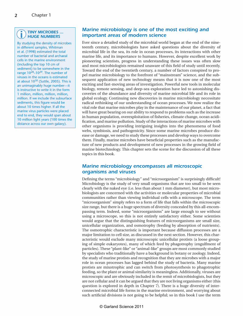
TINY MICROBES ... HUGE NUMBERS
By studying the density of microbes in different samples, Whitman et al. (1998) estimated the total number of bacterial and archaeal cells in the marine environment (including the top 10 cm of sediment) to be somewhere in the range 1028–1029. The number of viruses in the oceans is estimated at about 1030 (Suttle, 2005). This is an unimaginably huge number—it is instructive to write it in the form 1 million, million, million, million, million. If we include the subsurface sediments, this figure would be about 10 times higher. If all the marine virus particles were placed end to end, they would span about 10 million light years (100 times the distance across our own galaxy).
i
© Garland Science 2011
3 Microbes in the Marine Environment
“microbe” as a generic descriptor for all microscopic organisms (i.e. the bacteria, archaea, fungi, and unicellular protists), together with the viruses.
Marine microbes are found in all three domains of cellular lifeBiologists usually rely on the study of morphology and physiological properties to classify living organisms, but these characteristics have always proved frustrat-ingly unhelpful when dealing with microbes. Phylogenetic systems of classifica-tion depend on comparisons of the information content of their macromolecules, especially nucleic acids and proteins. If two organisms are very closely related, we expectthesequenceoftheindividualunitsinamacromoleculetobemoresimi-larthantheywouldbeintwounrelatedorganisms.Inthe1970s,CarlWoeseandcolleaguespioneeredtheuseofribosomalRNA(rRNA)sequencinginordertodevelop a better view of microbial diversity. Our view of the living world has since beenrevolutionizedbyadvancesinthisapproach,madepossiblebecauseoftheparalleladvancesinmolecularbiologicaltechniquesandcomputerprocessingof the large amounts of information generated. Because the secondary structure ofrRNAissoimportantintheribosomeandthevitalcellfunctionofproteinsyn-thesis,basesequencechangesintherRNAmoleculeoccurquiteslowlyinevolu-tion.Infact,somepartsofrRNAarehighlyconservedandsequencecomparisonscan be used to ascertain the similarity of organisms on a broad basis. The meth-odsandapplicationsofthismajortechniquearedescribedinChapter2.
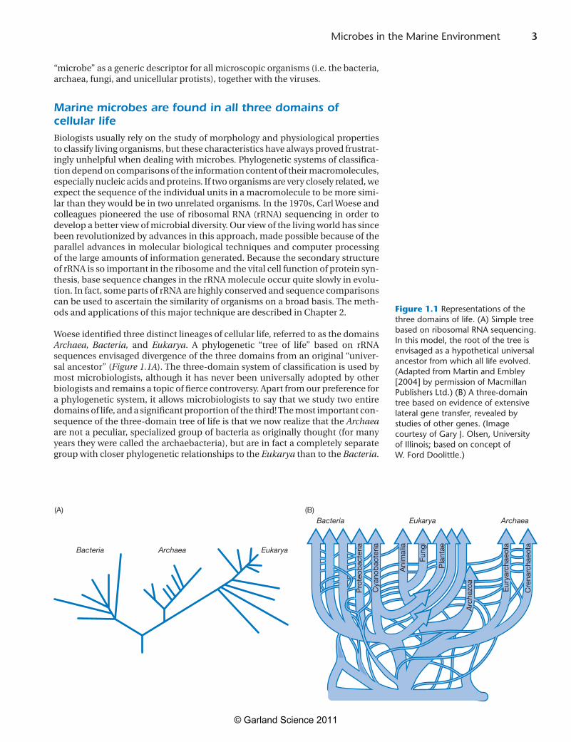
Woeseidentifiedthreedistinctlineagesofcellularlife,referredtoasthedomainsArchaea, Bacteria, and Eukarya. A phylogenetic “tree of life” based on rRNAsequencesenvisageddivergenceofthethreedomainsfromanoriginal“univer-sal ancestor” (Figure 1.1A). The three-domain system of classification is used by most microbiologists, although it has never been universally adopted by other biologistsandremainsatopicoffiercecontroversy.Apartfromourpreferencefora phylogenetic system, it allows microbiologists to say that we study two entire domains of life, and a significant proportion of the third! The most important con-sequenceofthethree-domaintreeoflifeisthatwenowrealizethattheArchaea arenotapeculiar,specializedgroupofbacteriaasoriginallythought(formanyyears they were called the archaebacteria), but are in fact a completely separate group with closer phylogenetic relationships to the Eukarya than to the Bacteria.
Archaea
Pro
teob
acte
ria
Cya
nob
acte
ria
Ani
mal
ia
Pla
ntae
Fung
i
Eur
yarc
haeo
ta
Cre
narc
haeo
ta
Arc
hezo
a
Bacteria EukaryaArchaea
Bacteria Eukarya(A)
Figure 1.1
(B)
Figure 1.1 Representations of the three domains of life. (A) Simple tree based on ribosomal RNA sequencing. In this model, the root of the tree is envisaged as a hypothetical universal ancestor from which all life evolved. (Adapted from Martin and Embley [2004] by permission of Macmillan Publishers Ltd.) (B) A three-domain tree based on evidence of extensive lateral gene transfer, revealed by studies of other genes. (Image courtesy of Gary J. Olsen, University of Illinois; based on concept of W. Ford Doolittle.)
© Garland Science 2011
4 Chapter 1
Eukaryotesaredistinguishedbyamembrane-boundnucleusandorganelleswithspecificfunctions.Mitochondriaoccurinalleukaryoticcells,withtheexceptionofafewanaerobicprotozoa,andcarryouttheprocessesofrespiratoryelectrontransport. In photosynthetic eukaryotes, chloroplasts carry out reactions forthe transfer of light energy for cellular metabolism. Various lines of evidence (especially the molecular analysis of the nucleic acids and proteins) support the hypothesis that theorganellesofeukaryoticcellshaveevolvedbyaprocessofendosymbiosis, in which one cell lives inside another cell for mutual benefit. This hypothesisproposesthattheoriginalsourceofmitochondriaineukaryoticcellsoccurredwhenprimitivecellsacquiredrespiratorybacteria(mostcloselyrelatedto proteobacteria) and that the chloroplasts evolved from endosymbiosis with cyanobacteria. Such interactions between different types of cell have continued throughoutevolution,andChapter10containsmanyexamplesofendosymbio-sis involving microbes.
Horizontal gene transfer confounds our understanding of evolutionSince it became possible to study the whole genome sequences of organisms,we have found increasing evidence of extensive lateral gene transfer (LGT; also knownashorizontalgenetransfer,HGT)betweenmicrobes.Suchtransferoccursmost commonly between related organisms, but transfer across bigger genetic distances also occurs—even between domains. Members of the Bacteria and Archaeacontainsomegeneswithverysimilarsequences,andmembersoftheEukarya contain genes from both of the other domains. Some members of the domain Bacteriahaveevenbeenshowntocontaineukaryoticgenes.Previously,evolution was explained only by the processes of mutation and sexual recombi-nation,butwenowknowthatthepaceofevolutionisacceleratedbythetransferandacquisitionofmodulesofgeneticinformation.Thisphenomenoniswide-spread in modern members of the Bacteria and Archaea and can occur via three processes.Duringtheprocessknownastransformation,cellsmaytakeupandexpressnakedDNA;whilstconjugationreliesoncell–cellcontactmediatedbypili. The most important source of LGT is the process of transduction by phages (viruses infecting bacteria); this is explored in detail in Chapter 7. The enormous diversity of marine viruses and the identification of a viral origin of genes in many marine organisms indicate how important this process has been through-out evolution.
Viruses are noncellular entities with great importance in marine ecosystemsVirus particles (virions) consist of a protein capsid containing the viral genome composedofeitherRNAorDNA.Becausetheyonlycontainonetypeofnucleicacid,virusesmustinfectlivingcellsandtakeoverthehost’scellularmachineryin order to replicate. It is often thought that viruses could have evolved (perhaps from bacteria) as obligate parasites that have progressively lost genetic infor-mation until they consist of only a few genes, or that they represent fragments ofhost-cellRNAorDNAthathavegainedindependencefromcellularcontrol.New ideas about the evolution of viruses are discussed in Research Focus Box 7.2. Thegenomeofvirusesoftencontainssequencesthatareequivalenttospecificsequencesinthehostcell.Virusesexistforeverymajorgroupofcellularorgan-isms (Bacteria, Archaea, Fungi, protists, plants, and animals), but at present we haveknowledgeofonlyatinyproportionofthevirusesinfectingmarinelife.Asdiscussed in Chapter 7, recognition of the abundance and diversity of marine viruses, and the role that they play in biogeochemical cycles and control of diver-sity in marine microbial communities, has been one of the most important dis-coveries of recent years.
WHAT HAPPENED TO THE PROKARYOTES?
Traditionally, members of the domains Bacteria and Archaea have been grouped together as “prokaryotes,” because they share a simple internal cellular structure, with their genetic material not bound by a nuclear membrane. However, this division of life is not supported by modern studies showing that Bacteria and Archaea are completely different phylogenetic groups. As pointed out by Pace (2009): “No-one can tell you what a prokaryote is, they can only tell you what it is not.” He argues that the prokaryote–eukaryote model is scientifically illogical and wrongly implies that prokaryotes gave rise to eukaryotes. Marine microbiology provides numerous examples that emphasize the fact that the prokaryotic designation is no longer appropriate—some marine bacteria are much larger than eukaryotic cells, some have complex intracellular structures, some show obvious multicellularity, and some differentiate during their lifecycle. Although many microbiologists defend continued use of the term “prokaryote” and reinterpretation of the concept in modern terms (see Whitman, 2009), I have decided not to use it in this book. However, readers should remember that the prokaryote concept is still deeply embedded, and you will find the term in many research papers and other books. Indeed, one of the most important reference works in microbiology is The Prokaryotes (Dworkin et al., 2007).
?
© Garland Science 2011
5 Microbes in the Marine Environment
Microbial processes shape the living worldProbably the most important overriding features of microbes are their excep-tional diversity and ability to occupy every conceivable habitat for life. Indeed, what we consider “conceivable” is challenged constantly by the discovery of new microbial communities in habitats previously thought of as inhospitable, or car-rying out processes that we had no idea were microbial in nature. Bacteria and archaeahaveshapedthesubsequentdevelopmentoflifeonEartheversincetheirfirst appearance—the metabolic processes that they carry out in the transforma-tion of elements, degradation of organic matter, and recycling of nutrients play a central role in innumerable activities that affect the support and maintenance of all other forms of life. Microbial life and the Earth have evolved together and the activities of microbes have affected the physical and geochemical properties of the planet. Indeed, they are actually the driving forces responsible for major planetaryprocesseslikechangesinthecompositionoftheatmosphere,oceans,soil,androcks.Thisisespeciallyrelevanttoourconsiderationofthemarineenvi-ronment, in view of the huge proportion of the biosphere that this constitutes. Despite the preponderance of microbes and the importance of their activities, they are unseen in everyday human experience. Microbes live and grow almost everywhere, using a huge range of resources, whereas plants and animals occupy only a small subset of possible environments and have a comparatively narrow range of lifestyles.
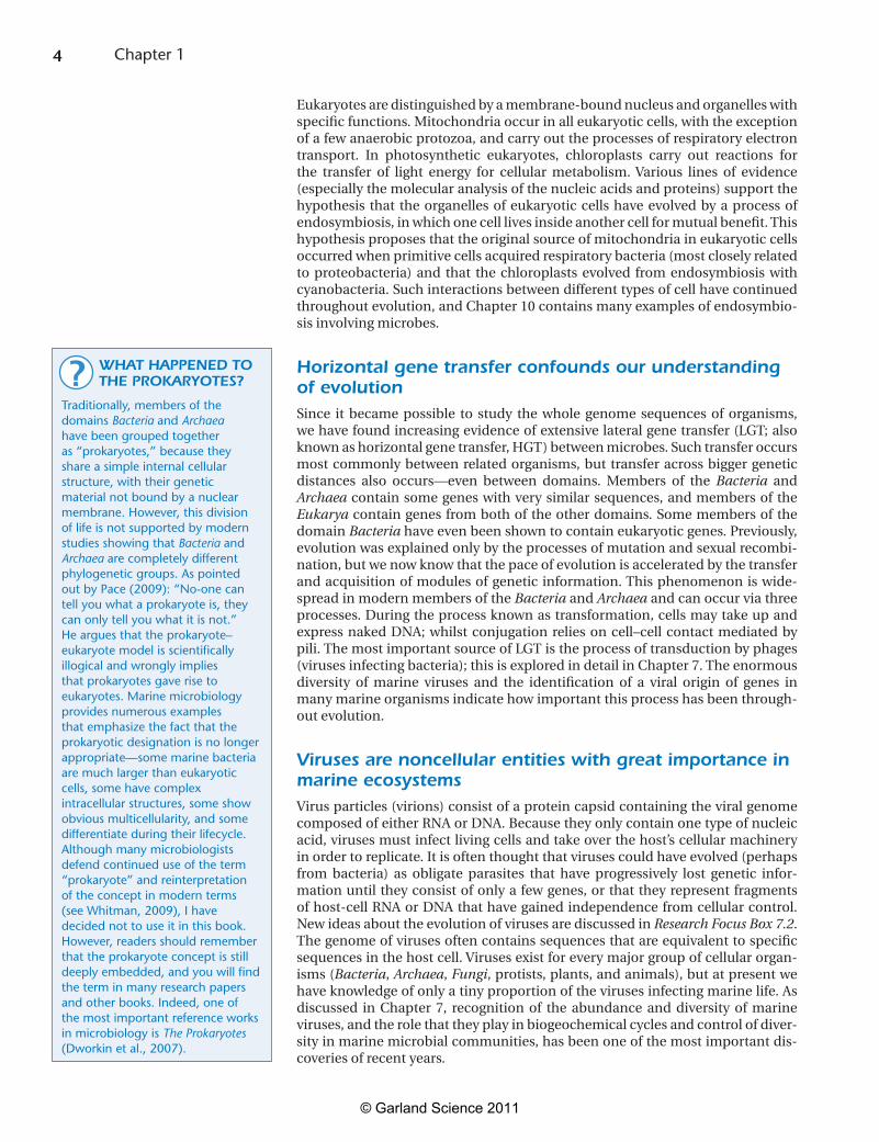
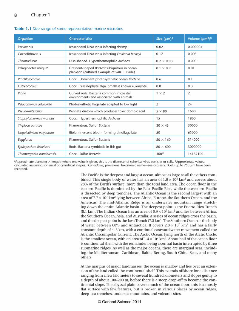
Marine microbes show great variation in sizeTable 1.1 shows the range of dimensions and volumes of some representative marine microbes. Even by the usual standards of microbiology, the most abun-dant microbes found in seawater are exceptionallysmall.Theirverysmallsizeisthemainreasonappreciationoftheirabundanceeludedusuntilquiterecently.As described in Chapter 2, recognition of the abundance of marine microbesdepended on the development of fine-pore filters and direct counting methods usingepifluorescencemicroscopyandflowcytometry.Smallcellsizehasgreatsignificanceintermsofthephysicalprocessesthataffectlife.Atthemicroscale,the rate of molecular diffusion becomes the most important mechanism for transport of substances into and out of the cell. Small cells feeding by absorption (osmotrophy)cantakeupnutrientsmoreefficientlythanlargercells.Thesurfacearea:volumeratio(SA/V)isthecriticalfactorbecauseascellsizeincreases,vol-ume(V)increasesmorequicklythansurfacearea(SA),asshowninFigure 1.2.
The most abundant ocean bacteria and archaea have very small cell volumes andlargeSA/Vratios.Themajorityaresmallerthanabout0.6mm in their largest dimension, and many are less than 0.3 mm, with cell volumes as low as 0.003 mm3. If nutrients are severely limiting, as they are in most of the oceans, selection will favor small cells. Since the first description of such small cells, termed “ultrami-crobacteria,” theirsizehasprovokedconsiderablecontroversy.Suchextremelysmall cells could result from a genetically fixed phenotype maintained through-out the cell cycle or because of physiological changes associated with starva-tion. The latter explanation is supported by the fact that some cultured bacteria become much smaller when starved. Most naturally occurring bacteria have been impossible to grow in culture—this is a central problem in marine microbi-ology, which we shall return to on several occasions in future chapters. Because of this, it has been difficult to determine whether small size is a genotypicallydetermined condition for marine bacteria. However, studies with some recently cultured marine bacteria from low-nutrient (oligotrophic) ocean environments show that addition of nutrients does not cause an increase in cell size. Smallcellsizealsohasimportantimplicationsformechanismsofactivemotilityand chemotaxis, because of the microscale effects of Brownian movement (bom-bardment by water molecules) and shear forces. Small marine bacteria have
IS IT TIME TO CHOP DOWN THE “TREE OF LIFE”?
The idea that relationships between all living organisms can be represented as a tree of life helped to shape Darwin’s theory of evolution by natural selection and has been deeply embedded in the philosophy of biology for more than 150 years. As the importance of endosymbiosis and LGT became better understood, some evolutionary scientists began to question the validity of the “tree of life” concept. A seminal paper by Doolittle (1999) argued that “Molecular phylogeneticists will have failed to find the ‘true tree’, not because their methods are inadequate or because they have chosen the wrong genes, but because the history of life cannot properly be represented as a tree.” Relationships are now envisaged as complex intertwined branches, more like a web (see Figure 1.1B ) or network of genomes (Dagan and Martin, 2009). However, this remains a controversial topic, and some have argued that analysis of genome sequences for “core genes” still supports the idea of a common ancestor and branching tree (Ciccarelli et al., 2006).
?
© Garland Science 2011
6 Chapter 1
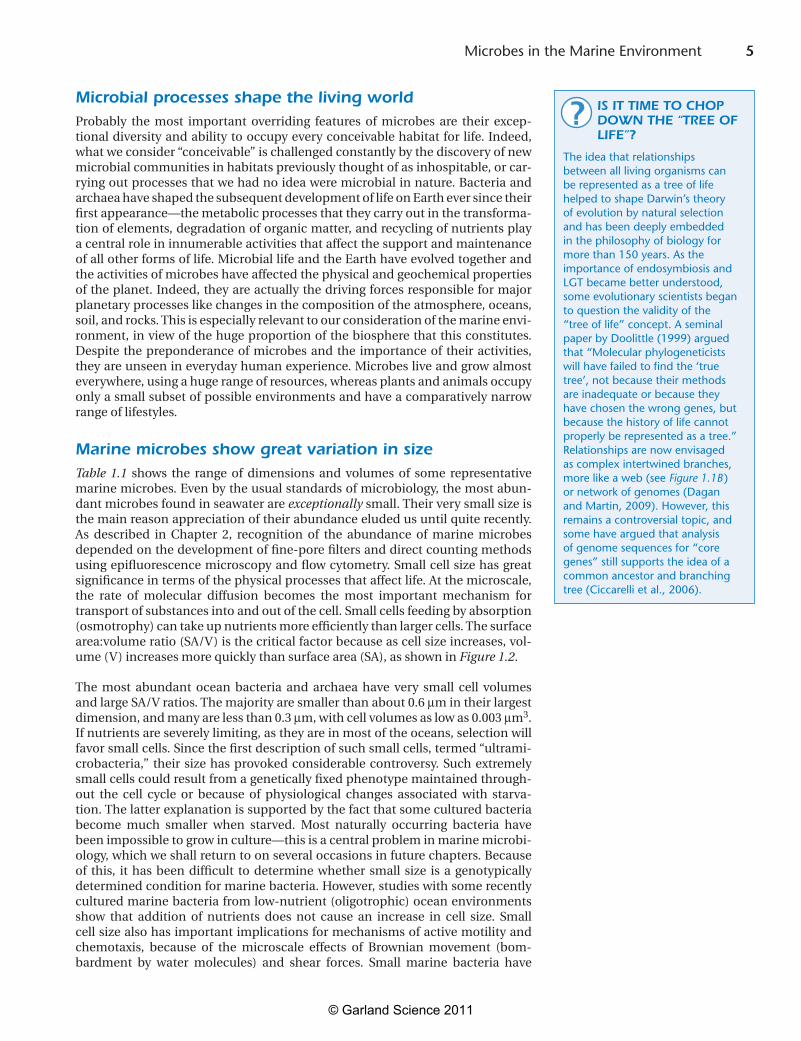
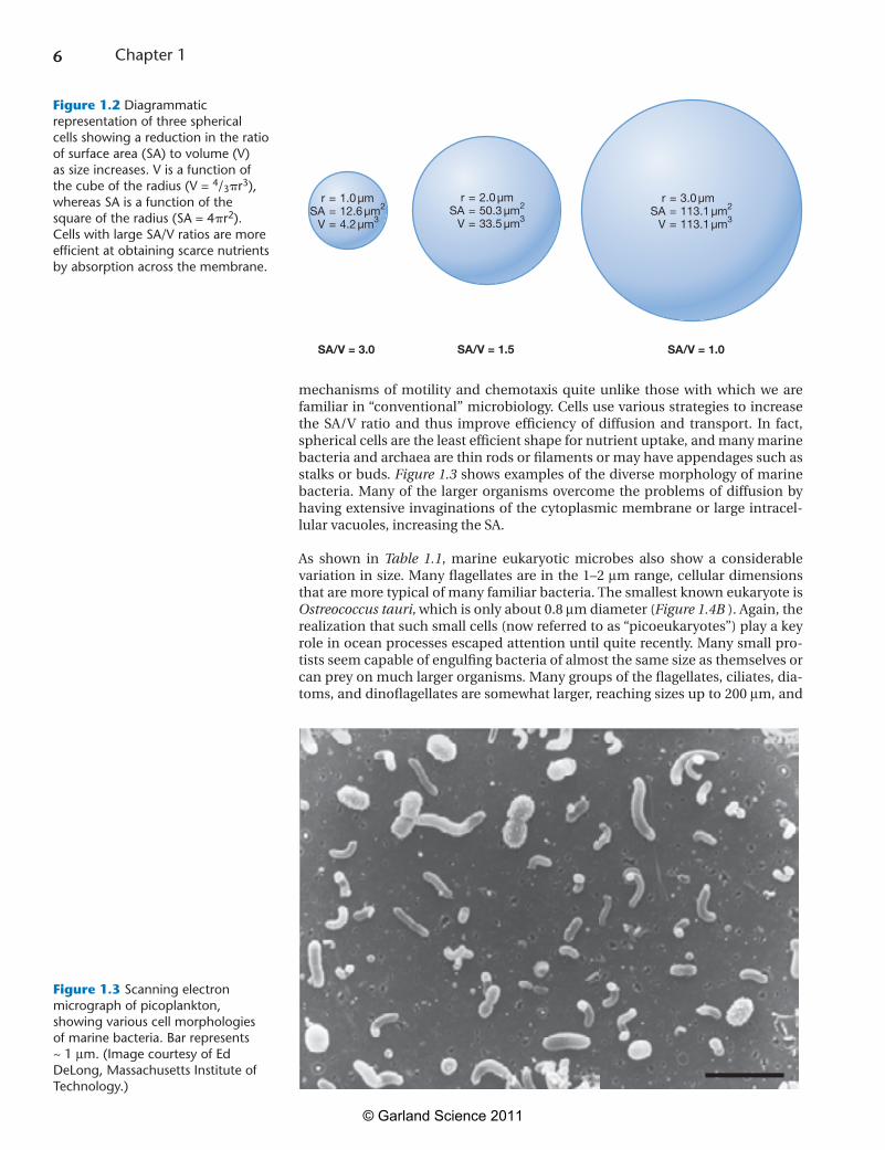
mechanisms of motility and chemotaxis quite unlike those with which we arefamiliar in “conventional” microbiology. Cells use various strategies to increase the SA/V ratio and thus improve efficiency of diffusion and transport. In fact,sphericalcellsaretheleastefficientshapefornutrientuptake,andmanymarinebacteria and archaea are thin rods or filaments or may have appendages such as stalksorbuds.Figure 1.3 shows examples of the diverse morphology of marine bacteria. Many of the larger organisms overcome the problems of diffusion by having extensive invaginations of the cytoplasmic membrane or large intracel-lularvacuoles,increasingtheSA.
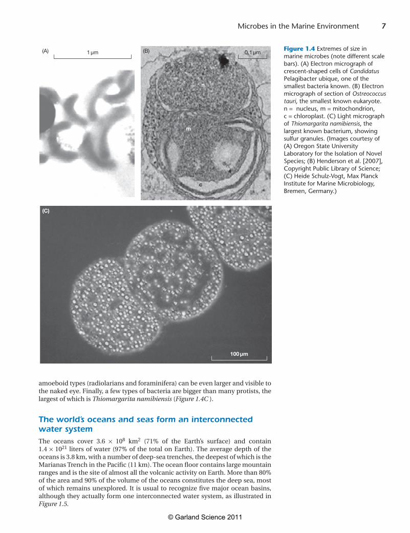
As shown in Table 1.1, marine eukaryotic microbes also show a considerablevariationinsize.Manyflagellatesareinthe1–2mm range, cellular dimensions thataremoretypicalofmanyfamiliarbacteria.ThesmallestknowneukaryoteisOstreococcus tauri, which is only about 0.8 mm diameter (Figure 1.4B).Again,therealizationthatsuchsmallcells(nowreferredtoas“picoeukaryotes”)playakeyroleinoceanprocessesescapedattentionuntilquiterecently.Manysmallpro-tistsseemcapableofengulfingbacteriaofalmostthesamesizeasthemselvesorcan prey on much larger organisms. Many groups of the flagellates, ciliates, dia-toms,anddinoflagellatesaresomewhatlarger,reachingsizesupto200mm, and
Figure 1.2
rSA
V
1.0 µm12.6 µm2
4.2 µm3
===
rSA
V
2.0 µm50.3 µm2
33.5 µm3
===
rSA
V
3.0 µm113.1 µm2
113.1 µm3
===
SA/V = 3.0 SA/V = 1.5 SA/V = 1.0
Figure 1.2 Diagrammatic representation of three spherical cells showing a reduction in the ratio of surface area (SA) to volume (V) as size increases. V is a function of the cube of the radius (V = 4/3r3), whereas SA is a function of the square of the radius (SA = 4r2). Cells with large SA/V ratios are more efficient at obtaining scarce nutrients by absorption across the membrane.
Figure 1.3 Scanning electron micrograph of picoplankton, showing various cell morphologies of marine bacteria. Bar represents ~ 1 mm. (Image courtesy of Ed DeLong, Massachusetts Institute of Technology.)
© Garland Science 2011
7 Microbes in the Marine Environment
amoeboid types (radiolarians and foraminifera) can be even larger and visible to thenakedeye.Finally,afewtypesofbacteriaarebiggerthanmanyprotists,thelargest of which is Thiomargarita namibiensis (Figure 1.4C ).
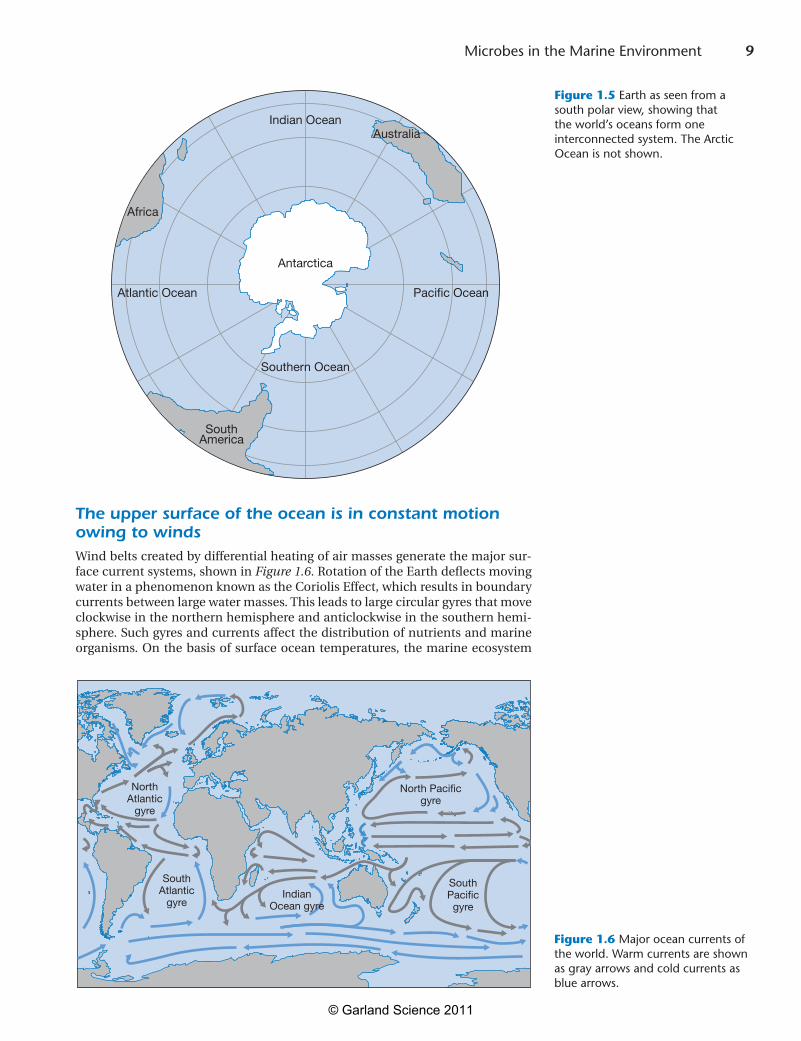
The world’s oceans and seas form an interconnected water systemThe oceans cover 3.6 ¥ 108 km2 (71% of the Earth’s surface) and contain1.4¥1021 litersofwater (97%of the totalonEarth).Theaveragedepthof theoceansis3.8km,withanumberofdeep-seatrenches,thedeepestofwhichistheMarianasTrenchinthePacific(11km).Theoceanfloorcontainslargemountainranges and is the site of almost all the volcanic activity on Earth. More than 80% oftheareaand90%ofthevolumeoftheoceansconstitutesthedeepsea,mostofwhichremainsunexplored.It isusualtorecognizefivemajoroceanbasins,although they actually form one interconnected water system, as illustrated in Figure 1.5.
Figure 1.4 Extremes of size in marine microbes (note different scale bars). (A) Electron micrograph of crescent-shaped cells of Candidatus Pelagibacter ubique, one of the smallest bacteria known. (B) Electron micrograph of section of Ostreococcus tauri, the smallest known eukaryote. n = nucleus, m = mitochondrion, c = chloroplast. (C) Light micrograph of Thiomargarita namibiensis, the largest known bacterium, showing sulfur granules. (Images courtesy of (A) Oregon State University Laboratory for the Isolation of Novel Species; (B) Henderson et al. [2007], Copyright Public Library of Science; (C) Heide Schulz-Vogt, Max Planck Institute for Marine Microbiology, Bremen, Germany.)
Figure 1.4
1 µm
100 µm
0.1 µm
c
m
n
(A)
(C)
(B)
© Garland Science 2011
8 Chapter 1
The Pacific is the deepest and largest ocean, almost as large as all the others com-bined.Thissinglebodyofwaterhasanareaof1.6¥108km2 and covers about 28%oftheEarth’ssurface,morethanthetotallandarea.Theoceanfloorintheeastern Pacific is dominated by the East Pacific Rise, while the western Pacific isdissectedbydeeptrenches.TheAtlanticOceanisthesecondlargestwithanarea of 7.7 ¥107km2lyingbetweenAfrica,Europe,theSouthernOcean,andtheAmericas. The mid-Atlantic Ridge is an underwater mountain range stretch-ingdowntheentireAtlanticbasin.ThedeepestpointisthePuertoRicoTrench(8.1km).TheIndianOceanhasanareaof6.9¥107km2andliesbetweenAfrica,theSouthernOcean,Asia,andAustralia.Aseriesofoceanridgescrossthebasin,andthedeepestpointistheJavaTrench(7.3km).TheSouthernOceanisthebodyof water between 60°S and Antarctica. It covers 2.0¥ 107 km2 and has a fairly constantdepthof4–5km,withacontinualeastwardwatermovementcalledtheAtlanticCircumpolarCurrent.TheArcticOcean,lyingnorthoftheArcticCircle,isthesmallestocean,withanareaof1.4¥107km2.Abouthalfoftheoceanflooris continental shelf, with the remainder being a central basin interrupted by three submarineridges.Aswellasthemajoroceans,therearemarginalseas,includ-ing the Mediterranean, Caribbean, Baltic, Bering, South China Seas, and many others.
Atthemarginsofmajorlandmasses,theoceanisshallowandliesoveranexten-sion of the land called the continental shelf. This extends offshore for a distance rangingfromafewkilometerstoseveralhundredkilometersandslopesgentlytoadepthofabout100–200m,beforethereisasteepdrop-offtobecomethecon-tinental slope. The abyssal plain covers much of the ocean floor; this is a mostly flat surface with few features, but is broken in various places by ocean ridges,deep-sea trenches, undersea mountains, and volcanic sites.
Table 1.1 Size range of some representative marine microbes
Organism Characteristics Size (m)a Volume (m3)b
Parvovirus Icosahedral DNA virus infecting shrimp 0.02 0.000004
Coccolithovirus Icosahedral DNA virus infecting Emiliania huxleyi 0.17 0.003
Thermodiscus Disc-shaped. Hyperthermophilic Archaea 0.2 0.08 0.003
Pelagibacter ubiquec Crescent-shaped Bacteria ubiquitous in ocean plankton (cultured example of SAR11 clade)
0.1 0.9 0.01
Prochlorococcus Cocci. Dominant photosynthetic ocean Bacteria 0.6 0.1
Ostreococcus Cocci. Prasinophyte alga. Smallest known eukaryote 0.8 0.3
Vibrio Curved rods. Bacteria common in coastal environments and associated with animals
1 2 2
Pelagomonas calceolata Photosynthetic flagellate adapted to low light 2 24
Pseudo-nitzschia Pennate diatom which produces toxic domoic acid 5 80 1600
Staphylothermus marinus Cocci. Hyperthermophilic Archaea 15 1800
Thiploca auracae Filamentous. Sulfur Bacteria 30 43 30000
Lingulodinium polyedrum Bioluminescent bloom-forming dinoflagellate 50 65000
Beggiatoa Filamentous. Sulfur Bacteria 50 160 314000
Epulopiscium fishelsoni Rods. Bacteria symbiotic in fish gut 80 600 3000000
Thiomargarita namibiensis Cocci. Sulfur Bacteria 300d 14137100
aApproximate diameter length; where one value is given, this is the diameter of spherical virus particles or cells. bApproximate values, calculated assuming spherical or cylindrical shapes. cCandidatus; provisional taxonomic name—see Glossary. dCells up to 750 m have been recorded.
© Garland Science 2011
9 Microbes in the Marine Environment
The upper surface of the ocean is in constant motion owing to windsWindbeltscreatedbydifferentialheatingofairmassesgeneratethemajorsur-face current systems, shown in Figure 1.6. Rotation of the Earth deflects moving waterinaphenomenonknownastheCoriolisEffect,whichresultsinboundarycurrents between large water masses. This leads to large circular gyres that move clockwiseinthenorthernhemisphereandanticlockwiseinthesouthernhemi-sphere. Such gyres and currents affect the distribution of nutrients and marine organisms. On the basis of surface ocean temperatures, the marine ecosystem
IndianOcean
SouthernOceanAtlanticOceanPacificOcean
Figure 1.5
Indian Ocean
Southern Ocean
Atlantic Ocean Pacific Ocean
Antarctica
SouthAmerica
Africa
Australia
Figure 1.5 Earth as seen from a south polar view, showing that the world’s oceans form one interconnected system. The Arctic Ocean is not shown.
Figure 1.6 Major ocean currents of the world. Warm currents are shown as gray arrows and cold currents as blue arrows.
Figure 1.6
NorthAtlantic
gyre
SouthAtlantic
gyre
North Pacificgyre
SouthPacificgyre
IndianOcean gyre
© Garland Science 2011
10 Chapter 1
canbedividedintofourmajorbiogeographicalzones,namelypolar,coldtem-perate,warmtemperate(subtropical),andtropical(equatorial).Theboundariesbetweenthesezonesarenotabsoluteandvarywithseason.
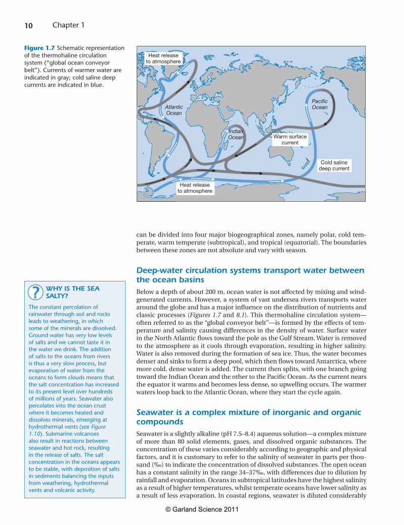
Deep-water circulation systems transport water between the ocean basinsBelowadepthofabout200m,oceanwaterisnotaffectedbymixingandwind-generated currents. However, a system of vast undersea rivers transports water around the globe and has a major influence on the distribution of nutrients and classic processes (Figures 1.7 and 8.1). This thermohaline circulation system—often referred to as the “global conveyor belt”—is formed by the effects of tem-perature and salinity causing differences in the density of water. Surface water intheNorthAtlanticflowstowardthepoleastheGulfStream.Waterisremovedto the atmosphere as it cools through evaporation, resulting in higher salinity. Waterisalsoremovedduringtheformationofseaice.Thus,thewaterbecomesdenserandsinkstoformadeeppool,whichthenflowstowardAntarctica,wheremore cold, dense water is added. The current then splits, with one branch going towardtheIndianOceanandtheothertothePacificOcean.Asthecurrentnearstheequatoritwarmsandbecomeslessdense,soupwellingoccurs.ThewarmerwatersloopbacktotheAtlanticOcean,wheretheystartthecycleagain.
Seawater is a complex mixture of inorganic and organic compoundsSeawaterisaslightlyalkaline(pH7.5–8.4)aqueoussolution—acomplexmixtureof more than 80 solid elements, gases, and dissolved organic substances. The concentration of these varies considerably according to geographic and physical factors, and it is customary to refer to the salinity of seawater in parts per thou-sand (‰) to indicate the concentration of dissolved substances. The open ocean hasaconstantsalinityintherange34–37‰,withdifferencesduetodilutionbyrainfall and evaporation. Oceans in subtropical latitudes have the highest salinity as a result of higher temperatures, whilst temperate oceans have lower salinity as a result of less evaporation. In coastal regions, seawater is diluted considerably
Figure 1.7
Heat releaseto atmosphere
Heat releaseto atmosphere
Warm surfacecurrent
Cold salinedeep current
AtlanticOcean
PacificOcean
IndianOcean
Figure 1.7 Schematic representation of the thermohaline circulation system (“global ocean conveyor belt”). Currents of warmer water are indicated in gray; cold saline deep currents are indicated in blue.
WHY IS THE SEA SALTY?
The constant percolation of rainwater through soil and rocks leads to weathering, in which some of the minerals are dissolved. Ground water has very low levels of salts and we cannot taste it in the water we drink. The addition of salts to the oceans from rivers is thus a very slow process, but evaporation of water from the oceans to form clouds means that the salt concentration has increased to its present level over hundreds of millions of years. Seawater also percolates into the ocean crust where it becomes heated and dissolves minerals, emerging at hydrothermal vents (see Figure 1.10). Submarine volcanoes also result in reactions between seawater and hot rock, resulting in the release of salts. The salt concentration in the oceans appears to be stable, with deposition of salts in sediments balancing the inputs from weathering, hydrothermal vents and volcanic activity.
?
© Garland Science 2011
11 Microbes in the Marine Environment
byfreshwaterfromriversandterrestrialrunoffandisintherange10–32‰.Con-versely,inenclosedareassuchastheRedSeaandArabianGulf,thesalinitymaybeashighas44‰.Inpolarareas,theremovaloffreshwaterbytheformationofice also leads to increased salinity. The major ionic components of seawater are sodium(55%w/v),chloride(31%),sulfate(8%),magnesium(4%),calcium(1%),and potassium (1%). Together, these constitute more than 99% of the weightof salts.There are four minor ions–namely bicarbonate, bromide, borate, andstrontium–which together make up just less than 1% of seawater. Many otherelements are present in trace amounts (<0.01%), including key nutrients suchas nitrate, phosphate, silicate and iron. The concentration of these is crucial in determining the growth of marine microbes and the net productivity of marine systems,asdiscussedinChapter9.
Theconcentrationofsaltshasamarkedeffectonthephysicalpropertiesofsea-water.Thefreezingpointofseawaterat35‰is–1.9°C,andseawaterincreasesindensityuptothispoint.Asnotedabove,thisresultsintheformationofmassesofcold,densewaterinpolarregions,whichsinktothebottomoftheoceanbasinsand are dispersed by deep-water circulation currents. Differences in the density of seawater create a discontinuity called the pycnocline, which separates the top few hundred meters of the water column from deeper water. This has great sig-nificance, because the gases oxygen and carbon dioxide are more soluble in cold water.
Oxygen is at its highest concentrations in the top 10–20 m of water, owing toexchange with the atmosphere and production of oxygen by photosynthesis. Concentration decreases with distance from the surface until it reaches a mini-mumbetween200and1000m,andbacterialdecompositionoforganicmattermay create conditions that are almost anoxic. Below this, the oxygen content increases again as a result of the presence of dense water (with increased solubil-ityatlowertemperature)thathassunkfrompolarregionsandbeentransportedon the thermohaline circulation system. This oxygen gradient varies greatly in different regions, and there are several regions where large bodies of hypoxic wateroccuratrelativelyshallowdepths—thesearetheoxygenminimumzones(see Plate 9.1).
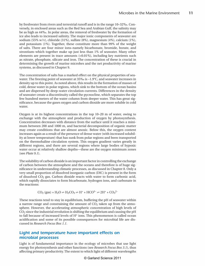
The solubility of carbon dioxide is an important factor in controlling the exchange of carbon between the atmosphere and the oceans and therefore is of huge sig-nificance in understanding climatic processes, as discussed in Chapter 8. Only a very small proportion of dissolved inorganic carbon (DIC) is present in the form of dissolved CO2 gas. Carbon dioxide reacts with water to form carbonic acid, which rapidly dissociates to form bicarbonate, hydrogen ions, and carbonate in the reactions:
CO2 (gas) + H2O H2CO3 H+ + HCO3– 2H+ + CO32–
Thesereactionstendtostayinequilibrium,bufferingthepHofseawaterwithina narrow range and constraining the amount of CO2 taken up from the atmo-sphere. However, the accelerating atmospheric concentration of high levels of CO2sincetheindustrialrevolutionisshiftingtheequilibriumandcausingthepHto fall because of increased levels of H+ ions. This phenomenon is called ocean acidification and some of its possible consequences for microbial life are dis-cussed in Research Focus Box 1.1.
Light and temperature have important effects on microbial processesLight is of fundamental importance in the ecology of microbes that use light energy for photosynthesis and other functions (see Research Focus Box 3.1), thus affecting primary productivity. The extent to which light of different wavelengths
© Garland Science 2011
12 Chapter 1
The effects of high levels of CO2 (together with other gases such as methane and nitrous oxide) in promoting global warming via the “greenhouse effect” are well known anda major topic of current sociopolitical concern. However, a less well-known effect of atmospheric CO2 is the pro-cess of ocean acidification (OA), which has been termed“the other CO2 problem” to bring it to the attention of the public(seeMitchell,2009).WhenCO2 is absorbed from the atmosphere, it leads to an increase in the concentration ofhydrogen ionsandhencea fall inpH(seeequationonp.11).ApproximatelyhalfoftheCO2 produced by human activities in the past 200 years has been absorbed by theoceans, leading to a fall in the average pH of ocean surface waterfromabout8.21to8.10.Althoughthisseemssmall,it represents a 30% increase in the hydrogen ion concen-tration because pH is a logarithmic scale. Some models predict that the average ocean pH of the oceans could fall to pH 7.9 by 2100—at which point hydrogen ions will bethree times the current levels—unless CO2 emissions are drastically reduced. This level is lower than has occurred for hundreds of millions of years; more importantly, this rate of change has probably never occurred in the history of theplanet(RoyalSociety,2005).Eveniftheworldsucceedsin controlling CO2 emissions, much of this change will be irreversible, because of the long time needed for mixing of deepwatersandnaturalbufferingprocessestotakeeffect.
One of the main effects of the altered seawater chemistry as aresultofOAislikelytobeoncalcifyingorganismsthatusecalcium carbonate (CaCO3)toconstructaskeletonorshell.As well as animals such as corals, crustaceans and mol-lusks,therearesomeveryimportantplanktonicmicrobesthat carry out calcification, especially the coccolithophores (seeChapter6).Orretal. (2005)concludedthatsuchcal-cifying organisms could begin to experience difficulties in maintaining their CaCO3 skeletonsasearlyas2050,espe-cially in polar oceans, where the state of CaCO3 saturation is lower.When considering the effect of OA on microbes,some examples of recent research given here illustrate how difficult it is to reach clear conclusions about the effect of OAonmicrobialprocesses.
When coccolithophores make the plates of calcite thatsurround the cell (see Figure 6.5B), they release CO2, but they also fix CO2 during photosynthesis. Thus, the balance between these processes is very important in the global car-bon cycle. Research has indicated that there is great varia-tion in the responses of different species (and strains within species) to changes in ocean pH and CO2 levels. Riebesell (2004)concludedthat increasedCO2 levels enhance pho-tosyntheticcarbonfixationofsomephytoplanktongroupsandpredictedthatcalcifyingplanktonmightbenefitatthe
expense of some other groups. Mesocosm studies (see Fig-ure 2.2) of the response of blooms of the coccolithophore Emiliania huxleyi showed a reduction in calcification rates when CO2 levels simulating end of the century conditions wereapplied(Delilleetal.,2005;Engeletal.,2005).How-ever, Iglesias-Rodriguez et al. (2008a) developed observa-tionsfromsedimentcoresfromasiteintheNorthAtlantic,indicating that coccolithophores have increased their cal-cification rates in response to rising CO2 levels—the aver-agecoccolithmasshasincreasedby~40%overthepast220years. In laboratory cultures, a strain of E. huxleyi increased photosynthesis and calcification rates by 100–150% athigh CO2 levels, a level much higher than that observed in previous studies. This paper has attracted a lot of interest because of the important conclusion that coccolithophores are adapting to a high CO2 ocean and the impact that this will have on predicting future trends. Following publica-tion,Riebeselletal.(2007)werehighlycriticaloftheexperi-mental setup, but Iglesias-Rodriguez et al. (2008b) havedefended their conclusions. In another mesocosm study, Riebesell et al. (2007) showed that CO2 uptake by phyto-planktonwas27%and39%higher,respectively,whenCO2 was at two or three times present-day levels. These authors also found that the ratio of carbon to nitrogen uptakeincreased at higher CO2 concentrations, whereas the car-bon–nitrogen ratio within the cells of the phytoplanktonwas unaltered. It seems that extra carbon incorporated through photosynthesis was rapidly lost from the cells to form transparent exopolymer particles that contribute to the formation of marine snow and the flux of organic carbon from the surface to the deep sea (see Figure 1.9). Thus, increased productivity might provide a partial nega-tivefeedbackmechanismbywhichsomeoftheincreasedCO2 dissolved in seawater is removed. Further evidence for a possible negative feedback mechanism comes from theobservationbyRamosetal.(2007)thatthecyanobacteriumTrichodesmium increases its rate of nitrogen fixation very markedlyathighCO2 levels. The authors suggest that this could enhance the productivity of oligotrophic oceans that are currently limited by nitrogen limitation and increase the flux of carbon in the biological pump.
From the few examples of conflicting results presented here,itisclearthatourknowledgeoftheeffectsofOAonthe physiology and diversity of marine microbes is very poorly understood. Experts have recently been coordinat-ing efforts to highlight the challenges for future research in this area and the need for coordinated methodological approaches(C-MORE,2009;EPOCA,2009).Microbeshaveadapted to previous changes in ocean chemistry that have occurred periodically over the past 3 billion years. How will they adapt to the extreme changes currently occurring?
BOX 1.1 RESEARCH FOCUS
Ocean acidification—“the other CO2 problem”How will microbes respond to rapid changes in ocean chemistry?
© Garland Science 2011
13 Microbes in the Marine Environment
penetrates seawater depends on a number of factors, notably cloud cover, the polar ice caps, dust in the atmosphere, and variation of the incident angle of solar radiation according to season and location on the Earth’s surface. Light is absorbed or scattered by organisms and suspended particles. Even in the clearest ocean water, photosynthesis is restricted by the availability of light of sufficient intensity to the upper 150–200 m.This is termed the photic or euphotic zone(fromtheGreek,“welllit”).Bluelighthasthedeepestpenetration,andphotosyn-theticmicrobesatthelowerpartofthephoticzonehavelight-harvestingsystemsthataretunedtocollectbluelightmostefficiently(seep.114).Inturbidcoastalwaters,duringseasonalplanktonblooms,theeuphoticzonemaybeonlyafewmeters deep.
Solar radiation also leads to thermal stratification of seawater. In tropical seas, the continual input of energy from sunlight leads to warming of the surface waters to 25–30°C, causing a considerable difference in density from that ofdeeperwaters.Thus,throughouttheyear,thereisamarkedthermoclineatabout100–150m,belowwhichthereisasuddenreductionintemperatureto10°Corless. Little mixing occurs between these layers. In polar seas, the water is perma-nently cold except for a brief period in the summer, when a slight thermocline results. Apart from this period, turbulence created by surface winds generatesmixing of the water to considerable depths. Temperate seas show the greatest seasonal variation in the thermocline, with strong winds and low temperatures leading to extensive mixing in the winter. The thermocline develops in the spring, leading to a marked shallow surface layer of warmer water in summer. As theseacoolsandwindincreases,thethermoclinebreaksdownagainintheautumn.Combined with seasonal variations in light intensity, these temperature stratifi-cation effects and vertical mixing have a great impact on rates of photosynthesis and other microbial activities.
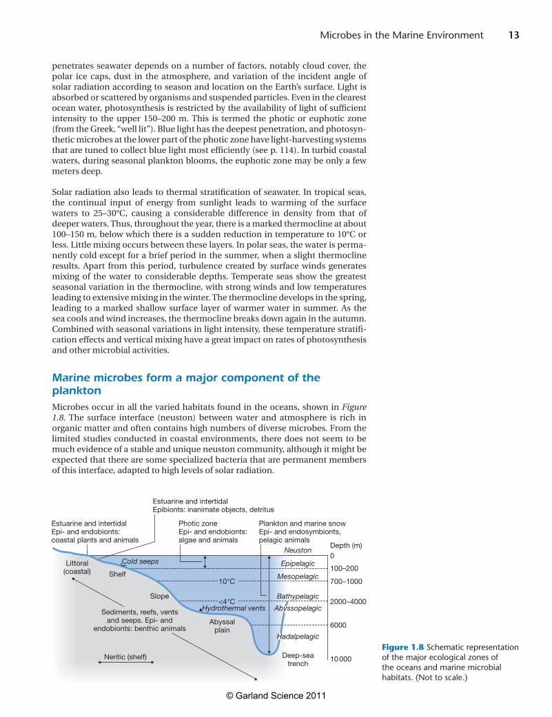
Marine microbes form a major component of the planktonMicrobes occur in all the varied habitats found in the oceans, shown in Figure 1.8. The surface interface (neuston) between water and atmosphere is rich in organic matter and often contains high numbers of diverse microbes. From the limited studies conducted in coastal environments, there does not seem to be muchevidenceofastableanduniqueneustoncommunity,althoughitmightbeexpectedthattherearesomespecializedbacteriathatarepermanentmembersof this interface, adapted to high levels of solar radiation.
Figure 1.8
0
100–200
2000–4000
700–1000
<4°C
10°C
6000
10 000
Depth (m)Neuston
Epipelagic
Bathypelagic
Mesopelagic
AbyssopelagicHydrothermal vents
Hadalpelagic
Deep-seatrench
Abyssalplain
Littoral(coastal)
Sediments, reefs, ventsand seeps. Epi- and
endobionts: benthic animals
Shelf
Slope
Estuarine and intertidalEpi- and endobionts:coastal plants and animals
Plankton and marine snowEpi- and endosymbionts,pelagic animals
Neritic (shelf)
Cold seeps
Photic zoneEpi- and endobionts:algae and animals
Estuarine and intertidalEpibionts: inanimate objects, detritus
Figure 1.8 Schematic representation of the major ecological zones of the oceans and marine microbial habitats. (Not to scale.)
© Garland Science 2011
14 Chapter 1
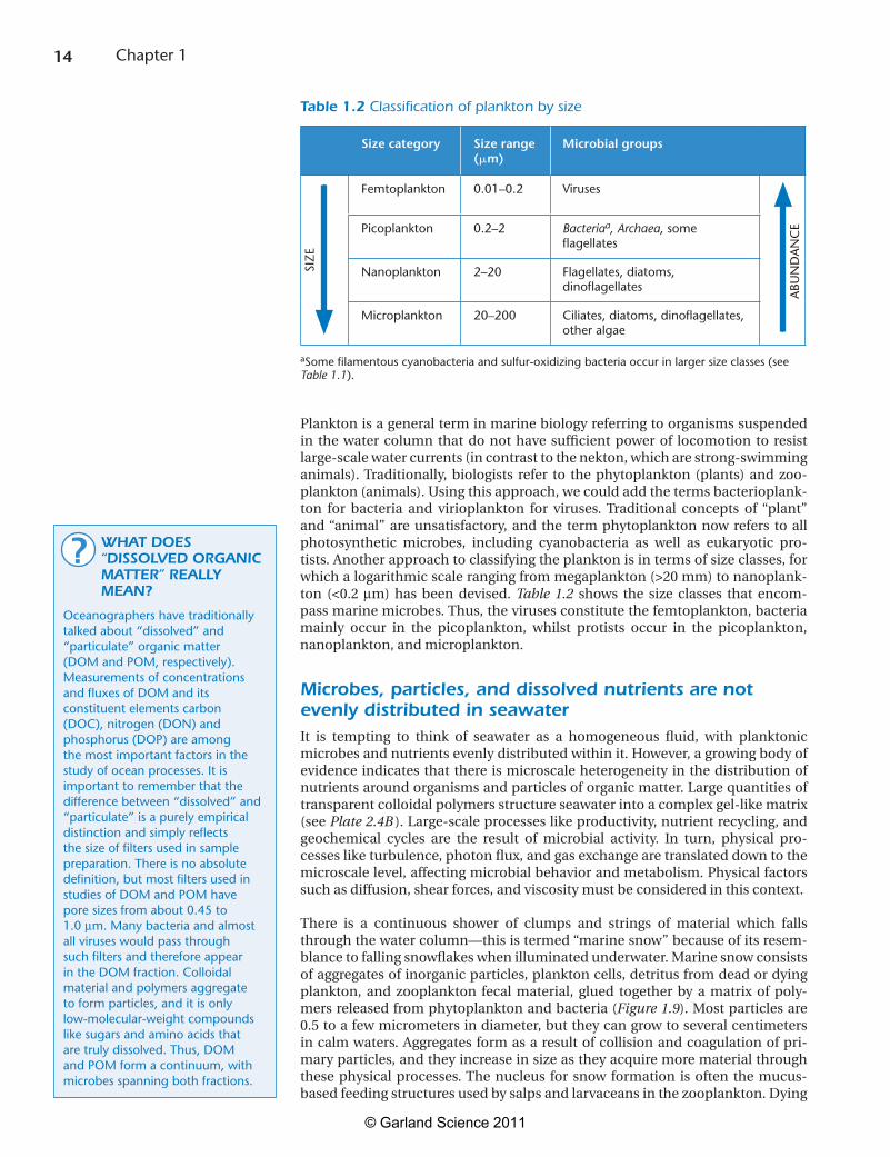
Planktonisageneralterminmarinebiologyreferringtoorganismssuspendedin the water column that do not have sufficient power of locomotion to resist large-scalewatercurrents(incontrasttothenekton,whicharestrong-swimminganimals).Traditionally, biologists refer to the phytoplankton (plants) and zoo-plankton(animals).Usingthisapproach,wecouldaddthetermsbacterioplank-ton for bacteria and virioplankton for viruses. Traditional concepts of “plant”and“animal” are unsatisfactory, and the term phytoplankton now refers to allphotosynthetic microbes, including cyanobacteria as well as eukaryotic pro-tists.Anotherapproachtoclassifyingtheplanktonisintermsofsizeclasses,forwhichalogarithmicscalerangingfrommegaplankton(>20mm)tonanoplank-ton (<0.2mm) has been devised. Table 1.2 shows the size classes that encom-passmarinemicrobes.Thus,thevirusesconstitutethefemtoplankton,bacteriamainly occur in the picoplankton, whilst protists occur in the picoplankton,nanoplankton,andmicroplankton.
Microbes, particles, and dissolved nutrients are not evenly distributed in seawaterIt is tempting to think of seawater as a homogeneous fluid, with planktonicmicrobes and nutrients evenly distributed within it. However, a growing body of evidence indicates that there is microscale heterogeneity in the distribution of nutrientsaroundorganismsandparticlesoforganicmatter.Largequantitiesoftransparentcolloidalpolymersstructureseawaterintoacomplexgel-likematrix(see Plate 2.4B).Large-scaleprocesseslikeproductivity,nutrientrecycling,andgeochemical cycles are the result of microbial activity. In turn, physical pro-cessesliketurbulence,photonflux,andgasexchangearetranslateddowntothemicroscale level, affecting microbial behavior and metabolism. Physical factors such as diffusion, shear forces, and viscosity must be considered in this context.
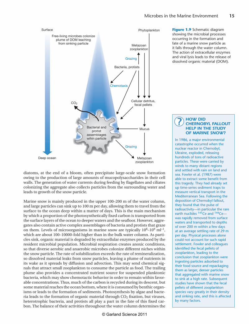
There is a continuous shower of clumps and strings of material which falls through the water column—this is termed “marine snow” because of its resem-blancetofallingsnowflakeswhenilluminatedunderwater.Marinesnowconsistsofaggregatesofinorganicparticles,planktoncells,detritusfromdeadordyingplankton, and zooplankton fecal material, glued together by a matrix of poly-mersreleasedfromphytoplanktonandbacteria(Figure 1.9). Most particles are 0.5 to a few micrometers in diameter, but they can grow to several centimeters incalmwaters.Aggregates formasaresultofcollisionandcoagulationofpri-maryparticles,andtheyincreaseinsizeastheyacquiremorematerialthroughthese physical processes. The nucleus for snow formation is often the mucus-basedfeedingstructuresusedbysalpsandlarvaceansinthezooplankton.Dying
Table 1.2 Classification of plankton by size
Size category Size range (m)
Microbial groups
Femtoplankton 0.01–0.2 Viruses
Picoplankton 0.2–2 Bacteriaa, Archaea, some flagellates
Nanoplankton 2–20 Flagellates, diatoms, dinoflagellates
Microplankton 20–200 Ciliates, diatoms, dinoflagellates, other algae
aSome filamentous cyanobacteria and sulfur-oxidizing bacteria occur in larger size classes (see Table 1.1).
WHAT DOES “DISSOLVED ORGANIC MATTER” REALLY MEAN?
Oceanographers have traditionally talked about “dissolved” and “particulate” organic matter (DOM and POM, respectively). Measurements of concentrations and fluxes of DOM and its constituent elements carbon (DOC), nitrogen (DON) and phosphorus (DOP) are among the most important factors in the study of ocean processes. It is important to remember that the difference between “dissolved” and “particulate” is a purely empirical distinction and simply reflects the size of filters used in sample preparation. There is no absolute definition, but most filters used in studies of DOM and POM have pore sizes from about 0.45 to 1.0 mm. Many bacteria and almost all viruses would pass through such filters and therefore appear in the DOM fraction. Colloidal material and polymers aggregate to form particles, and it is only low-molecular-weight compounds like sugars and amino acids that are truly dissolved. Thus, DOM and POM form a continuum, with microbes spanning both fractions.
?
SIZ
E
ABU
ND
AN
CE
© Garland Science 2011
15 Microbes in the Marine Environment
diatoms, at the end of a bloom, often precipitate large-scale snow formation owing to the production of large amounts of mucopolysaccharides in their cell walls. The generation of water currents during feeding by flagellates and ciliates colonizingtheaggregatealsocollectsparticlesfromthesurroundingwaterandleads to growth of the snow particle.
Marinesnowismainlyproducedintheupper100–200mofthewatercolumn,andlargeparticlescansinkupto100mperday,allowingthemtotravelfromthesurface to the ocean deep within a matter of days. This is the main mechanism by which a proportion of the photosynthetically fixed carbon is transported from the surface layers of the ocean to deeper waters and the seafloor. However, aggre-gatesalsocontainactivecomplexassemblagesofbacteriaandprotiststhatgrazeon them. Levels of microorganisms in marine snow are typically 108–109 ml–1, whichareabout100–10000-foldhigherthaninthebulkwatercolumn.Asparti-clessink,organicmaterialisdegradedbyextracellularenzymesproducedbytheresident microbial population. Microbial respiration creates anoxic conditions, sothatdiverseaerobicandanaerobicmicrobescolonizedifferentnicheswithinthesnowparticle.Therateofsolubilizationexceedstherateofremineralization,sodissolvedmaterialleaksfromsnowparticles,leavingaplumeofnutrientsinitswakeas itspreadsbydiffusionandadvection.Thismaysendchemicalsig-nalsthatattractsmallzooplanktontoconsumetheparticleasfood.Thetrailingplume also provides a concentrated nutrient source for suspended planktonicbacteria, which may show chemotactic behavior in order to remain within favor-able concentrations. Thus, much of the carbon is recycled during its descent, but some material reaches the ocean bottom, where it is consumed by benthic organ-isms or leads to the formation of sediments. Photosynthesis by algae and bacte-ria leads to the formation of organic material through CO2 fixation, but viruses, heterotrophic bacteria, and protists all play a part in the fate of this fixed car-bon. The balance of their activities throughout the water column determines the
Figure 1.9
Phytoplankton
CO2
Metazoanzooplankton
Cellular detritus,fecal pellets
Bacteria, protists
Bacterial andprotist
assemblageswihin particle
Free-living microbes colonizeplume of DOM leaching
from sinking particle
Mucus
Surface
Deep ocean
Grazing
Grazing
Chemotaxis
Metazoanzooplankton
Figure 1.9 Schematic diagram showing the microbial processes occurring in the formation and fate of a marine snow particle as it falls through the water column. The action of extracellular enzymes and viral lysis leads to the release of dissolved organic material (DOM).
HOW DID CHERNOBYL FALLOUT HELP IN THE STUDY OF MARINE SNOW?
In 1986, a major environmental catastrophe occurred when the nuclear reactor in Chernobyl, Ukraine, exploded, releasing hundreds of tons of radioactive particles. These were carried by winds to many distant regions and settled with rain on land and sea. Fowler et al. (1987) were able to extract some benefit from this tragedy. They had already set up time-series sediment traps to measure vertical transport in the Mediterranean Sea. Following the deposition of Chernobyl fallout, they found that the pulse of radioactivity—in particular the rare earth nuclides 141Ce and 144Ce—was rapidly removed from surface waters and transported to depths of over 200 m within a few days at an average settling rate of 29 m per day. Physical processes alone could not account for such rapid settlement. Fowler and colleagues identified the fecal pellets of zooplankton, leading to the conclusion that zooplankton were ingesting particles adsorbed to their food source and repackaging them as larger, denser particles that aggregated with marine snow to sink at a high rate. Subsequent studies have shown that the fecal pellets of different zooplankton species vary greatly in their density and sinking rate, and this is affected by many factors.
?
© Garland Science 2011
16 Chapter 1
proportionsoffixedcarbonthatareremineralizedtoCO2, transferred to higher trophic levels, or reach the sea floor. The discovery of this mechanism, termed the microbial loop, was one of the most important conceptual advances in bio-logical oceanography, and its significance is considered further in Chapter 8.
Microbes play a key role in the formation of sedimentsMuch of the continental shelf and slope is covered with terrigenous or lithog-enous sediments derived from erosion of the continents and transported into the ocean as particles of mud, sand, or gravel. The mineral composition reflects the natureoftherocksandthetypeofweathering.LargeriverssuchastheAmazon,Orinoco or Ganges transfer millions of tons of fine sediments to the ocean each year. Most of this mud settles along the continental margins or is funneled by submarine currents as dilute suspensions.
In the deep ocean, about 75% of the deep ocean floor is covered by abyssal clays andoozes.Abyssalclaysareformedbythedepositionofwind-blownterrestrialdust from the continents, mixed with volcanic ash and cosmogenic dust from meteorimpact.Theseaccumulateveryslowly—lessthan1mmper1000years—whilstoozesaccumulateatupto4cmper1000years.Biogenousoozescontainover 30% of material of biological origin, mainly shells of protistan plankton,mixedwithclay.Oozesareusuallyinsignificantintheshallowwatersnearcon-tinents. Calcareous oozes or muds cover nearly 50% of the ocean floor, espe-cially in theIndianandAtlanticOceans.Theyare formedbythedepositionofthe calcium carbonate shells (tests) of two main types of protist: the coccolitho-phoridsandtheforaminifera(seeChapter6).Siliceousoozesare formedfromthe shells (frustules) of diatoms and radiolarians, which are composed of opal-ine silica (SiO2.nH2O).Therateofaccumulationofbiogenousoozesdependsontherateofproductionoforganismsintheplankton,therateofdestructiondur-ing descent to the seafloor, and the extent to which they are diluted by mixing with other sediments. In the case of coccolithophorids and foraminifera, depth hasan importanteffectondissolutionof the tests.At relativelyhigh tempera-tures near the surface, seawater is saturated with CaCO3. As calcareous shellssink,CaCO3 becomes more soluble as a result of the increased content of CO2 in water at lower temperatures and higher pressures. The carbonate compensation depth is the depth at which carbonate input from the surface waters is balanced by dissolution in deep waters; this varies between 3000 m in polar waters and 5000mintropicalwaters.Forthisreason,calcareousoozestendnottoforminwaters more than 5000 m deep. Similarly, not all of the silica in the frustules of diatoms reaches the ocean floor because bacterial action has been shown to play alargepartinthedissolutionofdiatomshellsduringtheirdescent(seep.145).The rate of deposition of protist remains to the seabed is much more rapid than wouldbeassumedfromtheirsmallsize.Thisisbecausetheyareaggregatedintolargerparticlesthroughegestionasfecalpelletsaftergrazingbyzooplanktonandthrough the formation of marine snow as described above. In shallower waters near the continental shelf, the high input of terrigenous sediments mixes with and dilutes sediments of biogenous origin.
Remineralization of readily degradable organic matter in the water columnthrough microbial action means that only a small fraction of fixed carbon—prob-ablylessthan1%ofprimaryproduction—reachesthedeepoceanfloor.Oxygenonly penetrates the top few millimeters of sediments, and below this the sedi-ments are anoxic. Indeed, the overlying water column may be completely anoxic in some regions where high nutrient concentrations promote high oxygen con-sumptionbymicrobes.Forexample,theBlackSeacontainsnofreeoxygenfromadepthof150mtothebottom,at2000m,owingtosulfatereduction.TherearealsolargeanoxicbasinsoffthecoastsofVenezuelaandMexicoandintheEastern
LIFE IS ABUNDANT AND ACTIVE BENEATH THE SEA FLOOR
More than 70% of the Earth’s surface is covered by marine sediments, and this forms the largest global reservoir of organic carbon. The sediments in much of the Atlantic and Pacific Oceans are typically 500–1000 m thick (in some places they are much thicker), below which they become heavily compacted. The microbiology of deep marine sediments and subsurface rocks is an area of current active investigation using deep-core drilling, and microbes have been detected to a depth of 1.6 km in porous rocks that were laid down as sediments tens or hundreds of million years ago. Whitman et al. (1998) estimated that the deep subsurface sediments contained more than 1030 bacterial and archaeal cells—over half of the biomass on Earth. Improved methods enabling better extraction of DNA and lipids characteristic of the Archaea have revealed that this group dominates the deep biosphere more than 1 m below the surface (Lipp et al., 2008). Evidence that these populations are replicating and active is provided by the observations of Danovaro et al. (2008), who measured the abundance and biomass of viruses, showing that they play a major role in the lysis of bacterial and archaeal cells, releasing carbon and other nutrients for cycling within the sediments. Knowledge of the activities of these microbes and their fate over great periods of time, as they sink into the sediments, is essential to understand planetary biogeochemical processes.
i
© Garland Science 2011
17 Microbes in the Marine Environment
Mediterranean (see Research Focus Box 1.2).Asorganicmaterialsettlesdeeperinto anoxic sediments, it accumulates more rapidly than it is degraded and thus joins the geological cycle, reemerging millions of years later when uplifted in continentalrocksthroughtectonicprocesses.
There is increasing recognition of the importance of microbial activities in the sediment–water interface (SWI) and deep-sea benthic boundary layer (BBL),whichisalayerofhomogeneouswater10mormorethick,adjacenttothesedi-mentsurface.TheSWIincludeshighconcentrationsofparticulateorganicdebrisand dissolved organic compounds that become absorbed onto mineral particles. The structure and composition of the microbial habitat is modified by benthic “storms” and the action of animals such as worms and burrowing shrimps, which move and resuspend sediments, transporting oxygen into deeper layers.
As well as the constant“snowfall” of plankton-derived material, concentratednutrient inputs reach the seabed in the form of large animal carcasses. For example,time-lapsephotographyhasshownhowquicklyfallenwhalecarcassesattract colonies of animals, and microbiological studies accompanying these investigations have yielded novel bacteria, some with biotechnological applica-tions (see p. 308). The microbial communities and symbioses that develop are similar to those found at hydrothermal vents and cold seeps. Other types of sedi-ment that provide special habitats for microbes include those in salt marshes, mangroves, and coral reefs.
Studiesoftheextenttowhichcarbonfixedinthephoticzonefindsitswaytotheseabed, and its fate in sediments, are important in understanding the role of the oceans in the planetary carbon cycle. Microbial processes such as production and oxidation of methane and oxidation and reduction of sulfur compounds are of special interest. Studies of the diversity and activity of microbial life in the various types of sediment are yielding many new insights, mainly because of the applicationofmoleculartechniques,andaredescribedinsubsequentchapters.
Microbes colonize surfaces through formation of biofilmsInthelasttwodecades,thespecialphenomenathatgovernthecolonizationofsurfaces by microbes have come under intense scrutiny, with the growing recog-nition that such biofilm formation involves complex physicochemical processes and community interactions. Biofilms consist of a collection of microbes bound to a solid surface by their extracellular products, which trap organic and inorganic components. Inthemarineenvironment,allkindsofsurfaces includingothermicrobes,plants,animals,rocks,andfabricatedstructuresmaybecolonizedbybiofilms.Asaresultofmetabolicprocesses,ecologicalsuccessionresultsinthedevelopmentofmicroenvironmentsandcolonizationbymixedcommunitiesofbacteriaandprotiststoformlayeredstructuresknownasmicrobialmats,whichcan be several millimeters thick. These are particularly important in shallow and intertidal waters. The composition of microbial mats is affected by physical factors such as light, temperature, water content, and flow rate; and by chemi-cal factors such as pH, redox potential, the concentration of molecular oxygen and other chemicals (especially sulfide, nitrate, and iron), and dissolved organic compounds. Phototrophic bacteria and diatoms are major components of strati-fiedmicrobialmats,andthespeciescompositionandzonationaredeterminedby the intensity and wavelength of light penetration into the mat. Light normally onlypenetratesabout1mmintothematandanoxicconditionsdevelopbelowthis. The formation and diurnal variations of oxygen and sulfide gradients has a major effect on the distribution of organisms in the mat. Biofilm formation is considered in more detail in Chapter 3, and the economic importance in biofoul-ingisdiscussedinChapter13.
© Garland Science 2011
18 Chapter 1
An international consortium of scientists is studying themicrobiologyofdeephypersalineanoxicbasins(DHABs)aspart of the European Commission Biodeep project. There areatleastfourknownDHABs(calledBannock,L’Atalante,Discovery,Urania),occurringatdepthsof3.2–3.6kmintheEastern Mediterranean. It is thought that they were formed whentheMediterraneanSeaevaporatedabout250millionyearsago, leavinga layerofrocksalt,whichthenbecamecovered by sediments. Movement of the tectonic plates has exposed the salt deposits in a few areas. Here, the salts have dissolved to form dense pockets of highly concentratedsalt solutions, separated from the overlying seawater by extremely sharp environmental interfaces (chemoclines). These undersea brine lakes have extreme physical andchemicalconditions,notablythecomplete lackofoxygenand near-saturated solutions of salts (up to 10 times thesalinity of seawater).
Theinterfaceisjustafewmetersthick,andaccuratesam-plingwithinthischemoclineatsuchagreatdepthrequiresgreat ingenuity. Conventional remotely operated vehicles (ROVs) and manned submersibles cannot be used because of the damaging properties of the brines. Daffonchio et al. (2006) describe the use of a specially adapted samplingmodulewithaCTDprobe(seep.28)containingasensorto record the exact pressure at which sampling bottles are closed. Operators on the research vessel can observe entry into the narrow band of water at the interface with a cam-era. Daffonchio and colleagues found that the interface at the Bannock basin contained about 106 cells ml–1, com-paredwithabout104 cells ml–1 in both the overlying water andunderlyingbrine.Asinpreviousstudiesofthedeepsea(Karneretal.,2001), theproportionoforganismsbelong-ing to the Bacteria and Archaea in overlying seawater was about the same, but within the interface the proportion of Bacteria was much higher. The interface also showed sig-nificantly higher metabolic activity than the overlying or underlying layers. By careful fractionation of the samples from different depths, Daffonchio et al. (2006) found twopeaks in biomass and biodiversity, occurring at about 8%and 15–22% salinity. Because salt creates a high osmoticpotential,organismsgrowinginsuchhabitatsrequirespe-cial adaptations, and it might be expected that the diver-sity of bacterial types would be low. However, through the useofDNA-basedidentificationmethods,manypreviouslyunknown groups of Bacteria were identified, occupying narrow niches in the highly stratified interface.
Borinetal.(2009)alsousedthefine-scalesamplingtostudymicrobial processes in the Urania basin, which has lowersalt concentrations but exceptionally high levels of sulfide. Twochemoclinesoccurred, indicatingtwooverlyinglakesof brine with very little mixing. The first interface (about
2mthick),isaverysharpbarrierbetweenoxicandanoxicconditions, with differences in temperature and salinity and the presence of different electron acceptors. Again,bacterial abundance and diversity was higher along the chemoclines than in the seawater or in the uniform brines. The main group of Bacteria found belonged to the Delta-proteobacteria (sulfate reducers) and Epsilonproteobacteria (sulfideoxidizers),whilstthedeepest,mostsalinepartsofthe basin were dominated by archaea responsible for very highlevelsofmethaneproduction.Whatsustainsthisdensemicrobialcommunity?Atsuchadepth,photosynthesis isobviously impossible, and the chemocline acts as power-ful density barrier to nutrient particles settling through the watercolumn.Couldchemosynthesisberesponsible?Yaki-movetal.(2007)measuredcarbonfixationthroughuptakeof radioactively labeled bicarbonate and showed that this wasmuchhigherwithinthebrinelakethanintheoverly-ing water. By using gene probes to detect genes involved in theanaerobicoxidationofammonia(anammox,seep.69),it was shown that Crenarchaeotadominatethebrinelake.
In the Discovery Basin, the brine is an almost saturated solution of MgCl2 rather than NaCl. There is a steep gradi-ent of MgCl2 from 55 mM at the seawater face to 5.05 M at thelowerface.AlthoughMgisessentialinlowconcentra-tions, life would be inhibited by such high levels of MgCl2 because it isahighlychaotropicsalt (i.e. itweakenselec-trostatic interactionsthathelptostabilizebiologicalmol-ecules).Hallsworthetal.(2007)isolatedandculturedsomebacteria from the upper parts of the interface, but labora-tory tests showed that these would not grow—even after 18 months—above 1.26 M MgCl2. DNA corresponding tosulfate-reducing bacteria and methanogenic archaea was foundthroughouttheinterface,butmRNA(areliableindi-cator of active microbes because it only has a short life) was only found in the uppermost layer, with MgCl2 concentra-tionsbelow2.3M,suggestingthatthisisthelimitforlife.
As well as Bacteria and Archaea, Edgcomb et al. (2009)recentlyshowedthattheinterfaceinBannockandDiscov-ery Basins harbors an astonishing diversity of protists. They obtainedmorethan1500protistangenesequences,mostofwhich have never been described before.
Clearly, microbes thrive in environments that we previ-ously thought completely inhospitable to life. Such studies are important for our understanding of life and biogeo-chemical processes in the deep sea, as well as having bio-technological potential through discovery of new genes and compounds. They will also inform the search for life on other planets or moons, for example in the planned mis-sions to Mars and Europa.
BOX 1.2 RESEARCH FOCUS
Living on the edge—an insight into extraterrestrial life?Microbes are abundant at the interface of brine lakes in the deep sea
© Garland Science 2011
19 Microbes in the Marine Environment
Microbes in sea ice form an important part of the food chain in polar regions
Atthepoles,thetemperatureissolowduringthewinterthatlargeareasofsea-waterfreezetoformseaice,someofwhichformsadjacenttothecoastalshore-lineandsomeofwhichformsfloatingmassesofpackice.Seaiceformswhenthetemperatureislessthan–1.8°C,thefreezingpointofwaterat35‰salinity.Thefirststage insea-ice formation is theaccumulationofminutecrystalsof frazilice on the surface, which are driven by wind and wave action into aggregated clumpscalledgrease ice.Theseturn intopancake-shaped icefloesthat freezetogetherandformasolidicecover.Atthewintermaxima,thecombinedcover-agebyseaiceatthenorthandsouthpolarregionsisalmost10%oftheEarth’ssurface(1.8¥107km2intheAntarcticand1.5¥107km2intheArctic).Duringtheformationoffrazilice,planktonicmicrobesbecometrappedbetweentheicecrystals and wave motion transports more organisms into the grease ice during its formation.Nearthe ice–air interface, temperaturesmaybeas lowas–20°Cduring thedepthsof thepolarwinter,whilst the temperatureat the ice–waterinterfaceremainsfairlyconstantatabout–2°C.Whenseawaterfreezes,itformsa crystalline lattice of pure water, excluding salts from the crystal structure. The salinityoftheliquidphaseincreasesanditsfreezingpointdropsstillfurther.Thisverycold,high-density,high-salinity(upto150‰)waterformsbrinepocketsorchannelswithintheice,whichcanremainliquidto–35°C.Theicebecomeslessdense than seawater and rises above sea level, with the channels draining brine through the ice to the underlying seawater. Thus, sea ice is very different from freshwater glacial ice.
The structure of sea ice provides a labyrinth of different microhabitats for microbes, with variations in temperature, salinity, nutrient concentration, and lightpenetration.Thisenablescolonizationandactivemetabolismbydistinc-tive mixed communities of cold-adapted (psychrophilic) photosynthetic and heterotrophic protists and bacteria, as well as viruses. Microbial activities also alter the physicochemical conditions, mainly owing to the production of large amounts of cryoprotectant compounds and extracellular polymers, leading to the creation of additional microenvironments. The dominant photosynthetic organismsneartheice–seainterfacearepennatediatomsandsmalldinoflagel-lates.Thedensityofdiatomsinseaicemaybeupto1000timesthatinsurfacewaters.Throughphotosynthesis, themicroalgaemakeasmall,butsignificant,contribution to primary productivity in the polar regions. For example, the con-tribution of sea ice to primary productivity in the Southern Ocean is only about 5% of the total, but it extends the short summer period of primary production and provides a concentrated food source that sustains the food web during the winter.DuringtheAntarcticwinter,microalgaeontheundersurfaceofsea iceare the main source of food for grazing krill, shrimp-like crustaceans that arethemaindietoffish,birds,andmammalsintheSouthernOcean.Awiderangeof protists and heterotrophic bacteria have been found in sea ice, including new species with biotechnological potential. Some microbes remain active—albeit at a much reduced metabolic rate—even in the coldest parts of the ice and in “frost flowers”formedonthesurfaceoftheice,wheretheyaretrappedinpocketsofvery low temperatures and high salinity.
Microbial activity at hydrothermal vents provides an oasis of life in the deep sea
Hydrothermalventsformaspecializedandhighlysignificanthabitatformicrobes.They occur mainly at the mid-ocean ridges at the boundary of the Earth’s tectonic plates, where seafloor spreading and formation of new ocean crust is occurring. NumeroussuchsiteshavebeenstudiedinthePacificandAtlanticOceans.Sea-waterpermeatesthroughcracksandfissuresinthecrustandinteractswiththe
© Garland Science 2011
20 Chapter 1
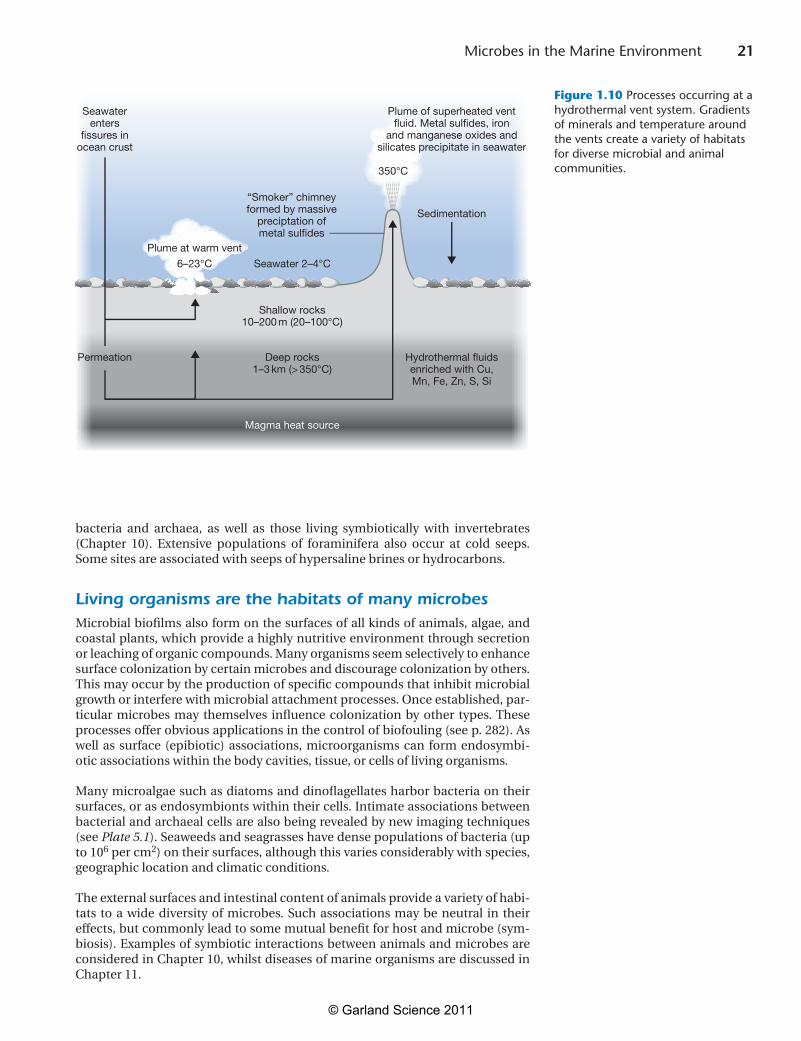
heatedunderlyingrocks,therebychangingthechemicalandphysicalcharacter-isticsofboththeseawaterandtherock.Thepermeabilitystructureoftheoceancrust and the location of the heat source determine the circulation patterns of hydrothermalfluids.Ascoldseawaterpenetratesintotheoceancrust,itisgradu-ally heated along its flow path, leading to the removal of magnesium from the fluidintotherock,withproductionofacidduringtheprocess.Thisleadstotheleachingofothermajorelementsandtransitionmetals fromtherock into thehydrothermal fluid, and sulfate in the seawater is removed by precipitation and reductiontohydrogensulfide.Asthepercolatingfluidsreachtheproximityofthemagmaheatsource,extensivechemicalreactionsoccurwithintherockandthepressurizedfluidsareheatedtoover350°C,becomingbuoyantandrisingtowardtheoceanfloor.Astheyrise,decompressioncausesthefluidstocoolslightly,andprecipitation of metal sulfides and other compounds occurs en route. The hydro-thermal fluid is injected into the ocean as plumes of mineral-rich superheated water.Thehottestplumes(upto350°C)aregenerallyblack,becauseofthehighcontent of metal sulfide and sulfate particles, and precipitation occurs as the hot plume mixes with the cold seawater. Some of these precipitates form chimney structurescalled“blacksmokers,”whilstothersaredispersedthroughthewaterand form sediments in the vicinity. In other parts of the vent field, the circula-tion of hydrothermal fluid may be shallower, leading to diffuse plumes of water heatedto6–23°C(Figure 1.10). The gradients of temperature and nutrients that exist at hydrothermal systems provide a great diversity of habitats for microbes suspended in the surrounding heated waters, in sediments, and attached to surfaces of the chimneys. Many of these are hyperthermophilic bacteria and archaea,whichcangrowattemperaturesupto121°C,whilstothersgrowatlowertemperatures further from the fluid emissions. Molecular studies are revealing an astonishing diversity of such organisms, many of which have biotechnologi-calapplications.Themicrobiologyofthedeepsubsurfacerocksbeneathventsisalso now under investigation, and many novel microbes and metabolic processes are being discovered. Microbial activity in the deep subsurface contributes to the chemical changes in composition during circulation of the hydrothermal fluids.
Hydrothermalventsystemswerefirstdescribedin1977,whenscientistsaboardthe submersible Alvin,fromWoodsHoleOceanographicInstitution,wereexplor-ingtheseabedabout2500mdeepneartheGalapagosIslands.Thediscoveryoflife around the vents was totally unexpected. The Alvin scientists observed dense communities of previously unknown animals, including tubeworms, clams,anemones, crabs, and many others (see Plate 10.3).Subsequentresearchshowedthat the warm waters near hydrothermal vents contain large populations of che-mosynthetic bacteria and archaea, which fix CO2 using energy from the oxidation of sulfides in the vent fluids. This metabolism supports a food chain with many trophiclevelsthatisindependentofphotosynthesis.Wenowknowthatmanyofthe animals at vent sites contain chemosynthetic bacteria as symbionts within their tissues or on their surfaces—these relationships are discussed in Chapter 10.Inaddition,bacterialpopulationsdirectlysupportthegrowthoffilter-feedinganimals,suchasclamsandmussels,orshrimps,whichgrazeonmicrobialmats.Thus, hydrothermal vents are an oasis of life in the deep sea. Previously, life was thought always to rely ultimately on the fixation of CO2 by photosynthesis, but the vent communities function without the input of material derived from the use of light energy. However, note that sulfide oxidation depends on dissolved oxygen in the water, and the origin of this is photosynthetic.
Cold seeps also support diverse life
Cold seeps are abundant along the continental shelf and slope, where the upwards percolation of fluids through the sediments is influenced by plate tectonics and othergeologicalprocesses.At thesesites,highconcentrationsofmethaneandsulfide support prolific chemosynthetic communities consisting of free-living
WHAT WILL HAPPEN WHEN THE SEA-ICE MELTS?
Global warming is affecting polar regions more rapidly than predicted by previous models. Although there is a large natural variation in the Arctic climate, since the 1970s, the extent of summer sea ice in the Arctic has declined by an average 8% per decade and ice packs have become thinner. The loss of cover in 2007 was especially severe and led to fears that a “tipping point” had been reached, but the loss has not been so severe in recent years. The loss of ice cover results in changes to wind patterns and warmer and less saline waters in the upper ocean. Most significantly, loss of summer ice also reduces the albedo effect, resulting in a positive feedback, which increases the absorption of solar energy by seawater rather than reflecting sunlight, further hastening the rise in temperature. An intensive research program to compare the microbiology of current and archived samples of deep-sea sediment has recently been initiated by Antje Boetius and colleagues at the Max Planck Institute of Marine Microbiology, Bremen. This is revealing important information about changes in microbial diversity and how the flux of particulate organic matter is affected by the loss of permanent ice cover. Besides producing major changes in marine ecology, the melting of summer sea ice means that shipping can now pass through the ice-free North West Passage and is enabling exploration for oil and gas in previously inaccessible parts of the Arctic Ocean.
?
© Garland Science 2011
21 Microbes in the Marine Environment
bacteria and archaea, as well as those living symbiotically with invertebrates (Chapter 10). Extensive populations of foraminifera also occur at cold seeps.Some sites are associated with seeps of hypersaline brines or hydrocarbons.
Living organisms are the habitats of many microbesMicrobialbiofilmsalsoformonthesurfacesofallkindsofanimals,algae,andcoastal plants, which provide a highly nutritive environment through secretion or leaching of organic compounds. Many organisms seem selectively to enhance surfacecolonizationbycertainmicrobesanddiscouragecolonizationbyothers.This may occur by the production of specific compounds that inhibit microbial growth or interfere with microbial attachment processes. Once established, par-ticular microbes may themselves influence colonization by other types.Theseprocessesofferobviousapplicationsinthecontrolofbiofouling(seep.282).Aswell as surface (epibiotic) associations, microorganisms can form endosymbi-otic associations within the body cavities, tissue, or cells of living organisms.
Many microalgae such as diatoms and dinoflagellates harbor bacteria on their surfaces, or as endosymbionts within their cells. Intimate associations between bacterialandarchaealcellsarealsobeingrevealedbynewimagingtechniques(see Plate 5.1). Seaweeds and seagrasses have dense populations of bacteria (up to106 per cm2) on their surfaces, although this varies considerably with species, geographic location and climatic conditions.
The external surfaces and intestinal content of animals provide a variety of habi-tats to a wide diversity of microbes. Such associations may be neutral in their effects, but commonly lead to some mutual benefit for host and microbe (sym-biosis). Examples of symbiotic interactions between animals and microbes are consideredinChapter10,whilstdiseasesofmarineorganismsarediscussedinChapter11.
Figure 1.10
Sedimentation
350°C
“Smoker” chimneyformed by massive
preciptation ofmetal sulfides
Plume of superheated ventfluid. Metal sulfides, iron
and manganese oxides andsilicates precipitate in seawater
Seawaterenters
fissures inocean crust
Permeation Deep rocks1–3 km (> 350°C)
Hydrothermal fluidsenriched with Cu,Mn, Fe, Zn, S, Si
Magma heat source
Shallow rocks10–200 m (20–100°C)
Seawater 2–4°C6–23°C
Plume at warm vent
Figure 1.10 Processes occurring at a hydrothermal vent system. Gradients of minerals and temperature around the vents create a variety of habitats for diverse microbial and animal communities.
© Garland Science 2011
22 Chapter 1
ConclusionsThis chapter has introduced the marine environment and the various types of marine microbes that are found. The adaptations of marine microbes to the dif-ferent physical, chemical, and biological conditions encountered in the wide range of marine habitats have led to the evolution of highly diverse microbes. The discovery that these tiny microbes are present in such large numbers and are largely responsible for the biogeochemical processes that shape our planet can beviewedasoneofthemostimportantadvancesofmodernscience.Subsequentchapters build on this introduction by exploring the mechanisms underlying this diversity of form and function.
ReferencesBorin,S.,Brusetti,L.,Mapelli,F.,etal.(2009)Sulfurcyclingandmethanogenesis primarily drive microbial colonization of thehighly sulfidic Urania deep hypersaline basin. Proc. Natl Acad. Sci. USA106:9151–9156.
Ciccarelli,F.D.,Doerks,T.,vonMering,C.,Creevey,C.J.,Snel,B.andBork,P.(2006)Towardautomaticreconstructionofahighlyresolved tree of life. Science311:1283–1287.
C-MORE(2009)RisingCO2, ocean acidification, and their impacts on marine microbes. Center for Microbial Oceanography: Research and Education, http://cmore.soest.hawaii.edu/oceanacidification/[Accessed10December2010].
Daffonchio, D., Borin, S., Brusa, T., et al. (2006) Stratifiedprokaryote network in the oxic/anoxic transition of a deep-seahalocline. Nature440:203–207.
Dagan, T. and Martin, W. (2009) Getting a better picture ofmicrobial evolution en route to a network of genomes. Philos. Trans. R. Soc. Lond. B Biol. Sci.364:2187–2196.
Danovaro, R., Dell’Anno, A., Corinaldesi, C., Magagnini, M.,Noble, R., Tamburini, C. andWeinbauer, M. (2008) Major viralimpact on the functioning of benthic deep-sea ecosystems. Nature454:1084–1087.
Delille, B., Harlay, J., Zondervan, I., et al. (2005) Response ofprimary production and calcification to changes of pCO2 during experimental blooms of the coccolithophorid Emiliania huxleyi. Global Biogeochem. Cycles19:GB2023.
Doolittle,W.F.(1999)Phylogeneticclassificationandtheuniversaltree. Science284:2124–2128.
Dworkin, M., Falkow, S., Rosenberg, E., Schleifer, K. H. andStackebrandt,E.(2007)The Prokaryotes. Springer,NewYork.
Edgcomb,V.,Orsi,W.,Leslin,C.,etal.(2009)Protistancommunitypatterns within the brine and halocline of deep hypersaline anoxic basins in the eastern Mediterranean Sea. Extremophiles 13:151–167.
Engel,A.,Zondervan,I.,Aerts,K.,etal.(2005)Testingthedirecteffect of CO2 concentration on a bloom of the coccolithophorid Emiliania huxleyi in mesocosm experiments. Limnol. Oceanogr. 50:493–507.
EPOCA(2009)EuropeanProjectonOceanAcidificationwebsite,accessedAugust28,2010,http://www.epoca-project.eu/.
Fowler,S.W.,Buat-Menard,P.,Yokoyama,Y.,Ballestra,S.,Holm,S.andVanNguyen,H.(1987)RapidremovalofChernobylfalloutfrom Mediterranean surface waters by biological activity. Nature 329:56–58.
Hallsworth, J. E.,Yakimov, M. M., Golyshin, P. N., et al. (2007)Limits of life in MgCl2-containing environments: chaotropicity defines the window. Environ. Microbiol.9:801–813.
Henderson,G.P.,Gan,L.andJensen,G.J.(2007)3-Dultrastructureof Ostreococcus tauri: electron cryotomography of an entire eukaryoticcell.PLoS ONE2(8):e749.
Iglesias-Rodriguez,M.D.,Halloran,P.R.,Rickaby,R.E.M.,etal.(2008a)Phytoplanktoncalcificationinahigh-CO2 world. Science 320:336–340.
Iglesias-Rodriguez, M. D., Buitenhuis, E. T., Raven, J. A., et al.(2008b) Response to comment on“Phytoplankton calcificationin a high-CO2 world”. Science322:1466.
Karner, M. B., De Long, E. F. and Karl, D. M. (2001) ArchaealdominanceinthemesopelagiczoneofthePacificOcean.Nature 409:507–510.
Lipp, J. S., Morono, Y., Inagaki, F. and Hinrichs, K. U. (2008)SignificantcontributionofArchaeatoextantbiomassinmarinesubsurface sediments. Nature454:991–994.
Martin,W.andEmbley,T.M.(2004)Evolutionarybiology:earlyevolution comes full circle. Nature431:134–137.
Mitchell, A. (2009) Sea Sick. The Hidden Ecological Crisis of the Global Ocean.OneWorldPublications,Oxford.
Orr, J. C., Fabry, V. J., Aumont,O., et al. (2005) Anthropogenicocean acidification over the twenty-first century and its impact on calcifying organisms. Nature437:681–686.
Pace, N. R. (2009) It’s time to retire the prokaryote. Microbiol. Today36:85–87.
Ramos,J.B.E.,Biswas,H.,Schulz,K.G.,LaRoche,J.andRiebesell,U. (2007) Effect of rising atmospheric carbon dioxide on themarine nitrogen fixer Trichodesmium. Global Biogeochem. Cycles 21:GB2028;doi:10.1029/2006GB002898.
Riebesell, U. (2004) Effects of CO2 enrichment on marine phytoplankton.J. Oceanogr. 60:719–729.
Riebesell,U.,Schulz,K.G.,Bellerby,R.G.J.,etal.(2007)Enhancedbiological carbon consumption in a high CO2 ocean. Nature 450:545–548.
Riebesell,U.,Schulz,K.G.,Bellerby,R.G.J.,Botros,M.,Fritsche,P., Meyerhofer, M., et al. (2007) Enhanced biological carbonconsumption in a high CO2 ocean. Nature450:545–548.
Royal Society (2005) Ocean acidification due to increasingatmospheric carbon dioxide. Policy document 12/05. TheRoyal Society, London, 2005, accessed August 28, 2010, http://royalsociety.org/.
Suttle,C.A.(2005)Virusesinthesea.Nature437:359–361.
Whitman,W. B. (2009)The modern concept of the procaryote. J. Bacteriol. 191:2000–2005.
Whitman, W. B., Coleman, D. C. and Wiebe, W. J. (1998)Prokaryotes: the unseen majority. Proc. Natl Acad. Sci. USA 95:6578–6583.
© Garland Science 2011
23 Microbes in the Marine Environment
Yakimov,M.M.,LaCono,V.,Denaro,R.,D’Auria,G.,Decembrini,F.,Timmis, K. N., Golyshin, P. N. and Giuliano, L., et al. (2007)Primaryproducingprokaryoticcommunitiesofbrine,interfaceandseawaterabovethehaloclineofdeepanoxiclakeL’Atalante,Eastern Mediterranean Sea. ISME J.1:743–755.
Further readingHistorical perspectives
Karl, D. M. and Proctor, L. M. (2008) Foundations of microbialoceanography. Oceanography 20:16–27.
Yayanos, A. A. (2003) Marine microbiology at Scripps.Oceanography16:63–75.
Microbial diversity and evolution
Caron, D. A., Worden, A. Z., Countway, P. D., Demir, E. andHeidelberg,K.B.(2008)Protistsaremicrobestoo:aperspective. ISME J.3:4–12.
Lawton,G.(2009)WhyDarwinwaswrongaboutthetreeoflife.New Scientist 2692:34–39.
Schulz,H.N.andJørgensen,B.B.(2001)Bigbacteria.Annu. Rev. Microbiol. 55:105–137.
Sherr,B.F.,Sherr,E.B.,Caron,D.A.,Vaulot,D.andWorden,A.Z.(2007)Oceanicprotists.Oceanography 20:130–134.
Microscale processes, marine snow, and dissolved organic matter
Azam, F. and Long, R. A. (2001) Sea snow microcosms. Nature 414:495–498.
Azam,F.andMalfatti,F.(2007)Microbialstructuringofmarineecosystems. Nat. Rev. Microbiol.5:782–791.
Grossart, H. P., Kiørboe,T.,Tang, K.W., Allgaier, M.,Yam, E. M.and Ploug, H. (2006) Interactions between marine snow andheterotrophic bacteria: aggregate formation and microbial dynamics. Aquat. Microb. Ecol.42:19–26.
Kjorbøe, T. (2001) Formation and fate of marine snow: small-scale processes with large-scale implications. Sci. Mar. 65 (S2):57–71.
Verdugo,P.,Alldredge,A.L.,Azam,F.,Kirchman,D.L.,Passow,U.andSantschi,P.H.(2004)Theoceanicgelphase:abridgeintheDOM-POM continuum. Marine Chem. 92:67–85.
Sea-ice microbiology and polar oceans
Arrigo,K.R.andThomas,D.N.(2004)Largescaleimportanceofsea ice biology in the Southern Ocean. Antarctic Sci.16:471–486.
Kirchman,D.L.,Moran,X.A.G.andDucklow,H.(2009)Microbialgrowth in the polar oceans—role of temperature and potential impact of climate change. Nat. Rev. Microbiol.7:451–459.
Mock,T. andThomas, D. N. (2005) Recent advances in sea-icemicrobiology. Environ. Microbiol. 7:605–619.
Sediments and subsurface microbiology
Jørgensen, B. B. and Boetius, A. (2007) Feast and famine—microbial life in the deep-sea bed. Nat. Rev. Microbiol. 5:770–781.
Hydrothermal vents
Martin, W., Baross, J., Kelley, D. and Russell, M. J. (2008)Hydrothermal vents and the origin of life. Nat. Rev. Microbiol. 6:805–814.
Reysenbach,A.-L.,Cady,S.L.(2001)Microbiologyofancientandmodern hydrothermal systems. Trends Microbiol. 9:79–86.
© Garland Science 2011

© Garland Science 2011