Embed Size (px)
Citation preview

Eur J Vasc Endovasc Surg (2011) 41, S1eS58
Management of Abdominal Aortic AneurysmsClinical Practice Guidelines of the European Societyfor Vascular Surgery
F.L. Moll a,*, J.T. Powell b, G. Fraedrich c, F. Verzini d, S. Haulon e,M. Waltham f, J.A. van Herwaarden a, P.J.E. Holt g, J.W. van Keulen a,h,B. Rantner c, F.J.V. Schlosser h, F. Setacci i, J.-B. Ricco j
aDepartment of Vascular Surgery, University Medical Center Utrecht, Utrecht, The Netherlandsb Imperial College, London, UKcUniversity Hospital Innsbruck, AustriadAzienda Ospedaliera di Perugia, ItalyeHopital Cardiologique, CHRU de Lille, Lille, Francef St Thomas’ Hospital, London, UKg St George’s Vascular Institute, London, UKhYale University - School of Medicine, New Haven, Connecticut, USAiUniversity of Siena, Siena, ItalyjUniversity of Poitiers, Poitiers, France
Submitted 4 September 2010; accepted 12 September 2010
*
10do
KEYWORDSAbdominal aorticaneurysms;Guidelines;Management;Clinical practice;Evidence-basedmedicine
Corresponding author. Tel.: þ31 88E-mail address: [email protected] (F.L.
78-5884/$36 ª 2010 Published by Ei:10.1016/j.ejvs.2010.09.011
Introduction
Purpose of these guidelines
The European Society for Vascular Surgery (ESVS) appointedthe AAA Guidelines Committee to write the current clinical
7556965; fax: þ31 887555017.Moll).
lsevier Ltd on behalf of European
practice guidelines document for surgeons and physicianswho are involved in the care of patients with abdominalaortic aneurysms (AAAs). Guideline development was rec-ommended in 1990 by the Institute of Medicine to improvedecision making for specific patients’ circumstances and todecrease the variability in appropriate and inappropriate
Society for Vascular Surgery.

S2 F.L. Moll et al.
health care between providers.1,2 Appropriate decision-making is critical to achieving excellent outcomes.
Abdominal aortic aneurysm disease is complex and hassignificant clinical practice variability, although a validevidence base is available to guide recommendations. Thesignificant increase in the quantity of scientific literatureconcerning abdominal aortic aneurysmal disease publishedin recent years along with the number of technical andmedical advances enables guideline recommendations withmore certainty and supporting evidence than before.Potential increases in health care costs and risks due toindustry and public-driven use of novel treatment optionsmake the current guidelines increasingly important.3e6
Many clinical situations of patients with AAAs have notbeen the subject of randomised clinical trials. Patient care,however, needs to be delivered and decisions have to bemade in these situations. Therefore, this document alsoprovides guidance for decisions when extensive level Ievidence is not available and recommendations are deter-mined on the basis of the currently available best evidencefor these situations. By providing information about therelevance and validity of the quality of evidence, thereader will be able to locate the most important andevidence-based information relevant to the individualpatient.7 To optimise the implementation of the currentdocument, the length of the guidelines has been kept asshort as possible to enable prompt access to the guidelineinformation. This clinical guidelines document is supposedto be a guide, not a document of rules, and allows flexibilityfor specific patients’ circumstances.
This is the resulting clinical practice guidelines docu-ment and provides recommendations for clinical care ofpatients with abdominal aortic aneurysms including pre-operative, perioperative and post-operative care.
Methods
PatientswithAAAsaredefinedasmaleor femalepatientswithasymptomatic, symptomatic or ruptured AAA with fusiformdilatation. This document does not cover patients witha saccular, infected or mycotic AAA or pseudoaneurysmalaortic dilatation. The AAA Guidelines Committee met inSeptember 2009 for the first time to discuss the purpose andmethods. The AAA Guidelines Committee has been consti-tutedwith incorporation ofmembers fromdifferent Europeancountries, from academic and private hospitals, vascular andendovascular specialists and patients tomaximise the supportfor the final guidelines document. Since Europe encompassesa variety of health care systems and political economies,health policy makers were not included.8
The AAA Guidelines Committee performed a systematicliterature search in MEDLINE, EMBASE and COCHRANE Librarydatabases for each of the different topics that are discussedin this guidelines document. The Guidelines Committee useda grading schema based on levels of evidence and grades ofrecommendation according to the levels of evidence fromthe Oxford Centre For Evidence-Based Medicine.9
The level of evidence classification provides informationabout the study characteristics supporting the recommen-dation and expert consensus, according to the categoriesshown in Table 1.
The recommendation grade indicates the strength ofa recommendation. Definitions of the grades of recom-mendation are shown in Table 2.
The AAA Guidelines Committee aimed to report as muchas possible the calculated estimates of effects with their95% confidence intervals. Every part of the guidelinesdocument has been prepared by at least two members ofthe Committee and has been reviewed by the entireCommittee. The initial guidelines document has beensubsequently reviewed by the AAA Guidelines ReviewCommittee. After incorporation of all comments andrecommendations, the guidelines have been provided tothe members of the ESVS. The final document has beenapproved by the ESVS.
Chapter 1 e Epidemiology
Definition of abdominal aortic aneurysms
Abdominal aortic aneurysm (AAA), which comes from theAncient Greek word ἀ�eύrysma, means a dilatation orwidening of the abdominal aorta. The most accepted defi-nition of an AAA is based on the diameter of the abdominalaorta: an abdominal aortic diameter of 3.0 cm or more,which usually is more than 2 standard deviations above themean diameter for both men and women, and is consideredto be aneurysmal.10e12 Other researchers have suggesteddefining abdominal aortic aneurysm as the maximum infra-renal aortic diameter being at least 1.5 times larger thanthe expected normal infra-renal aortic diameter tocompensate for individual variation in the diameter of theadjacent aorta.13e15
AAA can be defined as an abdominal aortic diameter of3.0 cm or more in either anterior-posterior or transverseplanes. Level 2c, Grade B.
Epidemiology
Prevalence and risk factorsPopulation screening studies offer the best evidenceregarding the prevalence of AAA. Several of these havebeen conducted as randomised trials to assess the benefitsof screening (MASS, Western Australia, Viborg and Chi-chester, the latter being the only one to include wom-en).16e19 Other evidence comes from the Rotterdam,Tromsø and other large epidemiological screeningstudies.20,21 Prevalence rates vary according to age, genderand geographical location (Table 3). Level 1a.
In keeping with ethnic and environmental risk factors,a screening study of US veterans (between 50 and 79 yearsold, n Z 73,451) showed the highest prevalence of AAA�3.0 cm was 5.9% and was found in white male smokersbetween 50 and 79 years.22 All the aneurysm populationscreening data (Table 3) are now dated and there is littlecontemporary information for 21st century prevalence,although there are some indications, at least in the USA,that the admission rate for aneurysm repair is declining.23
Important risk factors for AAA are advanced age, malegender and smoking.20e31 A positive family history for AAAespecially in male first-degree relatives, is also associated

Table 1 Level of evidence classification.
Level Therapy/Prevention, Aetiology/Harm
Prognosis Diagnosis
1a SR (with homogeneity) of RCTs
SR (with homogeneity) of inception cohort studies; CDRvalidated in different populations
SR (with homogeneity) of Level 1 diagnostic studies; CDR with 1b studies from different clinical centres
1b Individual RCT (with narrow Confidence Interval)
Individual inception cohort study with > 80% follow-up; CDR validated in a single population
Validating cohort study with good reference standards; or CDR tested within one clinical centre
1c All or none All or none case-series Absolute SpPins and SnNouts 2a SR (with homogeneity) of
cohort studies SR (with homogeneity) of either retrospective cohort studies or untreated control groups in RCTs
SR (with homogeneity) of Level >2 diagnostic studies
2b Individual cohort study (including low quality RCT; e.g., <80% follow-up)
Retrospective cohort study or follow-up of untreated control patients in an RCT; Derivation of CDR or validated on split-sample only
Exploratory cohort study with good reference standards; CDR after derivation, or validated only on split-sample or databases
2c "Outcomes" Research; Ecological studies
"Outcomes" Research
3a SR (with homogeneity) of case-control studies
SR (with homogeneity) of 3b and better studies
3b Individual Case-Control Study
Non-consecutive study; or without consistently applied reference standards
4 Case-series (and poor quality cohort and case-control studies)
Case-series (and poor quality prognostic cohort studies)
Case-control study, poor or non-independent reference standard
5 Expert opinion without explicit critical appraisal, or based on physiology,
Expert opinion without explicit critical appraisal, or based on physiology, bench
Expert opinion without explicit critical appraisal, or based on physiology, bench research or
bench research or "first principles"
research or "first principles" "first principles"
SR, systematic review; RCT, randomised controlled trial; CDR, clinical decision rule; SpPin, Specificity is so high that a Positive result rules-in the diagnosis; SnNout, Sensitivity is so high that a Negative result rules-out the diagnosis.
Management of Abdominal Aortic Aneurysms S3
with increased risk for AAA.29e31 Smoking is a strong riskfactor (odds ratio >3.0 in all studies), the associated riskbeing much higher than for either coronary artery diseaseor stroke.20e22,24,28 Level 2a.
Additionally, the following factors have been associatedwith AAA development: history of other vascularaneurysms,32e35 greater height,22 coronary artery
Table 2 Grades of recommendation
A Consistent level 1 studiesB Consistent level 2 or 3 studies or extrapolations from
Level 1 studiesC Level 4 studies or extrapolations from level 2 or
3 studiesD Level 5 evidence or troublingly inconsistent or
inconclusive studies of any level
“Extrapolations" are where data are used in a situation that haspotentially clinically important differences than the originalstudy situation.
disease,22,33 cerebrovascular disease,34 atherosclerosis,22
hypercholesterolemia,20,22 and hypertension,21,22,35,36
although the data for some of these factors are inconsistentand studies may not have been subject to multivariateadjustment, so that spurious associations may have beenreported. More recently, genome-wide association studieshave demonstrated the association with variants on chro-mosome 9p21. The presence of rs7025486[A] in the DAB21Pgene is associated with a 20% increased risk of developingAAA, odds ratio 1.21 [95%CI 1.14e1.28].37 Black or Asian raceand diabetes mellitus are negatively associated with AAAdevelopment.22,38 Level 2a-3b.
The evidence for other risk factors including homo-cysteinemia, high levels of lipoprotein (a) and plasminogenactivator inhibitor-1 is very weak.39 Level 4b.
Natural history
AAA growth ratesThe reported average growth rate of AAAs between 30 and55 mm ranges from 0.2 to 0.3 cm per year. Larger AAA
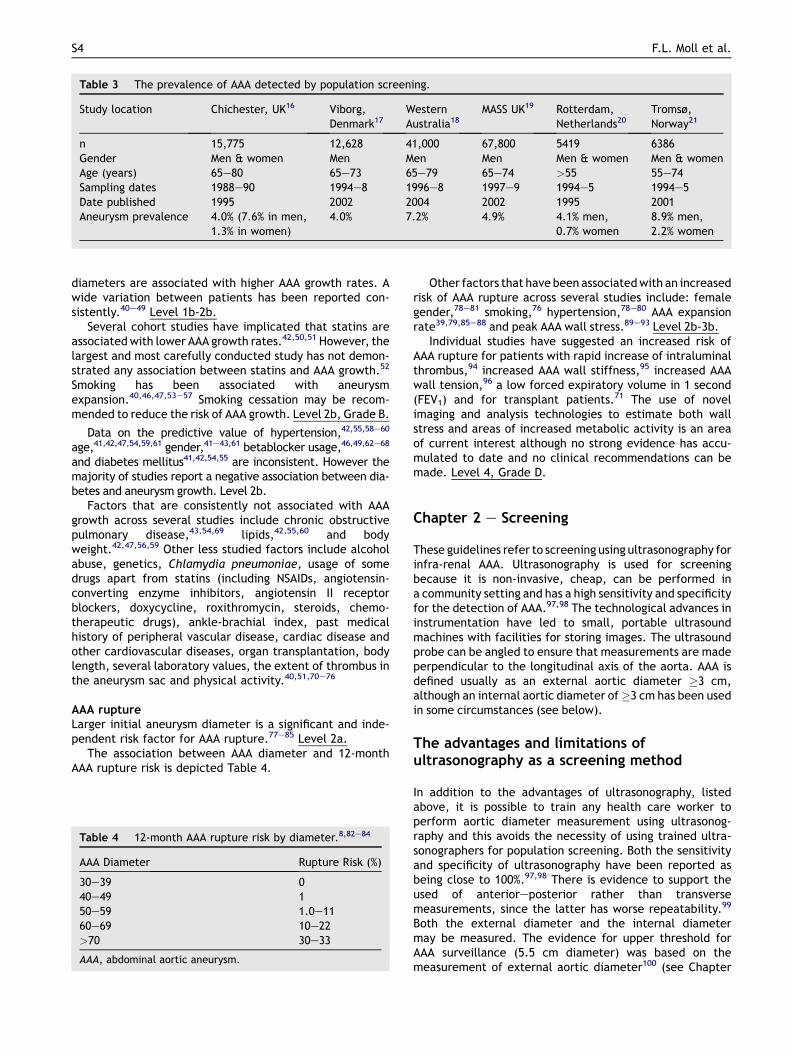
Table 3 The prevalence of AAA detected by population screening.
Study location Chichester, UK16 Viborg,Denmark17
WesternAustralia18
MASS UK19 Rotterdam,Netherlands20
Tromsø,Norway21
n 15,775 12,628 41,000 67,800 5419 6386Gender Men & women Men Men Men Men & women Men & womenAge (years) 65e80 65e73 65e79 65e74 >55 55e74Sampling dates 1988e90 1994e8 1996e8 1997e9 1994e5 1994e5Date published 1995 2002 2004 2002 1995 2001Aneurysm prevalence 4.0% (7.6% in men,
1.3% in women)4.0% 7.2% 4.9% 4.1% men,
0.7% women8.9% men,2.2% women
S4 F.L. Moll et al.
diameters are associated with higher AAA growth rates. Awide variation between patients has been reported con-sistently.40e49 Level 1b-2b.
Several cohort studies have implicated that statins areassociatedwith lower AAA growth rates.42,50,51 However, thelargest and most carefully conducted study has not demon-strated any association between statins and AAA growth.52
Smoking has been associated with aneurysmexpansion.40,46,47,53e57 Smoking cessation may be recom-mended to reduce the risk of AAA growth. Level 2b, Grade B.
Data on the predictive value of hypertension,42,55,58e60
age,41,42,47,54,59,61 gender,41e43,61 betablocker usage,46,49,62e68
and diabetes mellitus41,42,54,55 are inconsistent. However themajority of studies report a negative association between dia-betes and aneurysm growth. Level 2b.
Factors that are consistently not associated with AAAgrowth across several studies include chronic obstructivepulmonary disease,43,54,69 lipids,42,55,60 and bodyweight.42,47,56,59 Other less studied factors include alcoholabuse, genetics, Chlamydia pneumoniae, usage of somedrugs apart from statins (including NSAIDs, angiotensin-converting enzyme inhibitors, angiotensin II receptorblockers, doxycycline, roxithromycin, steroids, chemo-therapeutic drugs), ankle-brachial index, past medicalhistory of peripheral vascular disease, cardiac disease andother cardiovascular diseases, organ transplantation, bodylength, several laboratory values, the extent of thrombus inthe aneurysm sac and physical activity.40,51,70e76
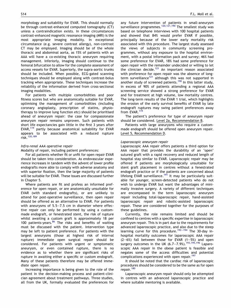
AAA ruptureLarger initial aneurysm diameter is a significant and inde-pendent risk factor for AAA rupture.77e85 Level 2a.
The association between AAA diameter and 12-monthAAA rupture risk is depicted Table 4.
Table 4 12-month AAA rupture risk by diameter.8,82e84
AAA Diameter Rupture Risk (%)
30e39 040e49 150e59 1.0e1160e69 10e22>70 30e33
AAA, abdominal aortic aneurysm.
Other factors that have been associatedwith an increasedrisk of AAA rupture across several studies include: femalegender,78e81 smoking,76 hypertension,78e80 AAA expansionrate39,79,85e88 and peak AAA wall stress.89e93 Level 2b-3b.
Individual studies have suggested an increased risk ofAAA rupture for patients with rapid increase of intraluminalthrombus,94 increased AAA wall stiffness,95 increased AAAwall tension,96 a low forced expiratory volume in 1 second(FEV1) and for transplant patients.71 The use of novelimaging and analysis technologies to estimate both wallstress and areas of increased metabolic activity is an areaof current interest although no strong evidence has accu-mulated to date and no clinical recommendations can bemade. Level 4, Grade D.
Chapter 2 e Screening
These guidelines refer to screening using ultrasonography forinfra-renal AAA. Ultrasonography is used for screeningbecause it is non-invasive, cheap, can be performed ina community setting and has a high sensitivity and specificityfor the detection of AAA.97,98 The technological advances ininstrumentation have led to small, portable ultrasoundmachines with facilities for storing images. The ultrasoundprobe can be angled to ensure that measurements are madeperpendicular to the longitudinal axis of the aorta. AAA isdefined usually as an external aortic diameter �3 cm,although an internal aortic diameter of�3 cm has been usedin some circumstances (see below).
The advantages and limitations ofultrasonography as a screening method
In addition to the advantages of ultrasonography, listedabove, it is possible to train any health care worker toperform aortic diameter measurement using ultrasonog-raphy and this avoids the necessity of using trained ultra-sonographers for population screening. Both the sensitivityand specificity of ultrasonography have been reported asbeing close to 100%.97,98 There is evidence to support theused of anterioreposterior rather than transversemeasurements, since the latter has worse repeatability.99
Both the external diameter and the internal diametermay be measured. The evidence for upper threshold forAAA surveillance (5.5 cm diameter) was based on themeasurement of external aortic diameter100 (see Chapter

Management of Abdominal Aortic Aneurysms S5
3: Decision-making). In contrast, the MASS trial, the largestof the population-based aneurysm screening trials, wasbased on the measurement of internal aortic diameter101
(http://aaa.screening.nhs.uk/Implementation_Guidance).The Viborg aneurysm screening trial17 and most otherscreening programmes have reported using external aorticdiameter. Since internal diameters are 2e5 mm smallerthan external diameters, there are two important issues tobe resolved.
Are the threshold aortic diameters of 3 cm and 5.5 cmbased on the internal aortic diameter safe? The MASS trialreports an increase of aneurysm ruptures in screenedpatients after 8 years of follow-up,102 so were the smallestaneurysms overlooked?
Which diameter, internal or external, is most reproduc-ibly measured in community screening programmes? This isimportant since, at best, the reproducibility of measure-ment of external aortic diameters is �2 mm.98e100
The evidence in favour of population screening forAAA in men
The four randomised trials of population screening are theChichester trial in the UK,16 the Viborg trial in Denmark,17
the Western Australia trial18 and the MASS trial in theUK.19 In each trial, populations were randomised to eitheran offer of aneurysm screening or to no offer of screening,and in each trial screening, was shown to reduce aneurysm-related mortality for men. These results, to 5 years, havebeen summarised in a Cochrane Review103 and the oddsratio in favour of screening for men was 0.60 [95%CI0.47e0.78]. A systematic review for the US Preventive TaskForce reported a similar benefit for screening men, oddsratio 0.53 [95%CI 0.42e0.68].104 The individual character-istics of the trials are summarised in Table 5. This table alsoserves to illuminate some of the differences between thetrials. In the Western Australia trial randomisation occurredseveral months ahead of the invitation for screening beingissued, so that about 2296 men had died before their invi-tations were issued; the uptake of screening was 63% ifestimated from the time of randomisation and 70% if esti-mated from the time of invitation. There also is one broadsimilarity between the trials, not listed in Table 3, in thatall trials were conducted in relatively advanced socioeco-nomic areas where a semi-rural hinterland is dotted withmedium or small size towns inhabited predominantly bypersons of Caucasian origin. None of the screening trialswere conducted, except small part, in very deprived largecity districts.
The longer-term follow-up of subjects in the MASS trialhas provided additional results. After 7 years of follow-upthe MASS trial reported an all-cause mortality benefit infavour of screening at the limits of statistical significance,hazard ratio 0.96 [95%CI 0.93e1.00];105 no all-causemortality benefit was observed in the Western Australiatrial after 5 years of follow-up. Very recently the MASS trialpublished 10-year results.101 These showed that aneurysm-related deaths were halved in the group invited forscreening at a cost of ₤100 for every man screened,although there is a suggestion from a report from the USAthat costs might be less than this.106 Overall there were 552
elective aneurysm repairs in the screened group (with anoperative mortality of 4%) versus 226 in the control group(with an operative mortality of 6%). However, after 8 yearsthere was a noticeable increase in ruptures in the screenedgroup. Although studies have reported that a single screenat age 65 years is sufficient, this may require re-evaluation,particularly as the population lives longer.107
Population screening of older men for AAA, in regionswhere the population prevalence is 4% or more, reducesaneurysm-related mortality by almost half within 4 years ofscreening, principally by reducing the incidence of aneu-rysm rupture. Level 1a, Recommendation A.
The evidence for screening in women
The population prevalence of AAA is three times higher inmen than in women. Therefore, not surprisingly, there is nogood evidence to support aneurysm screening in olderwomen. The only screening trial conducted in women wasin Chichester, UK,108 and is reported as part of the Chi-chester trial in Table 3. There was no reduction in theincidence of aneurysm rupture after either 5 or 10 years offollow-up. Given the previous low prevalence of aneurysmsdetected in women, this trial may not have had sufficientpower to detect any benefit from screening. Howeversmoking, the principal risk factor for AAA, has beenincreasing in women and the future incidence of AAA infemale smokers is unknown.
Population screening of older women for AAA does notreduce the incidence of aneurysm rupture. Level 1b,Recommendation B.
Population screening of older female smokers for AAAmay require further investigation. Level 3c, Recommenda-tion B.
Screening in other subgroupsConsideration has been given to the merits of screening bydifferent subgroups, including those relating to smoking,ethnicity, other cardiovascular disease and those having orhaving had relatives with AAA.
The US Preventive Services Task Force has recommendedaneurysm screening for men aged 65e75 years who haveever smoked, based on the strength of the associationbetween smoking and AAA.109 There is no good evidence tosupport this proposal, although it seems reasonable.
Ever-smoking increases the risk of developing AAA 4- to 5-fold. Screening only smokers might improve the cost-effec-tiveness of aneurysm screening. Level 5, Recommendation D.
The Society of Vascular Surgery recommends screeningmen aged 65 years with a family history of AAA.110 This isbased on reports from several countries of an increasedincidence of AAA amongst first-degree relatives of AAApatients. The best data for this comes from a Swedishpopulation study, when a family history of AAA increasedthe risk of AAA, odds ratio 1.9 [95%CI 1.6e2.2].31 Thebenefits of screening for AAA in the presence of a familyhistory of aneurysm has not been assessed formally.
A family history of AAA increases the risk of AAA about 2-fold. Screening of older men and women having a familyhistory of AAA might be recommended. Level 3a, Recom-mendation C.

Table 5 Summary of the population-based randomised screening trials.
Trial characteristics Chichester, UK16 Viborg, Denmark17 MASS UK101,c Western Australia18
Number randomised 15,775 12,628 67,800 41,000Gender Men & women Men Men MenAge (years) 65e80 65e73 65e74 65e79Dates recruited 1988e90 1994e8 1997e9 1996e8Date published 1995 2002 2002 2004% accepting screening 68% 76% 80% 70%d
Aneurysms found 4% (7.6% in men) 4% 4.9% 7.2%Place of screening Hospital Hospital Community CommunityIntervention policy At 6 cm At 5 cm At 5.5 cm measured
as internal diameterNone
Mean follow-up (months) 30.5 61 49 43AAA mortality 0.59 men only 0.31 0.58 0.72odds ratio screenedvs not (95%CI)a
(0.27e1.29) (0.13e0.79) (0.42e0.78) (0.39e1.32)
All-cause mortality Men only 1.07 0.97 0.98odds ratio Screenedvs not (95%CI)b
(0.93e1.22) (0.93e1.02) (0.91e1.04)
Other outcomes reported No aneurysm-related mortalitybenefits in women
Hospital deathsCostsQuality of life
Quality oflife CostsWorkload
Extended follow-up available Yes Yesa Pooled odds ratio overall 4 trials strongly in favour of screening, OR 0.57 (0.45e0.74), together with a halving of the incidence of
aneurysm rupture in screened populations.b Pooled odds ratio trend in favour of screening, OR 0.98 (0.95e1.02).c The MASS trial recently has published 10-year follow-up, demonstrating the cost-effectiveness of screening and a significant all-cause
mortality benefit but a rising incidence of AAA rupture in the screened group.d As percentage of those alive when invitation for screening was sent: randomisation predated this invitation by several months in
a large sector of subjects.
S6 F.L. Moll et al.
Screening those with a known family history of AAAshould be evaluated and include both men and womenabove 50 years of age.
Two studies, both from the UK, have reported a very lowincidence of aneurysms in subjects of Asian ethnicorigin.38,111 In particular in the Leicester screening pro-gramme among men aged 65 years of Asian origin the prev-alence of AAA (0.45%) was significantly lower than among theCaucasian population (4.69%). Screening Asian men for AAAmay not be cost-effective. Level 2b, Recommendation B.
There is no good evidence about the prevalence of AAAamong other ethnic groups represented in Europe orelsewhere.
There is evidence to suggest that the incidence of AAA ishigh (7e10%) among those with other forms of peripheralarterial disease.112,113
Opportunistic screening of patients with peripheralarterial disease should be considered. Level 2a, Recom-mendation B.
There is some evidence to suggest that screening ofpatients with hypertension is not very productive.
Can screening cause harm?There are three potential harms that may be caused byscreening.
First there is the anxiety and subsequent effects onquality of life associated with being told that you havesomething, potentially fatal, wrong with you. Both the MASSand Viborg trials report that subjects found to have an
aneurysm on screening experienced anxiety and a decreasedquality of life for a short period after screening. Such effectswere most marked in those with poor quality of life atbaseline but the effects resolved within a few months ofscreening.101,114
Second, and perhaps more importantly, there is themortality risk associated with intervention. If screening isto be conducted safely, the vascular surgical referralcentres for patients must have an audited low mortality forboth open and endovascular aneurysm repair (EVAR):115 forelective open repair the operative mortality must be lessthan 5% (as practised in the Chichester, Viborg and MASStrials), and for EVAR less than 2%. The early advantage ofEVAR, together with its increasing usage, is unlikely toresult in a greater survival advantage of populationscreening because there is a “catch-up” in mortality afterEVAR, so that after 2e3 years the overall mortality afteropen and endovascular repair is closely similar.116e119
Recent work clearly shows that most patients have a pref-erence for aneurysm repair by EVAR rather than by openrepair. The recent results showing the risk of late endograftrupture (0.7% per 100 person-years) were unknown at thetime of patient preference studies and may dampen someof the preferences for EVAR.119 However some patients stillprefer open repair since it avoids the need for long-termpost-repair surveillance.120,121 However some patients willnot be anatomically suitable for routine endografting.Therefore, to allow for both patient preferences anddiverse patient anatomy there is a continuing need for

Table 6 Surveillance frequency of screen-detected aneurysms.
UKSAT modelling study122 Surveillanceinterval (months)
Chichester16 Viborg17 MASS101 Western Australia18
3.0e3.9 cm 24 Annual scans Annual scans Annual scans No surveillance4.0e4.5 cm 12 3.0e4.4 cm 3.0e5.0 cm 3.0e4.4 cm policy4.5e5.0 6 then 3 monthly then 3 monthly>5.0 3 scans to 6.0 cm scans to 5.5 cm
Management of Abdominal Aortic Aneurysms S7
centres to provide elective AAA repair using both opensurgery and EVAR with low mortality.
Third, screening may cause an unacceptable burden onlocal vascular surgical services. The MASS and other trialshave shown that the rate of elective repairs doubles withthe advent of screening, although the burden of out-of-hours ruptures is reduced.101e103
Screen detection of an AAA causes a small but tempo-rary reduction in quality of life. Aneurysm screening shouldonly be conducted if the audited mortality from aneurysmrepair at the referral hospital is low. Level 2a, Recom-mendation B.
Referral hospital facilities to cope with an increasednumber of elective AAA repairs, both open and endovas-cular, must be in place before aneurysm screening starts.Level 5, Recommendation D.
Referral hospitals should offer both open and endovas-cular repair. Level 2c, Recommendation B.
Potential health benefits associated with screeningDetection of an aneurysm should be accompanied byreferral for cardiovascular risk assessment and lifestyleadvice. The benefits of stopping smoking, good control ofblood pressure and other relevant lifestyle and therapeuticchanges, including statins, are discussed in Chapter 3below.
An effective treatment to reduce or stop the growth ofsmall AAA has not yet been identified clearly. Systematicreview of the evidence to hand suggests that statins mayreduce aneurysm growth rates by about 50%, althougha large recent study found no such benefit associated withstatin therapy.122,123 Smoking cessation appears to reducegrowth rate by 20e30%.41
All subjects with a screen-detected aneurysm should bereferred for cardiovascular risk assessment with concomi-tant advice and treatment, including statins and smokingcessation therapy. Level 2c, Recommendation B.
The management of patients with screen-detectedaneurysm
Themanagement of patientswith AAAdetected on screeningdepends principally on the aneurysm diameter and theseissues are discussed in Chapter 3. Most people with aneu-rysms in the diameter range 3e5.5 cm will be kept underreview in surveillance programmes.
The frequency of resurveillance for those with smallaneurysmsA modelling exercise using data from the UK Small AneurysmTrial and Study has been the most scientific approach to
date of optimal resurveillance intervals.41 These intervalsare compared with the intervals used in the screening trialsin Table 6. There is consensus that the rescreening intervalis inversely related to the aneurysm diameter, but optimalrescreening intervals remain to be established. The NationalInstitute of Health Research in the UK has commissionedsuch research, which is in progress.
Rescreening intervals should shorten as the aneurysmenlarges. Level 2a, Recommendation B.
Evidence to support safe, cost-effective rescreeningintervals is awaited.
To prevent interval rupture, it is recommended thata vascular surgeon review patients within 2 weeks of theaneurysm reaching 5.5 cm or more in diameter. Level 5,Recommendation D.
Where should screening take place e hospital or localcentre?Screening can take place either in hospitals16,101 orcommunity care by visiting sonographers with portableultrasound equipment,18,100 or by a combination. Thesuccess of either model may depend on distribution of thescreened population (urban or rural) and the presence ofa suitable community network or general practitioners orcommunity medical facilities. There are no studies directlycomparing these approaches.
The screeningmodel chosen shouldbeflexible for the localpopulation characteristics. Level 4, Recommendation D.
When to screenAge is an important risk factor for AAA and all of therandomised trials screened at 65 years and older. This hasbeen chosen as an age when the prevalence of AAA is highenough for there to be a benefit for screening whilstbalancing risk of rupture at an earlier age against the costof repeat screening when older. A significant number ofruptures occur in those younger than 65 years, although theproportion reported varies from 5 to 18%.124,125 Data fromnational statistics could be used to determine the age ofscreening in individual countries.
No trial has assessed the optimum age at which there isgreatest benefit in terms of lives saved and cost-benefit. Ina simulation cohort model screening at 60 instead of 65was equally cost-effective with the advantage of more lifeyears gained.126 There may be an argument for earlierscreening and repeat screening for those at higher risk foraneurysm although in the model the benefit of treatinghigher risk groups was eliminated by their lower lifeexpectancy.127
The incidence of new AAA after a single normal scan at65 years is rare, and when present rarely reaches

S8 F.L. Moll et al.
a significant size, although the MASS trial has reported anincrease in late rupture (after 8 years) in those witha normal screen at 65 years.128 A negative result on a singlescan at 65 years greatly reduces the risk of future AAArupture.107,124,129e131
Men should be screened with a single scan at 65 yearsold. Level 1a, Recommendation A.
Screening should be considered at an earlier age forthose at higher risk for AAA. Level 4, Recommendation C.
Repeat screening should be considered only in thoseinitially screened at a younger age or at higher risk for AAA.Level 2b, Recommendation C.
When should patients be referred to a vascular surgeon?
Size, symptoms and growth rates.The size criteria for referral for patients have been set
between 5.5 cm and 6.0 cm diameter. These were based onearlier evidence that suggested that the annual rupturerate in patients with aneurysm 6.0 cm in diameter waslower than the mortality rate for elective surgery in mostcentres.125,132 The safety of surveillance for aneurysms lessthan 5.5 cm has since been confirmed in trials.100,133 Datafrom MASS trial suggests that size alone is the best indicatorof risk with symptoms and rapid expansion being poorindicators.134
Men should be considered for surgery when themaximum aortic diameter reaches 5.5 cm or more. Level1b, Recommendation A.
Increased risk groups.Female gender, smoking, hypertension and chronic
airway disease are associated with an increased risk ofsmall aneurysm rupture in some studies.78,135,136 Womenhave a 3- to 4- fold increased risk of rupture when undersurveillance100 and average aortic size at rupture is 5 mmsmaller in women than men,137 although operativeoutcomes tend to be worse for women than men.138
Patients with a higher risk of rupture should be consid-ered for surgery when the maximum aortic diameter rea-ches 5.0 cm. Level 3, Recommendation C.
How to optimise uptake of screening?Optimising uptake will reduce average cost per person ofscreening, although when modelled the attendance ratehad little effect on the cost-effectiveness ratio.129 Factorsthat may affect attendance include public awareness, thedemographics of the screened population, the locationfrom where invitations are sent, the use of written andtelephone invitations, the site of screening, scheduling ofappointment times, removal of financial barriers to attendand re-invitation strategies for non-attendees.139 It ispossible that invitations to screening coming from thefamily or general practitioner will be received morefavourably than those coming from a hospital or screeningprogramme. However, there are no studies evaluating theeffectiveness of these or other factors in AAA screeningprogrammes.
Screening programmes should be tailored to the localpopulation to maximise attendance. Invitation to screeningfrom the general or family practitioner might be receivedfavourably. Level 4, Recommendation D.
Patients reviewing these guidelines felt strongly thatuptake would be optimised by a better advertisingcampaign for screening, general practitioner invitationsand community screening.
Screening programmes should be well advertised. Level4, Recommendation B.
Problems with ultrasonographyUltrasound has high sensitivity and specificity if performedwith adequate quality assurance and false positives ornegatives must be minimised to ensure a benefit ofscreening. Ultrasound can reliably image the aorta in 99% ofsubjects,98 but difficulty visualising the aorta may occur insome cases and this must be recognised (1.2% in the MASStrial).101 The subject should be rescanned in a hospitalsetting by an experienced sonographer.
The incidence of false-positive scans is uncertain but issmall and of little clinical consequence as they are likely tobe detected on surveillance rescanning or confirmatory CT.
If screening programmes use relatively inexperiencedscreening staff and portable ultrasound devices, pro-grammes should be audited for quality control. Level 5,Recommendation D.
Detection of incidental pathologyThe incidence of incidental discovery of other pathologiesin screening programmes for AAA appears to be low (nonereported in MASS). In the MASS study iliac aneurysms werereferred if over 3 cm140 but there are no reported data onthe incidence detected.
Incidental pathology should be referred to the familypractitioner. Level 5, Recommendation D.
SummaryAlthough the evidence that screening programmes reducethe incidence of aneurysm rupture and are likely to becost-effective is very strong, there are still many practicalaspects relating to screening programmes which requirebetter evidence. These include techniques to optimise theuptake of screening, whether internal or external diam-eter should be measured, cost-effective surveillanceintervals, and the management of patients with smallaneurysms to reduce anxiety and cardiovascular risk.Merely mimicking the practice of the successful screeningtrials is not enough and there is an urgent need for furtherevidence around the practicalities of screeningprogrammes.
Chapter 3 e Decision-making for Elective AAARepair
These guidelines refer to the management of electiveinfra-renal AAA onlye for cases that are amenable totreatment by a standard, commercially available endog-raft, or by open repair utilising an infra-renal aortic clampplacement. Cases that will require the use of branched/fenestrated endografts, a suprarenal aortic clamp,suprarenal aneurysms and thoraco-abdominal aneurysmsshould be referred to units specialising in the treatment ofthese more complex, higher-risk cases.

Management of Abdominal Aortic Aneurysms S9
The threshold for repair of asymptomatic AAA
The management of AAA depends on the size or diameter ofthe aneurysm and is a balance between the risk of aneu-rysm rupture and the operative mortality for aneurysmrepair (Fig.1 and Fig. 2). A commonly used definition of AAAis when the maximum aortic diameter is �3.0 cm.
There is consensus that for very small aneurysms,3.0e3.9 cm, the risk of rupture is negligible. Therefore,these aneurysms do not require surgical intervention andshould be kept under ultrasound surveillance at regularintervals (see Chapter 2 Screening).
The management of aneurysms 4.0e5.5 cm in diameterhas been effectively determined by two large multi-centredrandomised controlled trials of early open elective surgeryversus surveillance, the UK Small Aneurysm Trial (UKSAT) andthe American Aneurysm Detection And Management study(ADAM)100,133 and a smaller trial of endovascular repair versussurveillance (CAESAR).141 Another trial of early endovascularrepair versus surveillance, PIVOTAL, focused only on the4.0e5.0 cm diameter range.142 All the trials had clearlydefined intervention policies for the surveillance groups inaddition to reaching the threshold diameter: these includedrapid growth (>1 cm/year and the development of symptomsreferable to the aneurysm). Neither trial of endovascularrepair versus surveillance enrolled many women.
In theUKSAT, 1090men andwomen, 60e76 years old,withasymptomatic small aneurysms (4.0e5.5 cm in diameter)were randomised either to early open surgery or to an
Figure 1 Management of AAA depending on
aneurysmsurveillanceprotocol.Mid-termresults reportedatthe end of the trial showed no significant difference in all-cause mortality at 5 years between the two groups, andresults were similar after 12 years of follow-up.100,143 Theaneurysm rupture rate was 1% per year in the surveillancegroup and the elective mortality rate for open surgery in theimmediate repair cohort was 5.6%. Most patients in thesurveillance group eventually underwent surgery because ofaneurysm enlargement. Cost-effectiveness analyses sug-gested that surveillancewas less costly than early surgery.137
The ADAM study recruited 1136 patients, nearly all male,with small aneurysms from Veterans’ Affairs hospitals in theUSA who were aged between 50 and 79 years old and wereconsidered to be fit for open AAA repair. In this population,both the rupture rate in the surveillance group (0.6% peryear) and the perioperative mortality rate in the surgerygroup (2.7%) were lower than in the UKSAT. As with UKSAT,the majority (60%) of the surveillance group underwentoperative AAA repair by the end of the study periodbecause of aneurysm enlargement. The findings of thesetwo trials, summarised in a recent Cochrane review (at 6years HR 1.11 [95%CI 0.91e1.34]), show the safety andhence benefits of a policy of surveillance for aneurysms4.0e5.5 cm in diameter.144
Early surgery with EVAR?In the UKSAT, the elective operative mortality rate was5.6%, in ADAM 2.7%. At the time of these trials, opinion was
size of aneurysm (continued in Figure 2).

Figure 2 Management of AAA for large aneurysms (continued from Figure 1).
S10 F.L. Moll et al.
divided as to whether patients undergoing surgery in unitswith lower mortality would derive a greater long-termbenefit from repair, potentially pushing the results infavour of early surgery. Subsequent analyses have demon-strated population-based perioperative mortality rateshigher even than those reported in the trials and thissupports a policy of small aneurysm surveillance in thegeneral population.145e147
The advent of endovascular aneurysm repair (EVAR),associated with an elective mortality rate of approximatelyone-third that of open repair (1e2%)117,118,147e149 hasenlivened debate about the relevance of these historicalresults in modern surgical practice. Would early endovas-cular intervention be associated with improved longer-termsurvival when compared with a surveillance group?
Two multicentre randomised controlled trials of earlyEVAR versus surveillance for small aneurysms have beenconducted.141,143 These trials again have shown the verylow rupture rate of small aneurysms with the early EVARgroups showing no mortality benefits at 3 years of follow-up, although the PIVOTAL trial used time to aneurysmrupture or aneurysm-related mortality as the primaryendpoint, rather than all-cause mortality, as well asfocussing only on AAA of 4.0e5.0 cm. PIVOTAL reported theprimary endpoint as an unadjusted hazard ratio of 0.99[0.14e7.06; p Z 0.99].150 There was no difference inoverall mortality with a hazard ratio of 1.01 [0.49e2.07;p Z 0.98]. In CAESAR, three years after randomisation,survival was similar in the two groups: 96.4% in the earlyEVAR arm vs 92.4% in the surveillance arm (p Z 0.6). Therewere no significant differences in aneurysm-relatedmortality (0.6% vs 0.6%; p Z 1), 30-day mortality (1% vs 0%;p Z 1), aneurysm rupture (0% vs 0.2% p Z 0.2) andsecondary procedure rates (9.3% vs 5.3%; p Z 0.4).
Therefore, these trials have not altered the currentrecommendations of surveillance as the preferred policy
for aneurysms 4.0e5.5 cm in diameter. These findings alsoare supported by the Chichester screening trial, wheresurveillance of men to an aneurysm diameter of 6 cm wasused safely and effectively and MASS where a 5.5 cminternal diameter was used.16,101
A policy of ultrasonographic surveillance of small aneu-rysms (4.0e5.5 cm) is safe and advised for asymptomaticaneurysms. Level 1a, Recommendation A.
When the threshold diameter (5.5 cm, measured byultrasonography, in males) is reached or symptoms developor rapid aneurysm growth is observed (>1 cm/year),immediate referral to a vascular surgeon is recommended.Level 3a, Recommendation B.
To prevent interval rupture, it is recommended thata vascular surgeon review patients within 2 weeks of theaneurysm reaching 5.5 cm or more in diameter. Level 5,Recommendation D. In some centres an earlier referral, atbetween 5.0 and 5.5 cm is an acceptable alternativepractice.
There remains someuncertainty about themanagement ofsmall aneurysms in defined subgroups (e.g. young patients,females, and those with limited life expectancy), see below.
Younger patients and women with AAAsNone of the randomised trials were powered to detectdifferences in all-cause mortality between subgroups byage or gender. No individual patient data meta-analyseshave been conducted to detect these possible differences.The randomised trials have recruited very few women, theexception being UKSAT. Again, no individual patient datameta-analysis has been conducted. However females withsmall aneurysms are three or four times more likely torupture whilst under surveillance than males, are less likelyto be offered emergency treatment and have worseoutcomes from subsequent interventions (AAArepair).100,151 Furthermore, females appear more likely to

Management of Abdominal Aortic Aneurysms S11
suffer AAA rupture at smaller aortic diameters than males.While there remains a paucity of data to definitivelysupport earlier intervention in females, that which doesexist would point towards a policy of surgery at a maximumaortic diameter, measured by ultrasonography, of closer to5.2 cm, rather than the 5.5 cm threshold used for men.152
Females should be referred to vascular surgeons forassessment at a maximum aortic diameter of 5.0 cm asmeasured by ultrasonography.
Aneurysm repair should be considered at a maximumaneurysm diameter of 5.2 cm in females. Level 3b,Recommendation C.
Patients with limited life expectancyThe benefit of intervention in patients with limited lifeexpectancy, or considered unfit for intervention remainsuncertain. There is no early benefit (up to 3 years) ofendovascular repair with respect to either aneurysm-related or all-cause mortality.152 For the frail patient withlife expectancy of more than 3 years, endovascular repairreduces aneurysm-related mortality and may attenuate all-cause mortality.119,152
Surveillance scan frequencyThe optimum frequency for ultrasonographic surveillancescans of aneurysms 3.0e5.5 cm in diameter has not beendetermined by randomised trials and is discussed further inthe chapter on screening and management of the patientwith small screen-detected AAA. A few centres use CTscanning for surveillance and on average CT reports higherdiameters than ultrasonography.
Concomitant aneurysmsIliac, femoral and popliteal aneurysms may safely bemonitored at 6-monthly intervals. Referral to a vascularsurgeon to discuss intervention can be recommended at thefollowing maximum diameters: Iliac 3 cm; femoral andpopliteal 2.5 cm. It should be noted that 85% of patientswith a femoral artery aneurysm, and 62% of those witha popliteal artery aneurysm, will have a concomitant AAA.These guidelines will not expand further on the evidence,or techniques, behind popliteal aneurysm repair.
Patients with an infra-renal AAA should have formalimaging through CT scanning of the iliac and commonfemoral arteries. Level 5, Recommendation D.
Medical optimisation of patients with AAAAAA is a disease of the ageing population and often presentsin patients with several comorbidities. Cardiac, respiratoryand renal comorbidities all have a significant effect on theoutcome of subsequent AAA repair.153 Therefore, severalpre-operative care strategies may improve early post-intervention morbidity and mortality.
Where patients have large aneurysms, medical optimi-sation should be initiated by vascular surgeons who mustdevelop robust referrals pathways with other hospitalspecialists. For small aneurysms, there is more time tointroduce beneficial lifestyle modifications and treatmentoptions. Community health services must be made aware ofthe necessity for this and referrals made to specialists fromwithin the community. All patients with a diagnosed
aneurysm should be medically managed to best currentevidence. As the evidence for therapeutic interventions inmedical optimisation is continually evolving, specialistsmanaging patients with AAA must remain conversant withthe current evidence in the field.
Several interventions have been tested in randomisedtrials of surgical patients, often those undergoing openvascular surgical procedures, but none are uniquely basedon aneurysm patients.
Optimising respiratory function
Smoking cessationSmoking cessation can provide for short-term improve-ments by reducing lung secretions and lung function can beimproved by physiotherapy or exercise programmes.154
Intensive smoking cessation therapy introduced 4e6weeks before surgery can reduce post-operative cardiaccomplications and length of hospital stay.155e157
Longer-term, chronic respiratory disease has beenshown to be associated with increased aneurysm expansionrates and higher rates of AAA rupture.158 The forced expi-ratory volume in 1 second in particular is correlated withsurgical outcome.154,159,160 In tandem with smoking cessa-tion programmes, the optimisation of pulmonary functionshould be a priority in the pre-optimisation of patientswith AAA.
Smoking cessation and physiotherapy can reduce post-operative complications. Level 2a, Recommendation A.
All patients undergoing AAA repair should have anassessment of their respiratory function (with referral toa respiratory physician to optimise respiratory disease priorto surgery if considered appropriate). Level 5, Recom-mendation D.
Pharmacotherapy for AAA patients
StatinsTwo randomised trials and a number of cohort studies havedemonstrated the effect of a short pre-operative course ofstatins to improve cardiac morbidity and mortality within30 days of vascular surgery.161e165 The recent trial of flu-vastatin (80 mg daily for 30 days before surgery andcontinued until at least 30 days after surgery) showed thattreatment with fluvastatin significantly halved both theprimary 30-day outcome of post-operative myocardialischemia and the secondary outcome of non-fatal myocar-dial infarction and cardiovascular death.162 Almost half ofthe patients in this trial underwent surgery for abdominalaortic aneurysm, spread evenly between open and endo-vascular repairs. These findings have been supported bya number of other trials.166e168
Statins should be started one month before interventionto reduce cardiovascular morbidity. Level 1a, Recommen-dation A.
Statins should be continued in the perioperativeperiod, for an indefinite duration. Level 3b, Recommen-dation C.
b-blockadeThe DECREASE research group previously conducted a clin-ical trial showing similar benefits for the use of pre-oper-ative bisoprolol, started about 1 month before surgery, in

S12 F.L. Moll et al.
vascular surgical patients of the highest cardiovascularrisk.169 Recently they suggested that there also may bea reduction in cardiovascular morbidity when bisoprolol isstarted well before surgery in intermediate-risk patients.170
There is no evidence that b-blockade reduces eitheraneurysm expansion rate or rupture risk.64,65,67,68,154
For many patients surgery cannot be delayed for 1month or more. Large clinical trials where b-blockade wasstarted a few days before surgery, such as POBBLE, POISEand MaVS, have indicated either no benefit or even harmfor perioperative beta-blockade.171e173 These trials all usedshort duration (perioperative) treatment with metoprolol ina number of different patient groups. These includedvascular surgical candidates and specifically AAA repairpatients. Both MaVS and POBBLE demonstrated thatpatients treated with metoprolol prior to surgery did nothave a lower rate of cardiac events or death in the peri-operative period (POBBLE adjusted risk ratio 0.87; 95%confidence interval, 0.48e1.55; MaVS relative risk reduc-tion 15.3%, 95% CI -38.3% to 48.2%).
These findings would suggest that short course b-blockade is not without significant complications andshould be avoided. These negative effects are linked withperioperative bradycardia or hypotensive episodes andmight be related to inadequate perioperative moni-toring.174 Longer-term b-blockade, when patients can beassessed for adequacy of effect preoperatively (aiming fora heart rate of 60e70 bpm) is a safer treatment strategy.
Only use b-blockade in the patients of highest cardiacrisk and if b-blockade can be started one month beforeintervention. Level 1b, Recommendation A.
b-blockers are recommended in patients with ischaemicheart disease or who have myocardial ischemia on stresstesting. Level 2a, Recommendation B.
Anti-platelet therapyThe evidence for anti-platelet therapy is, in part, based ona meta-analysis of primary and secondary prevention rand-omised trials.175 None of the trials investigated AAA patientsspecifically although those on secondary prevention didconsider patients with proven vascular disease. The resultssuggested that, in terms of secondary prevention, the use oflow-dose aspirin was associated with a reduction in majorcoronary events (RR 0$80 [0$73e0$88], p < 0$00001)including non-fatalmyocardial infraction (0$69 [0$60e0$80])and coronary heart disease-related mortality (0$87[0$78e0$98]). In terms of stroke, a significant reduction in allstroke (0$81 [0$71e0$92]) and ischaemic stroke (0$78[0$61e0$99]) was seen, but at a non-significantly increasedrisk of haemorrhagic stroke (1$67 [0$97e2$90]). A trend levelsignificance for a reduction in all vascular deaths wasdemonstrated (RR 0$91 [0$82e1$00], p Z 0$06) with nosignificant effect on non-vascular mortality (RR 0$85[0$66e1$08], p Z 0$2), yielding a 10% reduction in totalmortality (RR 0$90 [0$82e0$99], p Z 0$02).
Specific evidence in regard to the prevention of peri-operative cardiac events remains limited. It is a pragmaticrecommendation that all patients with AAA should bestarted on aspirin therapy at the time of AAA diagnosis andthis should be continued through the perioperative periodas the risk of significant haemorrhage appears low.176,177
Patients on warfarin therapy should stop this 5 to 7 days
prior to AAA repair to prevent haemorrhage and be placedon to low-molecular weight heparin unless there isa contraindication to their use (e.g. renal failure), in whichcase un-fractionated heparin should be used.
Patients with vascular disease should be started on low-dose aspirin therapy, unless specific contraindicationsexist. Level 1a, Recommendation A.
Patients with AAA should be on low-dose aspirin and thisshould be continued through the perioperative period.Level 3b, Recommendation C.
HypertensionBlood pressure control should be achieved from the time ofdiagnosis of AAA. The full guidelines for the management ofhypertension are outside the scope of these vascularsurgical guidelines, but are published by nationalbodies.178,179 All vascular specialists managing aorticaneurysms should have robust referral patterns establishedwith specialists in the management of hypertension,including complex or refractory cases.
Blood pressure control should be initiated for secondaryprevention to reduce cardiovascular morbidity. Level 2a,Recommendation B.
Vascular surgeons should be familiar with their currentnational guidelines for the management of hypertension.Recommendation A.
Treatment for patients with small aneurysms should beinitiated by community physicians with a target bloodpressure of less than 140/90 mmHg.
Robust referral pathways should exist for refractoryhypertension.
Pre-operative cardiac evaluation
Patients undergoing AAA repair have a high cardiac risk,which carries an associated mortality. Ischaemic cardiacevents are a major cause of perioperative morbidity andmortality in non-cardiac surgery with 10e40% of peri-operative deaths being due to myocardial infarction. Thiscan be effectively reduced through detailed pre-opera-tive cardiac assessment of patients to identify those atthe highest risk (for medical therapy see section 2above).
All patients undergoing AAA repair should be assessed forcardiac risk. A thorough medical history, resting ECG andassessment of cardiac symptoms is the starting point,eliciting details of previous myocardial infarction, anginapectoris (stable or unstable), congestive heart failure,diabetes mellitus, renal failure, and a history of transientischaemic attack (TIA) or cerebrovascular accident (CVA),all of which affect outcome.
Based on the planned operation (EVAR, laparoscopic oropen repair) and the patient’s symptoms, the cardiac riskassessment and initiation of cardio-protective medicationsshould follow the publication of the recent EuropeanSociety of Cardiology guidelines for perioperative cardiaccare.180,181 These have been recently summarised inreference to vascular surgery.182 Urgent referral toa cardiologist to consider optimisation of cardiac functionbefore aneurysm repair should be considered for allpatients of medium to high cardiac risk.

Management of Abdominal Aortic Aneurysms S13
Two trials have assessed the role of prophylactic coronaryrevascularisation in vascular surgical patientse CARP183 andDECREASE-V.184 The latter investigated a higher risk group ofpatients than CARP, with a large number of patients havingleft mainstem, or three-vessel disease and left ventricularejection fractions below 35%. Both studies demonstratedthat there was no difference in the primary outcomemeasures of mortality or myocardial infarction in patientswho had undergone revascularisation (either CABG or PCI) ornot prior to vascular surgical intervention.
All patients undergoing AAA repair should have a formalassessment of their cardiac risk. This includes a pre-oper-ative ECG in all cases. Level 1c, Recommendation A.
Patients undergoing open or laparoscopic AAA repair, inthe presence of cardiac risk factors, or a positive cardiachistory, should undergo a pharmacological stress echo ormyocardial perfusion scan prior to surgery. Level 2b,Recommendation B.
Patients undergoing EVAR, in the presence of cardiac riskfactors, or a positive cardiac history should have a trans-thoracic echocardiogram and consideration of a pharmaco-logical stress test or myocardial perfusion scan prior to AAArepair. Level 2c, Recommendation B.
Coronary revascularisation should be considered prior toAAA repair for patients who have ischaemic coronarysymptomatic or left main coronary artery disease. Level 1b,Recommendation B.
The role of ECG-gated coronary CT as a diagnosticadjunct should be actively evaluated by clinicians invascular surgical practice. No evidence-based recommen-dation can be made at present as to which patients willbenefit most from this technique.
Renal investigation and optimisation
Pre-operative renal function is a major determinant ofoutcome from AAA repair, whether by open or endovascularrepair.153,159,160,185e187 All patients should have their serumcreatinine measured and renal creatinine clearance (eGFR)estimated pre-operatively. If these lie significantly outsidethe normal range, a review by a renal physician for theoptimisation of medications prior to aneurysm repair mustbe undertaken. All patients should be adequately hydratedprior to AAA repair, especially where intravenous contrast isto be employed.
All patients must have serum creatinine measuredand eGFR estimated preoperatively. Level 2c, Recommenda-tion C.
Referral to a renal physician is advised where these areoutside the normal range.
All patients should be adequately hydrated prior to AAArepair.
AAA repair should only be undertaken in hospitals wherethere are the facilities for haemofiltration on-site 24 hoursa day. Level 5, Recommendation D.
Anaesthesia
The outcomes of AAA repair might be improved when theanaesthetic is performed by specialists in vascular anaes-thesia. Consequently, a pre-operative assessment by ananaesthetist familiar with the current literature on themanagement of patients with AAA is desirable in all patients.
The intra-operativemanagement of AAA repair by a specialistvascular anaesthetist also is desirable. There remains debateabout the best type of anaesthetic in EVAR; general orlocoregional. This is expanded in the operative repair chapter.These issues are considered in more detail in Chapter 5.
All medium and high risk patients being considered foran AAA repair should be reviewed by a specialist vascularanaesthetist prior to admission for surgery. Level 5,Recommendation D.
Risk indicesA number of mathematical models have been generated toaid surgeons in selecting patients for AAA repair. No systemhas been shown to be entirely reliable especially onexternal validation using different patient populations andmany of the models require recalibration. Specific tools forendovascular repair are becoming available, quantifyingthe risks of endoleak and mortality based on bothmorphological and anatomical criteria.
Where debate exists about a patient’s fitness, riskstratification based on physiological, and morphological forEVAR, parameters should be undertaken. Level 2c,Recommendation D.
The management of large aneurysmsLarge aneurysms (those with a maximum aortic diameter ofgreater than 5.5 cm) carry a significant rupture risk but thedata derive from studies of patients considered unfit for orrefusing intervention. One study reported annual rupturerisks of 10e20% at 6e7 cm; 20e40% at 7e8 cm; and 30e50% atgreater than 8 cm.85 Meta-analysis has indicated that therupture risk of AAA >6 cm in diameter is 27 per 100 person-years.86 Large aneurysms detected at screening, or throughimaging investigating another pathology, should be referredimmediately to a vascular surgeon directly for appropriateimaging and aneurysm repair, since the risk of intervalrupture is very high.
All aneurysms over 5.5 cm, or 5.2 cm in females, shouldbe referred for an urgent surgical opinion for imaging and toplan intervention before aneurysm rupture. Level 3a,Recommendation C.
In-patient management might be considered for aneu-rysms over 9 cm in diameter. Level 5, Recommendation D.
The management of iliac aneurysmsCoexisting iliac aneurysms should be treated concurrentlywith AAA, and aortoiliac aneurysms comprise up to 43% ofa specialist vascular surgeon’s workload.188 Isolated iliacaneurysms may be treated by either open or, preferen-tially, endovascular techniques. Intervention should beconsidered when the iliac diameter exceeds 3 cm. Furtherdetails can be found in Chapter 5.
Iliac aneurysms should be repaired once the diameterexceeds 3 cm. Level 3a, Recommendation C.
Endovascular treatment options should be considered inall patients and in defined subgroups this will include theconsideration for iliac branch graft placement. Level 3a,Recommendation C.
ImagingConcurrent with vascular surgical referral, formal vascularimaging is warranted to determine aneurysm, extent,
S14 F.L. Moll et al.
morphology and suitability for EVAR. This should normallybe through contrast-enhanced computed tomography (CT)unless a contraindication exists. In these circumstancescontrast-enhanced magnetic resonance imaging (MRI) is themost appropriate imaging modality. In exceptionalcircumstance (e.g. severe contrast allergy), non-contrastCT may be employed. Imaging should be of the wholethoracic and abdominal aorta, as 15% of patients with anAAA will have a co-existing thoracic aneurysm requiringmanagement. Inferiorly, imaging should continue to thefemoral bifurcation to allow for the complete assessment ofaccess vessels for EVAR. Superiorly, the supra-aortic trunksshould be included. When possible, ECG-gated scanningtechniques should be employed along with contrast-bolustracking when appropriate. These techniques increase thereliability of the information derived from cross-sectionalimaging modalities.
For patients with multiple comorbidities and poorfitness scores but who are anatomically suitable for EVAR,optimising the management of comorbidities (includingcoronary angioplasty, prescription of statins, physio-therapy to improve lung function etc) should be prioritisedahead of aneurysm repair: the case for compassionateaneurysm repair remains unproven. Such patients withshort life expectancies gain little in the first 3 years afterEVAR,119 partly because anatomical suitability for EVARappears to be associated with a reduced rupturerisk.120,189
Infra-renal AAA operative repairModality of repair, including patient preferences:
For all patients whether fit or unfit for open repair EVARshould be taken into consideration. As endovascular expe-rience increases in tandem with the advent of lower profileendografts more able to comply with adverse anatomy andwith superior fixation, then the large majority of patientswill be suitable for EVAR. These issues are discussed furtherin Chapter 5.
Where patients are fit and profess an informed pref-erence for open repair, or are anatomically unsuitable forEVAR (with standard endografts), or are unlikely toattend for post-operative surveillance, then open repairshould be offered as an alternative to EVAR. For patientswith aneurysms of 5.5e7.5 cm in diameter where effec-tive repair can only be performed by using a custom-made endograft, or fenestrated stent, the risk of rupturewhilst awaiting a custom graft is approximately 18 per100 patients-years.86 The risks and benefits of waitingmust be discussed with the patient. Intervention typemay be left to patient preference. For patients with thelargest aneurysms (those at highest risk of intervalrupture) immediate open surgical repair should beconsidered. For patients with urgent or symptomaticaneurysm, or even contained rupture, there is noevidence as to whether there are significant risks ofrupture in awaiting either a specific or custom endograft.Many of these patients therefore may be offered imme-diate open repair.
Increasing importance is being given to the role of thepatient in the decision-making process and patient-clini-cian agreement about treatment pathways. Three studies,all from the UK, formally evaluated the preferences for
any future intervention of patients in small-aneurysmsurveillance programmes.120,121,190 The smallest study wasbased on telephone interviews with 100 hospital patientsand showed that 84% would prefer EVAR if possible,principally because of the lower early mortality riskassociated with this procedure. The largest study assessedthe views of subjects in community screening pro-grammes, without any exposure to the hospital environ-ment, with a postal information pack and survey; 46% hadsome preference for EVAR, 18% had some preference foropen repair with the remainder undecided or willing to letthe clinician decide.121 An important reason associatedwith preference for open repair was the absence of long-term surveillance121 although this was not supported inanother study of screened patients.190 In this latter study,in excess of 90% of patients attending a regional AAAscreening service showed a strong preference for EVARand for treatment at high volume, low mortality centres.The long-term results of the EVAR 1 trial, which highlightsthe erosion of the early survival benefits of EVAR by lateendograft ruptures may swing patient preferences awayfrom EVAR.119
The patient’s preference for type of aneurysm repairshould be considered. Level 2a, Recommendation B.
Patients with large aneurysms who require a custom-made endograft should be offered open aneurysm repair.Level 5, Recommendation D.
Laparoscopic aneurysm repairLaparoscopic AAA repair offers patients a third option forAAA repair that provides the durability of an ‘open’sutured graft with a rapid recovery and reduced length ofhospital stay similar to EVAR. Laparoscopic repair may beoffered if patients are morphologically unsuitable forstent graft placement in centres without a fenestratedendograft practice or if the patients are concerned aboutlifelong EVAR surveillance.191 It may be particularly suit-able for younger, screen-detected patients who do notwish to undergo EVAR but want the advantages of mini-mally invasive surgery. A variety of different techniquesare encompassed in the term laparoscopic aneurysmrepair including total-laparoscopic repair, hand-assistedlaparoscopic repair and robotic-assisted laparoscopicrepair. These are considered together for the purposes ofthese guidelines.
Currently, the role remains limited and should beconfined to centres with a specific expertise in laparoscopicaneurysm repair. This is in part due to the requirements foradvanced laparoscopic practice, and also due to the steeplearning curve for this procedure.192e194 The 30-day in-hospital mortality outcomes for laparoscopic AAA repair(2e6%) fall between those for EVAR (1e5%) and opensurgery outcomes in the UK (6.7e7.9%).193,195,196 Laparo-scopic AAA repair in the obese patient is feasible andnegates some of the access difficulties and potentialcomplications experienced with open repair.197
It should be noted that the cardiac risk of laparoscopicprocedures should be considered to be the same as for openrepair.180
Laparoscopic aneurysm repair should only be attemptedin centres with an advanced laparoscopic practice andwhere suitable mentoring is available.

Management of Abdominal Aortic Aneurysms S15
Procedures should initially only be carried out undersupervision from someone experienced in laparoscopicaneurysm repair. Facilities to deal with emergency surgicalconversion should be available at all times. Its role remainslimited, but in selected patients it might represent a thirdoption for AAA repair. Level 4, Recommendation C.
Hospital-volume, surgeon-volume and co-dependency ofother specialtiesAAA repair should be undertaken in centres with sufficientexperience of elective AAA repair. Current evidence wouldsuggest that, as relationships exist for both open repair andEVAR between annual workload (volume of AAA repairs) andoutcome, this means a minimum of 50 elective infra-renalAAA repairs per annum.145,147,198,199 Similar relationshipsbetween volume and outcome have been reported for non-elective aneurysm repair.200e202 The best results are ach-ieved in hospitals performing high volumes of elective andemergency aneurysm surgery by high-volume specialistvascular surgeons.
AAA repair should only be performed in hospitals per-forming at least 50 elective cases per annum, whether byopen repair or EVAR. Level 2c, Recommendation B.
Surgeon experience and specialisationSufficient evidence exists to suggest that elective AAArepair should only be performed by vascular specialistswho undertake a high annual volume of AAA repairs.203
This is true for both open repair and EVAR. This meansthat general surgeons with a vascular interest shouldconsider the long-term feasibility of continuing to performany arterial surgery and aortic surgery in particular. It isalso possible that many vascular surgeons will no longerhave the experience or support services to safely under-take aneurysm repair, particularly open repair, and shouldconsider referral to aneurysm specialists in an appropriateunit.
Symptomatic AAASymptomatic aneurysms may present with abdominal, backpain or embolic events. These aneurysms are thought tohave a higher rupture risk than asymptomatic aneurysms.The management of these cases is through urgent surgicalrepair on the next available elective operating list. Repairshould preferentially be with EVAR, where anatomicallysuitable.200e202
Symptomatic aneurysms should be repaired on the nextavailable elective operating list as they have a higher risk ofrupture. Level 5, Recommendation D.
Where morphologically suitable, patients should beoffered EVAR, which has a lower operative mortality forsymptomatic cases than open repair. Level 2c, Recom-mendation B.
Evidence needed and evidence in progressA great number of questions remain unanswered in themanagement of AAA. Whilst optimising the outcome ofoperative repair and screening to prevent AAA rupturerepresent effective management at the far end of thespectrum of aneurysmal disease, the ultimate goal is to puteffective primary care strategies and pharmacological
management in place to prevent expansion much earlier inthe disease process.
The role of ACE inhibition remains poorly defined in themanagement of AAA. The influence of ACE inhibitors onaneurysm expansion and rupture rates and any affect onthe outcome of subsequent repair is poorly understood,with conflicting evidence being available. An RCT iscurrently underway to clarify these questions. Experi-mental studies are providing the basis for the evaluation ofother drugs, including thiazoledinediones in the manage-ment of AAA.
Chapter 4 e Pre- and Perioperative Imaging
Pre-operative imaging
Several imaging modalities can be used in the preproce-dural care of patients with an AAA, such as digitalsubtraction angiography (DSA), (duplex) ultrasound, intra-vascular ultrasound (IVUS), computed tomography angiog-raphy (CTA), and magnetic resonance angiography (MRA).These specific imaging techniques, all with their ownindications, advantages and limitations, will be discussedhere.
Duplex ultrasound
Preoperatively, ultrasound is the modality of choice forthe detection and surveillance of an AAA in an asymp-tomatic patient.204e206 Ultrasound is relatively cheap, non-invasive, widely available, and reliable. The specificityand sensitivity of ultrasound for the detection of AAAs inasymptomatic patients is almost 100%.204,207,208 A disad-vantage of ultrasound is that the aorta can not bevisualised properly due to obesity or bowel gas ina minority of AAA patients. Moreover, the determination ofaortic diameters by ultrasound is subject to operatorvariability.209
It is therefore advisable to perform imaging, additionalto ultrasonography, if an AAA is approaching a size requiringintervention, or if rapid growth is suspected. Level 2,Recommendation A.
Investigation of the supra- and infra-renal borders of anAAA, the presence of periaortic disease, and of additionaliliac aneurysms is not reliable on ultrasound.208,210,211
Ultrasound is not suitable for the pre-operative work-upof an AAA patient and other imaging modalities willtherefore have to be used.
Contrast-enhanced ultrasound has no proven additionalvalue in the pre-operative work-up of AAA patients. Level5, Recommendation D.
Digital substraction angiography (DSA)
DSA was commonly used as a pre-operative work-upmodality in the past. Advantages of DSA are the visual-isation of the true lumen of the aortoiliac arteries and itssidebranches. Direct intervention prior to aneurysmrepair for significant problems, as renal or iliac arterystenosis, is possible while performing DSA. DSA, however,

S16 F.L. Moll et al.
has some major drawbacks: it images the true lumen ofvessels, and the actual size of vessels and aneurysms canbe underestimated due to the presence of thrombus.Moreover, DSA is invasive and exposes patients to iodin-ated contrast. It is for those reasons that DSA has lost itsimportance as a primary pre-operative work-up modalityfor AAAs.
DSA is not recommended as a routine pre-operativeimaging modality. Level 5, Recommendation D.
Intravascular ultrasound (IVUS)
IVUS, another invasive method, can also be used pre-operatively. An advantage of IVUS is that no contrast isused and IVUS can measure aortic diameters and lengthsaccurately.212,213 Moreover, post-processing of IVUSimages currently is possible. Besides being an invasivetechnique, there are several other disadvantages ofIVUS: it is not widely available, and requires significantskill and experience in both the performance andinterpretation.
Computed tomography angiography (CTA)
The use of sequential computed tomography (CT)provides more information about an AAA and thesurrounding structures including venous anomalies, ret-roaortic left renal vein and renal anomalies such asa horseshoe kidney. CT is also adequate to identifyinflammatory aortic aneurysms, but CT is not optimal toprovide detailed information about the arterial anatomyand its sidebranches.214
On the contrary, CTA is both a powerful tool for planningEVAR and open surgical repair.215 Multidetector CT systemswith, for example, 16-, 32-, 64-, 128- and even 256-detector rows are currently available. An advantage of CTsystems with 128- or 256-detector rows over systems with16- or less detector rows is the decreased scanning time,making the use of less contrast agency possible. The aorticborders are very clear on images acquired with 128- or 256-detector row CT systems, but represent the aorta in thesystole, diastole, or somewhere in between.216 Aorticimages acquired with a 16- or less detector CT scannerresult in a less clear border of the aorta, but this more orless represents the mean size of the aorta during thecardiac cycle.
CTA is a fast, and reproducible modality, and providesall necessary detailed anatomical information for opera-tion planning.217 CTA is able to visualise the entire rele-vant arterial anatomy including the surroundinganatomy.218,219 CTA can provide 3D information anddynamic images, which has become more valuable sincethe introduction of endovascular aneurysm repair(EVAR).216 CTA therefore currently is the primary pre-operative imaging modality in most centres. CTA currentlyis the primary pre-operative imaging modality. Level B,Recommendation 2c.
In addition, several CTA findings have been reported tobe predictive of rupture of the aneurysm such as aorticblebs and discontinuous aortic calcifications.220 The disad-vantages of CTA include the radiation burden and the usenephrotoxic contrast agents.
Magnetic resonance angiography (MRA)
CTA is most often used, but MR and MRA can also be of usein the pre-operative assessment.221 The use of contrast inMR imaging of the aorta is not necessary, but enhances theimage quality. Most MR contrast agents in clinical use arechelates of gadolinium. Benefits of MRA are the high soft-tissue contrast, the possibility of arterial wall movementand flow quantification, and the ability to evaluate both thevascular lumen and its wall.208,216 MRA is comparable toCTA for measurement of aneurysm diameter and accuratein determining the extent of aortic involvement in inflam-matory aneurysms.222 An MRA is obtained without the use ofiodinated contrast material or radiation burden, which isimportant as MRA usually requires more than one sequenceto generate a complete overview of the entire imagedarea.223 Since MR techniques have no ionizing radiation,MRA can be used to demonstrate multiple phases ofvascular contrast, including arterial, venous and delayedphases. Drawbacks of MRA are its susceptibility to arte-facts, and the acquisition of images can be time-consuming. The costs of MRAs are generally highercompared to CTA, and MRA can be contraindicated inpatients with claustrophobia or implantations.
Post-processing of CTA and MRA images has become moreimportant with the introduction and the increasing use ofEVAR. Multiplanar, centre lumen line (CLL), and 3D recon-structions provide valuable information in the pre-operativeplanning of AAA patients.224 These reconstructions arevaluable to investigate whether a patient is suitable forEVAR. ACLL is required for appropriate stent graft sizing priorto EVAR. It allows for diameter measurements perpendicularto the aorta, and correct lengthmeasurements alongside theaorta.224 Aortic 3D reconstruction andaCLL arenecessary forthe pre-operative investigation of aortic angulations.225
Furthermore, reconstructed images can be used to preop-eratively determine the most optimal C-arm position duringEVAR as well.224
Perioperative imaging
Several imaging modalities can be used during both openand endovascular exclusion of an aortic aneurysm. Duplexultrasound and DSA can be used to investigate the proximaland distal anastomosis and the patency of possibly insertedvisceral vessels after the open or laparascopic exclusion ofan AAA.
Digital substraction angiography (DSA)
The main use of angiography is during and after EVAR. Theperformance of this procedure on a radiolucent operatingtable is therefore strongly recommended. A perioperativeDSA will be obtained prior to proximal stent graft deploy-ment, with the C-arm in the most optimal position. The C-arm needs to be angulated perpendicular to the aneurysmneck and perpendicular to the armpit of themost distal renalartery. Furthermore, to avoid parallax, it is important tokeep the area of interest centred in the screen when a non-flat detector is used. Possible additional DSAs may beacquired prior to distal stent graft deployment. A completion

Management of Abdominal Aortic Aneurysms S17
angiography will have to be obtained after the placement ofa stent graft, to investigate the stent graft position, thepatency of sidebranches and the presence of endoleaks.
Iodinated contrast agents are the medium of choice forangiography, but they carry the risk of nephrotoxicity oranaphylaxis.226 Low-osmolality iodinated contrast agentsare generally preferred since they reduce the incidence ofsuch adverse events, when compared to high-osmolalityagents. Carbon dioxide arteriography is a non-nephrotoxicalternative, but the obtained images are frequentlyinadequate.227 Gadolinium is another non-nephrotoxiccontrast medium, and is considered to be an alternative foriodinated contrast agents in patients with renalinsufficiency.226
An alternative for perprocedural angiography is IVUS,allowing for perioperative real-time diameter and lengthmeasurements. Perioperatively, IVUS can be a useful tool inpatients without, or with an indecisive, pre-operative CTAor MRA. IVUS can help in reducing the amount of peri-operative contrast used. However, as discussed earlier, thistechnique is not widely available, difficult to perform, andadds time to the procedure.
Quite recently, a newer imaging technique, the on-tableangiographic CT modality has been introduced. Thisimaging technique acquires CT-like images and might helpin the detection of complications which are possibly missedby unipolar angiography. Currently, the field of view ofthese techniques is still limited and the acquired imageshave a lower resolution, compared to CTA. Nevertheless,the on-table angiographic CT is still evolving and isa promising technique for the near future.228,229
Chapter 5 e Management of Non-ruptured AAA
Open repair of non-ruptured AAA
Perioperative management
AntibioticsA single shot prophylactic treatment with systemic antibi-otics is recommended in any arterial reconstructivesurgery. Thus wound infection and early graft infection areprohibited in almost three-quarters of patients. Medicationshould be administered within 30 min prior to skin incision.There is no clear evidence for an advantage of first- orsecond-generation cephalosporins, penicillin � b-lacta-mase inhibitor, or aminoglycosides.230,231
A single shot antibiotic prophylaxis in patients withabdominal aneurysm repair is recommended to avoid earlygraft infection and wound infection. Level 2c, Recommen-dation B.
Body temperatureHypothermia (<36 �C) is a risk factor for perioperativecomplications. Elmore et al. saw that patients who werehypothermic had lower cardiac output and platelet counts,higher prothrombin times and APACHE II scores, and greaterincidences of sinus tachycardia and ventricular arryth-mias.232 Therefore maintenance of body temperatureeither by air-warming blankets or warmed inhaled gassesand fluids during aneurysm repair is beneficial.233,234
Body temperature should be kept at a physiologicallevel (>36 �C) during AAA repair to avoid perioperativecomplications. Level 3b, Recommendation B.
Intraoperative fluid resuscitation and blood conservationLoss of fluid during aortic surgery is on one hand due toblood loss, and on the other hand extracellular loss, due tothe development of tissue edema, typically 1 L per hourduring surgery and continuing into the immediate post-operative period. Especially before ‘declamping’ anadequate volume regimen is important to avoid thedeclamping shock with the blood release into a vasodilatedischaemic periphery. Although there are 38 randomisedtrials following the question of the best fluid managementduring aortic surgery, there is not enough evidence on thebenefits of any particular individual or combination fluidtherapy. Crystalloid solutions and colloids are commonlyused with few differences in important outcomes, such asthe need for allogenic blood transfusion, complications oforgan failure, and length of post-operative hospital stay.235
Intraoperative blood salvage during aortic aneurysmrepair either with red-blood-cell processors or haemofil-tration devices is widely used. Although the centrifugationproduct of the cell processors is pure and efficient, plate-lets and clotting factors are lost. A review of the availableliterature shows that cell salvage techniques are not able toreduce the need of transfusion and do not help to reducecosts236 in AAA repair. The use of cell salvage and ultrafil-tration devices might nevertheless be recommended iflarge blood loss is likely, and if the risk of transfusion-related complications or disease transmission from bankedblood is considered high. Transfusions of red blood cellsshould be considered if blood loss is ongoing and if thehaematocrit is lower than 30%.237
No specific fluid-replacement strategy has been shown tobe superior to another in the use of abdominal aortic surgery.A combination therapy from crystalloid and colloid solutionsis most commonly used. Level 1a, Recommendation B.
In case of an expected large blood loss and if the risk oftransfusion-related disease transmission is considered high,the use of cell salvage and ultrafiltration devices might berecommended. Red blood cells should be transfused ifblood loss is ongoing and if the haematocrit is lower than30%. Level 2b, Recommendation B.
Fast-track surgeryWhat was initally introduced in colorectal surgery has nowbecome more common in patients with abdominal aorticsurgery. The fast-tracking multidisciplinary programme aimsto reduce periprocedural ischaemic complications and tofacilitate early rehabilitation. In elective open aortic aneu-rysm repair severe complications such as myocardial infarc-tion, pneumonia and acute renal failure can be observed inabout 60% of patients.161,238 Following the conventionalconcept of perioperative management, the median durationof ventilator use is 1.3 days and the median length of stay onintensive care unit is 3.2 days.239 Meanwhile some highvolume centres follow a fast-track regimen, consisting of:
- Patient education and instruction preoperatively- Shortening of the pre-operative fasting to 2 h beforethe surgery

S18 F.L. Moll et al.
- No bowel washout- Increased temperature of the operation room to 22 �C- Pain control by pre-operatively inserted epiduralcatheter
- Enteral feeding and ambulation on the evening of thesurgery
- Restriction of intravenous fluid application to 1L/24 h
Brustia et al. applied this concept among 323 unselectedpatients for open abdominal aortic surgery and found thatthey could significantly improve perioperative outcome (noneed of stay on a intermediate care unit, restoration of theambulation on the evening of the surgery) with a medianpost-operative discharge home after 3 days.243 Muehling etal.244 also report on the fast track programme, in a rando-mised trial with 82 patients they found that the traditionalgroup had a significantly higher need for assisted post-operative ventilation (33.3% vs 5.4%). In addition themedian length of stay on an intermediate care unit could beshortened and the rate of post-operative medical compli-cations was significantly lower in the fast-track group(16.2% vs 35.7%).242,245
Both authors conclude, that with the fast track programpost-operative morbidity after aortic aneurysm repair canbe optimised.243, 245
Fast-track surgery can positively influence perioperativeoutcome after AAA repair. Appropriate outpatient pre-operative work-up with admission close to the time coupledwith judicious fluid management and early mobilisation canlead to improved outcomes and reduced ICU/total lengthsof stay. Level 2b, Recommendation B.
Type of incision for open repairAccess for open repair is either through a trans-abdominalor retroperitoneal approach; for the former there areoptions of transverse or vertical incisions and the length ofincision may be important to patient recovery. Althoughdevelopment of incisional hernia is uncommon, a small trialhas indicated that transverse incisions has been reported toreduce the incidence of incisional hernias.246 Comparison oflong vertical trans-abdominal incisions versus retroperito-neal incisions have been made in three small underpoweredrandomised trials, conducted 15e20 years ago.247e249 Thefirst two trials, conducted in the USA, found that retro-peritoneal incisions may be associated with an improvedpost-operative course and shorter length of hospital stay,whereas the third, an Australian trial found no differences.No meta-analysis has been conducted. More recent workindicates that shorter trans-abdominal incisions are safeand may be associated with an improved post-operativecourse and shorter length of hospital stay.250
There has been little recent good quality research toimprove the outcomes of open repair and with theincreasing use of EVAR, the opportunity for a large trial ofmini-laparotomy may have been missed. The increasing useof EVAR also means that the technical challenges associatedwith open repair may be heightened.
In the absence of convincing evidence favouring any onetype of incision, the incision for open repair should betailored to the patient needs and local expertise. Forinstance, the presence of a hostile abdomen provides anindication for the retroperitoneal approach, as does
juxtarenal extension of the aneurysm, inflammatory aneu-rysms or horseshoe kidney. Level 2b, Recommendation C.
Graft configuration
There are several prosthetic grafts available for aorticreplacement: knitted or woven Dacron, impregnated withcollagen, albumin or gelatine if needed, and polytetra-fluoroethylene (ePTFE). All materials show excellentpatency and long-term results, so that the surgeon’s pref-erence and the costs determine the aortic graftchoice.251e253 The literature following the question of theoptimal prosthesis for elective aortic replacement iscontroversial. Prager et al. found a comparable long-termpatency for PTFE and Dacron, but PTFE had a higher inci-dence of early graft failure and graft infection.254
Because of the convincing handling characteristics,knitted Dacron is the material most commonly chosen. Theneed for preclotting can be avoided by using impregnatedDacron grafts, which makes these materials first choicegrafts in the case of a ruptured aortic aneurysm.
Aneurysm size and extent determine the configuration ofthe graft. Because operative time is shorter, tube grafts arepreferred to bifurcated grafts. A further advantage of thetube graft is the opportunity of a reduced dissection withless risk of injury to adjacent structures such as the ureter,iliac veins, or parasympathetic nerves. In the case of add-tional iliac artery aneurysms or a concomitant arterialocclusive disease, indication for a bifurcated graft is given,if necessary all the way to the groins. In such cases, a higherincidence of wound infection, graft limb thrombosis, andanastomotic aneurysm has been reported.255
Available prosthetic graft materials for abdominal aorticaneurysm repair are comparable concerning patency andlong-term results. Level 3b, Recommendation B.
If the iliac arteries are unaffected (aneurysm formationor arterial occlusive disease) tube grafts should be usedbecause of the shorter operative time and the reduced riskof adjacent injuries of the neighbouring structures. Level2b, Recommendation A.
Pelvic circulationThe status of pelvic blood supply should, if ever possible,be investigated pre-operatively to avoid post-operativeproblems such as buttock claudication and colonicischaemia.
In patients with AAA, the inferior mesenteric artery ispatent in more than half of cases.256 The ligation ofa patent inferior mesenteric artery is among the mostreported risk factors for the development of a colonicischaemia.257 The decision towards ligation is controversial.The artery can be ligated if:
- It has good backflow on release- Pulsations of the mesenteric arcade branches aresatisfactory
- At least one hypogastric artery is patent.
In the case of impaired sigmoid colon perfusion, partic-ularly if the hypogastric arteries are diseased or excludedfrom the circulation, the inferior mesenteric artery needs

Table 7 Perioperative complications following openaneurysm repair242
Cardiac complications in 5.4% of patients:arrythmia 3%myocardial infarction 1.4%congestive heart failure 1%
Pulmonary complications in 4.2% of patientspneumonia 3%adult respiratory distress syndrome 1%pulmonary embolism 0.2%
Renal complications with renal insufficiency in 1.7% ofpatients
Sepsis in 0.7% of patientsStroke in 0.4% of patients
Local complications were observed as:Wound complications in 3.3%Intestinal obstruction and ischemia in 2%Retroperitoneal bleeding in 0.4%Amputation in 0.1%274
Management of Abdominal Aortic Aneurysms S19
to be reimplanted. In questionable cases, Doppler signalsfrom the bowel can assess bowel viability.258,259
To grant sufficient perfusion of the pelvic organs, atleast one hypogastric artery should be preserved duringaneurysm repair. Failure to accomplish this might causea variety of problems such as erectile dysfunction, symp-tomatic hip and buttock claudication, in rare occasionscolon ischaemia, buttock necrosis, or spinal cord (caudaequina) ischaemia. With increasing endovascular tech-niques in aortic aneurysm repair, the hypogastric artery isfrequently embolised prior to aneurysm repair. Literaturereports indicate that the incidence of buttock claudicationis about 30% (178 of 634 patients in one study) afterhypogastric artery embolisation: 31% of unilateral emboli-sations (99 of 322) and 35% of bilateral embolisations.260
In the presence of impaired pelvic and sigmoid colonicperfusion, the inferior mesenteric artery needs to bereimplanted during aortic aneurysm repair. The perfusionof one hypogastric artery or the inferior mesenteric arteryis mandatory to avoid post-operative complications. Level3, Recommendation B.
Perioperative mortality and morbidityDepending on the study design and patient selection theperioperative 30-day mortality rate after open aorticaneurysm repair differs widely and ranges between 1% and8%, with selected centres of excellence reporting a 1%mortality rate. In multiple population-based series andstate- or nation-wide databases perioperative mortalityrates reaches 8%.240e242,255,261e271
Peri-operative ICU carePatients undergoing open aneurysm repair should bemanaged in critical care areas that are experienced in post-operative fluid optimisation. The optimisation of cardiacoutput and non-invasive or invasive moitoring has beenshown to reduce the post-operative complication rate andmortality in surgical patients, including AAA repair. ITUlength of stay and total length of stay are also reduced.Recent evidence has shown that these benefits extend to 15years post-surgery.272 Furthermore, the early identificationand proactive management of post-surgical complicationshas been shown to significantly reduce early surgicalmortality.273
Outcome after open aortic aneurysm repairMany authors focused on risk factors for post-operativedeath following elective surgical repair. Brady et al.investigated in the UK Small Aneurysm Trial that animpaired pre-operative lung (assessed by FEV1) and renalfunction (assessed by creatinine level) were strongly asso-ciated with post-operative death. The cut-off levels for anincreased perioperative mortality rate for FEV1 ranked at2.2 L, for creatinine at 104 mmol/L, respectively. Age didnot matter in the fully adjusted model.238
Hertzer and colleagues presented data of the openaneurysm repair in the Cleveland Clinic between 1989 and1998. The overall 30-day mortality rate was 1.2%. Onehundred and fifty (13%) of the 1135 patients experiencedperioperative complications (Table 7).
These data are much better than early results fromJohnston in 1989 from 666 patients who underwent surgeryfor non-ruptured abdominal aortic aneurysm. Johnstonreports a much higher percentage of cardiac (15.1%) andalso pulmonary complications (8.4%). Renal damage wasreported in 5.4% of patients. In addition the authors reportone case of paraplegia. The percentage of ischaemic colitisreached 0.6% and 11% of patients suffered from a prolongedpost-operative ileus.255
Schlosser et al. performed a study on the relationshipbetween gender and age and themortality risk after electiveabdominal aneurysm repair. They saw that themortality risksafter elective AAA repair was strongly age related: 28-daymortality ranged from 3.3% to 27.1% in men and 3.8%e54.3%in women, 5-year mortality from 12.9% to 78.1% in men and24.3%e91.3% in women. Female gender, increased age andprior hospitalisation for congestive heart failure were inde-pendently and significantly associated with higher 28-dayand 1-year mortality in patients with elective AAA repair.Higher age, diabetes mellitus and previous hospital admis-sion for congestive heart failure or cerebrovascular accidentwere associated with higher 5-year mortality. The authorsassume from their findings that a general threshold of 55 mmfor surgery might not be justified for all patients.275
Similarly, in a study by Hertzer et al., the long-termmortality rate was influenced by age of more than 75 years,or previous history of congestive heart failure, chronicpulmonary disease, or renal insufficiency. A worse outcomewas observed in men than women, in patients with a historyof congestive heart failure, chronic pulmonary disease, orrenal insufficiency. KaplaneMeier method survival rateestimates were 75% at 5 years and 49% at 10 years. In thelong-term follow-up only 0.4% of patients experiencedcomplications that were related to their aortic replace-ment graft (graft infection, graft limb occlusion,pseudoaneurysm).242
Conrad et al. performed a more detailed analysis onthe long-term durability of the grafts after open elective

S20 F.L. Moll et al.
aneurysm repair. Among their 540 patients they sawa operative mortality of 3% and post-operative complica-tions in 13% of patients. A history of myocardial infarc-tion, and renal insufficiency served as negative predictorsfor the perioperative outcome. There were 13 graft-related complications (2%), consisting of seven anasto-motic pseudoaneurysms, four graft limb occlusions andtwo graft infections after a medain follow-up of 7.2years.265
Biancari et al. did a retrospective study of 208 patientsafter aortic aneurysm repair (elective and ruptured) toassess the number of late graft-related complications ina follow-up of 15 years. The total number of complicationsobserved was 15.4% with pseudoaneurysms being the mostfrequent complications: 2.9% proximal, 8.7% distal and 3.4%bilateral pseudoaneurysms, a limb occlusion occurred in5.3% of the patients.266
To assess the efficacy of elective aneurysm repair, Becket al. performed a study to develop a risk prediction modelfor the mortality during the first year after electiveabdominal aortic aneurysm repair. The analysis of the dataof the Vascular Study Group of Northern New Englandshowed that a combination of age, chronic pulmonarydisease, renal insufficiency and need for suprarenalclamping had a significant impact on the 1-year mortalityafter open aortic aneurysm repair.267
A history of congestive heart failure, chronic pulmonarydisease, or renal disease is associated with increased 30-day mortality and reduced long-term survival after electiveAAA repair. Level 2a.
Endovascular repair of non-ruptured AAA
Pre-operative evaluationEndovascular aneurysm repair (EVAR) is a minimally inva-sive surgery for the treatment of AAA based on the use ofa stent graft, usually deployed inside the aneurysm throughfemoral access to exclude the AAA sac from the circulation.EVAR requires adequate aortic and iliac fixation sites foreffective sealing and fixation. These requirements shouldbe carefully assessed and verified prior to surgery withadequate aortoiliac imaging to select suitable patients forendografting.
Potential advantages of EVAR over open repair (OR)include reduced operative time, avoidance of generalanaesthesia, less trauma and post-operative pain, reducedhospital length of stay and less need for intensive care unit(ICU), reduced blood loss and reduced immediate post-operative mortality. Potential disadvantages include therisk of incomplete AAA sealing, with the development ofcontinuous refilling of the aneurysm sac, either because thegraft does not seal completely at the extremities (Type Iendoleak), between segments (Type III endoleak), orbecause of backfilling of the aneurysm from other smallvessels in the aneurysm wall (Type II endoleak). To monitorthe developments of endoleaks and sac behaviour, patientsafter EVAR may require repetitive imaging to check for thepresence of late-occurring complications. In addition, ifEVAR is unsuccessful or complications arise during theprimary endovascular procedure, conversion to OR may benecessary, therefore a thorough patient evaluation should
be completed prior to EVAR to assess the risk of bothprocedures.
Comorbid diseaseRegardless of the type of surgery, coronary artery disease(CAD) is the leading cause of early and late mortality afterAAA repair and a substantial proportion of patients withAAA have underlying CAD. Renal insufficiency, diabetesmellitus and chronic obstructive pulmonary disease (COPD)may also influence morbidity and mortality, and thereforetheir careful evaluation and treatment optimisation shouldbe obtained prior to aortic surgery.
Pre-operative evaluation of cardiac morbidityRandomised controlled trials, large registries and singlecenter series comparing EVAR with OR have shown that theminimally invasive approach has lower early morbidity andmortality116,117,148,276e279 with low incidence of primaryconversion to OR after EVAR, between 0.9 and 5.9%.280e285
The DREAM trial reported an operative mortality rate of4.6% percent in the open repair group and 1.2% in theendovascular repair group, with a higher rate of moderateand severe systemic complications in the open surgicalarm. However, cardiac complication rate in this trialresulted similarly in the two groups (5.7% for OR vs 5.3% forEVAR), underlining that even EVAR should be considereda procedure with intermediate to high risk of cardiaccomplications.117
Before the planned endovascular procedure, a detailedcardiac history should therefore be obtained, and patientsshould be screened for all cardiovascular risk factors. Level2, Recommendation B.
In the presence of an active cardiac disease, repre-sented by unstable coronary occlusive disease (unstable orsevere angina, myocardial infarction within 1 month),decompensated heart failure (new onset, worsening, orNew York Heart Association [NYHA] Class IV), significantarrhythmia (atrio-ventricular [AV] block, poorly controlledatrial fibrillation, new onset ventricular tachycardia), orsevere valvular heart disease (symptomatic, aortic valvearea <1 cm2 or pressure gradient >40 mm Hg), electiveopen or endovascular aortic surgery should be deferreduntil optimal management of cardiac comorbidity has beenreached.
Patients with severe cardiac morbidities should haveaneurysm repair deferred until optimal management ofthese morbidities. Level 2b, Recommendation B.
In patients with a history of coronary artery disease, asthose with previous myocardial infarction, previous coro-nary intervention, or present stable angina pectoris, or withother cardiovascular risk factors such as history of cere-brovascular accident or transient ischaemic attack, age>70 years, chronic heart failure, and chronic obstructivepulmonary disease (defined as a forced expiratory volumein 1 second < 70% of age and gender predictive value, ormedication use), or renal insufficiency, further testing maybe advisable. Pre-operative stress testing should be doneaccording to the number of cardiac risk factors identified atpre-operative screening. Patients without cardiac riskfactors usually do not benefit from additional cardiac stresstesting, as those with 1 or 2 risk factors, according to the
Management of Abdominal Aortic Aneurysms S21
Dutch Echocardiographic Cardiac Risk Evaluation(DECREASE II) trial.286 Patients with three or more riskfactors should undergo additional testing and eventuallyinvasive treatment if indications are consistent withestablished guidelines.180,287
Cardiac stress testing prior to EVAR is recommended inpatients with three or more clinical factors for cardiacdisease. Level 2b, Recommendation B.
In case of percutaneous cardiac revascularisation, theneed for long-term dual anti-platelet therapy should betaken into consideration for the choice between OR andEVAR in anatomically suitable patients. The endovascularaneurysm treatment can be carried out without discontin-uation of the anti-aggregation, given the low risk ofbleeding, mostly associated with the estimated risk ofconversion to OR during or immediately after theprocedure.288
Minimal invasive AAA repair can be carried out underdual anti-platelet treatment after drug-eluting coronarystenting. Level 5, Recommendation D.
Patients at high cardiac risk after maximal therapy aswell as those who require AAA treatment immediately aftercardiac intervention should be better treated with EVAR, ifanatomically suitable. Level 4, Recommendation C.
Pulmonary diseaseDependency on home oxygen and COPD have been identi-fied as co-morbidities associated with poor outcome for anymajor surgical procedure.289,290 In addition, this conditionis known to be associated with an increased prevalence ofAAA291,292 and is an independent predictor of AAArupture.135 Thus, patients with severe COPD and AAA are atan increased risk of rupture and have an apparently higherrisk for any type of intervention.
Between 7% and 11% of patients with COPD have ananeurysm and failure to optimise COPD management isassociated with increased morbidity and mortality. If COPDis severe, formal pulmonary consultation is recommendedfor prediction of short- and long-term prognosis and opti-misation of medical therapy. In general, smoking cessationfor at least 2 weeks prior to aneurysm repair can bebeneficial and administration of pulmonary bronchodilatorsfor at least 2 weeks prior to aneurysm repair is recom-mended for patients with a history of symptomatic COPD orabnormal pulmonary function studies.
A recent retrospective study conducted by Jonker293
found that patients with AAA and chronic obstructivepulmonary disease had improved outcomes after EVARcompared to those undergoing open repair. In-hospitaldeath and major complications occurred in 30% of patientsafter open repair compared with 12% after EVAR.
Renal protection strategiesPre-operative renal dysfunction is a well-known determi-nant of early mortality after aneurysm repair.263,294e296
The Lifeline registry of EVAR, publishing the results on2664 EVAR patients collected under four multicenterInvestigational Device Exemption (IDE) clinical trials inUnited States, found that renal failure was not an inde-pendent risk factor for aneurysm-related death (HR1.775; 95% CI Z 0.524 6.013, p Z 0.3569), while itrepresented an independent predictor for all-cause
mortality at 5 years (HR Z 1.566, 95% CI Z 1.062e2.311,p Z 0.0237).297
According to the U.S. National Kidney Foundationguidelines, estimates of glomerular filtration rate (GFR) arethe best overall indices of renal function.298 Recent studieshave described the higher prognostic value of glomerularfiltration rate (GFR) compared with serum creatinine (SC)alone in patients undergoing endovascular aortic repair.185
EVAR is a procedure at increased risk for the develop-ment of renal complications, mostly related to adminis-tration of contrast agents (contrast-induced nephropathy,CIN), embolic debris dislodgement with catheters andwires, and potential early and late risk of arterial occlusionmainly attributable to graft impingements on the renalostia or coverage by suprarenal bare stents.
Risk of CIN, defined as an increase of 25% of the baselineserum creatinine or an absolute increase of at least 0.5 mg/dL (44.2 mmol/L) of serum creatinine, occurring between 24and 72 h after contrast administration, and not imputableto other factors, is quantifiable in 0.6e2.3% in the generalpopulation. It is more frequent in patients with pre-existentrenal insufficiency with an estimated glomerular filtrationrate (eGFR) �30 mL/min, diabetes mellitus, older age,reduced left ventricle systolic function, advanced heartfailure, acute myocardial infarction, and shock. Volumeand type of contrast medium, concomitant use of nephro-toxic medications, hypotension, dehydration, hypo-albuminemia, anemia, and the use of intra-aortic balloonpump represent the most occurring modifiable risk factorsfor CIN.
Volume supplementation remains the cornerstone forthe prevention of CIN. Current evidence suggests that thecombination of intravenous and oral volume supplementa-tion effectively prevents CIN in low- and moderate-riskpatients. Normal isotonic (0.9%) saline should be started12 h before (or at least in the morning of) the contrastprocedure with an infusion rate of 1 ml/kg of body weightper hour and be continued for 24 h. Level 2a, Recommen-dation A.
In addition, patients should be encouraged to drinkplenty of fluids after a successfully completed EVAR.Addition of an antioxidant drug, the N-acetyl-cysteine(NAC), at an oral dose of 600e1200 mg b.i.d., has beenshown to decrease the risk of CIN at least in high riskpatients, although this has not been shown in patientsundergoing EVAR specifically. In the meta-analysis byKshirsagar et al.,299 16 randomised trials with a total of1538 patients were included. The authors concluded thatthe heterogeneity of the current literature limits anymeaningful conclusion on the benefit of NAC for CIN. Inanother meta-analysis by Isenbarger et al.,300 seven studiesselected from 19 were included, involving 805 studysubjects. The odds of developing CIN were significantlylower in the NAC group (ORZ 0.37; 95% CI 0.16e0.84), witha resulting number of needed-to-treat patients of nine. TheREMEDIAL trial suggested that the strategy of volumesupplementation by sodium bicarbonate plus NAC seems tobe superior to the combination of normal saline with NACalone or with the addition of ascorbic acid in preventing CINin patients at medium to high risk.301
Use of non-ionic, low- or iso-osmolar contrast media areusually preferred in patients with pre-existing renal

Table 8 Minimal requirements for standard commerciallyavailable endografts.
Proximal aortic neck
Neck diameter >17 mm, < 32 mmAngle between the suprarenal aorta and the juxtarenalaorta <60�
Angle between the juxtarenal aorta and the long axis of theaneurysm sac <60�e90�
Neck length >10 mm;Neck thrombus covering <50% of the proximal neckcircumference
Neck dilated <3 mm within 10 mm of the most caudal renalartery
Focal neck enlargement <3 mm within 15 mm from themost caudal renal artery
Neck calcification <50% of the proximal neck circumference
Aortic bifurcation
Aortic bifurcation diameter >20 mm in case of a bifurcatedgraft
Iliac artery
Iliac luminal diameter > 7 mmAngle between the long axis of the aneurysm and the iliacaxis <60�
Iliac calcification: non extensively circumferentialIliac neck diameter <22 mmIliac neck length >15 mm
S22 F.L. Moll et al.
insufficiency, while no evidence is supporting their prefer-ential use in patients with normal renal function. A meta-analysis of prospective comparison trials found a nearlytwofold higher incidence of CIN with high osmolar contrastmedia, but it has to be underlined that these studies did notroutinely include prophylactic volume expansion or otherpharmacologic prophylaxis.302
In the meta-analysis of Kelly et al. published in 2008,303
fenoldopam, as ascorbic acid, prostaglandin I, dopamine,and theofilline, did not show any beneficial effect on theincidence of CIN. N-acetyl-cysteine reduced acutenephropathy with a relative risk of 0.66 (95%CI Z 0.44e0.88), while furosemide increased it witha relative risk of 3.27 (95%CI, 1.48 to 7.26).
Direct intra-arterial fenoldopam infusion with specifi-cally designed delivery systems may have the advantage ofproviding a higher local effective dose with potentiallygreater renal effects, while limiting systemic adverseeffects due to renal first-pass elimination. These effectshave been found to be beneficial in a prospective registry(Be-RITe!), where a reduction of 71% on the expected CIN inhigh risk patients was observed.304
Use of non-ionic, low- or iso-osmolar contrast media areto be preferred in patients with pre-existing renal insuffi-ciency. Level 1b, Recommendation B.
Pre- and post-operative NAC administration for 3 daysmay be protective for those patients at high risk of devel-oping CIN. Level 1b, Recommendation C.
Morphological criteriaThe increased use of EVAR has been affected by limitationsof the related technology, although the percentage of AAAdeemed suitable for EVAR has been growing over the pastdecade, due to improvements in graft design. However,long-term durability is still being questioned especially incase of adverse anatomy, rendering the pre-operativeanatomical evaluation crucial for late success of EVAR.According to the instructions for use of the commerciallyavailable standard endografts, main anatomical charac-teristics and indications may vary according to graft model;minimal requirements are listed in Table 8.
Graft model choiceAppropriately sized aortic endograft should be selected onthe basis of patient anatomy: according to the instructionfor use of abdominal endografts, generally the deviceshould be oversized 15e20% with respect to the aortic neckdiameter to guarantee optimal seal. Level 2a, Recommen-dation A.
Several devices are available today to treat abdominalaneurysm, differing with respect to design, modularity,metallic composition and structure of the stent, thick-ness, porosity, methods of attaching the fabric to thestent and the presence or absence of an active method offixing the device to the aortic wall. The overall perfor-mance among the current generations of aortic devices isquite similar and data appear to confirm low complicationrate. An ideal stent graft incorporating all the advantagesand no drawbacks is unreliable. Randomised trialscomparing different devices would be challenging giventhe different anatomical requirements specific for eachdevice.
Non-randomised comparisons of the results of differentgrafts have been published. At the Cleveland Clinic theauthors reviewed different devices specific outcomes fromtheir 6-year single series including 703 EVAR finding nodifferences in risk for aneurysm-related death, conversion,secondary intervention, migration, freedom from rupture,and Type I or III endoleaks.305
The European Registry Eurostar compared the outcomesof relatively new stent grafts (AneuRx, Excluder, Talent andZenith) versus the earlier EVT/Ancure, Stentor (MinTec, LaCiotat, France) and Vanguard in 6787 patients. All newdevices carried a lower risk of migration, kinking, occlusionand secondary intervention, conversion.306
A direct comparison between bifurcated versus aorto-uni-iliac (AUI) stent grafts may be very unreliable becauseit is recognised that AUI can be used to treat a largeproportion of aneurysms, and are often used in older, unfitpatients with larger aneurysms or in symptomatic orrupture settings. The RETA Registry reported alarmistunfavourable outcomes for the early outcomes in 263 AUIversus 733 bifurcated/tubular endografts implanted in UKcentres. All in-hospital complications, reinterventions,conversions, and technical failure were significantly morefrequent in the AUI group.307
A more recent attempt to compare results amongdifferent EVAR devices in patients enrolled in 2 randomisedcontrolled trials on EVAR has been recently published. Twobifurcated devices, Talent and Zenith, implanted within theEVAR 1 and 2 trials were compared. Authors failed to findany convincing device-specific differences between AAArelated outcomes.308

Management of Abdominal Aortic Aneurysms S23
Type of anaesthesiaThe commonest type of anaesthesia used for the inter-vention is general anaesthesia, chosen in 61% of thecases, followed by regional (34%) and local anaesthesia(8%).309 In more recent publications, however, a clearpreference for local anaesthesia has been underlined.Some authors reported that epidural anaesthesia is indeedfeasible in a high percentage of patients in whom it isattempted; it ensures comparable outcomes to generalanaesthesia and may be associated with shorter period ofhospitalisation.310
Verhoeven et al.311 suggested that a strategy based onthe preferential use of local anaesthesia for EVAR,restricting regional anaesthesia or general anaesthesia onlyto those with predefined contraindications, is feasible andappears to be well tolerated. A more recent literaturereview312 compared the impact of the type of anaesthesia(locoregional versus general anaesthesia) on the outcomesfollowing EVAR. This review suggested that locoregionalanaesthesia can improve post-operative outcomesfollowing EVAR by reducing hospital stay, ICU stay,mortality, and morbidity. The retrospective analysis of 91consecutive patients who underwent EVAR under local,epidural and general anaesthesia conducted by Bettex,313
reported that local anaesthesia is a safe anaestheticmethod for the endovascular repair of infra-renal abdom-inal aneurysm, offering several advantages such assimplicity, stable haemodynamics, and reduced consump-tion of intensive care resources, and hospital beds.
The preferential use of local anaesthesia for EVAR,restricting regional anaesthesia or general anaesthesia onlyto those with predefined contraindications, is feasible andappears to be well tolerated. Level 3b, Recommendation B.
Percutaneous accessTechnology today available allows arteriotomy repair witha percutaneous suture device even after the use of large-bore introducers.314e317
Torsello et al. reported the first large, non-randomisedseries in order to assess the feasibility of percutaneousaccess, also after using sheaths up to 27F.318 Subse-quently, a German randomised study concluded that thesuccess rate with the percutaneous technique rangedbetween 71.4% and 96%, depending on patient volume andselection. In the same study, cost analysis revealed nosignificant differences, with higher instrumentation costswith the percutaneous approach and longer mean opera-tion time and hospital stay with the cutdownprocedure.319
The main risk factors for failure of the closure device arerepresented by obesity, calcified femoral arteries, scarredgroin, and kinking of both iliac arteries and aorta.320e322
Analysing the results of percutaneous technique in a largesingle centre experience, Torsello et al. found a primarytechnical success of 96.1% in 500 consecutive patients. Theneed for early conversion correlated with femoral arterycalcification (OR 74.5, 95% CI 17.8 to 310.7; p < 0.001) andoperator experience (OR 43.2, 95% CI 9.8 to 189.0;p < 0.001). The risk of late complications was significantlyhigher in the presence of a groin scar (OR 48.8, 95% CI 9.2 to259.0; p < 0.001), while sheath size and obesity playeda minor role in influencing the results.323
Percutaneous approach for EVAR may provide a lessinvasive aortic access and can facilitate shorter hospitalstay in selected patients. Level 3, Recommendation D.
Management of accessory renal arteriesAccessory renal arteries are frequently encountered whenpatients are evaluated for endovascular abdominal aorticaneurysm repair. Approximately 15%e30% of all adultpatients have renal accessory arteries.324 From the pre-operative CT angiogram, it can be possible to size theaccessory renal artery and evaluate the amount of renalparenchyma dependent from this vessel. It is usuallybelieved that preservation of accessory renal arteriesshould be taken into consideration for vessels > 3 mm indiameter, or supplying circulation to more than one-thirdof the kidney. Recently, some authors reported thatocclusion of accessory renal arteries is not associated withclinically significant signs or symptoms, even in patientswith mild or moderate renal insufficiency. Sacrifice ofaccessory renal arteries has not been found to lead todetectable renal infarction, either clinically or radio-graphically. Moreover, accessory renal arteries were notfound to contribute to endoleaks even without prophylacticembolisation.325,326
Most often the occlusion of accessory renal arteriesduring EVAR is not associated with clinically significant signsor symptoms of renal infarct, does not contribute to anyincrease in endoleak rate and should not be embolised pre-operatively. Level 4, Recommendation C.
Management of concomitant iliac aneurysmsDilation of one or both common iliac arteries (CIAs), makingthem unsuitable for adequate distal sealing and thereforecompromising the success of endovascular repair and thefeasibility of the procedure, may be present in up to 40% ofEVAR patients.327e330 Coil embolisation of hypogastricartery, followed by endograft extension into the externaliliac artery (EIA), is usually performed to prevent type 2endoleak. The sacrifice of hypogastric artery may rarelyresult in severe morbidity and mortality, caused by bowelor even spinal ischemia, particularly in the presence ofbilateral hypogastric occlusion and/or concomitantatherosclerotic occlusive disease, while it does not defi-nitely reduce the risk of Type 2 endoleak. Hypogastricembolisation is usually preferred over simple coverage ofits ostium by the endograft to prevent the risk of Type 2endoleak, but coils should be placed as proximal as possibleto spare collateral circulation. Usually, the procedure iscarried out as a single stage together with EVAR, since itwas found to increase operative time without increasingsignificantly the operative risk.331 Literature data show thatapproximately one-third of patients with hypogastricocclusion have symptoms of pelvic ischaemia: buttockclaudication is fortunately the most common, occurring inabout 80% of symptomatic patients, with impotence inabout 10% and colonic ischaemia in 6e9% of all the pelvicischaemic complications.260,332e338 Bilateral hypogastricinterruption, where incidence of ischaemic complicationsmay increase, is to be avoided at least in standard riskpatients. Fortunately, life threatening pelvic or intestinalischaemia seem to occur very rarely, while a more severeand frequent buttock claudication and erectile dysfunction

S24 F.L. Moll et al.
can be expected in bilateral versus unilateral hypogastricocclusion.
Repair with a bifurcated iliac endograft, the iliac sidebranch device (IBD) has recently emerged as an alternative,flow-preserving, endovascular technique to address thisproblem. The use of an IBD in maintaining antegrade flow toat least one hypogastric artery for aortoiliac aneurysmrepair was shown to be feasible and safe in some prelimi-nary clinical series.339e342
Preservation of flow to at least one hypogastric artery isrecommended in standard risk patients. Level 2c, Recom-mendation B.
Hypogastric embolisation is usually preferred oversimple coverage of its ostium by the endograft to preventthe risk of Type 2 endoleak, but coils should be placed asproximal as possible to spare collateral circulation. Level 4,Recommendation C.
Pararenal aneurysms: fenestrated graftsShort or diseased proximal aortic necks represent the majorcause for precluding EVAR in AAA patients in up to 40% ofthe cases.329,343
Endografts with fenestrations, openings within thefabric to accommodate visceral arteries, have been intro-duced and are now commercially available in Europe.Preliminary single-centre experiences show promisingresults, at least in units with extensive experience in aorticand visceral vessels disease endovascular treatment. Ina recent review, early mortality of the procedure resultedranging between 0% and 8.5%, with a reintervention rate of7.9e24%.344 In the largest published single-centre series,from the Cleveland Clinic, 119 high-risk patients have beenaddressed with this technique with a resulting mortality of1/119, and a renal occlusion rate of 10/231.345 The resultsof multicentre trials in the USA and France are confirmingthese promising results.346,347
In case of more proximal aneurysms or thoraco-abdominal aneurysms, the technique has evolved providingtoday branched grafts, where fenestrations are beingsubstituted by short internal-external sidebranches orspiral external branches to provide a better seal betweenthe aortic graft and the stent graft used for the visceralvessel.
In case of short or diseased neck the use of endografts withfenestrations shows promising results but should be per-formed with appropriate training and in centers with exten-sive experience in EVAR. Level 3, Recommendation C.
Post-operative patient managementThe management of infra-renal AAAs has changed in thelast decade with the introduction of endovascular tech-niques. EVAR is less invasive than open repair, and some ofthe reported advantages of EVAR are lower perioperativemorbidity and mortality, shorter hospital stay, lower bloodloss, and faster recovery.116,117,348
Post-operative analgesic treatment consists mainly ofanti-inflammatory non-steroidal analgesic and/or intrave-nous boli of morphin. Patients in the recovery room can betransferred as soon as possible or by the end of the day tothe regular ward, and are free to drink clear fluids. Regulardiet as well as free ambulating are resumed on the firstpost-operative day. Patients at increased risk of a cardiac
event following EVAR should be considered for electrocar-diogram (ECG) monitoring and measurement of post-oper-ative troponin levels, since troponin elevation is predictiveof adverse outcomes.349,350 Otherwise, troponin measure-ment is only recommended for patients with post-operativeECG changes, chest pain, or other signs of cardiovasculardysfunction. The comparison of early and intermediateresults in patients suitable for open and endovasculartechnique proposed by Garcia-Madrid et al.351 showed thatpatients in the open surgery group had a longer length ofstay in the post-operative monitoring unit (median 17 h vs.2 h in the EVAR group) and in the ward before discharge(median 6 days vs. 2 days).
Perioperative mortality and morbidityPerioperative mortality of EVAR has decreased notably inrecent years with the widespread adoption of newtechnologies.
In 2004 the first level I evidence for early outcomes ofEVAR was provided by the results of the UK EVAR and theDutch DREAM trials randomising patients with aneurysmgreater than 5.5 cm or 5 cm in diameter to either opensurgery or EVAR.116,276 Both trials showed a 2.5-foldreduction in surgical 30-day mortality following EVAR: 4.6%vs. 1.2% in the open vs. endovascular group, respectively(p Z 0.10) for the DREAM trial; 4.7% vs. 1.7%, in the openvs. EVAR group, respectively (pZ 0.009) in the EVAR 1 trial.The last published RCT on EVAR patients, the OVER trial(Open Versus Endovascular Repair) from the Veterans’Affairs Cooperative Study Group showed a lower perioper-ative mortality rate at 0.5% in the EVAR group.148 A recentmetanalysis352 concluded that according to RCTs, EVAR,compared to open repair, reduces operative mortality (OR0.35; 95% CI: 0.19e0.63).
Higher perioperative mortality rates resulted from EVARregistries focusing on old devices not more in use today andearly experience of the operators. Since its start in 1996,the European RETA registry reported in 2001 a 4.0%mortality within 30 days for the 389 EVAR performed withbifurcated or tubular devices in 31 vascular units.353 Thelarger EUROSTAR,354 showed a 2.3% mortality at 30 daysover 4392 EVAR mainly based on commercially availabledevices performed up to 2002.
Non-randomised but controlled studies suggest a definiteadvantage of EVAR vs. open surgery in terms of perioper-ative mortality, with rates <2.0% in multicentre trials in theUSA. There was a 1.7% 30-day mortality for 573 patientstreated with the Guidant Ancure system355 and 2% for 416patients treated with the Aneurx device;356 1.0% 30-daymortality for 235 patients treated with the Gore Excluderdevice,357 for 352 patients treated with Zenithdevice,358,359 and for 192 patients treated with Powerlinkdevices;360 and 0.8% mortality in 240 patients withTalent device.361
A recent Medicare population study using administrativedata from 45,000 Medicare beneficiaries undergoing elec-tive EVAR in the USA showed a 1.2% 30-day mortality withEVAR and 4.8% with open surgery (relative risk 0.25; 95% CI0.22 to 0.29; p < 0.001).118 The absolute advantage of EVARvs. open repair increased with increasing age: from 2.1%absolute risk reduction at 67e69 years to 8.5% at 85 years orolder.
Management of Abdominal Aortic Aneurysms S25
Failure rate and perioperative morbidityPerioperative adverse outcomes of EVAR include aneurysmrupture, technical failure, local vascular, device- orprocedure-related complications and medical complica-tions (myocardial infarction, pneumonia, acute renalfailure, deep vein thrombosis, pulmonary embolism, colonischemia, etc).
Technical failure is uncommon with last generationdevices and increased experience in EVAR. A meta-analysisof 28,862 EVAR procedures performed before 2003 showedthat primary conversion to open surgery was required in 3.8%of patients.362 However, rates of complications decreasedsignificantly over time (from 1992 to 2002) according to themeta-regression analysis. Immediate failures with primaryconversion are reported in 1.8% of patients in all the 3 RCTson EVAR116,117,148 and in 1.6% of patients in the recentanalysis of 45,000, propensity-score matched Medicarebeneficiaries treated by EVAR from 2001 to 2004.
EVAR has the advantage of reduced median proceduretimes (2.9 h vs. 3.7 h), blood loss (200 vs. 1000 mL), trans-fusion requirement (0 units vs. 1.0 units), durationof mechanical ventilation (3.6 h vs. 5.0 h), hospital stay(3 days vs. 7 days) and intensive care unit stay (1 day vs. 4days) when compared to open surgery. However substantialexposure to fluoroscopy (median 23.0 min vs. 0 min) andcontrast (median 132.5 mL vs. 0 mL) is required.148 In addi-tion, EVAR may be associated with substantial 30-day inter-vention rate five times more often than open repair. Thirty-day reintervention rates after EVAR were 9.8% in EVAR 1 trialand 18% in EVAR 2 trial.189 Reinterventions are often relatedto the presence of immediate endoleak: a condition uniqueto stentgrafts (persistence of blood flowoutside of the lumenof the stent graft but within the aneurysm sac). Reportedincidence of endoleaks within 30 days postoperatively mayreach 40% in selected experiences.363 Type I and III endoleaksare always considered clinically significant and should betreated as soon as they are diagnosed, as spontaneous reso-lution over time cannot be expected. In these cases theaneurysm sac is considered at high risk of rupture due to thecontinuous column of pressure between the aorta and theendograft and increasedpressurisation of the aneurysmsac islikely. For Type II endoleaks detected at the time of EVAR,further treatment is not immediately indicated, since spon-taneous resolution is possible.364e367 Close imaging follow-upwith CT scan is mandatory.
Due to the minimally invasive approach, elective EVARprocedures reveal reduced systemic complications. Ina Medicare propensity matched analysis of EVAR vs. opensurgery patients, all medical complications were 2% lesslikely after EVAR than after open repair.365 The combinedincidence of mortality and severe complications and that ofmortality and moderate/severe complications at 30 days inEVAR patients were 4.7% and 18.1%, respectively in theDREAM trial.276
EVAR has been associated with a lower incidence ofperioperative cardiac arrhythmias, myocardial ischaemiaand cardiac events368 with respect to open repair: 3.3% vs.7.8% in the state-wide review of Anderson et al. on EVARpatients treated before 2002;369 7% vs. 9.4% myocardialinfarction according to Medicare data.335
Colon ischaemia has been reported as occurring in asmany as 1.4% of patients after EVAR;370 however, this rate
resulted lower than that after open repair, according tolarge Medicare beneficiaries data.363
Wald et al. showed that acute renal failure in the post-operative period was significantly better using EVAR thanopen repair (OR 0.42; 95% CI 0.33e0.53).371 Administrativedata also showed a reduction in the incidence of acuterenal failure (5.5% vs. 10.9%) and need for dialysis (0.4% vs.0.5%) among patients treated with EVAR.365 Accuratesurveillance of renal function in all the patients after EVARis recommendable.
The potential of cytokine release after aneurysm sacthrombosis may be responsible for a ‘post-implantation’syndrome, a rare phenomenon lasting up to 10 days afterEVAR consisting in fever, malaise, back or abdominal painwith a transient rise in C-reactive protein (CRP) levels,leucocyte concentrations, and body temperature. Surveil-lance and aspirin are recommended in these cases.372
Local vascular or device-related complications mayoccur in 9%e16% after EVAR and have been found in 16% ofpatients enrolled in the DREAM trial.276
Most of these complications are due to groin and woundcomplication due to access injuries. Some are related toinefficiency or inexperience with closure devices systems.Ischemic limb complications may occur for limb occlusion orthrombosis especially when unsupported stent grafts areused in patients with aortoiliac disease, inappropriate stentgraft oversizing, or small distal aorta. Distal embolisationusing a lower-profile introducer system is now rare.
Chapter 6 e Management of Ruptured AAA
Open repair of ruptured AAA
Indications for open AAA repairThe incidence of ruptured abdominal aortic aneurysmsranges between 5.6 and 17.5 per 100,000 person-years inWestern countries373e375 and seems to have declined in thelast decade. The number of aneurysm ruptures dropped from18.7/100,000 (in 1994) to 13.6/100,000 (in 2003) in theUSA.376 The overall mortality rate of patients is stillextremely high with an approximately 80e90%.373,377,378 Theoperative mortality of ruptured aortic aneurysm has notimproved significantly in recent years, with mortality ratesstill ranging from 32% to 80%.379e385
Definition of ruptured and symptomatic abdominal aorticaneurysmsAAA rupture is defined as bleeding outside the adventitia ofa dilated aortic wall. Rupture is further classified into freerupture in the peritoneal cavity and retroperitoneal rupturewhere the retroperitoneal tissue provides tamponade andreduces temporarily the volume of blood loss. Differentia-tion between symptomatic and ruptured aneurysms iscritical. Symptomatic AAAs are those that have becomepainful but without breach of the aortic wall. The inclusionof symptomatic AAAs in data on ruptured AAAs will artifi-cially improve the results of outcome series.
Pre-operative evaluationSince the screening of AAA has become more and moreroutine, the number of emergency surgeries has decreasedin recent years.376

S26 F.L. Moll et al.
If a patient with known aortic aneurysm is admitted tothe hospital with signs of shock and symptoms that might belinked to an aneurysm rupture, further diagnostic does notseem mandatory and the patient should be immediatelytransferred to the operating room. Depending on thehospital settings, emergency ultrasound scanning can bedone to confirm the suspected diagnosis.
Lloyd et al. performed a time-to-death study in patientswith ruptured AAA who did not undergo surgery for severalreasons. The authors saw that the majority of patients(87.5%) survived more than 2 h after admission to thehospital, with a median time interval of about 11 h. Theconclusion from these data is that most patients witha ruptured abdominal aortic aneurysm who reach thehospital alive are sufficiently stable to undergo computedtomography for further therapy setting.386
The timing of surgery for patients with symptomatic butunruptured aneurysms remains more controversial. An emer-gentopen repair under less favourable circumstances includesa higher risk of perioperative complications.387e389 Patientsthat may benefit from pre-operative preparation have to beidentified. An individually adapted approach within 2 daysmight be beneficial for selected patients.387e390
Immediate repair is recommended in patients with docu-mented aneurysm rupture. Level 1c, Recommendation A.
In symptomatic but unruptured AAA an optimisation ofthe patient and delayed repair of less than 48 h might bediscussed. Level 3b, Recommendation C.
Perioperative management
Permissive hypotensionAgainst the initial idea of aggressive fluid resusciation in themanagement of haemorrhagic shock, there is considerableevidence that vigorous fluid replacement may exacerbatebleeding.391e399 In 1991 Crawford published his experienceswith 180 patients and found a survival benefit in those withhypotensive resuscitation with a target systolic blood pres-sure of 50e70 mm Hg and fluid restriction to allow clotformation and avoid the development of an iatrogenic coa-gulopathy.400 In Hardman’s review the correlation betweenhypotension at admission and mortality was investigated.The infusion of more than 3.5 L of fluid prior to surgery wasassociated with an increased relative risk of death by factor3.54. Since the relative risk of death associated with bloodpressure (per 10 mm Hg) was 0.91 it can be speculated thatthe volume of infused fluid has a more significant impact onthe risk of death than systolic blood pressure.401
Van der Vliet et al. published the first series of patientswith ruptured AAA in which a protocol of permissive hypo-tension has been adopted in daily routine using nitrates whenindicated. The aim was to limit prehospital intravenous fluidadministration to 500 mL and to maintain systolic bloodpressure at a rangeof50e100mmHg following admission. Thedesired systolic blood pressure range was reached in 46% ofthe caseswhereas in 54%, a systolic bloodpressure higher than100 mm Hg was recorded for a period longer than 60 min.402
Currently, there is no prospective study on the effect ofhypotensive resuscitation in patients with ruptured AAAavailable in humans. Surgery in case of aneurysm ruptureneeds to be performed in general endotracheal anaes-thesia. The anaesthesiologist and the surgeon need to
dovetail their acts, since vasodilation on induction willoften lead to sudden hypotension with the need of rapidbleeding control through the surgeon.
Hypotensive resuscitation might have a beneficial effecton the survival in case of abdominal aortic aneurysmrupture. Systolic blood pressure should range between 50and 100 mmHg depending on the patient’s condition atadmission. Level 4, Recommendation C.
Perioperative mortality and morbidity
Abdominal compartment syndromeA compartment syndrome is defined as a ‘condition inwhich increased tissue pressure in a confined anatomicspace, causes decreased blood flow leading to ischemia anddysfunction’ and ‘may lead to permanent impairment offunction’.403 Though the abdominal compartmentsyndrome lacks a uniformly accepted definition, anabdominal pressure of more than 20 mm Hg in the presenceof organ dysfunction is normally used to describe this crit-ical state. It is observed in 10e55% of patients afteremergent aneurysm repair.404e406 Measurement of theintra-abdominal pressure can either be performed byurinary bladder pressure, which is the most frequently usedtechnique, by gastric pressure or by invasive methods likecatheterisation of the pressure in the vena cava.407 Thedebate regarding the timing of and criteria for decom-pression is ongoing. The balance between effective tam-ponade of bleeding and the unfavourable physiologicaleffects of compartment syndrome is delicate. In Meldrum’sseries, where decompression was performed at a bladderpressure of >20 mm Hg, the survival rate was 71%.408 Inpast years the concept of temporary abdominal closurewith impermeable mesh or Silastic sheeting of a vacuum-assisted closure were prompted. Rasmussen et al. saw intheir case-control study that patients who needed meshclosure had a higher mortality rate than did the patientswho underwent primary closure (56% vs 9%). However, thepatients who underwent mesh closure at the initial opera-tion had a lower mortality rate (51% vs 70%) and were lesslikely to develop a multi-organ failure (11% vs 70%) than thepatients who underwent mesh closure after a secondoperation in the post-operative period for abdominalcompartment syndrome.409 The authors generated a list ofpredictors of poor outcome that warrant initial meshclosure in the initial operation for aneurysm rupture:
- Haemoglobin of less than 10 g- Pre-operative cardiac arrest- Systolic blood pressure of <90 mm Hg for more than18 min
- More than 3.5 L of fluid resuscitation per hour at theoperation
- Temperature less than 33�C- Base deficit of greater than 13
Kimball et al. saw in a retrospective analysis of 122patients with ruptured abdominal aortic aneurysm that pre-operative hypotension, blood loss of at least 6 L, or intra-operative resuscitation with at least 12 L predictedmortality. They saw a statistically significant survivalbenefit in the first 24 h after surgery for patients who were

Management of Abdominal Aortic Aneurysms S27
treated with a vacuum-pack technique (0% vs 21% standardprimary closure), this could however not be reconfirmedafter 30 days (32% vs 40%).410
Among the temporary abdominal closure techniques, thevacuum-assisted device delivers best results concerningtime of definite abdominal closure rate, discharge from theimmediate care unit and survival.411,412
An increased abdominal pressure serves as a negativepredictive factor for the survival after open repair ofa ruptured AAA. Measurement of the intra-abdominalpressure is recommended and in case of elevated levels(>20 mm Hg) in combination with organ dysfunctiondecompressive surgery should immediately be performed.Temporary abdominal closure systems can positively influ-ence outcome. Level 2c, Recommendation A.
Endovascular repair of ruptured AAA
Patients who are critically ill with ruptured AAA (rAAA)could be the most likely to benefit from a less invasiveprocedure. However, over a decade since the feasibilitywas first demonstrated, EVAR of rAAA has not been widelyadopted. The broad application of EVAR in rAAA settingsencounters a number of barrier issues, notably aneurysmmorphology, logistics, and stent graft requirement.Currently, there is no level evidence to support the wide-spread adoption of EVAR in an unselected population ofpatients who present with rAAA. A few population basedstudies are now supporting EVAR for rupture, although alloverstate the effect due to selection bias.200e202
Pre-operative management
FeasibilityThe anatomical suitability for EVAR of rAAA is commonlyreported at 60% (range 18e83%);413e421 the ongoing rand-omised Amsterdam Acute Aneurysm Trial has recentlypresented data on 83 enrolled patients with proven rAAAand showed that 46% were suitable for EVAR but only 35%were treated.414 The wide range of feasibility quoted in theliterature is a result of the different stent graft systems andanatomic criteria used. Many groups accept the sameanatomic criteria of rAAA as in elective EVAR cases.However, more often, since the primary goal of treatmentfor rAAA is to save the patient’s life, more liberalmorphologic criteria have also been accepted, particularlyin regard to the proximal seal zone length. The hypothesisis that the morbidity/mortality associated with immediateEVAR and eventually delayed conversion to OR after EVARfailure is better than that of OR as first option in emergencysettings. With the newer stentgraft systems that generallyuse strong fixation modes, have a wide range of sizes, andcan be accommodated in sharp angulations, a greaternumber of rAAAs will be suitable for EVAR.
LogisticsImplementing endovascular management of patients withrAAA is a complex process. Good logistics, adequate trainingof physician and staff and versatile stentgrafts are prereq-uisites for this type of treatment program. The organisationrequired to cover an EVAR service 24 h per day around theyear with proficiency and equipment for emergency repair is
one of the major drawbacks preventing the extensivedissemination of endovascular approach to rAAA.
Multidisciplinary algorithm and protocolsParamount to the effective endovascular treatment of a rAAAis the development of a common set of rules that facilitatesthe synchronous passage of the patient through the emer-gency department and imaging service to the endovascularsuite. The decision toproceedwith emergent EVARor invasiveimaging studies, placement of an aortic occlusion balloon, useof local anaesthesia and criteria for feasibility are some of themost debated topics and they depend on the comfort level ofthe operative team and the condition of the patient.
The set-up of standardised protocols for endovasculartreatment of rAAA including a multidisciplinary approachhas been demonstrated successfully and should beemployed.422 Level 2c, Recommendation A.
Dedicated equipment, angiography suite andpersonnel
Unlike open repair, EVAR in emergency settings requiresa dedicated and readily available multidisciplinary staffwith trained experience as well as dedicated specifictechnology. An on-call endovascular team of vascularsurgeons, radiologists, radiology technicians with experi-ence in open and endovascular repair of rAAA as well asanaesthesiologists, transport personnel and operating roomnurses, must be readily available at all times.
The hospital should have a dedicated endovascular suitein which open repair can also be performed. This can beprovided with a mobile imaging unit or preferably a fixedfluoroscopic imaging unit in an operating room.
Equipment for EVAR and open repair should be presentall the time. A ‘rupture kit’ for EVAR of rAAA should bemaintained, with an inventory of preferred and most usablestent grafts components with which the treating surgeonhas experience. Large-diameter main-body devices withshort and long limb lengths should suffice in most emergentcases.422 Level 4, Recommendation C.
ImagingAlthough ultrasonography (US) can detect an aortic aneu-rysm, it is not a sensitivemodality for the detection of ruptureand has not been validated to assess aortic morphologyfeasibility for EVAR. Main reasons for which patients withrAAA may need pre-operative computed tomography (CT)examination before proceeding to EVAR are as follows.
Confirm true ruptureThe results of emergent EVAR for any suspected rAAA maybe different when applied to patients with unstable condi-tions, in those with aneurysm rupture but stable hemody-namic or in those with impending rupture, or symptomaticaneurysms in whom the proof of aortic wall integrity is notdemonstrated. Since EVAR does not allow direct intra-operative inspection of aortic integrity, without the proof ofrupture by pre-operative CT scan the prevalence and posi-tive results of rAAA by EVAR can be overestimated.
Assess anatomical suitabilityA pre-operative CT scan is generally suggested for allconscious patients in a haemodynamically stable condition.

S28 F.L. Moll et al.
The management is different for unstable or unconsciouspatients who generally mandate immediate repair. Thepresence of a multi-slice CT scanner in the emergencydepartment can greatly facilitate rapid imaging and severalcurrent studies have shown that a CT scan can now beobtained in 10e15 min. It is generally accepted that thepatient should remain stable during the anatomical imagingthat is necessary prior to emergent repair.
CT scanning in patients with rAAA is not a totally benignintervention, particularly in critically ill patients: even withthe most advanced technology, the diagnosis-to-CT scandelay is likely to be crucial if EVAR is to affect animprovement in survival from rAAA. The delay incompleting and interpreting emergency CT scan remainsone of the principal threats to improving the survival fromrAAA by endovascular approach. The lack of broad promptavailability and good quality pre-operative CT scans inmany community settings decreases the feasibility ofemergent EVAR.418
Delay needed for imaging may not be the only disad-vantage of being treated by EVAR using CT scan in emer-gency settings: patients with rAAA are relatively elderly andsome will be in shock and hence at increased risk ofcontrast nephropathy. The risk is amplified by the twofoldcontrast exposure required for pre-operative CT andprocedural angiography.
Some investigators have eliminated today the need ofroutine pre-operative CT scan as a prerequisite for allendovascular treatment in rAAA patients, especially whenhighly unstable, using in these cases the intra-operativeangiogram for device selection.423,424With the availabilityof a large inventory of devices, size matching becomes lessof an issue: the degree of diameter oversizing and devicelength may be effectively adjustable with the type of self-expanding modular stentgrafts.
EVAR should be considered as a treatment option forruptured AAA, provided that anatomy is suitable, and thecentre is appropriately equipped and the team experiencedin emergency endovascular aneurysm procedures. Level 2b,Recommendation B.
Intraoperative management
ResuscitationApproximately 25% of patients with rAAA will arrive in anhypotensive state. Fluid resuscitation should be restrictedto an amount needed to maintain patient’s consciousnessand systolic blood pressure of 50e100 mm Hg (permissivehypotension). Experience has shown that systolic arterialpressures of 50e70 mm Hg are well tolerated for shortperiods and limit internal bleeding and its associated loss ofplatelets and clotting factors.402,425e428 Resuscitationefforts should be preferentially managed with the use ofblood products.
Whether or not pharmacological lowering of bloodpressure is beneficial remains to be conclusively shown.
Aortic occlusion balloonThe placement of an aortic occlusive balloon during EVARfor rAAA can be used to control hemodynamic instabilityfrom ongoing blood loss.429 The use should be limited only
when there is severe circulatory collapse. Aortic balloonocclusion poses risks of renal and splanchnic ischaemia,distal embolisation and do not prevent bleeding from ilio-femoral arteries and can adversely impact the angiogramquality. Occlusive balloons can be placed via femoral orbrachial access. Level 4, Recommendation C.
AnaesthesiaUse of local anaesthesia has been advocated to preventcirculatory collapse caused by the induction of generalanaesthesia and to promote peritoneal tamponade. Theloss of abdominal wall muscle tone and compensatedsympathetic activation during the induction of generalanaesthesia can in fact promote ongoing blood loss.430
Whether general anaesthesia is used to eliminate motionand improve fluoroscopic imaging to permit precise graftdeployment remains controversial. As an alternative, localanaesthesia supplemented by sedation can be used. Level4, Recommendation C.
Stent graft systemBoth uniliac and bi-iliac device configurations have beensuccessfully used in EVAR for rAAA, without any evidence ofsignificant superiority of one over the other. Aortouniliac(AUI) stent grafts have the advantages of allowing expedi-tious introduction and deployment, and rapidly controllingbleeding by decreasing the intra-aneurysmal pressure.These stent grafts may also offer broader applicability byrequiring only favourable unilateral iliac anatomy andallowing exclusion of contralateral iliac aneurysms.However, a femoro-femoral crossover bypass graft isrequired with AUI stent grafts preventing the use of localanaesthesia, increasing the rate of wound infections andthe risk for graft occlusion.431 In addition, with the newavailable devices, if difficulty is encountered with contra-lateral limb deployment a bi-iliac stent graft can be easilyconverted to an AUI device with the placement ofa converter across the flow divider. It cannot be over-emphasised that the devices used for rAAAs should besystems that the operator routinely uses for elective EVARand with which he or she has significant experience.
Pre-operative fluid administration should be restrictedto a minimum to maintain hypotensive haemostasis. Level2b, Recommendation A.
Patients who are unconscious or in whom a systolic bloodpressure cannot be maintained should be immediatelytransferred to the operating room. The decision to proceedwith emergency open repair, placement of an aortic occlu-sion balloon or invasive imaging studies should depend on thecomfort level of the surgeon and conditions of the patient.Level 4, Recommendation C.
Perioperative mortality and morbidity
MortalityMortality rates lower than open repair have been observedwith EVAR for rAAA ranging from 18% to 53% with severalstudies reporting a mortality rate of 20% or less.432e434
Unfortunately, the studies are based on very small samplesize and selected populations of patients. Patient selectionmay be one important reason for the variation in outcomesthat have been published. Another factor might be the
Management of Abdominal Aortic Aneurysms S29
differences in operative technique and experience. Also, theopen surgery group for rAAA is likely to contain more complexcases, such as those with pararenal diseases and moreunstable patients, unfit for imaging delay. This may bemisleadingwhencomparingoutcomesofEVARvs.ORfor rAAA.To date no results from a complete RCT comparing open toEVAR for rAAA are available. The only RCT that so far has beenpublished435 was suspended after randomizing only 32 of the103 admitted patients because of logistical problems there-fore providing inconclusive results. The trial concluded thatthere was no superiority of one technique over the other and30-day mortality was similar after OR and EVAR (53% on anintention-to-treat basis). Moderate or severe operativecomplications occurred in 77% in the EVAR group and in 80% inthe OR group. Blood loss, ICU stay and hospital stay weresignificantly reduced. The Swedvasc registry436 reported 1132AAA repairs during 2006 from 33 hospitals, 16 of which per-formed EVAR. Out of 84 acute aneurysm repairs, 56 wereperformedwith EVAR, but only 37were true rAAA. Overall 30-day mortality was 11% among the overall 56 acute cases andwas 18% in EVAR vs. 23% in OR. In patients in shock, 30-daymortality raised to 29% after EVAR and 46% after OR.
MorbidityAlthough technical success rates of 96e100% can beobtained, emergent EVAR raises also the risk of a number ofcomplications.
Abdominal compartment syndromeAbdominal compartment syndrome (ACS) has beendescribed in as many as 20% of patients undergoing EVAR forrAAA and is a major cause of mortality. It is advantageous tokeep a high index of suspicion for this entity. Avoidance ofsystemic heparinisation to decrease the ongoing bleedingfrom collateral vessels can be useful. If one or more factorsassociated with development of ACS (need for an aorticballoon, presence of severe coagulopathy, massive trans-fusion requirements, conversion of a bifurcated stent graftto aortouniliac) an on-table laparotomy may be warrantedto alleviate the hypotension, improve ventilatory compli-ance and oliguria.437
In addition to routine physiologic monitoring, patientswho have undergone EVAR for rAAA should have hourlybladder pressures recorded to help in the early diagnosis ofACS.406 Level 3, Recommendation B.
End-organ ischaemiaEnd-organ ischaemia (visceral, spinal cord, renal) are amongthe most feared complications after emergency EVAR; theyare often caused by embolisation or ischaemia/reperfusionafter placement of an aortic occlusion balloon. Spinal cordischemia has been observed in as many as 11.5% of patientsundergoing EVAR for rAAA,438 hypogastric artery occlusionand prolonged functional aortic occlusion being the majorcausative factors. In addition, the use of contrast mediumeither for pre-operative CT scan or for intra-operativeprocedure, is associated with a risk of renal failureaugmented by hypoperfusion, hypotension and embolisation.
EndoleakThe development of Type I endoleak has been observed in5e25% of patients.414,427,435,439,440
This range may be the result of the different anatomiccriteria in determining patient eligibility for EVAR. Thehigher rates of Type I endoleak support the use of morestringent anatomic criteria for EVAR in rAAA. Type I endo-leaks are considered unacceptable since they do not allowthe EVAR repair to prevent rupture. The development oflate endoleak after EVAR for rAAA should also be investi-gated but data on durability are lacking.
Chapter 7 e Follow-up after AAA Repair
Follow-up after open AAA repair
The true benefit of AAA repair depends on its impact on thepatient’s long-term survival, but most reports have focusedalmost exclusively on the early post-operative period. Latesurvival and freedom from complications such as rupture,recurrent aneurysm formation, graft infection, aortoen-teric fistula, graft migration should be considered as anindex of durability and long-term success of the open orendovascular procedures. This chapter refers to thesurvival of patients and management of late complicationsoccurring after AAA repair.
Long-term management after open surgery
Survival and functional outcomeFive-year survival rates after non-ruptured abdominal aorticaneurysm (AAA) repair range from 60% to 75% compared withapproximately 80% in the age- and gender-matched generalpopulation.242,441e446 Overall, survival after AAA repair isreduced compared with that of a matched populationbecause of greater associated comorbidity in patients withAAA.447e449 The main causes of late death after AAA repairare cardiac disease (44%), cancer (15%), rupture of anotheraneurysm (11%), and stroke (9%).441,442,449 A series of 263consecutive patients with AAA who had systematic coronaryangiograms has shown that the presence of an AAA was anindicator of coronary disease.450 But so far there have beenno randomised studies to ascertain the value of prophylacticcoronary artery bypass for enhancing life expectancy afterAAA repair.
All patients treated for an AAA should receive the bestmedical treatment including aspirin, statins, an ACE-inhibitor and b-blockers if tolerated. Level 2a, Recom-mendation B.
Stroke is another factor which is contributing to anincreased mortality among patients with AAA, not onlybecause of the coexistence of carotid disease but alsobecause of the increased prevalence of hypertensionamong the patients with AAA.441,451
Para-anastomotic aneurysmPara-anastomotic aneurysms after AAA repair include falseaneurysms resulting from a disruption of the anastomosisand true aneurysms that develop adjacent to the anasto-mosis. The aetiology of para-anastomotic aneurysm ismultifactorial. The break of a suture, the type of prostheticmaterial, degeneration of the artery and infection shouldbe suspected in all patients with pseudoaneurysms. Szilagyianalysed a 15-year experience with open aortic repair in

S30 F.L. Moll et al.
which anastomoses in the femoral region were at highestrisk (3%), followed by the iliac (1.2%) and infra-renal aorta(0.2%).452 But this study done prior to CT imaging may havemissed many of the intra-abdominal para-anastomoticaneurysms. In another study, Edwards et al.453 havesystematically followed patients after aortic surgery withserial duplex-scan and have reported an incidence of para-anastomotic aortic aneurysm of 10% at 10-year follow-up.Ylonen et al.454 confirmed also that after 10 years, 20% ofpatients may have an anastomotic femoral pseudoaneur-ysm. There are no studies on the natural history of para-anastomotic aneurysms but because of the risk ofrupture,453 elective repair should be carried out on largepara-anastomotic aneurysms. Redo surgery using a trans-peritoneal or a retroperitoneal approach can be chal-lenging, and stent grafting when anatomically possible isthe preferred approach.455,456 Redo femoral surgery is doneusing an interposition graft.
As para-anastomotic aortic aneurysm is not accessible toclinical examination, Post-operative surveillance protocols,including use of colour duplex ultrasound or CT imaging isrecommended at regular intervals after open AAA repair (at5 years, 10 years, 15 years). Level 3b, Recommendation B
Natural history of common iliac artery after openAAA repair and tube graft insertion
Aortoaortic grafts have long been advocated instead of aor-tobiiliac grafts for surgical repair of AAA. Yet preferential useof tube grafts or bifurcated grafts remains controversial.Proponents of bifurcated grafts point out that these graftsprevent subsequent aneurysmal change of the common iliacarteries (CIA). In a retrospective study of 438 patients, Huanget al.457 have shown that the expansion rate of an iliac arteryaneurysm was 0.29 cm/year. These results along with thefact that no iliac aneurysm under 3.8 cm ruptured aftera mean follow-up of 3.7 years provide some useful informa-tion. In a prospective multicenter study, Hassen-Khodja etal.458 have shown that with reasonably long follow-up (4.8years), no patient with a CIA less than 25 mm in diameter atthe time of initial surgery will require repeat procedure forsubsequent aneurysmal dilatation below an aortic tubegraft. Indeed, most CIA do not expand much after tube graftinsertion. This was confirmedby Ballota et al.459 who showedno rupture or significant progression of CIA >25 mm ina prospective study of 201 patients receiving a tube graft andfollowed 7.1 years.
Tube graft placement during AAA surgery is justifiedeven for moderate common iliac artery dilatation <25 mm.Common iliac arteries with a pre-operative diameter �25 mm warrant insertion of a bifurcated graft during AAArepair. Level 2b, Recommendation B
Graft infectionThe reported incidence of prosthetic graft infection variesbetween 0.3% and 6%.460e463 A frequency influenced by theanatomical location of the involved prosthesis but all graftsare at risk of infection either at implantation or later byhaematogenous seeding during endoscopic procedures withbiopsy and dental procedures. Intra-abdominal aortoaortic oraortoiliac bypass grafts mostly used to treat an aortoiliac
aneurysm develop infection in less than 1% of cases.464,465 Incontrast the presence of prosthetic material in the groinincreases the rate of infection to 2e4%465 Other predisposingfactors include surgical revision and emergency surgery. Thediagnosis of vascular graft infection can be challenging forintracavitary grafts and for infections causedby low-virulenceorganisms. Presentations can be quite diverse includinggeneralised sepsis, groin purulence, pseudoaneurysm forma-tion.466 Staphylococcal organisms are the most frequentbacterial isolates, with S. epidermidis emerging as the mostcommon organism recovered from infected prosthetic graftsfollowed by S. aureus and E. coli.467
Potential risk of late infection by hematogenous seedingmakes antibiotic prophylaxis recommended for patientswith a prosthetic graft prior to endoscopy with biopsy anddental procedures. Level 4, Recommendation C.
Infections associatedwith prosthetic-enteric fistula (PEF)should be considered differently because they represent aninitial mechanical problem followed by contamination of theexposed prosthesis. PEF are rare, less than 1%.466e468 Ina systematic review of the literature, Berqvist et al.,469
identified 1135 cases from papers on complications havingPEF. Although the duodenum was most frequently affected,all parts of small and large bowel have beenimplicated.466e470 The development of PEF can occur at anytime after primary surgery. Bleeding is the dominantsymptom with herald bleeding in half of the patients andgeneralised sepsis in about 25% of those. Bleeding is morecommon when the anastomosis erodes into the GI tract,while sepsis and abscess formation may be more commonwith paraprosthetic fistula involving the body of the graft.Diagnostic delay is typical. The diagnosis of PEF is one ofexclusion and is occasionally confirmed by endoscopy or CTscanning.471,472 Normal findings on endoscopy do not excludeAEF and a sensitivity of 50% has been reported.472
Any gastrointestinal bleeding in a patient having anaortic graft should prompt the evaluation of a prosthetic-enteric fistula. Level 1c, Recommendation B.
Computed tomography (CT) usually provides the mostinformation about the nature of the problem, extent ofinfection, and other associated abnormalities. CT hasa sensitivy and a specificity of 90e100% when done foradvanced graft infection.473e475 But with low grade graftinfection, CT sensitivity and specificity drop to 65%.476
Magnetic resonance imaging (MRI) can differentiate fluidand inflammation from haematoma, which CT scanningcannot. Recent studies have suggested the interest of thecombination of fluorodeoxyglucose-positron emissiontomography (FDG-PET) and CT scanning.477
Critical issue: There is a need to search for functionaltests that could provide assessment of graft infection.
Treatment traditionally includes excision of all infec-ted graft material with extra-anatomic reconstruction,particularly in the presence of extensive contamination,but several recent advances prompted a reassessment ofthese principles. First, in many cases, the causativeorganism in vascular graft infection shifted from the high-virulence S. aureus to the low-virulence S. epidermidis.Second, reports emerged of the successful treatmentof infection without complete graft removal. Third,cryopreservation techniques allowed the use of in situpreserved aortic tissue, and in situ autogenous venous

Management of Abdominal Aortic Aneurysms S31
conduits are being used in this setting. Fourth, pros-theses impregnated with antimicrobial agents becameavailable.
With these remarks in mind, the following observationsshould be made. Extra-anatomical bypass followed byinfected graft removal remains the procedure with thelargest experience. But mortality (11e44%), graft rein-fection (3e37%) and aortic stump disruption (3e24%)remain high. Only a few papers with in-situ prostheticreconstruction using antibiotic or silver bonded graftshave been published, all with low mortality and lowamputation rates but with a high risk of new graft rein-fection.478e480 Considering series of in situ aortic recon-struction with an aortic allograft, mortality rates areranging from 9% to 56% with a low amputation rate butwith a significant risk of late stenosis or aneurismal dila-tation of the allograft and a risk of disruption of the aorticanastomosis appearing in almost every report.481e487
Finally, in situ aortic reconstruction with autogenoussuperficial femoral vein, first described by Clagett etal.,488 and Nevelsteen et al., 489,490 is the more recenttechnique used for aortic graft infection with a mortalitybetween 7% and 32%, a low rate of recurrent infection andvariable rates of venous morbidity. But most recent seriesrestrict the use of this technique to stable patients withless virulent organisms and without enteric fistula490,491
making comparison difficult. On the other hand, recentreports have shown that unstable patients with general-ised sepsis and bleeding could benefit from expeditiousprocedure to control bleeding, including a ‘temporary’stent graft as a bridge before a more definitiveprocedure.492
In a recent meta-analysis of the reported outcomescomparing these four techniques for the management ofaortic graft infection, O’Connor et al.493 concluded thatextra-anatomical bypass followed by infected graftremoval494 had the highest rate of adverse event followedby in situ autogenous vein, in situ cryopreserved allograftsand in situ antibiotic-bonded prostheticgrafts.483,487,495,496 This conclusion should be interpretedwith caution. First, many of these studies are retrospec-tive with variable data reporting. Second, there is no clearoutcome endpoint. Third, the authors have included serieswith primary aortic infection as well as infected graftswith a global outcome and not according to the pathology.In these series, the most advanced grafts infections, andthose caused by the most virulent organisms are generallytreated by an extraanatomical bypass followed bycomplete graft excision.
Unstable patients might benefit from expeditiousprocedures to control bleeding, including ‘temporary’ stentgraft. Level 4, Recommendation C.
Stable patients with infection caused by high virulenceorganisms with enteric fistula should receive a stagedprocedure with extraanatomical revascularisation first,followed by graft excision, debridement of the infectedfield, aortic stump closure with an omental flap and closureor diversion of the gastrointestinal tract. Level 2c,Recommendation B.
In situ revascularisation using autogenous superficialfemoral vein or aortoiliac allograft should be used in patientswithout enteric fistula. Level 2c, Recommendation C.
Antibiotic-bonded prosthetic in situ reconstructionshould only be favoured in selected patients with limitedcontamination. Level 2c, Recommendation C.
Critical issue: There is a need to search for infection-resistant aortic prostheses.
Limb occlusionOne of the advantage of open AAA repair is its durability.Hallett et al.497 reviewed 307 patients who underwent openrepair with a cumulative 10-year incidence of 3% of graftthrombosis. Biancari266 reported at a median follow-up of 8years, graft limb occlusion in 5.3% of patients but witha large number of aortofemoral grafts (49%) in this series.Conrad et al.265 reported from a series of 152 open AAArepairs under post-operative surveillance by CT scanning forgraft limb occlusion (2.6%) at 7 years. In this series only 12%of the patients received a graft extending to the femoralartery. Stenotic limbs can be successfully treated bystenting. Treatment of an occluded limb includes throm-bectomy or lytic therapy with secondary endovascular orsurgical intervention.
Follow-up of patients after open AAA surgery shouldinclude, in addition to clinical examination, a colour duplexultrasound with ABI on a regular basis. Level 2a, Recom-mendation B.
Impaired sexual functionRetrograde ejaculation and impotence may result after AAArepair due to injury of autonomic nerves during aortoiliacdissection.498 In the ADAM trial, 40% of men had impotencebefore AAA repair499 and less than 10% developed newimpotence in the first year after AAA repair. But theproportion reporting new impotence increased over timesuch that by 4 years after AAA repair,more than 60% reportedhaving impotence, which underscore the multifactorialaetiology of impotence in this age group. Careful preserva-tionof thenerves along the left sideof the aorta andcross theleft common iliac artery has been shown to reduce thiscomplication.500 Other causes of post-operative impotenceinclude reduction of pelvic blood flow due to internal iliacocclusion or embolisation.
In patients with AAA, aortoiliac reconstructions shouldbe performed using a nerve-sparing technique, with pres-ervation or improvement of pelvic blood supply. Level 2b,Recommendation C.
Long-term complications related to the incisionLike any intra-abdominal operation, open AAA repair is alsoassociated with a risk for incisional hernia and adhesiveintestinal obstruction. In an observational study of 45,660Medicare beneficiaries comparing EVAR and open AAAsurgery with propensity-score methods, Schermerhorn etal.118 found that the incidence of laparotomy-relatedcomplications requiring intervention within 4 years wassignificantly higher after open repair with lysis of adhesions(1.5% vs. 0.5%, p < 0.001), and repair of abdominal inci-sional hernia (5.8% vs. 1.1%, p < 0.001), a finding thatappears to be significantly more common after opentreatment of AAA than aortic occlusive disease with a 2.8-fold increased risk of incisional hernia (p < 0.001).501
Retroperitoneal incisions for AAA repair have also been

S32 F.L. Moll et al.
associated with weakened lateral abdominal wall muscu-lature and a bulge in a significant number of patients.502
Surgical exposure of the femoral arteries is uncommon foropen AAA repair, but the incidence of post-operativeseroma and femoral nerve injury are well documented inthese patients when a bifurcated aortofemoral graft isneeded.
Critical issue: Patients with AAA appear to havea significant of risk for both inguinal and incisional herniacompared to patients with peripheral aortic occlusivedisease. A large prospective multicentre study is needed toconfirm this.
Follow-up after endovascular AAA repair
Randomised trials276,503 have shown reductions in peri-operative mortality and morbidity with endovascular repairof abdominal aortic aneurysm (EVAR) as compared withopen repair (OR). Long-term survival rates however weresimilar for the two procedures with clinically significantcomplications occurring more frequently after EVAR,including certain procedure specific complications, such asendoleaks requiring lifelong careful follow-up. A significantnumber of new complications and subsequent reinterven-tions continue to be reported up to 8 years after the orig-inal EVAR procedure.119,504
Overall survival and long-term outcomes after EVARThere is no controversy concerning the short-term benefitof EVAR as compared to open AAA repair, but there isconcern that the long-term outcome may be less favour-able. In the EVAR 1 trial,503 a lower aneurysm-relatedmortality rate after EVAR did appear to be maintained at 4-year follow-up (4% in the EVAR group versus 7% in the ORgroup), but in terms of overall mortality this was cancelledout by excess mortality from other causes at around 28% inboth groups. Comparable results were found in the DREAMtrial,276 with lower aneurysm-related deaths at 2 years inthe EVAR group (2.1% vs. 5.7%) but comparable survival forOR (89.6%) and EVAR (89.7%) groups. Aneurysm-relatedmortality is a concept created to measure the efficacy ofaneurysm repair in preventing death from aneurysmrupture from a population-based and health economyperspective. The entire 3% mortality difference in aneu-rysm-related deaths between OR and EVAR is generated inthe first 30 post-operative days, as any death in that periodis ‘aneurysm-related’ by definition. In the EVAR and DREAMtrials, the overall survival curves appeared to converge inthe second year after randomisation. But as in this patientpopulation, the reported 5-year mortality rates are 30% orhigher,505e507 the first-years benefits can be considered ashighly relevant even if not maintained in the periodthereafter. In this setting, the EVAR 1 and DREAM trialsshowed a significant improvement in the quality of lifeafter EVAR during the first 3 months following the proce-dure, but this difference disappears thereafter. Scher-merhorn et al.118 in a propensity analysis of Medicarebeneficiaries undergoing OR and EVAR compared 22,830matched patients in each cohort and found that latesurvival was similar in the two cohorts although the survivalcurves did not converge until after 3 years, and the survival
advantage was more durable among older patients. By 4years, rupture was also more likely to occur in the EVARgroup than in the OR group (1.8% vs. 0.5%, p < 0.001) aswas intervention related to AAA (9.0% vs. 1.7%, p < 0.001),including both major reinterventions (e.g., open repairwith in-line or extraanatomical bypass, conversion to openrepair, or repair of an infected graft), 1.6% vs. 0.6%,p < 0.001 and minor reinterventions (7.8% vs. 1.3%,p < 0.001).
All patients receiving an aortic stentgraft should be kepton the best medical treatment including statins (withaspirin, ACE-inhibitor or b-blockers if considered appro-priate) for secondary prevention of cardiovascular disease.Level 2a, Recommendation B.
EndoleakIn 1997, White et al.514 proposed the term ‘endoleak’ todescribe ‘persistent blood flow within the aneurysm sac butoutside the stent graft’. They differentiated early orprimary endoleak, observed during the first 30 days afterEVAR, and late or secondary endoleak, developing laterduring follow-up. Schlosser et al.508 have shown the role ofendoleaks as the main cause of rupture in 160 of 235patients. Endoleak Type I caused rupture in 88, endoleakType II in 23, endoleak Type III in 26, and endotension in 9.In this analysis of AAA ruptures following EVAR collectedfrom the MEDLINE and Embase databases, endoleak typewas not specified in 14 of the patients with rupture due toendoleak.
Endoleak is frequent after EVAR and has been reportedin nearly one in four patients at some time during follow-up.509e511 It is one of the most common abnormalitiesidentified on late imaging and used to justify lifelongfollow-up of these patients. The most frequent endoleaksare Type 2 endoleaks perfused by aortic branches. Mostfrequently, they connect an inflow source with an outflowvessel, thus limiting the increase of sac pressure. When anoutflow path does not exist, the net effect is a highermean pressure in the sac with a potential risk forcomplications.512
Further categorisation of endoleak requires informationregarding the course of the blood flow into the aneurysmalsac. Four types of endoleak (Table 9) have beendescribed.513,514,533
Type I endoleak is indicative of a persistence perigraftchannel of blood flow caused by inadequate seal at theproximal (Type IA) or distal (Type IB) end of the stent graft.A Type I endoleak may also refer to inadequate seal of aniliac occluder (Type IC). Incidence of Type I endoleakincreases with difficult anatomical situations, such as shortor angulated necks, and landing zones with calcifications.Type I endoleak is associated with significant pressureelevation in the sac and has been linked to a continued riskof rupture. Analysis of 4291 patients enrolled in the Euro-star registry in 2002, showed that Type I and Type IIIendoleaks with structural disintegration of stentgrafts werethe most commonly documented findings at the time ofrupture.515
The development of a proximal Type I endoleak duringfollow-up is evidence either of the inadequacy of fixationor dilatation of the neck of the AAA. From a subset ofEVAR 1 trial patients, increase in aortic neck size was

Table 9 Classification for endoleaks and endotension.
Endoleaks (Type) Source of perigraft flow
I Attachment siteA Proximal end of the stentgraftB Distal end of the stentgraftC Iliac occluder
II Branch leaks without attachment site leaksA Simple: one patent branchB Complex: two or more patent branches
III Stentgraft defectA Junctional leak or modular disconnectB Fabric holes
IV Stentgraft fabric porosity <30 days after placement
Endoleaks (Time of detection) Primary, present from time of EVARSecondary, appearing after prior negative CTAa
Endotension AAA enlargement with increased intrasacpressure after EVAR without visualised endoleak on delayed contrast CTA.
From White et al.514, Chaikof et al.533, Veith et al.513a CTA: Computed tomographic scan with delayed imaging.
Management of Abdominal Aortic Aneurysms S33
much greater after EVAR than after OR.516 When nomigration is depicted, Type I endoleaks can be treated byballoon dilatation or deployment of a palmaz stent.517
But if migration of the stentgraft occurred, this isunlikely to be efficacious and the choice is betweenconversion to open repair especially in patients with largeaneurysms fit for open surgery518 or deployment ofa proximal cuff or a fenestrated stent graft across therenal arteries.
On occasion, some Type I endoleak may seal spontane-ously by the time of the first post-operative surveillancestudy. But even if sealing has occurred, Type I endoleakmay have serious consequence because systemic pressurecan be transmitted through clot. This explains why coilembolisation for Type I or Type II endoleaks may be inef-fective to prevent rupture.513
Management of secondary distal Type I endoleak isgenerally more simple. In most cases, it is sufficient toextend the stentgraft limbs into the distal common orexternal iliac artery. When extending into the externaliliac artery, consideration should be given to embolisa-tion of the proximal internal iliac artery trunk to preventback bleeding into the aneurysmal sac. If the contra-lateral internal iliac artery is occluded, it may beadvisable to use a branched stent graft to secure bloodflow at least in one internal iliac artery. Secondary distalType I endoleak can also be in relation with theshrinkage of the aneurysm sac, creating upward forcespulling the distal iliac limb into the aneurysm sacgenerating sac pressurisation and potential rupture. It isrecommended, to avoid this complication, that the iliaclimb be extended at least 3 cm into the common iliacartery.
All Type I endoleaks should be treated. Level 2b,Recommendation B.
Type II endoleak is attributed to retrograde flow fromthe inferior mesenteric artery (IMA) (IIa), lumbar arteries(IIb), or other collateral vessels of the aneurysm sac. Originand outflow sources of any Type II endoleak should bespecified to avoid any confusion with Type I endoleak, butdetection of a Type II endoleak may be difficult becausethese endoleaks are often associated with low flow. Sidebranch reperfusion is observed on post-operative imagingin 20% of patients.519,520 Between 50% and 80% of suchleaks resolve spontaneously within the first six monthsafter operation and no treatment is indicated at thistime,365,367,521,522 but a minority persists or are delayedand these may cause concern. Type II endoleaks althoughoften benign and associated with aneurysm stability or sacshrinkage, an indication of low pressure in the aneurysmalsac,513 can also lead to increased sac diameter withintrasac pressure in the systemic range and a risk ofrupture.
Treatment of these Type II endoleaks associated with sacenlargement is recommended.520,523,524 A variety ofmethods have been proposed to abolish side branchreperfusion. Coil embolisation by transarterial super-selective catheterisation of the branches through thesuperior gluteal artery or superior mesenteric artery or bytranslumbar routes is the less invasive option.365 Morerecent techniques involve entering the aneurysm sac witha microcatheter and embolisation of both the feeding anddraining vessels. Additional coils are also deployed withinthe sac itself to prevent recurrence.525 Mansueto et al.526
have shown promising results using transcatheter trans-caval embolisation. CT scan guided translumbar approachhas also been reported.527 Should embolisation fail, lapa-roscopic retroperitoneal clipping of the side branches ispossible but require advanced laparoscopic experience.528
Laparotomy with ligation of the feeding side branches,

S34 F.L. Moll et al.
laparotomy and suturing of the side branch ostia within theaneurysmal sac, but leaving the stentgraft intact and finallyconversion to open repair are other alternatives. Someendoleaks could not be detected with even optimal CTscanning, but MRI with a blood pool contrast agent canimprove visualisation of Type II and Type IVendoleaks.529,530
CT scans with delayed arterial phase are the preferredmethod to detect type 2 endoleaks. Level 2a, Recommen-dation B.
Critical issue: Some endoleaks could not be detectedwith even optimal CT scanning. New techniques concerningvisualisation of endoleak, including MRI with a blood poolcontrast agent, should be developed.
Type II endoleaks without increased sac diameter can beobserved. Level 2b, Recommendation B.
Endovascular or laparoscopic treatment is recommendedfor Type II endoleaks with increased sac diameter �10 mm,with conversion to open surgery in case of failure. Level 2b,Recommendation B.
Type III endoleak is caused by component disconnec-tion (IIIa), or fabric disruption (IIIb). Modular disconnec-tion is usually related to insufficient overlap between thestent graft components. It can be treated by endovas-cular deployment of a covered stent to bridge the gapbetween the two components that have separated. Itshould be noted however that disconnection often occursas a result of migration and angulation of the stent graft.In this case, consideration should be given to conversionto OR.
Treatment is recommended for Type III endoleaks. Level2b, Recommendation B.
Type IV endoleak is caused by blood flow through anintact but porous fabric and observed during the first 30days after graft implantation. This definition is not appli-cable to fabric-related endoleaks observed after the first30-day period (Type IIIb endoleaks).
If an endoleak is visualised in imaging studies but theprecise source cannot be determined, the endoleak shouldbe categorised as of undefined origin.
Treatment is not recommended for Type IV endoleaks.Level 2b, Recommendation B.
Endotension: The term endotension was intended todescribe ‘sufficient pressure to cause rupture’531 It is rec-ognised that an AAA can enlarge after EVAR, even in theabsence of a detectable endoleak and that may lead torupture. The problem is that we do not know how muchpressure is necessary to cause rupture and if continuouspressure is less hazardous or as hazardous as pressure thatvaries throughout the cardiac cycle.532 An additionalconsideration is the presence or absence of an endoleak.The AAA sac may be pressurised via a low flow endoleak orindirectly via a clot (virtual endoleak), this explains whysome AAA enlarge even when no endoleak can be detectedand why endotension may occur without an endoleak.531 Inaddition, endotension may also be caused by a real endoleakwhich cannot be visualised with current imaging techniques.
Expansion of an aneurysm is evidence that the pressurewithin the sac is greater than in the surrounding tissues.Measurement of pressure within the aneurysmal sac, eitherby translumbar puncture of the sac or by passing a catheterbetween the stentgraft and the artery wall has been done,
but neither technique is wholly reliable. Consequently themajority of authors favour a pragmatic approach, if there isno endoleak but if the aneurysm is continuing to expand,whatever the pressure within the aneurysm, considerationmay be given to OR or implantation of a new stent graft.
An enlarging abdominal aortic aneurysm after endovas-cular abdominal aortic repair without evidence of anendoleak and with an increase in diameter �10 mm shouldusually be repaired surgically or with a new stent graft.Level 2b, Recommendation B.
Post-operative device migrationDevice migration after EVAR is defined as a movement of>10 mm relative to anatomic landmark with the useof three-dimensional CT reconstruction using a centre-lineof flow or any migration leading to symptoms or requiringintervention.533e535 Migration has been described with allcurrent stent grafts including unibody design, modularconfigurations, infra-renal and suprarenal fixation and stentgrafts with a longitudinal columnar support. Most seriesevaluating the prevalence of device migration have repor-ted an increase after 24 months.535e537 It can be asymp-tomatic and detected on CTA scan by the presence ofa Type I endoleak with repressurisation of the aneurysm sacthat can lead to rupture. Multiple factors affect stent graftmigration: aortic neck and AAA morphology, accuracy ofdeployment, post-operative neck enlargement, proximalattachment failure, and characteristics of stent grafts. Allthese factors contribute to migration.
The length of the proximal neck above the AAA is animportant factor. Instructions foruse (IFU) formostdevices callfora15mmneck length.Tonnessenetal.535 foundthat thepre-operative neck length was shorter in patients with stent graftmigration (22 � 2.1 mm vs. 31.2 mm� 1.2, pZ 0.02).
Proximal neck angulation (>45�) also seems to predis-pose to migration. The diameter of the proximal neck mayalso be predictive of migration. Cao et al.536 identified aninitial neck diameter of >25 mm as an increased risk fordevelopment of neck dilatation in the future. Data fromConners et al.537 suggest also that > 20% device oversizingwas associated with late aortic neck dilatation and subse-quent stent graft migration. But most studies that investi-gated neck dilatation are flawed by poor methodology.None of the studies described a positive relationshipbetween the degree of oversizing and the incidence ofendoleaks. Oversizing up to 25% seems to decrease the riskof proximal endoleaks, and they are conflicting dataregarding the risk of graft migration when oversizing isabove this limit.538 In addition, devices with limited radialforce will not tolerate as much oversizing without graftinfolding. Nonparallel aortic neck (conical vs. straight) andthe presence of thrombus in the aortic neck have been alsoassociated with an increased risk of distal migration.539
Proximal neck dilatation as a cause of stent graft migra-tion has been the subject of an ongoing debate. Rodway etal.516 have shown from a subset of EVAR 1 trial patients thatthe increase in aortic neck size was much greater 2 yearsafter EVAR versus open repair, and large aortic necks may beat higher risk for dilatation as the aortic wall is often morediseased and weaker.540,541 Resch et al.542 have shown thatgraft design and characteristics of the device includingsuprarenal fixation, presence of hooks or barbs, or radial

Management of Abdominal Aortic Aneurysms S35
force alone, can also influence device migration. Malina etal. have shown543 on cadaveric aorta that barbs and hooksincreased the proximal fixation tenfold. Heikkinen et al.544
were first to report on the potential importance of iliacfixation, and Benhardash et al.545 found that positionalstability of suprarenal and infra-renal stent graft devicesmayrely heavily on iliac fixation and recommended that the iliaclimb be extended at least 3 cm into the common iliac arteryand preferably down to the iliac bifurcation.
Critical issues: Excellent results of EVAR for infra-renalAAA are primarily achieved in patients with favourableanatomy. Late neck dilatation following EVAR is a majorcause of concern because of the potential loss of proximalfixation and seal.
Component separationIn a modular stentgraft system, there is the potential forindividual components to separate. Component separationand dislocation were more prevalent in first generationstentgrafts, but even today radiological surveillance by plainfilms and CTA is essential to identify junctional componentseparation. In addition, shrinking of the aneurysm sac,creating upward forces on the iliac limbs can also generatecomponent separation. Such a junction separation can leadto a Type III endoleak with sac pressurisation and requireseither a bridging stent graft or an aorto-uniiliac conver-sion.545,546 Fractures of the bare suprarenal stent struts havebeen described. They can result in separation from the mainbody of the stentgraft that are often associated with devicemigration. Material fatigue was also noted more often inpatients with significant aortic neck angulation.546
Implications for surveillance after EVARThe modes of failure after stent grafting are therefore welldocumented, and it is mandatory that all patients arerecruited into a programme of systematic surveillance toassure the continued efficacy of the repair and to detectcomplications. The principal concerns are graft-relatedendoleak, aneurysm enlargement and migration of thestents at the aortic and iliac landing zones, and modulardisconnections. Methods for surveillance are plain radiog-raphy, duplex ultrasonography (DU), contrast-enhancedcomputed tomography (CTA), magnetic resonance imaging(MRI) and sac pressure measurements, but as shown bySchlosser et al.,508 rupture may occur in patients in whomno endoleak was seen during follow-up.
Plain radiographyPlain radiography using a standardised protocol with ante-roposterior and lateral projections is very accurate inassessing stent fractures andmodular disconnections. Devicemigration can also be depicted, but radiographs are obvi-ously limited for the evaluation of aneurysm diameter andendoleaks. It is therefore not a stand-alone modality duringfollow-up.547
Colour duplex ultrasonographyIn a recent bivariate meta-analysis of 21 published studiescomparing CTA with colour duplex ultrasonography (DU) andcontrast-enhanced DU (CDU), Mirza et al.548 have shown thatthe pooled sensitivity and specificity of DU for endoleakdetection was 0.77 and 0.94 respectively. In comparison, the
pooled sensitivity and specificity of CDU was 0.98 and 0.88,respectively. These results should be interpreted withcaution due to the heterogeneity and small sizes of theanalysed trials, but this study confirms that CDU is probablya safe and sensitive modality for endoleak detection.Detection of flow direction of endoleaks is a specific advan-tage of DU compared to CTA, and very useful for furtherendoleak management. Parent et al.549 reported the rela-tionship between the Doppler waveform and the outcome oftype II endoleaks. A ‘to and fro’ pattern was associated withspontaneous Type II endoleak seal and a monophasic orbiphasic waveform was associated with endoleak persis-tence. But based on the lack of information about stentgraftintegrity and migration, DU is not a stand-alone follow-upmodality for surveillance after EVAR.
Contrast medium-enhanced CTCTA with delayed images is the most widely used modalityfor follow-up after EVAR and currently the best method fordetecting endoleaks. Although some controversy exists,most authors suggest that the sensitivity of CTA is superiorto that of DU for endoleak detection.550,551 CTA is the goldstandard for measurement of the AAA diameter. Thesensitivity and specificity rates for endoleak detection withCTA are better than those with conventional angiographyand DU.552 But detection of endoleak is very dependent onthe CT protocol. The Eurostar study553 suggested thatdelayed-phase CT with 3 mm slices was probably the besttechnique to demonstrate collateral reperfusion. Imagingof the patient after endoleak embolisation with coils, glueor other radiopaque material is challenging with CTA, andnon-enhanced CT should be performed before CTA to assistin distinguishing embolic material from endoleaks. Themajor concerns of the frequent use of CTA are contrastagent-induced nephrotoxicity,554 cumulative amount ofexposure to ionizing radiation with potential lifetimecancer risk,555 and cost. CTA can almost be a stand-alonemodality for lifelong follow-up after EVAR but with thepotential risk of radiation and nephrotoxicity.
Magnetic resonance imaging (MRI)MRI and MRI angiography are an alternative to CTA. Reli-ability of MRI for the measurement of aortic diameter anddetection of endoleaks is comparable to that of CTA556 witha better analysis of endoleaks on three-dimensional gado-linium-enhanced dynamic and delayed gradient-echosequences. The advantages of MRI versus CTA are related tothe lack of exposure to the ionizing radiation and lownephrotoxicity of MRI contrast medium. Disadvantages ofMRI are its lack of wide availability, difficulty assessingdevice integrity, contraindication in patients with cardiacpacemakers, and artefacts from stainless steel compo-nents; thus it is contraindicated with some of the currentstentgrafts. MRI is not a stand-alone modality for surveil-lance after EVAR.
Direct sac pressure measurementDirect pressuremeasurement in the aneurysm sac after EVARhas been reported. Although invasive, it is a reliable tech-nique for the measurement of pressure inside the AAA.557
Non-invasive AAA sac pressure measurement with implant-able wireless pressure sensing systems has been developed

S36 F.L. Moll et al.
and is feasible, but mural aortic thrombus can affect pres-sure transmission. Okhi et al.558 have shown the value of thistechnique in evaluation of the completeness of EVARprocedures; however, sac pressure did not predict the fate ofthe AAA during follow-up.559 Studies that used the Remonsystem (CardioMEMS, Atlanta, Georgia)560 showed goodcorrelation between reduction of sac pressure and shrinkageof the AAA in small series with short follow-up. In case ofendotension, the sensor may assist in therapeutic manage-ment. But remote pressure sensing does not provide anyinformation about device integrity and is therefore unlikelyto be a stand-alone modality after EVAR.
Nuclear medicine and experimental studiesNuclear medicine scans for detection of endoleaks havebeen studied. Technetium Tc99m sulphur colloid imagingwas unable to demonstrate endoleaks with rapid or slowflow. Many series with serum markers for AAA have beenpublished. Matrix metalloproteinase (MMP-9) activity hasbeen shown to change after EVAR and may have a role forlong-term follow-up. Lack of decreasing of MMP-9 levelsafter EVAR may predict aneurysm expansion and could havea role as an enzymatic marker for endoleaks.567 P-plas-mineantiplasmin complexes have been also reported asa serum marker associated with the expansion of AAA.567 Atpresent, the value of serum markers for follow-up afterEVAR or endoleak treatment cannot be established.
Redefining post-operative surveillance after EVARSurveillance protocols for EVAR that are the current stan-dard of care were derived from early trials without long-term data available and codified in the instructions for usefor the devices. They include serial CTA and plain abdom-inal radiographs at 1, 6 and 12 months and yearly there-after.568,569 As previously stated major concerns with thisprotocol are the potential carcinogenic effects of thecumulative radiation dose and contrast load on renalfunction. In addition, the cost associated with this yearly
Figure 3 Simplified surveillance protoc
lifelong protocol represents a third of the total costs ofEVAR during a 5-year period.475 Recent prospective multi-centre studies series with 5-year follow-up359,570,571 iden-tify a patient cohort with cumulative absence of endoleakat 12 months and significant aneurysm shrinkage with a lowsubsequent risk for aneurysm-related complications. ButSternbergh et al.475 also demonstrated also that theabsence of endoleak does not confer immunity for allaneurysm-related complication with a 10.5% risk of anyaneurysm-related complication at 5-year. On the basis ofthese data, we propose the following surveillance protocolfor patient undergoing EVAR (Fig. 3).
Recommendations for surveillance after EVARAll patients should have a CTA and plain radiographs withanteroposterior and lateral projections at 30 days post-procedure. Level 2c, Recommendation A.
If there is any endoleak or less than one stent compo-nent or iliac overlap, CTA at 6 months and 12 months withplain radiographs should be done with adequate treatmentif indicated. Level 2b, Recommendation B.
In patients with no early endoleak and good componentoverlap, the traditional 6-month CTA could be omitted, buta CTA and plain radiographs should be done at 12-month.Level 2b, Recommendation B.
At 12 months, if there is no endoleak and a stable orshrinking AAA, a yearly DU is recommended with plainradiographs using a standardised protocol with ante-roeposterior and lateral projections to assess devicemigration, stent fractures and modular disconnections. Ifthe patient’s body habitus preclude an adequate DU, thena non-contrast CT with plain radiographs can besubstituted. Level 2b, Recommendation B.
Any increasing aneurysmdiameter or new endoleak, afterprior imaging studies have suggested complete aneurysm sacexclusion, should prompt complete imaging with CTA andplain radiographs. Level 2b, Recommendation B.
Follow-up with DU, non-contrast CT imaging, and plainradiographs seems reasonable for patients with renal
ol for abdominal aortic stent grafts.

Management of Abdominal Aortic Aneurysms S37
insufficiency at any time after EVAR. Level 3b, Recom-mendation C.
Critical issuesA set of plain radiographs with anteroposterior andlateral projections was retained in all phases of thisprotocol to identify potential failure in the metallicsupport system of different devices (stent fractures, barbseparation, stent detachment) and assess adequacy ofcomponent overlap.
In some institutions, less than one stent component oriliac overlap are indications for secondary treatment beforea Type I or III endoleak is observed.
New endoleaks may be identified as late as seven yearsfollowing EVAR and justify lifelong follow-up of thesepatients. Further research with new stentgrafts is neededto confirm this lifelong follow-up.
There is a need to develop post-operative surveillanceprotocols, including optimal use of DU, contrast-enhanced DU, and CT imaging at various time periodsafter EVAR.
There is a need to study the effectiveness of pressuresensors in reduction of post-operative surveillance costs.
Limb occlusion and kinkingAs observed in the EVAR 1 trial, stentgrafts are at a higherrisk for limb thrombosis than prostheses placed during OR(2.3% vs. 0.2%, Odds ratioZ12.02, p Z 0.003). Any distor-tion of the limbs of the stent graft used in EVAR may resultin graft limb thrombosis. In a review of the Eurostar registryand over an 8-year period, post-operative stentgraft kink-ing was seen in 3.7% of cases and was significantly associ-ated with Type I endoleak, Type III endoleaks (midgraft),graft thrombosis, graft migration, and conversion to openrepair.572 Patent symptomatic kinked stentgrafts canusually be treated by an additional stenting, whereas anoccluded limb typically requires surgery with constructionof a femoro-femoral crossover bypass. Standard mechanicalballoon thrombectomy is less likely to be successful withEVAR grafts because of the angulation produced by thestents and the related risk of component or sealing zonedisruption.
Follow-up of patients after endovascular AAA surgeryshould include a colour duplex ultrasound with ABI ona regular basis. Level 3, Recommendation B.
Stentgraft infectionThe risk of graft infection after EVAR is low. The EUROSTARregistry reported only three procedures for endograftinfection in 2846 patients followed up to 5 years, a rate of0.1%.509 The EVAR 1 trial showed a comparable incidencebetween OR (0.4%) and EVAR (0.2%, odds ratio Z 0.48,p Z 0.49) over a 4-year follow-up period.116 Similarly,Schermerhorn et al.118 found at 4-year in a cohort of 45,660patients, comparable rates of graft infection amongpatients treated by EVAR and those who underwent OR(0.2% vs. 0.3%, p Z 0.13). As seen in OR, stent graftinfection after EVAR may present in association witha prosthetic-enteric fistula.562 Prevention of stentgraftinfection has focused on the use of antiseptic principles,
including meticulous sterile technique and prophylacticantibiotics. Treatment strategies are similar to thosedescribed for graft infection after OR.
Renal failure after EVAREVAR attenuates the perioperative renal injury associatedwith OR, but in the long term, renal function actuallydeteriorates more quickly after EVAR573 with a fall in theglomerular filtration rate independently associated withEVAR when compared with OR. The aetiology is probablymultifactorial. Implicated factors include essentially therepeated renal contrast agent injury resulting from yearlyCTA, and the potential role of suprarenal bare stent fixationwith the risk of renal artery trauma, stent-induced stenosisand aortic neck thromboembolism following endovascularmanipulation; however, studies have failed to demonstratethis.563
Quality of lifeUtilities for a given health state represent the preferencethat individuals have for a certain health state. Utilitiesare usually used to estimate quality-adjusted life years.In most studies, utilities were calculated using EuroQuol-5D. Both randomised trials, EVAR 1 and DREAM,276,503
demonstrated an initial dip in utilities due to the inva-sive nature of both EVAR and OR, but with OR the dip wasmore than that with EVAR at 4e6 weeks after the inter-vention. After 1 year, the utilities returned to baselinefor both EVAR and OR with the exception of the DREAMtrial, where utility scores for OR were better than forEVAR.564
Economics and cost-effectivenessSeveral studies have been carried out to evaluate the cost-effectiveness of EVAR as compared to OR.276,503,561,565,566 Inmost of these studies, only the hospital cost was included.Both randomised trials, EVAR 1 and DREAM,276,503 haveshowed that the cost for EVAR is higher than that of OR. Inaddition, during follow up, the cost of EVAR is increased bya third due to the imaging requirements and more commonreinterventions.
Critical issuesThere is a need to develop cost-effectiveness strategies forEVAR and there is a need to develop robust, simple risk-scoring systems.
Evidence Needed
This chapter focuses on the specific areas where moreevidence may improve future treatment strategies anddecision-making in the care of patients with AAAs.
Aneurysm growth and rupture risk may be moreaccurately predicted in the future by risk scoring whichincludes genetic testing and measurement of mechan-ical and metabolic properties of the aorta. The poten-tial slowing effect of statins and ACE inhibitors on AAAgrowth rates needs confirmation by randomisedplacebo-controlled trials. Genome-wide associationstudies and molecular proteomics may identify newmechanistic pathways, which can be targeted

S38 F.L. Moll et al.
therapeutically to effect a reduction in aneurysmgrowth and rupture risk.
Although the evidence that AAA screening programmesreduce the incidenceof aneurysmrupture andare likely tobecost-effective is very strong, there are still many practicalaspects which require better evidence. These include tech-niques to optimise the uptake of screening; whether internalor external diameter should be measured; cost-effectivesurveillance intervals; and the management of patients withsmall aneurysms to reduce anxiety and cardiovascular risk.Consideration has been given to the merits of screening bydifferent subgroups. The value of population screening ofolder female smokers for AAA requires further investigation.Screening can take place either in hospitals or communitycare by visiting sonographers with portable ultrasoundequipment or by a combination. Studies are needed thatdirectly compare these approaches. Optimal safe, cost-effective rescreening intervals remain to be established.
The development of faster CT scanners and innovativepost-processing algorithms today provide new possibilitiesfor dynamic imaging. To date, the clinical relevance ofdynamic imaging has not been proven, but dynamic changesof the aorta have to be taken into account in stentgraftselection and future stentgraft design. ECG-gated coronaryCT as a pre-operative diagnostic adjunct should be activelyevaluated by clinicians in vascular surgical practice.
The management of AAA depends on the size or diam-eter of the aneurysm and is a balance between the risk ofaneurysm rupture and the operative mortality for aneurysmrepair. There still remains some uncertainty about themanagement of small aneurysms in specific subgroupsincluding young patients, females, and patients withlimited life expectancy, which requires future evaluation.
There has been little recent good quality research toimprove the outcomes of open AAA repair and the oppor-tunity for a large trial of mini-laparotomy may have beenmissed. The timing of surgery for patients with symptom-atic but unruptured aneurysms remains controversial.Patients that may benefit from surgery in an electivesetting with pre-operative preparation have to be identi-fied. After open AAA surgery, better imaging modalities arerequired for the diagnosis of graft infection.
Thewidespreadadoptionof EVAR inpatientswith rupturedAAA requires confirmation by randomised controlled trials.Branched and fenestrated endografts show promising resultsfor the treatment of aortic disease involving visceral vesselsand need to be studied more extensively to improve futureendografts and treatment strategies.
On-table angiographic CT is evolving and might help inthe intra-operative detection of complications which arepossibly currently missed by unipolar angiography.
Endoleaks after EVAR are currently often not beingdiagnosed accurately, even with optimal CT scanning. Newtechniques should be evaluated to further increase thesensitivity of imaging modalities for diagnosing endoleaks,which may include MRI with a blood pool contrast agent andcontrast-enhanced duplex ultrasonography.
Post-operative surveillance protocols can be furtherimproved by evaluation of different follow-up imagingmodalities, reintervention strategies and lengths of follow-up interval periods. Evaluation of treatment strategies toreduce late neck dilatation following EVAR is important to
prevent loss of proximal fixation and seal. Better sustain-ability of endovascular stent-grafts is required to furtherreduce the risk of complications after EVAR. Withincreasing insight in predictors of the clinical course ofpatients after EVAR, it may be more and more possible totailor treatment to each patient’s unique characteristics,which will subsequently lead to an improved prognosis.
Summary and Conclusions
The Management of Abdominal Aortic Aneurysms ClinicalPractice Guidelines of the European Society for VascularSurgery provides recommendations for clinical care ofpatients with abdominal aortic aneurysms including pre-operative, perioperative and post-operative care.
Abdominal aortic aneurysm (AAA) can be defined as anabdominal aortic diameter of 3.0 cm in either anterior-posterior or transverse planes. Prevalence rates of AAA varyaccording to age, gender and geographical location.Important risk factors for AAA are advanced age, malegender, smoking and a positive family history for AAAs.
The reported average growth rate of AAAs between 3.0and 5.5 cm ranges from 0.2 to 0.3 cm per year. Larger AAAdiameters are associated with higher AAA growth rates. Awide variation between patients has been reportedconsistently. Smoking cessation may be recommended toreduce the rate of AAA growth.
Larger initial aneurysm diameter is a significant andindependent risk factor for AAA rupture. Other factors thathave been associated with an increased risk of AAA ruptureinclude female gender, smoking and hypertension.
Population screening of older men for AAA, in regionswhere the population prevalence is 4% or more, reducesaneurysm-related mortality by almost half within 4 years ofscreening, principally by reducing the incidence of aneu-rysm rupture. Screening only smokers might improve thecost-effectiveness of aneurysm screening. Populationscreening of older women for AAA may not reduce theincidence of aneurysm rupture.
Population screening of older female smokers for AAAmay require further investigation. Screening of older menand women having a family history of AAA might be rec-ommended. Opportunistic screening of patients withperipheral arterial disease should be considered. Thescreening model chosen should be flexible for the localpopulation characteristics. Men should be screened witha single scan at 65 years old. Screening should be consid-ered at an earlier age for those at higher risk for AAA.Repeat screening should be considered only in thoseinitially screened at a younger age or at higher risk for AAA.
Screening programmes should be well advertised andtailored to the local population to maximise attendance.Invitation to screening from the general or family practi-tioner might be received favourably. Incidental pathologyshould be referred to the family practitioner. If screeningprogrammes use relatively inexperienced screening staffand portable ultrasound devices, programmes should beaudited for quality control.
Screen detection of an AAA causes a small but temporaryreduction in quality of life. Aneurysm screening should onlybe conducted if the audited mortality from aneurysm repair

Management of Abdominal Aortic Aneurysms S39
at the referral hospital is low. Referral hospital facilitiesmust be in place before AAA screening starts to cope withan increased number of elective AAA repairs, both open andendovascular.
All subjects with a screen-detected aneurysm should bereferred for cardiovascular risk assessment with concomi-tant advice and treatment, including statins and smokingcessation therapy. Rescreening intervals should shorten asthe aneurysm enlarges.
When the threshold diameter (5.5 cm, measured byultrasonography, in males) is reached or symptoms developor rapid aneurysm growth is observed (>1 cm/year),immediate referral to a vascular surgeon is recommended.To prevent interval rupture, it is recommended thata vascular surgeon review patients within 2 weeks of theaneurysm reaching 5.5 cm or more in diameter. In somecentres an earlier referral, at between 5.0 and 5.5 cm is anacceptable alternative practice. In-patient managementmight be considered for aneurysms over 9 cm in diameter. Apolicy of ultrasonographic surveillance of small aneurysms(4.0e5.5 cm) is safe and advised for asymptomatic aneu-rysms. Patients with a higher risk of rupture should beconsidered for surgery when the maximum aortic diameterreaches 5.0 cm. There remains some uncertainty about themanagement of small aneurysms in defined subgroups (e.g.young patients, females, and those with limited lifeexpectancy). Females should be referred to vascularsurgeons for assessment at a maximum aortic diameter of5.0 cm as measured by ultrasonography, and aneurysmrepair should be considered at a maximum aneurysmdiameter of 5.2 cm in females.
Several pre-operative care strategies may improveearly post-intervention morbidity and mortality. Smokingcessation and physiotherapy can reduce post-operativecomplications. All patients undergoing AAA repair shouldhave an assessment of their respiratory function. Statinsshould be started one month before intervention toreduce cardiovascular morbidity. Statins should becontinued in the perioperative period, for an indefiniteduration. b-blockers are recommended in patients withischaemic heart disease or who have myocardial ischemiaon stress testing and can be started 1 month beforeintervention. Patients with vascular disease should bestarted on low-dose aspirin therapy, unless specificcontraindications exist, and the aspirin should becontinued through the perioperative period. Blood pres-sure control should be initiated for secondary preventionto reduce cardiovascular morbidity. Vascular surgeonsshould be familiar with their current national guidelinesfor the management of hypertension.
All patients undergoing AAA repair should have a formalassessment of their cardiac risk. This includes a pre-oper-ative ECG in all cases. Patients undergoing open or lapa-roscopic AAA repair, in the presence of cardiac risk factors,or a positive cardiac history, should undergo a pharmaco-logical stress echo or myocardial perfusion scan prior tosurgery. Patients undergoing EVAR, in the presence ofcardiac risk factors, or a positive cardiac history shouldhave a trans-thoracic echocardiogram and consideration ofa pharmacological stress test or myocardial perfusion scanprior to AAA repair. The role of ECG-gated coronary CT asa diagnostic adjunct should be actively evaluated by
clinicians in vascular surgical practice. Coronary revascu-larisation should be considered prior to AAA repair forpatients who have ischaemic coronary symptomatic or leftmain coronary artery disease. No evidence-based recom-mendation can be made at present as to which patients willbenefit most from this technique.
A history of congestive heart failure, chronic pulmonarydisease, or renal insufficiency serve as negative predictorsfor the 30-day mortality and also for the long time survivalafter elective open AAA repair. All patients must haveserum creatinine measured and GFR estimated preopera-tively. Referral to a renal physician is advised where theseare outside the normal range. All patients should beadequately hydrated prior to AAA repair.
All medium- and high-risk patients being considered foran AAA repair should be reviewed by a specialist vascularanaesthesiologist prior to admission for surgery. Wheredebate exists about a patient’s fitness, risk stratificationbased on physiological and morphological parametersshould be undertaken.
Open and endovascular treatment options should beconsidered in all patients with incorporation of thepatient’s preference and anatomical suitability. Patientswith large aneurysms who require a custom-made endog-raft should be offered open aneurysm repair. Patients athigh cardiac risk as well as those who require AAA treat-ment immediately after cardiac intervention should bebetter treated with EVAR, if anatomically suitable. Lapa-roscopic aneurysm repair has a limited role and should onlybe attempted in centres with an advanced laparoscopicpractice and where suitable mentoring is available.
AAA repair should only be performed in hospitals per-forming at least 50 elective cases per annum, whether byopen repair or EVAR. Symptomatic aneurysms should berepaired on the next available elective operating list, sincethey may have a higher risk of rupture. Where morpholog-ically suitable, patients should be offered EVAR, which hasa lower operative mortality for symptomatic cases thanopen repair.
Several imaging modalities can be used in the pre-procedural care of patients with an abdominal aorticaneurysm, such as digital subtraction angiography(DSA), duplex ultrasound, intravascular ultrasound (IVUS),computed tomography angiography (CTA), and magneticresonance angiography (MRA). DSA is not recommended asa routine pre-operative imaging modality. CTA is a fastand reproducible modality, and provides all necessarydetailed anatomical information for operation planning.CTA can provide 3-D information and dynamic images,which has become more valuable since the introduction ofEVAR. CTA therefore currently is the primary pre-opera-tive imaging modality in most centres. The main use ofangiography is during and after EVAR. An alternative forperiprocedural angiography is IVUS, allowing for peri-operative real time diameter and length measurements.IVUS can help in reducing the amount of perioperativecontrast used.
Open non-ruptured AAA repair
A single shot antibiotic prophylaxis in patients withabdominal aneurysm repair is recommended to avoid early

S40 F.L. Moll et al.
graft infection and wound infection. Body temperatureshould be kept at a physiological level (<36 �C) during AAArepair to avoid perioperative complications. No specificfluid replacement strategy has been shown to be superior toanother in the use of abdominal aortic surgery. A combi-nation therapy from crystalloid and colloid solutions is mostcommonly used. The use of cell salvage and ultrafiltrationdevices might be recommended in case of an expectedlarge blood loss and if the risk of disease transmission fromtransfusion is considered high.
Fast-track surgery can positively influence perioperativeoutcome after AAA repair. Appropriate outpatient pre-operative work-up with admission close to the time coupledwith judicious fluid management and early mobilisation canlead to improved outcomes and reduced high-dependencycare and total lengths of stay.
In the absence of convincing evidence favouring any onetype of incision, the incision for open repair should betailored to the patient needs and local expertise. Availableprosthetic graft materials for AAA repair are comparableconcerning patency and long-term results. If the iliacarteries are unaffected (aneurysm formation or arterialocclusive disease) tube grafts should be used because ofthe shorter operative time and the reduced risk of adjacentinjuries of the neighbouring structures. Tube graft place-ment during AAA surgery is justified even for moderatecommon iliac artery dilatation <25 mm. Common iliacarteries with a pre-operative diameter � 25 mm warrantinsertion of a bifurcated graft during AAA repair.
When there is suspicion of impaired pelvic and sigmoidcolonic perfusion, the inferior mesenteric artery needs tobe reimplanted during aortic aneurysm repair. The perfu-sion of one hypogastric artery or the inferior mesentericartery is mandatory to avoid post-operative complications.
Endovascular non-ruptured AAA repair
An appropriately sized aortic endograft should be selectedon the basis of patient anatomy. Generally, the deviceshould be oversized 15e20% with respect to the aorticneck diameter. The preferential use of local anaesthesiafor EVAR is feasible and appears to be well tolerated,whilst restricting regional anaesthesia or general anaes-thesia to those with predefined contraindications. Percu-taneous approach for EVAR may provide a less invasiveaortic access and can facilitate shorter hospital stay inselected patients.
Preservation of flow to at least one hypogastric artery isrecommended in standard risk patients. Hypogastric embo-lisation is usually preferred over simple coverage of itsostium by the endograft to prevent the risk of Type 2 endo-leak, but coils should be placed as proximal as possible tospare collateral circulation. In cases with a short or diseasedneck the use of endografts with fenestrations shows prom-ising results but should be performed with appropriatetraining and in centres with extensive experience in EVAR.
Open ruptured AAA repair
Immediate repair is recommended in patients with docu-mented aneurysm rupture. Hypotensive resuscitation might
have a beneficial effect on the survival in case of abdominalaortic aneurysm rupture. Systolic blood pressure shouldrange between 50 and 100 mm Hg depending on thepatient’s condition on admission. An increased abdominalpressure serves as a negative predictive factor for thesurvival after open repair of a ruptured abdominal aorticaneurysm. Measurement of the intra-abdominal pressure isrecommended and in case of elevated levels (>20 mm Hg)in combination with organ dysfunction decompressivesurgery should immediately be performed. Temporaryabdominal closure systems can positively influenceoutcome.
Endovascular ruptured AAA repair
The widespread adoption of EVAR in patients with rupturedAAA requires confirmation by randomised controlled trials.Currently, there is no level I evidence yet. A few recentpopulation-based studies support EVAR for rupture,although the effect is likely to be overstated due toselection bias.
The set-up of standardised protocols for endovasculartreatment of ruptured AAA including a multidisciplinaryapproach has been demonstrated successfully and shouldbe employed. Equipment for EVAR and open repair shouldbe present all the time.
The placement of an aortic occlusive balloon duringEVAR for ruptured AAA can be used to control haemody-namic instability from ongoing blood loss, although the useshould be limited to situations when there is severecirculatory collapse. Pre-operative fluid administrationshould be restricted to a minimum to maintain hypoten-sive haemostasis.
Patients who are unconscious or in whom a systolic bloodpressure cannot be maintained should be immediatelytransferred to the operating room. The decision to proceedwith emergency open repair, placement of an aorticocclusion balloon or invasive imaging studies should dependon the comfort level of the surgeon and conditions of thepatient.
Follow-up after open AAA repair
All patients treated for an AAA should receive the bestmedical treatment including aspirin and statins. Post-operative surveillance protocols, including use of colourduplex ultrasound or CT imaging is recommended at regularintervals after open AAA repair to evaluate for para-anas-tomotic aortic aneurysm (at 5 years, 10 years, 15 years).Any gastrointestinal bleeding in a patient having an aorticgraft should prompt the evaluation of a prosthetic-entericfistula. Patients with AAA appear to have a relatively highrisk for both inguinal and incisional hernia.
Follow-up after endovascular AAA repair
All patients receiving an aortic endograft should be kepton the best medical treatment including aspirin andstatins.
CTA with delayed images is the most widely usedmodality for follow-up after EVAR and currently the best

Management of Abdominal Aortic Aneurysms S41
method for detecting endoleaks. All patients should havea CTA and plain radiographs with anteroposterior andlateral projections at 30 days post-procedure. If there isany endoleak or less than one stent component or iliacoverlap, CTA at 6 months and 12 months with plainradiographs should be done with adequate treatment ifindicated. In patients with no early endoleak and goodcomponent overlap, the traditional 6-month CTA could beomitted, but a CTA and plain radiographs should be doneat 12 months. At 12 months, if there is no endoleak anda stable or shrinking AAA, a yearly Doppler ultrasound(DU) is recommended with plain radiographs usinga standardised protocol with anteroposterior and lateralprojections to assess device migration, stent fracturesand modular disconnections. If the patient’s body habitusprecludes an adequate DU, then a non-contrast CT withplain radiographs can be substituted. Any increasinganeurysm diameter or new endoleak, after prior imagingstudies have suggested incomplete aneurysm sac exclu-sion, should prompt complete imaging with CTA and plainradiographs. Follow-up with DU, non-contrast CTimaging, and plain radiographs seems reasonable forpatients with renal insufficiency at any time after EVAR.Follow-up of patients after endovascular AAA surgeryshould include also a colour duplex ultrasound with ABIon a regular basis.
Treatment of endoleaks differs per type. All Type Iendoleaks should be treated during follow-up. Type IIendoleaks without increased sac diameter can be observed.Endovascular or laparoscopic treatment is recommendedfor Type II endoleaks with sac diameter increase �10 mm.Conversion to open surgery may be required in case offailure of the reintervention. Treatment is recommendedfor Type III endoleaks. Treatment is not generally recom-mended for Type IV endoleaks. Endotension, an enlargingAAA after endovascular abdominal aortic repair withoutevidence of an endoleak, and with an increase in diameter�10 mm should usually be repaired surgically or with a newendograft.
Excellent results of EVAR for infra-renal AAA areprimarily achieved in patients with favourable anatomy.Post-operative surveillance protocols can be furtherimproved by evaluation of different follow-up imagingmodalities, reintervention strategies and lengths offollow-up interval periods. Evaluation of treatment strat-egies to reduce late neck dilatation following EVAR isimportant to prevent loss of proximal fixation and seal.Better durability of endovascular stent-grafts is requiredto further reduce the risk of complications after EVAR.With increasing insight into the predictors of the clinicalcourse of patients after EVAR, it may be more and morepossible to tailor treatment to each patient’s uniquecharacteristics, which will subsequently lead to animproved prognosis.
Guidelines Committee
Authors
Frans L. Moll, MD, PhD, chairman, University Medical CenterUtrecht, Utrecht, The Netherlands;
Gustav Fraedrich, MD, University Hospital Innsbruck,Austria;
Janet T. Powell, MD, PhD, Imperial College, London,United Kingdom;
Jean-Baptiste Ricco, MD, PhD, University of Poitiers,Poitiers, France;
Fabio Verzini, MD, University of Perugia, Perugia, Italy.
Co-authors
Felix J.V. Schlosser, MD, PhD
� Introduction� Chapter 1 e Epidemiology� Evidence needed� Summary
Peter J.E. Holt, MD, PhD
� Chapter 2 e Screening� Chapter 3 e Decision making for elective AAA repair
Matthew Waltham, MD, PhD
� Chapter 2 e Screening� Chapter 3 e Decision making for elective AAA repair
Joost A. van Herwaarden, MD, PhD
� Chapter 4 e Pre- and perioperative imaging
Jasper W. van Keulen, MD, PhD
� Chapter 4 e Pre- and peroperative imaging
Barbara Rantner, MD, PhD
� Chapter 5 e Management of non-ruptured AAAOpen AAA repair
� Chapter 6 e Management of ruptured AAAOpen AAA repair
Francesco Setacci, MD
� Chapter 5 e Management of non-ruptured AAAEndovascular AAA repair
� Chapter 6 e Management of ruptured AAAEndovascular AAA repair
Stephan Haulon, MD, PhD� Chapter 7 e Follow-up after AAA repair
Reviewers
Professor Gerry Fowkes, University of Edinburgh, UK;Anne Cheetham, patient & public panel from the Circula-tion Foundation;Professor Frank Lederle, University of Minnesota, USA.
Funding
This work was funded exclusively by the ESVS in the form oftravel grants for the meetings of the writing group.
Conflicts of Interest
Frans L. Moll is a consultant for Medtronic and Nellix.

S42 F.L. Moll et al.
Gustav Fraedrich: None declared.Jasper W. van Keulen: None declared.Joost A. van Herwaarden is a consultant for Medtronic, Incand 3Mensio Medical Imaging.Janet T Powell: None declared.Jean-Baptiste Ricco: None declared.Fabio Verzini: None declared.Felix J.V. Schlosser: None declared.Peter J.E. Holt: None declared.Matthew Waltham: None declared.Barbara Rantner: None declared.Francesco Setacci: None declared.Stephan Haulon is a consultant for Cook Medical and GeneralElectric.
References
1 Committee to Advise the Public Health Service on Clinical Prac-tice Guidelines, Institute of Medicine In: Field MJ, Lohr KN,editors. Clinical practice guidelines: directions of a newprogram. Washington, DC: National Academy Press; 1990.
2 Field M, Lohr K. Guidelines for clinical practice: from devel-opment to use. Washington, DC: National Academy Press; 1992.
3 Dubois RW, Dean BB. Evolution of clinical practice guidelines:evidence supporting expanded use of medicines. Dis Manag2006;9(4):210e23.
4 Sood R, Sood A, Ghosh AK. Noneevidence-based variablesaffecting physicians’ test-ordering tendencies: a systematicreview. Neth J Med 2007;65(5):167e77.
5 Manchanda P, Honka E. The effects and role of direct-to-physician marketing in the pharmaceutical industry: an inte-grative review. Yale J Health Policy Law Ethics 2005;5(2):785e822.
6 Win HK, Caldera AE, Maresh K, Lopez J, Rihal CS,Parikh MA, et al. EVENT Registry Investigators. Clinicaloutcomes and stent thrombosis following off-label use ofdrug-eluting stents. JAMA 2007;297(18):2001e9.
7 Fonarow GC, Abraham WT, Albert NM, Stough WG,Gheorghiade M, Greenberg BH, et al. OPTIMIZE-HF Investiga-tors and Hospitals. Association between performancemeasures and clinical outcomes for patients hospitalized withheart failure. JAMA 2007;297(1):61e70.
8 The AGREE collaboration. Development and validation of aninternational appraisal instrument for assessing the quality ofclinical practice guidelines: the AGREE project. Qual SafHealth Care 2003;12:18e23.
9 Centre for Evidence-Based Medicine. Levels of evidence, http://www.cebm.net/levels_of_evidence.asp [accessed 01.10. 10].
10 Steinberg I, Stein HL. Arterosclerotic abdominal aortic aneu-rysms. report of 200 consecutive cases diagnosed by intrave-nous aortography. JAMA 1966;195:1025.
11 McGregor JC, Pollock JG, Anton HC. The value of ultrasonog-raphy in the diagnosis of abdominal aortic aneurysm. ScottMed J 1975;20:133e7.
12 Wanhainen A, Thermudo R, Ahlstrom H, Lind L, Johansson L.Thoracic and abdominal aortic dimension in 70-years old menand women e a population-based whole-body MRI study. J VascSurg 2008;47:504e12.
13 Sterpetti A, Schultz R, Feldhaus R, Cheng S, Peetz D. Factorsinfluencing enlargement rate of small abdominal aortic aneu-rysms. J Surg Res 1987;43:211e9.
14 Collin J, Walton J, Araujo L, Lindsell D. Oxford screeningprogramme for abdominal aortic aneurysm in men aged 65 to74 years. Lancet 1988;2:613e5.
15 Sonesson B, Lanne T, Hansen F, Sandgren T. Infrarenal aorticdiameter in the healthy person. Eur J Vasc Surg 1994;8:89e95.
16 Scott RA, Wilson NM, Ashton HA, Kay DN. Influence ofscreening on the incidence of ruptured abdominal aorticaneurysm: 5-year results of a randomised controlled study. BrJ Surg 1995;82:1066e70.
17 Lindholt JS, Juul S, Fasting H, Henneberg EW. Screening forabdominal aortic aneurysms: single centre randomisedcontrolled trial. BMJ 2005;330:750e3.
18 Norman PE, Jamrozik K, Lawrence-Brown MM, Le MT,Spencer CA, Tuohy RJ, et al. Population based randomisedcontrolled trial on impact of screening on mortality fromabdominal aortic aneurysm. BMJ 2004;329:1259e62.
19 Multicentre Aneurysm Screening Study Group. The MulticentreAneurysm Screening Study (MASS) into the effect of abdominalaortic aneurysm screening on mortality in men: a randomisedcontrolled trial. The Lancet 2002;360:1531e9.
20 Pleumeekers HJ, Hoes AW, van der Does E, van Urk H,de Jong PT, Grobbee DE. Aneurysms of the abdominal aorta inolder adults. The Rotterdam Study. Am J Epidemiol 1995;142:1291e9.
21 Singh K, Bonaa KH, Jacobsen BK, Bjork L, Solberg S. Prevalenceand risk factors for abdominal aortic aneurysms in a pop-ulation-based study: the Tromsø Study. Am J Epidemiol 2001;154:236e44.
22 Lederle FA, Johnson GR, Wilson SE, Chute EP, Littooy FN,Bandyk D, et al. Prevalence and associations of abdominalaortic aneurysm detected through screening. AneurysmDetection and Management (ADAM) Veterans Affairs Coopera-tive Study Group. Ann Intern Med 1997;126:441e9.
23 Levin DC, Rao VM, Frangos AJ, Sunshine JH. Endovascularrepair vs open surgical repair of abdominal aortic aneurysms:comparative utilization trends from 2001 to 2006. J Am CollRadiol 2009;6:506e9.
24 Lederle FA, Johnson GR, Wilson SE, Chute EP, Hye RJ,Makaroun MS, et al. The aneurysm detection and managementstudy screening program: validation cohort and final results.Aneurysm Detection and Management Veterans Affairs Cooper-ative Study Investigators. Arch Intern Med 2000;160:1425e30.
25 Vardulaki KA, Walker NM, Day NE, Duffy SW, Ashton HA,Scott RA. Quantifying the risks of hypertension, age, sex andsmoking in patients with abdominal aortic aneurysm. Br J Surg2000;87:195e200.
26 Steickmeier B. Epidemiology of aortic disease: aneurysm,dissection, occlusion. Radiologe 2001;41:624e32.
27 Scott RA, Bridgewater SG, Ashton HA. Randomised clinical trialof screening for abdominal aortic aneurysm in women. Br JSurg 2002;89:283e5.
28 Wilmink TB, Quick CR, Day NE. The association betweencigarette smoking and abdominal aortic aneurysms. J Vasc Surg1999;30:1099e105.
29 Johansen K, Koepsell T. Familial tendency for abdominal aorticaneurysms. JAMA 1986;256:1934e6.
30 van Vlijmen-van Keulen CJ, Pals G, Rauwerda JA. Familialabdominal aortic aneurysm: a systematic review of a geneticbackground. Eur J Vasc Endovasc Surg 2002;24:105e16.
31 Larsson E, Granath F, Swedenborg J, Hultgren R. A population-based case-control study of the familial risk of abdominalaortic aneurysm. J Vasc Surg 2009;49:47e50.
32 Allardice JT, Allwright GJ, Wafula JM, Wyatt AP. High preva-lence of abdominal aortic aneurysm in men with peripheralvascular disease: screening by ultrasonography. Br J Surg 1988;75:240e2.
33 Shapira OM, Pasik S, Wassermann JP, Barzilai N, Mashiah A.Ultrasound screening for abdominal aortic aneurysms inpatients with atherosclerotic peripheral vascular disease. JCardiovasc Surg (Torino) 1990;31:170e2.
34 MacSweeney ST, O’Meara M, Alexander C, O’Malley MK,Powell JT, Greenhalgh RM. High prevalence of unsuspectedabdominal aortic aneurysm in patients with confirmed

Management of Abdominal Aortic Aneurysms S43
symptomatic peripheral or cerebral arterial disease. Br J Surg1993;80:582e4.
35 Graham LM, Zelenock GB, Whitehouse Jr WM, Erlandson EE,Dent TL, Lindenauer SM, et al. Clinical significance of arte-riosclerotic femoral artery aneurysms. Arch Surg 1980;115:502e7.
36 Baxter BT, Terrin MC, Dalman RL. Medical management of smallabdominal aortic aneurysms. Circulation 2008;117:1883e9.
37 Helgadottir A, Thorleifsson G, Magnusson KP, Gretarsdottir S,Steinthorsdottir V, Manolescu A, et al. The same sequencevariant on 9p21 associates with myocardial infarction,abdominal aortic aneurysm and intracranial aneurysm. NatGenet 2008;40:217e24.
38 Salem MK, Rayt HS, Hussey G, Rafelt S, Nelson CP, Sayers RD,et al. Should Asian men be included in abdominal aorticaneurysm screening programmes? Eur J Vasc Endovasc Surg2009;38:748e9.
39 Sofi F, Marcucci R, Giusti B, Pratesi G, Lari B, Sestini I, et al.High levels of homocysteine, lipoprotein (a) and plasminogenactivator inhibitor-1 are present in patients with abdominalaortic aneurysm. Thromb Haemost 2005;94:1094e8.
40 Thompson AR, Cooper JA, Ashton HA, Hafez H. Growth rates ofsmall abdominal aortic aneurysms correlate with clinicalevents. Br J Surg 2010;97:37e44.
41 Brady AR, Thompson SG, Fowkes FG, Greenhalgh RM, Powell JT.Abdominal aortic aneurysm expansion: risk factors and timeintervals for surveillance. Circulation 2004;110:16e21.
42 Schouten O, van Laanen JH, Boersma E, Vidakovic R,Feringa HH, Dunkelgrun M, et al. Statins are associated witha reduced infrarenal abdominal aortic aneurysm growth. Eur JVasc Endovasc Surg 2006;32:21e6.
43 Brown PM, Sobolev B, Zelt DT. Selective management ofabdominal aortic aneurysms smaller than 5.0 cm in a prospec-tive sizing program with gender-specific analysis. J Vasc Surg2003;38:762e5.
44 McCarthy RJ, Shaw E, Whyman MR, Earnshaw JJ, Poskitt KR,Heather BP. Recommendations for screening intervals for smallaortic aneurysms. Br J Surg 2003;90:821e6.
45 Santilli SM, Littooy FN, Cambria RA, Rapp JH, Tretinyak AS,d’Audiffret AC, et al. Expansion rates and outcomes for the3.0-cm to the 3.9-cm infrarenal abdominal aortic aneurysm. JVasc Surg 2002;35:666e71.
46 Lindholt JS, Heegaard NH, Vammen S, Fasting H,Henneberg EW, Heickendorff L. Smoking, but not lipids, lipo-protein(a) and antibodies against oxidized LDL, is correlated tothe expansion of abdominal aortic aneurysms. Eur J VascEndovasc Surg 2001;21:51e6.
47 Lindholt JS, Juul S, Vammen S, Lind I, Fasting H, Henneberg EW.Immunoglobulin A antibodies against Chlamydia pneumoniaeare associatedwith expansion of abdominal aortic aneurysm. BrJ Surg 1999;86:634e8.
48 Lindholt JS, Heickendorff L, Antonsen S, Fasting H,Henneberg EW. Natural history of abdominal aortic aneurysmwith and without coexisting chronic obstructive pulmonarydisease. J Vasc Surg 1998;28:226e33.
49 Stonebridge PA, Draper T, Kelman J, Howlett J, Allan PL,Prescott R, et al. Growth rate of infrarenal aortic aneurysms.Eur J Vasc Endovasc Surg 1996;11:70e3.
50 Sukhija R, Aronow WS, Sandhu R, Kakar P, Babu S. Mortalityand size of abdominal aortic aneurysm at long-term follow-upof patients not treated surgically and treated with and withoutstatins. Am J Cardiol 2006;97:279e80.
51 Schlosser FJ, Tangelder MJ, Verhagen HJ, van der Heijden GJ,Muhs BE, van der Graaf Y, et alSMART study group. Growthpredictors and prognosis of small abdominal aortic aneurysms.J Vasc Surg 2008;47:1127e33.
52 Ferguson CD, Clancy P, Bourke B, Walker PJ, Dear A,Buckenham T, et al. Association of statin prescription with
small abdominal aortic aneurysm progression. Am Heart J2010;159:307e13.
53 Brady AR, Thompson SG, Greenhalgh RM, Powell JT. Cardio-vascular risk factors and abdominal aortic aneurysm expan-sion: only smoking counts. US small aneurysm trialparticipants. Br J Surg 2003;90:491e2.
54 Chang JB, Stein TA, Liu JP, Dunn ME. Risk factors associatedwith rapid growth of small abdominal aortic aneurysms.Surgery 1997;121:117e22.
55 Vega de CM,GomezR, Estallo L, Rodriguez L, BaquerM, Barba A.Growth rate and associated factors in small abdominal aorticaneurysms. Eur J Vasc Endovasc Surg 2006;31:231e6.
56 Norman P, Spencer CA, Lawrence-Brown MM, Jamrozik K. C-reactive protein levels and the expansion of screen-detectedabdominal aortic aneurysms in men. Circulation 2004;110:862e6.
57 Eriksson P, Jones KG, Brown LC, Greenhalgh RM, Hamsten A,Powell JT. Genetic approach to the role of cysteine proteasesin the expansion of abdominal aortic aneurysms. Br J Surg2004;91:86e9.
58 Simoni G, Beghello A, Buscaglia M, Ermirio D, Caprio J.[Growth rate of abdominal aortic aneurysms. Ultrasoundsstudy and clinical outcome]. Minerva Cardioangiol 2002;50:371e7.
59 Wilmink AB, Hubbard CS, Day NE, Quick CR. The incidence ofsmall abdominal aortic aneurysms and the change in normalinfrarenal aortic diameter: implications for screening. Eur JVasc Endovasc Surg 2001;21:165e70.
60 Schewe CK, Schweikart HP, Hammel G, Spengel FA, Zollner N,Zoller WG. Influence of selective management on the prog-nosis and the risk of rupture of abdominal aortic aneurysms.Clin Investig 1994;72:585e91.
61 Solberg S, Singh K, Wilsgaard T, Jacobsen BK. Increased growthrate of abdominal aortic aneurysms in women. The Tromsøstudy. Eur J Vasc Endovasc Surg 2005;29:145e9.
62 Biancari F, Mosorin M, Anttila V, Satta J, Juvonen J, Juvonen T.Ten-year outcome of patients with very small abdominal aorticaneurysm. Am J Surg 2002;183:53e5.
63 Lindholt JS, Heickendorff L, Henneberg EW, Fasting H. Ser-umelastin-peptides as a predictor of expansion of smallabdominal aortic aneurysms. Eur J Vasc Endovasc Surg 1997;14:12e6.
64 Gadowski GR, Pilcher DB, Ricci MA. Abdominal aortic aneurysmexpansion rate: effect of size and beta-adrenergic blockade. JVasc Surg 1994;19:727e31.
65 Cronenwett JL, Sargent SK, Wall MH, Hawkes ML, Freeman DH,Dain BJ, et al. Variables that affect the expansion rate andoutcome of small abdominal aortic aneurysms. J Vasc Surg1990;11:260e8.
66 Propanolol Aneurysm Trial Investigators. Propanolol for smallabdominal aortic aneurysms: results of a randomised trial. JVasc Surg 2002;35:72e9.
67 Leach SD, Toole AL, Stern H, DeNatale RW, Tilson MD. Effect ofbeta-adrenergic blockade on the growth rate of abdominalaortic aneurysms. Arch Surg 1998;123:606e9.
68 Lindholt JS, Henneberg EW, Juul S, Fasting H. Impaired resultsof a randomised double blinded clinical trial of propranololversus placebo on the expansion rate of small abdominal aorticaneurysms. Int Angiol 1999;18:52e7.
69 Spencer C, Jamrozik K, Kelly S, Bremner P, Norman P. Is therean association between chronic lung disease and abdominalaortic aneurysm expansion? ANZ J Surg; 2003.
70 Englesbe MJ, Wu AH, Clowes AW, Zierler RE. The prevalenceand natural history of aortic aneurysms in heart and abdominalorgan transplant patients. J Vasc Surg 2003;37:27e31.
71 Lindholt JS, Ashton HA, Scott RA. Indicators of infection withChlamydia pneumoniae are associated with expansion ofabdominal aortic aneurysms. J Vasc Surg 2001;34:212e5.

S44 F.L. Moll et al.
72 Vammen S, Lindholt JS, Ostergaard L, Fasting H, Henneberg EW.Randomised double-blind controlled trial of roxithromycin forprevention of abdominal aortic aneurysm expansion. Br J Surg2001;88:1066e72.
73 Curci JA, Mao D, Bohner DG, Allen BT, Rubin BG, Reilly JM,et al. Preoperative treatment with doxycycline reduces aorticwall expression and activation of matrix metalloproteinases inpatients with abdominal aortic aneurysms. J Vasc Surg 2000;31:325e42.
74 Baxter BT, Pearce WH, Waltke EA, Littooy FN, Hallett Jr JW,Kent KC, et al. Prolonged administration of doxycycline inpatients with small asymptomatic abdominal aortic aneu-rysms: report of a prospective (Phase II) multicenter study. JVasc Surg 2002;36:1e12.
75 Mosorin M, Juvonen J, Biancari F, Satta J, Surcel HM,Leinonen M, et al. Use of doxycycline to decrease the growthrate of abdominal aortic aneurysms: a randomised, double-blind, placebo-controlled pilot study. J Vasc Surg 2001;34:606e10.
76 Dalman RL, Tedesco MM, Myers J, Taylor CA. AAA disease:mechanism, stratification, and treatment. Ann NY Acad Sci2006;1085:92e109.
77 Darling RC, Messina CR, Brewster DC, Ottinger LW. Autopsystudy of unoperated abdominal aortic aneurysms. The case forearly resection. Circulation 1997;56:II161e4.
78 Brown LC, Powell JT. Risk factors for aneurysm rupture inpatients kept under ultrasound surveillance. UK small aneu-rysm trial participants. Ann Surg 1999;230:289e96.
79 Brown PM, Zelt DT, Sobolev B. The risk of rupture in untreatedaneurysms: the impact of size, gender, and expansion rate. JVasc Surg 2003;37:280e4.
80 Cronenwett JL, Murphy TF, Zelenock GB, Whitehouse Jr WM,Lindenauer SM, Graham LM, et al. Acturial analysis of variablesassociated with rupture of small abdominal aortic aneurysms.Surgery 1985;98:472e83.
81 Norman PE, Powell JT. Abdominal aortic aneurysm: the prog-nosis in women is worse than in men. Circulation 2007;115:2865e9.
82 Reed WW, Hallett Jr JW, Damiano MA, Ballard DJ. Learning fromthe last ultrasound. A population-based study of patients withabdominal aortic aneurysm. Arch Intern Med 1997;157:2064e8.
83 Scott RA, Tisi PV, Ashton HA, Allen DR. Abdominal aorticaneurysm rupture rates: a 7-year follow-up of the entireabdominal aortic aneurysm population detected by screening.J Vasc Surg 1998;28:124e8.
84 Conway KP, Byrne J, Townsend M, Lane IF. Prognosis ofpatients turned down for conventional abdominal aorticaneurysm repair in the endovascular and sonographic era:szilagyi revisited? J Vasc Surg 2001;33:752e7.
85 Lederle FA, Johnson GR, Wilson SE, Ballard DJ, Jordan Jr WD,Blebea J, et al. Rupture rate of large abdominal aortic aneu-rysms in patients refusing or unfit for elective repair. JAMA2002;287:2968e72.
86 Powell JT, Brown LC, Greenhalgh RM, Thompson SG. TheRupture rate of large abdominal aortic aneurysms: is thismodified by anatomical suitability for endovascular repair?Ann Surg 2008;247:173e9.
87 Limet R, Sakalihassan N, Albert A. Determination of theexpansion rate and incidence of rupture of abdominal aorticaneurysms. J Vasc Surg 1991;14:540e8.
88 Hatakeyama T, Shigematsu H, Muto T. Risk factors for ruptureof abdominal aortic aneurysm based on three-dimensionalstudy. J Vasc Surg 2001;33:453e61.
89 Fillinger MF, Marra SP, Raghavan ML, Kennedy FE. Predictionof rupture risk in abdominal aortic aneurysm during obser-vation: wall stress versus diameter. J Vasc Surg 2003;37:724e32.
90 Fillinger MF, Raghavan ML, Marra SP, Cronenwett JL,Kennedy FE. In vivo analysis of mechanical wall stress andabdominal aortic aneurysm rupture risk. J Vasc Surg 2002;36:589e97.
91 Venkatasubramaniam AK, Fagan MJ, Mehta T, Mylankal KJ,Ray B, Kuhan G, et al. A comparative study of aortic wall stressusing finite element analysis for ruptured and non-rupturedabdominal aortic aneurysms. Eur J Vasc Endovasc Surg 2004;28:168e76.
92 Vande Geest JP, Wang DH, Wisniewski SR, Makaroun MS,Vorp DA. Towards a noninvasive method for determination ofpatient-specific wall strength distribution in abdominal aorticaneurysms. Ann Biomed Eng 2006;34:1098e106.
93 Truijers M, Pol JA, Schultze kool LJ, van Sterkenburg SM,Fillinger MF, Blankensteijn JD. Wall stress analysis in smallasymptomatic, symptomatic and ruptured abdominal aorticaneurysms. Eur J Vasc Endovasc Surg 2007;33:401e7.
94 Stenbaek J, Kalin B, Swedenborg J. Growth of thrombus maybe a better predictor of rupture than diameter in patients withabdominal aortic aneurysms. Eur J Vasc Endovasc Surg 2000;20:466e9.
95 Sonesson B, Sandgren T, Lanne T. Abdominal aortic aneurysmwall mechanics and their relation to risk of rupture. Eur J VascEndovasc Surg 1999;18:487e93.
96 Hall AJ, Busse EF, McCarville DJ, Burgess JJ. Aortic wall tensionas a predictive factor for abdominal aortic aneurysm rupture:improving the selection of patients for abdominal aorticaneurysm repair. Ann Vasc Surg 2000;14:152e7.
97 Lederle FA, Walker JM, Reinke DB. Selective screening forabdominal aortic aneurysm with physical examination andultrasound. Arch Int Med 1988;148:1753e6.
98 Lindholt JS, Vammen S, Juul S, Henneberg EW, Fasting H. Thevalidity of ultrasonographic scanning as a screening method forabdominal aortic aneurysm. Eur J Vasc Endovasc Surg 1999;17:472e5.
99 Ellis M, Powell JT, Greenhalgh RM. Limitations of ultrasonog-raphy for the surveillance of abdominal aortic aneurysms. Br JSurg 1991;78:614e6.
100 The UK Small Aneurysm Trial Participants. Mortality results forrandomised controlled trial of early elective surgery or ultra-sonographic surveillance for small abdominal aortic aneu-rysms. Lancet 1998;352:1649e55.
101 Multicentre Aneurysm Screening Study Group. The MulticentreAneurysm Screening Study (MASS) into the effect of abdominalaortic aneurysm screening on mortality in men: a randomisedcontrolled trial. Lancet 2002;360:1531e9.
102 Thompson SG, Ashton HA, Gao L, Scott RA. Multicentre Aneu-rysm Screening Study Group. Screening men for abdominalaortic aneurysm: 10-year mortality and cost-effectivenessresults from the Multicentre Aneurysm Screening Study. BMJ2009;338:1538e41.
103 Cosford PA, Leng GC. Screening for abdominal aortic aneu-rysm. Cochrane Database Syst Rev 2007;(2) [Art No:CD002945].
104 Fleming C, Whitlock EP, Beil TL, Lederle FA. Screening forabdominal aortic aneurysm: a best-evidence systematicreview for the US Preventive Services Task Force. Ann InternMed 2005;142:203e11.
105 Kim LG, Scott AF, Ashton HA, Thompson SG. A sustainedmortality benefit from screening for abdominal aortic aneu-rysm. Ann Intern Med 2007;146:699e706.
106 Lee ES, Pickett E, Hedayati N, Dawson DL, Pevec WC. Imple-mentation of an aortic screening program in clinical practice. JVasc Surg 2009;49:1107e11.
107 Crow P, Shaw E, Earnshaw JJ, Poskitt KR, Whyman MR,Heather BP. A single normal ultrasonographic scan at 65 yearsrules out significant aneurysm disease in men for life. Br J Surg2001;88:941e4.

Management of Abdominal Aortic Aneurysms S45
108 Scott RA, Bridgewater SG, Ashton HA. Randomised clinical trialof screening for abdominal aortic aneurysm screening inwomen. Br J Surg 2002;89:283e5.
109 US Preventive Services Task Force. Screening for abdominalaortic aneurysm. Ann Intern Med 2005;142:198e202.
110 Kent KC, Zwolak RM, Jaff MR, et alSociety for Vascular Surgery.Screening for abdominal aortic aneurysm: a consensus state-ment. J Vasc Surg 2004;39:267e9.
111 Spark JI, Baker JL, Vowden P, Wilkinson D. Epidemiology ofabdominal aortic aneurysms in the Asian community. Br J Surg2001;88:382e4.
112 MacSweeney ST, O’Meara M, Alexander C, O’Malley MK,Powell JT, Greenhalgh RM. High prevalence of unsuspectedabdominal aortic aneurysm in patients with confirmed symp-tomatic cerebral or peripheral arterial disease. Br J Surg 1993;80:582e4.
113 Alund M, Mani K, Wanhainen A. Selective screening forabdominal aortic aneurysm among patients referred to thevascular laboratory. Eur J Vasc Endovasc Surg 2008;35:669e74.
114 Marteau TM, Kim LG, Upton J, Thompson SC, Scott AP. Poorerself assessed health in a prospective study of men with screendetected abdominal aortic aneurysm: a predictor or a conse-quence of screening? J Epidemiol Community Health 2004;58:142e6.
115 Greenhalgh RM, Powell JT. Screening for abdominal aorticaneurysm can save lives but only if operative mortality is low.BMJ 2007;335:732e3.
116 EVAR Trial Participants. Endovascular aneurysm repair versusopen repair in patients with abdominal aortic aneurysm (EVARtrial 1): randomised controlled trial. Lancet 2005;365:2179e86.
117 PrinssenM,VerhoevenEL, Buth J, Cuypers PW, van SambeekMR,Balm R, et al. A randomised trial comparing conventional andendovascular repair of abdominal aortic aneurysms. N Engl JMed 2004;351:1607e18.
118 Schermerhorn ML, O’Malley AJ, Jhaveri A, Cotterill P,Pomposelli F, Landon BE. Endovascular vs. open repair ofabdominal aortic aneurysms in the Medicare population. NEngl J Med 2008;358:464e74.
119 The United Kingdom EVAR Trial Investigators. Endovascularversus open repair of abdominal aortic aneurysm. New Engl JMed 2010;362:1863e71.
120 Winterborn RJ, Amin I, Lyratzopoulos G, Walker N,Varty K, Campbell WB. Preferences for endovascular(EVAR) or open surgical repair among patients withabdominal aortic aneurysms under surveillance. J VascSurg 2009;49:576e81.
121 Reise JA, Sheldon H, Earnshaw J, Naylor AR, Dick F, Powell JT,et al. Patient preference for surgical method of abdominalaortic aneurysm repair: postal survey. Eur J Vasc EndovascSurg 2010;39:55e61.
122 Guessous I, Periard D, Lorensetti, Cornus J, Ghali WA. Theefficacy of pharmacotherapy for decreasing the expansion rateof abdominal aortic aneurysms: a systematic review and meta-analysis. PLOS ONE 2008;3:e1895.
123 Ferguson CD, Clancy P, Bourke B, Walker PJ, Buckenham T,Norman P, et al. Association of statin prescription with smallabdominal aortic aneurysm progression. Am Heart J 2010;159:307e13.
124 Lederle FA, Johnson GR, Wilson SE, Littooy FN, Krupski WC,Bandyk D, et al. Yield of repeated screening for abdominalaortic aneurysm after a 4-year interval. Aneurysm detectionand management veterans Affairs Cooperative study investi-gators. Arch Intern Med 2000;160:1117e21.
125 Scott RA, Wilson NM, Ashton HA, Kay DN. Is surgery necessaryfor abdominal aortic aneurysm less than 6 cm in diameter?Lancet 1993;342(8884):1395e6.
126 Choksy SA, Wilmink AB, Quick CR. Ruptured abdominal aorticaneurysm in the Huntingdon district: a 10-year experience.Ann R Coll Surg Engl 1999 Jan;81(1):27e31.
127 Wanhainen A, Mani K, Bjorck M. The value of a nationwidevascular registry in understanding contemporary time trendsof abdominal aortic aneurysm repair. Scand J Surg 2008;97:142e5.
128 Thompson SG, Ashton HA, Gao L, Scott RA. Screening men forabdominal aortic aneurysm: 10 year mortality and costeffectiveness results from the randomised Multicentre Aneu-rysm Screening Study. Br Med J 2009;338:b2307.
129 Wanhainen A, Lundkvist J, Bergqvist D, Bjorck M. Cost-effec-tiveness of different screening strategies for abdominal aorticaneurysm. J Vasc Surg 2005;41:741e51 [discussion 751].
130 Scott RA, Vardulaki KA, Walker NM, Day NE, Duffy SW,Ashton HA. The long-term benefits of a single scan forabdominal aortic aneurysm (AAA) at age 65. Eur J Vasc Endo-vasc Surg 2001;21:535e40.
131 Emerton ME, Shaw E, Poskitt K, Heather BP. Screening forabdominal aortic aneurysm: a single scan is enough. Br J Surg1994;81:1112e3.
132 Bernstein EF, Chan EL. Abdominal aortic aneurysm in high-riskpatients. Outcome of selective management based on size andexpansion rate. Ann Surg 1984;200:255e63.
133 Lederle FA, Wilson SE, Johnson GR, Reinke DB, Littooy FN,Acher CW, et al. Aneurysm Detection and ManagementVeterans Affairs Cooperative Study Group. Immediate repaircompared with surveillance of small abdominal aortic aneu-rysms. N Engl J Med 2002;346:1437e44.
134 Scott RA, Kim LG, Ashton HAMulti-centre Aneurysm ScreeningStudy Group. Assessment of the criteria for elective surgery inscreen-detected abdominal aortic aneurysms. J Med Screen2005;12:150e4.
135 Cronenwett JL, Murphy TF, Zelenock GB, Whitehouse Jr WM,Lindenauer SM, Graham LM, et al. Actuarial analysis of vari-ables associated with rupture of small abdominal aorticaneurysms. Surgery 1985 Sep;98(3):472e83.
136 Fillinger MF, Racusin J, Baker RK, Cronenwett JL,Teutelink A, Schermerhorn ML, et al. Anatomic character-istics of ruptured abdominal aortic aneurysm on conven-tional CT scans: implications for rupture risk. J Vasc Surg2004;39:1243e52.
137 Health service costs and quality of life for early electivesurgery or ultrasonographic surveillance for small abdominalaortic aneurysms. Uk small aneurysm trial participants. Lancet1998;352:1656e60.
138 Giles KA, Schermerhorn ML, O’Malley AJ, et al. Risk predictionfor perioperative mortality of endovascular vs open repair ofabdominal aortic aneurysms using the Medicare population. JVasc Surg 2009;50:256e62.
139 Jepson R, Clegg A, Forbes C, Lewis R, Sowden A, Kleijnen J.Systematic review of the determinants of screening uptakeand interventions for increasing uptake. Health Technol Assess2000;4:1e133.
140 Vardulaki KA, Walker NM, Couto E, Day NE, Thompson SG,Ashton HA, et al. Late results concerning feasibility andcompliance from a randomised trial of ultrasonographicscreening for abdominal aortic aneurysm. Br J Surg 2002;89:861e4.
141 Cao P. Comparison of surveillance vs aortic endografting forsmall aneurysm repair (caesar) trial: study design and prog-ress. Eur J Vasc Endovasc Surg 2005;30:245e51.
142 Ouriel K. The pivotal study: a randomised comparison ofendovascular repair versus surveillance in patients withsmaller abdominal aortic aneurysms. J Vasc Surg 2009;49:266e9.
143 Powell JT, Brown LC, Forbes JF, Fowkes FG, Greenhalgh RM,Ruckley CV, et al. Final 12-year follow-up of surgery versus

S46 F.L. Moll et al.
surveillance in the uk small aneurysm trial. Br J Surg 2007;94:702e8.
144 Ballard DJ, Filardo G, Fowkes G, Powell JT. Surgery for smallasymptomatic abdominal aortic aneurysms. Cochrane Data-base Syst Rev; 2008. CD001835.
145 Holt PJ, Poloniecki JD, Loftus IM, Michaels JA, Thompson MM.Epidemiological study of the relationship between volume andoutcome after abdominal aortic aneurysm surgery in the ukfrom 2000 to 2005. Br J Surg 2007;94:441e8.
146 First vascunet database report. European Society for VascularSurgery; 2007.
147 Holt PJ, Poloniecki JD, Khalid U, Hinchliffe RJ, Loftus IM,Thompson MM. Effect of endovascular aneurysm repair on thevolume-outcome relationship in aneurysm repair. Circ Car-diovasc Qual Outcomes 2009;2:624e32.
148 Lederle FA, Freischlag JA, Kyriakides TC, Padberg Jr FT,Matsumura JS, Kohler TR, et al. Outcomes following endo-vascular vs open repair of abdominal aortic aneurysm:a randomised trial. JAMA 2009;302:1535e42.
149 Greenhalgh RM, Brown LC, Kwong GP, Powell JT, Thompson SG.Comparison of endovascular aneurysm repair with open repairin patients with abdominal aortic aneurysm (evar trial 1), 30-day operative mortality results: randomised controlled trial.Lancet 2004;364:843e8.
150 Ouriel K, Clair DG, Kent KC, Zarins CK. Endovascular repaircompared with surveillance for patients with small abdominalaortic aneurysms. J Vasc Surg; 2010.
151 McPhee JT, Hill JS, Eslami MH. The impact of gender onpresentation, therapy, and mortality of abdominal aorticaneurysm in the united states, 2001e2004. J Vasc Surg 2007;45:891e9.
152 Forbes TL, Lawlor DK, DeRose G, Harris KA. Gender differencesin relative dilatation of abdominal aortic aneurysms. Ann VascSurg 2006;20:564e8.
153 Patterson BO, Holt PJ, Hinchliffe R, Loftus IM, Thompson MM.Predicting risk in elective abdominal aortic aneurysm repair:a systematic review of current evidence. Eur J Vasc EndovascSurg 2008;36:637e45.
154 Lindholt JS, Heegaard NH, Vammen S, Fasting H,Henneberg EW, Heickendorff L. Smoking, but not lipids, lipo-protein(a) and antibodies against oxidised ldl, is correlated tothe expansion of abdominal aortic aneurysms. Eur J VascEndovasc Surg 2001;21:51e6.
155 Moller AM, Villebro N, Pedersen T, Tonnesen H. Effect ofpreoperative smoking intervention on postoperative compli-cations: a randomised clinical trial. Lancet 2002;359:114e7.
156 Lindstrom D, Sadr Azodi O, Wladis A, Tonnesen H, Linder S,Nasell H, et al. Effects of a perioperative smoking cessationintervention on postoperative complications: a randomisedtrial. Ann Surg 2008;248:739e45.
157 Thomsen T, Tonnesen H, Moller AM. Effect of preoperativesmoking cessation interventions on postoperative complica-tions and smoking cessation. Br J Surg 2009;96:451e61.
158 Smoking, lung function and the prognosis of abdominal aorticaneurysm. The uk small aneurysm trial participants. Eur J VascEndovasc Surg 2000;19:636e42.
159 Brady AR, Fowkes FG, Greenhalgh RM, Powell JT, Ruckley CV,Thompson SG. Risk factors for postoperative death followingelective surgical repair of abdominal aortic aneurysm: resultsfrom the uk small aneurysm trial. On behalf of the uk smallaneurysm trial participants. Br J Surg 2000;87:742e9.
160 Samy AK, Murray G, MacBain G. Prospective evaluation of theglasgow aneurysm score. J R Coll Surg Edinb 1996;41:105e7.
161 Durazzo AE, Machado FS, Ikeoka DT, De Bernoche C,Monachini MC, Puech-Leao P, et al. Reduction in cardiovascularevents after vascular surgery with atorvastatin: a randomisedtrial. J Vasc Surg 2004;39:967e75 [discussion 975e966].
162 Schouten O, Boersma E, Hoeks SE, Benner R, van Urk H,van Sambeek MR, et al. Fluvastatin and perioperativeevents in patients undergoing vascular surgery. N Engl JMed 2009;361:980e9.
163 Hoeks SE, Scholte Op, Reimer WJ, Schouten O, Lenzen MJ,van Urk H, et al. Statin use in the elderly: results froma peripheral vascular survey in the netherlands. J Vasc Surg2008;48:891e5 [discussion 895e896].
164 Feringa HH, Schouten O, Karagiannis SE, Brugts J, Elhendy A,Boersma E, et al. Intensity of statin therapy in relation tomyocardial ischemia, troponin t release, and clinical cardiacoutcome in patients undergoing major vascular surgery. J AmColl Cardiol 2007;50:1649e56.
165 Welten GM, Chonchol M, Hoeks SE, Schouten O, Dunkelgrun M,van Gestel YR, et al. Statin therapy is associated withimproved outcomes in vascular surgery patients with renalimpairment. Am Heart J 2007;154:954e61.
166 Hindler K, Shaw AD, Samuels J, Fulton S, Collard CD, Riedel B.Improved postoperative outcomes associated with preopera-tive statin therapy. Anesthesiology 2006;105:1260e72 [quiz1289e1290].
167 Le Manach Y, Perel A, Coriat P, Godet G, Bertrand M, Riou B.Early and delayed myocardial infarction after abdominal aorticsurgery. Anesthesiology 2005;102:885e91.
168 Kertai MD, Boersma E, Westerhout CM, Klein J, VanUrk H, Bax JJ, et al. A combination of statins and beta-blockers is independently associated with a reduction inthe incidence of perioperative mortality and nonfatalmyocardial infarction in patients undergoing abdominalaortic aneurysm surgery. Eur J Vasc Endovasc Surg 2004;28:343e52.
169 Poldermans D, Boersma E, Bax JJ, Thomson IR, van de Ven LL,Blankensteijn JD, et al. The effect of bisoprolol on perioper-ative mortality and myocardial infarction in high-risk patientsundergoing vascular surgery. Dutch echocardiographic cardiacrisk evaluation applying stress echocardiography study group.N Engl J Med 1999;341:1789e94.
170 SchoutenO,PoldermansD, Visser L, KertaiMD, Klein J, vanUrkH,et al. Fluvastatin and bisoprolol for the reduction of perioper-ative cardiac mortality and morbidity in high-risk patientsundergoing non-cardiac surgery: rationale and design of thedecrease-iv study. Am Heart J 2004;148:1047e52.
171 Yang H, Raymer K, Butler R, Parlow J, Roberts R. The effects ofperioperative beta-blockade: results of the metoprolol aftervascular surgery (mavs) study, a randomised controlled trial.Am Heart J 2006;152:983e90.
172 Brady AR, Gibbs JS, Greenhalgh RM, Powell JT, Sydes MR.Perioperative beta-blockade (pobble) for patients undergoinginfrarenal vascular surgery: results of a randomised double-blind controlled trial. J Vasc Surg 2005;41:602e9.
173 Devereaux PJ, Yang H, Yusuf S, Guyatt G, Leslie K, Villar JC,et al. Effects of extended-release metoprolol succinate inpatients undergoing non-cardiac surgery (poise trial): a rand-omised controlled trial. Lancet 2008;371:1839e47.
174 Bangalore S, Wetterslev J, Pranesh S, Sawhney S, Gluud C,Messerli FH. Perioperative betablockers in patients havingnon-cardiac surgery: a meta-analysis. Lancet 2008;372:1962e76.
175 Baigent C, Blackwell L, Collins R, Emberson J, Godwin J, Peto R,et al. Aspirin in the primary and secondary prevention of vasculardisease: collaborative meta-analysis of individual participantdata from randomised trials. Lancet 2009;373:1849e60.
176 Willard JE, Lange RA, Hillis LD. The use of aspirin in ischemicheart disease. N Engl J Med 1992;327:175e81.
177 Smout J, Stansby G. Current practice in the use of antiplateletagents in the peri-operative period by uk vascular surgeons.Ann R Coll Surg Engl 2003;85:97e101.

Management of Abdominal Aortic Aneurysms S47
178 Williams B, Poulter NR, Brown MJ, Davis M, McInnes GT,Potter JF, et al. British hypertension society guidelines forhypertension management 2004 (bhs-iv): summary. BMJ 2004;328:634e40.
179 Hypertension ESo. European society of cardiology guidelinesfor the management of arterial hypertension. J Hypertens2003;21:1011e53.
180 Poldermans D, Bax JJ, Boersma E, De Hert S, Eeckhout E,Fowkes G, et al. Guidelines for pre-operative cardiac riskassessment and perioperative cardiac management in non-cardiac surgery: the task force for preoperative cardiac riskassessment and perioperative cardiac management in non-cardiac surgery of the european society of cardiology (esc) andendorsed by the european society of anaesthesiology (esa).Eur Heart J 2009;30:2769e812.
181 Poldermans D, Bax JJ, Boersma E, De Hert S, Eeckhout E,Fowkes G, et al. Guidelines for pre-operative cardiac riskassessment and perioperative cardiac management in non-cardiac surgery: the task force for preoperative cardiac riskassessment and perioperative cardiac management in non-cardiac surgery of the european society of cardiology (esc) andendorsed by the european society of anaesthesiology (esa).Eur J Anaesthesiol 2010;27:92e137.
182 Schouten O, Sillesen H, Poldermans D. New guidelines from theeuropean society of cardiology for perioperative cardiac care:a summary of implications for elective vascular surgerypatients. Eur J Vasc Endovasc Surg; 2009.
183 McFalls EO, Ward HB, Moritz TE, Goldman S, Krupski WC,Littooy F, et al. Coronary-artery revascularization beforeelective major vascular surgery. N Engl J Med 2004;351:2795e804.
184 Poldermans D, Schouten O, Vidakovic R, Bax JJ, Thomson IR,Hoeks SE, et al. A clinical randomised trial to evaluate thesafety of a noninvasive approach in high-risk patients under-going major vascular surgery: the decrease-v pilot study. J AmColl Cardiol 2007;49:1763e9.
185 Azizzadeh A, Sanchez LA, Miller 3rd CC, Marine L, Rubin BG,Safi HJ, et al. Glomerular filtration rate is a predictor ofmortality after endovascular abdominal aortic aneurysmrepair. J Vasc Surg 2006;43:14e8.
186 Samy AK, Murray G, MacBain G. Glasgow aneurysm score.Cardiovasc Surg 1994;2:41e4.
187 Barnes M, Boult M, Maddern G, Fitridge R. A model to predictoutcomes for endovascular aneurysm repair using preoperativevariables. Eur J Vasc Endovasc Surg 2008;35:571e9.
188 Karthikesalingam A, Hinchliffe RJ, Holt PJ, Boyle JR, Loftus IM,Thompson MM. Endovascular aneurysm repair with preserva-tion of the internal iliac artery using the iliac branch graftdevice. Eur J Vasc Endovasc Surg; 2009.
189 Endovascular aneurysm repair and outcome in patients unfitfor open repair of abdominal aortic aneurysm (evar trial 2):randomised controlled trial. Lancet 2005;365:2187e92.
190 Holt PJ, Gogalniceanu P, Murray S, Poloniecki JD, Loftus IM,Thompson MM. Screened individuals’ preferences in thedelivery of abdominal aortic aneurysm repair. Br J Surg 2010;97:504e10.
191 Coggia M, Cerceau P, Di Centa I, Javerliat I, Colacchio G,Goeau-Brissonniere O. Total laparoscopic juxtarenal abdom-inal aortic aneurysm repair. J Vasc Surg 2008;48:37e42.
192 Coggia M, Javerliat I, Di Centa I, Alfonsi P, Colacchio G,Kitzis M, et al. Total laparoscopic versus conventionalabdominal aortic aneurysm repair: a case-control study. J VascSurg 2005;42:906e10 [discussion 911].
193 Ferrari M, Adami D, Del Corso A, Berchiolli R, Pietrabissa A,Romagnani F, et al. Laparoscopy-assisted abdominal aorticaneurysm repair: early and middle-term results ofa consecutive series of 122 cases. J Vasc Surg 2006;43:695e700.
194 Coggia M, Javerliat I, Di Centa I, Colacchio G, Cerceau P,Kitzis M, et al. Total laparoscopic infrarenal aortic aneurysmrepair: preliminary results. J Vasc Surg 2004;40:448e54.
195 Kolvenbach R. Hand-assisted laparoscopic abdominal aorticaneurysm repair. Semin Laparosc Surg 2001;8:168e77.
196 Cau J, Ricco JB, Corpataux JM. Laparoscopic aortic surgery:techniques and results. J Vasc Surg 2008;48:37Se44S [discus-sion 45S].
197 Coscas R, Coggia M, Di Centa I, Javerliat I, Cochennec F,Goeau-Brissonniere O. Laparoscopic aortic surgery in obesepatients. Ann Vasc Surg 2009;23:717e21.
198 Holt PJ, Michaels JA. Does volume directly affect outcome invascular surgical procedures? Eur J Vasc Endovasc Surg 2007;34:386e9.
199 Holt PJ, Poloniecki JD, Gerrard D, Loftus IM, Thompson MM.Meta-analysis and systematic review of the relationshipbetween volume and outcome in abdominal aortic aneurysmsurgery. Br J Surg 2007;94:395e403.
200 Holt PJ, Karthikesalingam A, Poloniecki JD, Hinchliffe RJ,Loftus IM, Thompson MM. Propensity scored analysis ofoutcomes after ruptured abdominal aortic aneurysm. Br J Surg2010;97:496e503.
201 Giles KA, Hamdan AD, Pomposelli FB, Wyers MC, Dahlberg SE,Schermerhorn ML. Population-based outcomes followingendovascular and open repair of ruptured abdominal aorticaneurysms. J Endovasc Ther 2009;16:554e64.
202 Giles KA, Pomposelli FB, Hamdan AD, Wyers MC,Schermerhorn ML. Comparison of open and endovascularrepair of ruptured abdominal aortic aneurysms from the acs-nsqip 2005-07. J Endovasc Ther 2009;16:365e72.
203 Young EL, Holt PJ, Poloniecki JD, Loftus IM, Thompson MM.Meta-analysis and systematic review of the relationshipbetween surgeon annual caseload and mortality for electiveopen abdominal aortic aneurysm repairs. J Vasc Surg 2007;46:1287e94.
204 Lederle FA, Walker JM, Reinke DB. Selective screening forabdominal aortic aneurysms with physical examination andultrasound. Arch Intern Med 1988;148(8):1753e6.
205 Lederle FA, Wilson SE, Johnson GR, Reinke DB, Littooy FN,Acher CW, et al. Variability in measurement of abdominalaortic aneurysms. Abdominal Aortic Aneurysm Detection andManagement Veterans Administration Cooperative StudyGroup. J Vasc Surg 1995;21(6):945e52.
206 Lindholt JS, Vammen S, Juul S, Henneberg EW, Fasting H. Thevalidity of ultrasonographic scanning as screening method forabdominal aortic aneurysm. Eur J Vasc Endovasc Surg 1999;17(6):472e5.
207 Quill DS, Colgan MP, Sumner DS. Ultrasonic screening for thedetection of abdominal aortic aneurysms. Surg Clin North Am1989;69(4):713e20.
208 Sparks AR, Johnson PL, Meyer MC. Imaging of abdominal aorticaneurysms. Am Fam Physician 2002;65(8):1565e70.
209 Jaakkola P, Hippelainen M, Farin P, Rytkonen H, Kainulainen S,Partanen K. Interobserver variability in measuring the dimen-sions of the abdominal aorta: comparison of ultrasound andcomputed tomography. Eur J Vasc Endovasc Surg 1996;12(2):230e7.
210 Lamah M, Darke S. Value of routine computed tomography inthe preoperative assessment of abdominal aneurysm replace-ment. World J Surg 1999;23(10):1076e80.
211 Vowden P, Wilkinson D, Ausobsky JR, Kester RC. A comparisonof three imaging techniques in the assessment of an abdominalaortic aneurysm. J Cardiovasc Surg (Torino) 1989;30(6):891e6.
212 van Essen JA, van der LA, Gussenhoven EJ, Leertouwer TC,Zondervan P, van Sambeek MR. Intravascular ultrasonographyallows accurate assessment of abdominal aortic aneurysm: anin vitro validation study. J Vasc Surg 1998;27(2):347e53.
S48 F.L. Moll et al.
213 van Essen JA, Gussenhoven EJ, van der LA, Huijsman PC, vanMuiswinkel JM, van Sambeek MR, et al. Accurate assessment ofabdominal aortic aneurysm with intravascular ultrasoundscanning: validation with computed tomographic angiography.J Vasc Surg 1999;29(4):631e8.
214 Papanicolaou N, Wittenberg J, Ferrucci Jr JT, Stauffer AE,Waltman AC, Simeone JF, et al. Preoperative evaluation ofabdominal aortic aneurysms by computed tomography. AJRAm J Roentgenol 1986;146(4):711e5.
215 Zoetelief J, Geleijns J. Patient doses in spiral CT. Br J Radiol1998;71(846):584e6.
216 van Keulen JW, van PJ, Prokop M, Moll FL, vanHerwaarden JA. Dynamics of the aorta before and afterendovascular aneurysm repair: a systematic review. Eur JVasc Endovasc Surg 2009;38(5):586e96.
217 Balm R, Eikelboom BC, van Leeuwen MS, Noordzij J. Spiral CT-angiography of the aorta. Eur J Vasc Surg 1994;8(5):544e51.
218 Armon MP, Yusuf SW, Latief K, Whitaker SC, Gregson RH,Wenham PW, et al. Anatomical suitability of abdominal aorticaneurysms for endovascular repair. Br J Surg 1997;84(2):178e80.
219 BeebeHG,KritprachaB, Serres S, Pigott JP, PriceCI,WilliamsDM.Endograftplanningwithoutpreoperativearteriography:aclinicalfeasibility study. J Endovasc Ther 2000;7(1):8e15.
220 Boules TN, Compton CN, Stanziale SF, et al. Can computedtomography scan findings predict "impending" aneurysmrupture? Vasc Endovascular Surg 2006;40:41e7.
221 Petersen MJ, Cambria RP, Kaufman JA, LaMuraglia GM,Gertler JP, Brewster DC, et al. Magnetic resonance angiog-raphy in the preoperative evaluation of abdominal aorticaneurysms. J Vasc Surg 1995;21(6):891e8.
222 Tennant WG, Hartnell GG, Baird RN, Horrocks M. Radiologicinvestigation of abdominal aortic aneurysm disease: compar-ison of three modalities in staging and the detection ofinflammatory change. J Vasc Surg 1993;17:703e9.
223 Durham JR, Hackworth CA, Tober JC, Bova JG, Bennett WF,Schmalbrock P, et al. Magnetic resonance angiography in thepreoperative evaluation of abdominal aortic aneurysms. Am JSurg 1993;166(2):173e7.
224 van Keulen JW, Moll FL, van Herwaarden JA. Tips and tech-niques for optimal stent graft placement in angulated aneu-rysm necks. J Vasc Surg 2010;52(4):1081e6.
225 van Keulen JW, Moll FL, Tolenaar JL, Verhagen HJ, vanHerwaarden JA. Validation of a new standardized method tomeasure proximal aneurysm neck angulation. J Vasc Surg2010;51(4):821e8.
226 Ailawadi G, Stanley JC, Williams DM, Dimick JB, Henke PK,Upchurch Jr GR. Gadolinium as a nonnephrotoxic contrastagent for catheter-based arteriographic evaluation of renalarteries in patients with azotemia. J Vasc Surg 2003;37(2):346e52.
227 Diaz LP, Pabon IP, Garcia JA, de la Cal Lopez MA. Assessment ofCO2 arteriography in arterial occlusive disease of the lowerextremities. J Vasc Interv Radiol 2000;11(2 Pt 1):163e9.
228 Nordon IM, Hinchliffe RJ, Malkawi AH, Taylor J, Holt PJ,Morgan R, et al. Validation of DynaCT in the morphologicalassessment of abdominal aortic aneurysm for endovascularrepair. J Endovasc Ther 2010;17(2):183e9.
229 van Keulen JW, Moll FL, Verhagen HJ, van Herwaarden JA.Commentary: DynaCT and its use in patients with rupturedabdominal aortic aneurysm. J Endovasc Ther 2010;17(2):190e1.
230 Stewart A, Eyers PS, Earnshaw JJ. Prevention of infection inarterial reconstruction. Cochrane Database Syst Rev 2006;3[CD003073].
231 Stewart AH, Eyers PS, Earnshaw JJ. Prevention of infection inperipheral arterial reconstruction: a systematic review andmeta-analysis. J Vasc Surg 2007;46:148e55.
232 Elmore JR, Franklin DP, Youkey JR, Oren JW, Frey CM.Normothermia is protective during infrarenal aortic surgery. JVasc Surg 1998;28:984e92.
233 Gregorini P, Cangini D. Control of body temperature duringabdominal aortic surgery. Acta Anaesthesiol Scand 1996;40:187e90.
234 Muth CM, Mainzer B, Peters J. The use of countercurrent heatexchangers diminishes accidental hypothermia duringabdominal aortic aneurysm surgery. Acta Anaesthesiol Scand1996;40:1197e202.
235 Toomtong P, Suksompong S. Intravenous fluids for abdominalaortic surgery. Cochrane Database Syst Rev; 2010 [CD000991].
236 Alvarez GG, Fergusson DA, Neilipovitz DT, Hebert PC. Cellsalvage does not minimize perioperative allogeneic bloodtransfusion in abdominal vascular surgery: a systematicreview. Can J Anaesth 2004;51:425e31.
237 Carless PA, Henry DA, Moxey AJ, O’connell DL, Brown T,Fergusson DA. Cell salvage for minimising perioperative allo-geneic blood transfusion. Cochrane Database Syst Rev; 2006[CD001888].
238 Tang T, Walsh SR, Fanshawe TR, Gillard JH, Sadat U, Varty K,et al. Estimation of physiologic ability and surgical stress (E-PASS) as a predictor of immediate outcome after electiveabdominal aortic aneurysm surgery. Am J Surg 2007;194:176e82.
239 Becquemin JP, Chemla E, Chatellier G, Allaire E, Melliere D,Desgranges P. Peroperative factors influencing the outcome ofelective abdominal aorta aneurysm repair. Eur J Vasc EndovascSurg 2000;20:84e9.
240 Ernst CB. Abdominal aortic aneurysm. N Engl J Med 1993;328:1167e72.
241 Hirsch AT, Haskal ZJ, Hertzer NR, et al. ACC/AHA 2005practice guidelines for the management of patients withperipheral arterial disease (lower extremity, renal,mesenteric, and abdominal aortic): a collaborative reportfrom the American Association for Vascular Surgery/Societyfor Vascular Surgery, Society for Cardiovascular Angiog-raphy and Interventions, Society for Vascular Medicine andBiology, Society of Interventional Radiology, and theACC/AHA Task Force on Practice Guidelines (WritingCommittee to Develop Guidelines for the Management ofPatients With Peripheral Arterial Disease): endorsed by theAmerican Association of Cardiovascular and PulmonaryRehabilitation; National Heart, Lung, and Blood Institute;Society for Vascular Nursing; TransAtlantic Inter-SocietyConsensus; and Vascular Disease Foundation. Circulation2006;113:e463e654.
242 Hertzer NR, Mascha EJ, Karafa MT, O’Hara PJ, Krajewski LP,Beven EG. Open infrarenal abdominal aortic aneurysm repair:the Cleveland Clinic experience from 1989 to 1998. J Vasc Surg2002;35:1145e54.
243 Brustia P, Renghi A, Fassiola A, et al. Fast-track approach inabdominal aortic surgery: left subcostal incision with blendedanesthesia. Interact Cardiovasc Thorac Surg 2007;6:60e4.
244 Muehling B, Schelzig H, Steffen P, Meierhenrich R, Sunder-Plassmann L, Orend KH. A prospective randomised trialcomparing traditional and fast-track patient care in electiveopen infrarenal aneurysm repair. World J Surg 2009;33:577e85.
245 Muehling B, Halter G, Lang G, et al. Prospective randomisedcontrolled trial to evaluate "fast-track" elective open infrare-nal aneurysm repair. Langenbecks Arch Surg 2008;393:281e7.
246 Fassiadis N, Roidl M, Hennig M, South LM, Andrews SM.Randomised clinical trial of vertical or transverse laparotomyfor abdominal aortic aneurysm repair. Br J Surg 2005;92(10):1208e11.
247 Cambria RP, Brewster DC, Abbott WM, Freehan M,Megerman J, LaMuraglia G, et al. Transperitoneal versus

Management of Abdominal Aortic Aneurysms S49
retroperitoneal approach for aortic reconstruction:a randomised prospective study. J Vasc Surg 1990;11(2):314e24 [discussion 24e25].
248 Sicard GA, Reilly JM, Rubin BG, Thompson RW, Allen BT,Flye MW, et al. Transabdominal versus retroperitoneal inci-sion for abdominal aortic surgery: report of a prospectiverandomised trial. J Vasc Surg 1995;21(2):174e81.
249 Sieunarine K, Lawrence-Brown MM, Goodman MA. Comparisonof transperitoneal and retroperitoneal approaches forinfrarenal aortic surgery: early and late results. CardiovascSurg 1997;5(1):71e6.
250 Laohapensang K, Rerkasem K, Chotirosniramit N. Mini-lapa-rotomy for repair of infrarenal abdominal aortic aneurysm. IntAngiol 2005;24(3):238e44.
251 Friedman SG, Lazzaro RS, Spier LN, Moccio C, Tortolani AJ. Aprospective randomised comparison of Dacron and polytetra-fluoroethylene aortic bifurcation grafts. Surgery 1995;117:7e10.
252 Prager MR, Hoblaj T, Nanobashvili J, et al. Collagen- versusgelatine-coated Dacron versus stretch PTFE bifurcation graftsfor aortoiliac occlusive disease: long-term results ofa prospective, randomised multicenter trial. Surgery 2003;134:80e5.
253 Lord RS, Nash PA, Raj BT, et al. Prospective randomised trial ofpolytetrafluoroethylene and Dacron aortic prosthesis. I. Peri-operative results. Ann Vasc Surg 1988;2:248e54.
254 Prager M, Polterauer P, Bohmig HJ, et al. Collagen versusgelatin-coated Dacron versus stretch polytetrafluoroethylenein abdominal aortic bifurcation graft surgery: results ofa seven-year prospective, randomised multicenter trial.Surgery 2001;130:408e14.
255 Johnston KW, Scobie TK. Multicenter prospective study ofnonruptured abdominal aortic aneurysms. I. Population andoperative management. J Vasc Surg 1988;7:69e81.
256 Seeger JM, Coe DA, Kaelin LD, Flynn TC. Routine reimplantationofpatent inferiormesenteric arteries limits colon infarctionafteraortic reconstruction. J Vasc Surg 1992;15:635e41.
257 Brewster DC, Franklin DP, Cambria RP, Darling RC, Moncure AC,Lamuraglia GM, et al. Intestinal ischemia complicatingabdominal aortic surgery. Surgery 1991;109:447e54.
258 Johnston KW. Multicenter prospective study of nonrupturedabdominal aortic aneurysm. Part II. Variables predictingmorbidity and mortality. J Vasc Surg 1989;9:437e47.
259 Jack L, Cronenwett WJ. Rutherford’s vascular surgery. 7th ed.Saunders; 2010.
260 Rayt HS, Bown MJ, Lambert KV, Fishwick NG, McCarthy MJ,London NJ, et al. Buttock claudication and erectile dysfunc-tion after internal iliac artery embolization in patients prior toendovascular aortic aneurysm repair. Cardiovasc InterventRadiol 2008;31:728e34.
261 Brewster DC, Cronenwett JL, Hallett Jr JW, Johnston KW,Krupski WC, Matsumura JS. Guidelines for the treatment ofabdominal aortic aneurysms. Report of a subcommittee of theJoint Council of the American Association for Vascular Surgeryand Society for Vascular Surgery. J Vasc Surg 2003;37:1106e17.
262 Huber TS, Wang JG, Derrow AE, Dame DA, Ozaki CK,Zelenock GB, et al. Experience in the United States with intactabdominal aortic aneurysm repair. J Vasc Surg 2001;33:304e10.
263 Dardik A, Lin JW, Gordon TA, Williams GM, Perler BA. Resultsof elective abdominal aortic aneurysm repair in the 1990s:a population-based analysis of 2335 cases. J Vasc Surg 1999;30:985e95.
264 Blankensteijn JD, Lindenburg FP, van der GY, Eikelboom BC.Influence of study design on reported mortality and morbidityrates after abdominal aortic aneurysm repair. Br J Surg 1998;85:1624e30.
265 Conrad MF, Crawford RS, Pedraza JD, et al. Long-term dura-bility of open abdominal aortic aneurysm repair. J Vasc Surg2007;46:669e75.
266 Biancari F, Ylonen K, Anttila V, Juvonen J, Romsi P, Satta J.Durability of open repair of infrarenal abdominal aorticaneurysm: a 15-year follow-up study. J Vasc Surg 2002;35:87e93.
267 Beck AW, Goodney PP, Nolan BW, Likosky DS, Eldrup-Jorgensen J, Cronenwett JL. Predicting 1-year mortality afterelective abdominal aortic aneurysm repair. J Vasc Surg 2009;49:838e43.
268 Adams DF, Fraser DB, Abrams HL. The complications of coro-nary arteriography. Circulation 1973;48:609e18.
269 Killeen SD, Andrews EJ, Redmond HP, Fulton GJ. Providervolume and outcomes for abdominal aortic aneurysm repair,carotid endarterectomy, and lower extremity revasculariza-tion procedures. J Vasc Surg 2007;45:615e26.
270 Eckstein HH, Bruckner T, Heider P, Wolf O, Hanke M,Niedermeier HP, et al. The relationship between volume andoutcome following elective open repair of abdominal aorticaneurysms (AAA) in 131 German hospitals. Eur J Vasc EndovascSurg 2007;34:260e6.
271 Henebiens M, van den Broek TA, Vahl AC, Koelemay MJ.Relation between hospital volume and outcome of electivesurgery for abdominal aortic aneurysm: a systematic review.Eur J Vasc Endovasc Surg 2007;33:285e92.
272 Rhodes A, Cecconi M, Hamilton M, Poloniecki J, Woods J,Boyd O, et al. Goal-directed therapy in high-risk surgicalpatients: a 15-year follow-up study. Intensive Care Med 2010;36(8):1327e32.
273 Ghaferi AA, Birkmeyer JD, Dimick JB. Variation in hospitalmortality associated with inpatient surgery. N Engl J Med 2009;361:1368e75.
274 Chowdhury MM, Dagash H, Pierro A. A systematic review of theimpact of volume of surgery and specialization on patientoutcome. Br J Surg 2007;94:145e61.
275 Schlosser FJ, Vaartjes I, van der Heijden GJ, Moll FL,Verhagen HJ, Muhs BE, et al. Mortality after electiveabdominal aortic aneurysm repair. Ann Surg 2010;251:158e64.
276 Blankensteijn JD, de Jong SE, Prinssen M, van der Ham AC,Buth J, van Sterkenburg SM, et al. Two-year outcomes afterconventional or endovascular repair of abdominal aorticaneurysms. N Engl J Med 2005;352:2398e405.
277 Sicard GA, Zwolak RM, Sidawy AN, White RA, Siami FSSocietyfor Vascular Surgery Outcomes Committee. Endovascularabdominal aortic aneurysm repair: long-term outcomemeasures in patients at high-risk for open surgery. J Vasc Surg2006;44:229e36.
278 Bush RL, Johnson ML, Hedayati N, Henderson WG, Lin PH,Lumdsen AB. Performance of endovascular aortic aneurysmrepair in high-risk patients: results from the Veterans AffairsNational Surgical Quality Improvement Program. J Vasc Surg2007;45:227e33.
279 Jean-Baptiste E, Hassen-Khodja R, Bouillanne PJ,Haudebourg P, Declemy S, Batt M. Endovascular repair ofinfrarenal abdominal aortic aneurysms in high-risk-surgicalpatients. Eur J Vasc Endovasc Surg 2007;34:145e51.
280 Verhoeven ELG, Tielliu IFJ, Prins TR, Zeebregts CJAM, VanAndringa De Kempenaer MG, Cina CS, et al. Frequency andoutcome of re-interventions after endovascular repair forabdominal aortic aneurysm: a prospective cohort study. Eur JVasc Endovasc Surg 2004;28:357e64.
281 Lyden SP, Mcnamara JM, Sternbach Y, Illig KA, Waldman DL,Grenn RM. Technical considerations for late removal of aorticendograft. J Vasc Surg 2002;36:674e8.
282 Chaikov EL, Lin PH, Brinkman WT, Dodson TF, Weiss VJ,Lumdsen AB, et al. Endovascular repair of abdominal aortic

S50 F.L. Moll et al.
aneurysms: risk stratified outcomes. Ann Surg 2002;235:833e41.
283 Verzini F, Cao P, De Rango P, Parlani G, Xanthopoulos D,Iacono G, et al. Conversion to open repair after endograftingfor abdominal aortic aneurysm: causes, incidences and results.Eur J Vasc Endovasc Surg 2006;31:136e42.
284 Tiesenhausen K, Hessinger M, Konstantiniuk P, Tomka M,Baumann A, Thalhammer M, et al. Surgical conversion ofabdominal aortic stent-grafts. Outcome and technical consid-erations. Eur J Vasc Endovasc Surg 2006;31:36e41.
285 Jimenez JC, Moore WS, Quinones-Baldrich WJ. Acute andchronic conversion after endovascular aortic aneurysm repair:a 14-year review. J Vasc Surg 2007;46:642e7.
286 Poldermans D, Bax JJ, Schouten O, Neskovic AN, Paelinck B,Rocci G, et al. Should major vascular surgery be delayedbecause of preoperative cardiac testing in intermediate-riskpatients receiving beta-blocker therapy with tight heart ratecontrol? J Am Coll Card 2006;48(5):964e9.
287 Eagle KA, Guyton RA, Davidoff R, Edwards FH, Ewy GA,Gardner TJ, et al. American College of Cardiology; AmericanHeart Association. ACC/AHA 2004 guideline update for coro-nary artery bypass graft surgery: a report of the AmericanCollege of Cardiology/American Heart Association Task Forceon Practice Guidelines (Committee to Update the 1999Guidelines for Coronary Artery Bypass Graft Surgery). Circu-lation 2004 Oct 5;110(14):e340e437.
288 Fraker Jr TD, Fihn SD, Gibbons RJ, Abrams J, Chatterjee K,Daley J, et al. American College of Cardiology; American HeartAssociation; American College of Cardiology/American HeartAssociation Task Force on Practice Guidelines Writing Group.2007 chronic angina focused update of the ACC/AHA 2002Guidelines for the management of patients with chronic stableangina: a report of the American College of Cardiology/Amer-icanHeart Association Task Force on PracticeGuidelinesWritingGroup to develop the focused update of the 2002 Guidelines forthe management of patients with chronic stable angina. Circu-lation 2007 Dec 4;116(23):2762e72.
289 Jackson MCV. Preoperative pulmonary evaluation. Arch InternMed 1988;148:2120e7.
290 Wakefield TW, Stanley JC. Cardiopulmonary assessment formajor vascular reconstructive procedures. In: Haimovici H,editor. Haimovici’s vascular surgery. 4th ed. Chicago: Black-well Sciences; 1996. p. 209e21.
291 Van Laarhoven CJHM, Borstlap ACW, van BergeHenegouwen DP,Palmen FMLHG, Verpalen MCPJ, Schoemaker MC. Chronicobstructive pulmonary disease and abdominal aortic aneu-rysms. Eur J Vasc Surg 1993;7:386e90.
292 Bengtsson H, Bergqvist D, Ekberg O, Janzon L. A populationbased screening of abdominal aortic aneurysms. Eur J VascSurg 1991;5:53e7.
293 Jonker FH, Schlosser FJ, Dewan M, Huddle M, Sergi M,Dardik A, et al. Patients with abdominal aortic aneurysm andchronic obstructive pulmonary disease have improvedoutcomes with endovascular aneurysm repair compared withopen repair. Vascular 2009 NoveDec;17(6):316e24.
294 Rinckenbach S, Hassani O, Thaveau F, Bensimon Y, Jacquot X,Tally SE, et al. Current outcome of elective open repair forinfrarenal abdominal aortic aneurysm. Ann Vasc Surg 2004;18:704e9.
295 Upchurch Jr GR, Proctor MC, Henke PK, Zajkowski P, Riles EM,Ascher MS, et al. Predictors of severe morbidity and deathafter elective abdominal aortic aneurysmectomy in patientswith chronic obstructive pulmonary disease. J Vasc Surg 2003;37:594e9.
296 Pronovost P, Garrett E, Dorman T, Jenckes M, Webb III TH,Breslow M, et al. Variations in complication rates and oppor-tunities for improvement in quality of care for patients having
abdominal aortic surgery. Langenbecks Arch Surg 2001;386:249e56.
297 Lifeline Registry of EVAR Publications Committee. Lifelineregistry of endovascular aneurysm repair: long-term primaryoutcome measures. J Vasc Surg 2005;42:1e10.
298 US Renal Data System. USRDS 2000 annual data report.National Institutes of Health. National Institute of Diabetes,Digestive and Kidney Diseases; 2000.
299 Kshirsagar AV, Poole C, Mottl A, Shoham D, Franceschini N,Tudor G, et al. N-acetylcysteine for the prevention of radi-ocontrast induced nephropathy: a meta-analysis of prospec-tive controlled trials. J Am Soc Nephrol 2004 Mar;15(3):761e9.
300 Isenbarger DW, Kent SM, O’Malley PG. Meta-analysis ofrandomised clinical trials on the usefulness of acetylcysteinefor prevention of contrast nephropathy. Am J Cardiol 2003Dec 15;92(12):1454e8.
301 Briguori C, Airoldi F, D’Andrea D, Bonizzoni E, Morici N,Focaccio A, et al. Renal insufficiency following contrast mediaadministration trial (REMEDIAL): a randomised comparison of 3preventive strategies. Circulation 2007 Mar 13;115(10):1211e7 [Epub 2007 Feb 19].
302 Barrett B, Carlisle EJ. Metaanalysis of the relative nephro-toxicity of high- and low-osmolality iodinated contrast media.Radiology 1998;188:171e8.
303 Kelly AM, Dwamena B, Cronin P, Bernstein SJ, Carlos RC. Meta-analysis: effectiveness of drugs for preventing contrast-induced nephropathy. Ann Intern Med 2008 Feb 19;148(4):284e94.
304 Weisz G, Filby SJ, Cohen MG, Allie DE, Weinstock BS,Kyriazis D, et al. Safety and performance of targeted renaltherapy: the Be-RITe! Registry. J Endovasc Ther 2009 Feb;16(1):1e12.
305 Ouriel K, Clair DG, Greenberg RK, Lyden SP, O’Hara PJ, Sarac TP,et al. Endovascular repair of abdominal aortic aneurysm: devicespecific outcome. J Vasc Surg 2003;37:991e8.
306 Van Marrewijk CJ, Leurs LJ, Vallabhaneni SR, Harris PL,Buth J, Laheij RJF. Risk-adjusted outcome analysis of endo-vascular abdominal aortic aneurysm repair in a large pop-ulation: how do stent-graft compare? J Endovasc Ther 2005;12:417e29.
307 Thomas SM, Beard JD, Ireland M, Ayers S, on behalf of theVascular Society of Great Bitain and Ireland and the BritishSociety of Interventional Radiology. Results from theprospective registry of endovascular treatment of abdominalaortic aneurysm (RETA): midterm results to five years. Eur JVasc Endovasc Surg 2005;29:563e70.
308 The Evar Trial Participants. Secondary interventions andmortality following Endovascular Aortic Aneurysm Repair:device specific results from the UK EVAR Trials. Eur J VascEndovasc Surg 2007;34:281e90.
309 Ricotta 2nd JJ, Malgor RD, Oderich GS. Endovascular abdom-inal aortic aneurysm repair: part I. Ann Vasc Surg 2009NoveDec;23(6):799e812.
310 Cao P, Zannetti S, Parlani G, Verzini F, Caporali S, Spaccatini A,et al. Epidural anesthesia reduces length of hospitalizationafter endoluminal abdominal aortic aneurysm repair. J VascSurg 1999 Oct;30(4):651e7.
311 Verhoeven EL, Cina CS, Tielliu IF, Zeebregts CJ, Prins TR,Eindhoven GB, et al. Local anesthesia for endovascular abdom-inal aortic aneurysm repair. J Vasc Surg 2005 Sep;42(3):402e9.
312 Sadat U, Cooper DG, Gillard JH, Walsh SR, Hayes PD. Impact ofthe type of anesthesia on outcome after elective endovascularaortic aneurysm repair: literature review. Vascular 2008;16(6):340e5.
313 Bettex DA, Lachat M, Pfammatter T, Schmidlin D, Turina MI,Schmid ER. To compare general, epidural and local

Management of Abdominal Aortic Aneurysms S51
anaesthesia for endovascular aneurysm repair (EVAR). Eur JVasc Endovasc Surg 2001 Feb;21(2):179e84.
314 Howell M, Villareal R, Krajcer Z. Percutaneous access andclosure of femoral artery access sites associated with endo-luminal repair of abdominal aortic aneurysms. J Endovasc Ther2001;8:68e74.
315 Torsello G, Kasprzak B, Tessarek J, Neumann FU. PerkutaneGefabnahttechnik zur Aortenstentimplantation. Gefabchir-urgie 2001;6:108e10.
316 Haas PC, Kracjer Z, Dietrich EB. Closure of large percutaneousaccess sites using the Prostar XL percutaneous vascular surgerydevice. J Endovasc Surg 1999;6:168e70.
317 Papazoglou K, Christu K, Iordanides T, Balitas A,Giakoystides D, Giakoystides E, et al. Endovascular abdominalaortic aneurysm repair with percutaneous transfemoral pros-theses deployment under local anesthesia: initial experiencewith a new, simple-to-use tubular and bifurcated device in thefirst 27 cases. Eur J Vasc Endovasc Surg 1999;17:202e7.
318 Torsello J, Tessarek B, KasprzakKlenk E. Aortenaneur-ysmabehandlung mit komplett perkutaner Technik: Eine Zwi-schenbilanz nach Behandlung von 80 Patienten. Dtsch MedWochennschr 2002;127:1453e7.
319 Torsello GB, Kasprzak B, Klenk E, Tessarek J, Osada N,Torsello GF. Endovascular suture versus cutdown for endo-vascular aneurysm repair: a prospective randomised pilotstudy. J Vasc Surg 2003;38(1):78e82.
320 Teh LG, Sieunarine K, van Schie G, Goodman MA, Lawrence-Brown M, Prendergast FJ, et al. Use of the percutaneousvascular surgery device for closure of femoral access sitesduring endovascular aneurysm repair: lessons from our expe-rience. Eur J Vasc Endovasc Surg 2001;22:418e23.
321 Traul DK, Clair DG, Gray B, O’Hara PJ, Ouriel K, et al. Percu-taneous endovascular repair of infrarenal abdominal aorticaneurysms: a feasibility study. J Vasc Surg 2000;32:770e6.
322 Nehler MR, Lawrence WA, Whitehill TA, Charette SD, Jones DN,Krupski WC, et al. Iatrogenic vascular injuries from percuta-neous vascular suturing devices. J Vasc Surg 2001;33:943e7.
323 Eisenack M, Umscheid T, Tessarek J, Torsello GF, Torsello GB.Percutaneous endovascular aortic aneurysm repair:a prospective evaluation of safety, efficiency, and risk factors.J Endovasc Ther 2009;16(6):708e13.
324 Kabalin JN. Surgical anatomy of retroperitoneum, kidneys, andureters. In: Walsh PC, Retick, editors. Campbell’s urology. 8thed. Philadelphia: Saunders; 2002. p. 27e31.
325 Aquino RV, Rhee RY, Muluk SC, Tzeng EY, Carrol NM,Makaroun MS. Exclusion of accessory renal arteries duringendovascular repair of abdominal aortic aneurysms. J VascSurg 2001;34:878e83.
326 Karmacharya J, Parmer SS, Antezana JN, Fairman RM, Woo EY,Velazquez OC, et al. Outcomes of accessory renal arteryocclusion during endovascular aortic repair. J Vasc Surg 2006;43:8e13.
327 Wyers MC, Schermerhorn ML, Fillinger MF, Powell RJ,Rzucidlo EM, Walsh DB, et al. Internal iliac occlusion withoutcoil embolization during endovascular abdominal aorticaneurysm repair. J Vasc Surg 2002;36:1138e45.
328 Bergamini TM, Rachel ES, Kinney EV, Jung MT, Kaebnick HW,Mitchell RA. External iliac artery-to-internal iliac arteryendograft: a novel approach to preserve pelvic inflow in aor-toiliac stent grafting. J Vasc Surg 2002;35:120e4.
329 Carpenter JP, Baum RA, Barker CF, Golden MA, Mitchell ME,Velazquez OC, et al. Impact of exclusion criteria on patientselection for endovascular abdominal aortic aneurysm repair.J Vasc Surg 2001;34:1050e4.
330 Farahmand P, Becquemin JP, Desgranges P, Allaire E,Marzelle J, Roudot-Thoraval F. Is hypogastric artery emboli-zation during endovascular aortoiliac aneurysm repair (EVAR)
innocuous and useful? Eur J Vasc Endovasc Surg 2008;35:429e35.
331 Lee WA, Nelson PR, Berceli SA, Seeger JM, Huber TS. Outcomeafter hypogastric artery bypass and embolization duringendovascular aneurysm repair. J Vasc Surg 2006;44(6):1162e8.
332 Mehta M, Veith FJ, Darling RC, Roddy SP, Ohki T, Lipsitz EC,et al. Effects of bilateral hypogastric artery interruptionduring endovascular and open aortoiliac aneurysm repair. JVasc Surg 2004;40:698e702.
333 Criado FJ. The hypogastric artery in aortoiliac stent-grafting:is preservation of patency always better than interventionalocclusion? J Endovasc Ther 2002;9:493e4.
334 Yano OJ, Morrissey N, Eisen L, Faries PL, Soundararajan K,Wan S, et al. Intentional internal iliac artery occlusion tofacilitate endovascular repair of aortoiliac aneurysms. J VascSurg 2001;34:204e11.
335 Unno N, Inuzuka K, Yamamoto N, Sagara D, Suzuki M, Konno H.Preservation of pelvic circulation with hypogastric arterybypass in endovascular repair of abdominal aortic aneurysmwith bilateral iliac artery aneurysms. J Vasc Surg 2006;44:1170e5.
336 Razavi MK, DeGroot M, Olcott 3rd C, Sze D, Kee S, Semba CP,et al. Internal iliac artery embolization in the stent-grafttreatment of aortoiliac aneurysms: analysis of outcomes andcomplications. J Vasc Interv Radiol 2000;11:561e6.
337 Karch LA, Hodgson KJ, Mattos MA, Bohannon WT, Ramsey DE,McLafferty RB. Adverse consequences of internal iliac arteryocclusion during endovascular repair of abdominal aorticaneurysms. J Vasc Surg 2000;32:676e83.
338 Kritpracha B, Pigott JP, Price CI, Russell TE, Corbey MJ,Beebe HG. Distal internal iliac artery embolization: a proce-dure to avoid. J Vasc Surg 2003;37:943e8.
339 Malina M, Dirven M, Sonesson B, Resch T, Dias N, Ivancev K.Feasibility of a branched stent-graft in common iliac arteryaneurysms. J Endovasc Ther 2006;13:496e500.
340 Tielliu IF, Bos WT, Zeebregts CJ, Prins TR, Van Den Dungen JJ,Verhoeven EL. The role of branched endografts in preservinginternal iliac arteries. J Cardiovasc Surg (Torino) 2009;50(2):213e8.
341 Ziegler P, Avgerinos ED, Umscheid T, Perdikides T, Erz K,Stelter WJ. Branched iliac bifurcation: 6 years experience withendovascular preservation of internal iliac artery flow. J VascSurg 2007;46(2):204e10.
342 Verzini F, Parlani G, Romano L, De Rango P, Panuccio G, Cao P.Endovascular treatment of iliac aneurysm: concurrentcomparison of side branch endograft versus hypogastricexclusion. J Vasc Surg 2009;49(5):1154e61.
343 Moritz JD, Rotermund S, Keating DP, Oestmann JW. Infrarenalabdominal aortic aneurysms: implications of CT evaluation ofsize and configuration for placement of endovascular aorticgrafts. Radiology 1996;198(2):463e6.
344 Monahan TS, Schneider DB. Fenestrated and branched stentgrafts for repair of complex aortic aneurysms. Semin Vasc Surg2009;22:132e9.
345 O’Neill S, Greenberg RK, Haddad F, Resch T, Sereika J, Katz E.A prospective analysis of fenestrated endovascular grafting:intermediate term outcomes. Eur J Vasc Endovasc Surg 2006;32:115e23.
346 Greenberg RK, Sternbergh 3rd WC, Makaroun M, Ohki T,Chuter T, Bharadwaj P, et al. Fenestrated Investigators.Intermediate results of a United States multicenter trial offenestrated endograft repair for juxtarenal abdominal aorticaneurysms. J Vasc Surg 2009;50(4):730e7. e1.
347 Haulon S, Amiot S, Magnan PE, Becquemin JP, Lermusiaux P,Koussa M. An analysis of the french multicentre experience offenestrated aortic endografts: medium-term outcomes. AnnSurg 2010;251(2):357e62.

S52 F.L. Moll et al.
348 Aljabri B, Al Wahaibi K, Abner D, Mackenzie KS, Corriveau MM,Obrand DI, et al. Patient-reported quality of life after abdominalaortic aneurysm surgery: a prospective comparison of endovas-cular and open repair. J Vasc Surg 2006;44(6):1182e7.
349 Ali ZA, Callaghan CJ, Ali AA, Sheikh AY, Akhtar A, Pavlovic A,et al. Perioperative myocardial injury after elective openabdominal aortic aneurysm repair predicts outcome. Eur JVasc Endovasc Surg 2008;35:413e9.
350 Kertai MD, Boersma E, Klein J, Van Urk H, Bax JJ,Poldermans D. Long-term prognostic value of asymptomaticcardiac troponin T elevations in patients after major vascularsurgery. Eur J Vasc Endovasc Surg 2004;28:59e66.
351 Garcıa-Madrid C, Josa M, Riambau V, Mestres CA, Muntana J,Mulet J. Endovascular versus open surgical repair of abdominalaortic aneurysm: a comparison of early and intermediateresults in patients suitable for both techniques. Eur J VascEndovasc Surg 2004;28(4):365e72.
352 Chambers D, Epstein D, Walker S, Fayter D, Paton F, Wright K,et al. Endovascular stents for abdominal aortic aneurysms:a systematic review and economic model. Health TechnolAssess 2009;13(48):1e189. 215-318, iii.
353 Thomas SM, Gaines PA, Beard JD. Vascular Surgical Society ofGreat Britain and Ireland; British Society of InterventionalRadiology. Short-term (30-day) outcome of endovasculartreatment of abdominal aortic aneurism: results from theprospective registry of endovascular treatment of abdominalaortic aneurism (RETA). Eur J Vasc Endovasc Surg 2001;21(1):57e64.
354 Peppelenbosch N, Buth J, Harris PL, van Marrewijk C,Fransen G, EUROSTAR Collaborators. Diameter of abdominalaortic aneurysm and outcome of endovascular aneurysmrepair: does size matter? A report from EUROSTAR. J Vasc Surg2004;39(2):288e97.
355 Moore WS, Matsumura JS, Makaroun MS, Katzen BT,Deaton DH, Decker M, et al. EVT/Guidant Investigators.Five-year interim comparison of the Guidant bifurcatedendograft with open repair of abdominal aortic aneurysm.J Vasc Surg 2003;38(1):46e55.
356 Zarins CK, White RA, Moll FL, Crabtree T, Bloch DA,Hodgson KJ, et al. The AneuRx stent graft: four-year resultsand worldwide experience 2000. J Vasc Surg 2001;33(2 suppl):S135e45.
357 Matsumura JS, Brewster DC, Makaroun MS, Naftel DC. Amulticenter controlled clinical trial of open versus endovas-cular treatment of abdominal aortic aneurysm. J Vasc Surg2003;37:262e71.
358 Greenberg RK, Chuter TA, Sternbergh 3rd WC, Fearnot NE.Zenith AAA endovascular graft: intermediate-term results ofthe US multicenter trial. J Vasc Surg 2004;39:1209e18.
359 Greenberg RK, Chuter TA, Cambria RP, SternberghWC 3rd ,Fearnot NE. Zenith abdominal aortic aneurysm endovasculargraft. J Vasc Surg 2008;48:1e9.
360 Carpenter JP. The Powerlink bifurcated system for endovas-cular aortic aneurysm repair: four-year results of the USmulticenter trial. J Cardiovasc Surg 2006;47:239e43.
361 Criado FJ, Fairman RM, Becker GJ. Talent LPS PivotalClinical Trial investigators. Talent LPS AAA stent graft:results of a pivotal clinical trial. J Vasc Surg 2003;37(4):709e15.
362 Franks SC, Sutton AJ, Bown MJ, Sayers RD. Systematic reviewand meta-analysis of 12 years of endovascular abdominalaortic aneurysm repair. Eur J Vasc Endovasc Surg 2007;33(2):154e71.
363 Veith FJ, Baum RA, Ohki T, Amor M, Adiseshiah M,Blankensteijn JD, et al. Nature and significance of endoleaksand endotension: summary of opinions expressed at an inter-national conference. J Vasc Surg 2002;35(5):1029e35.
364 Maldonado TS, Rosen RJ, Rockman CB, Adelman MA,Bajakian D, Jacobowitz GR, et al. Initial successful manage-ment of type I endoleak after endovascular aortic aneurysmrepair with n-butyl cyanoacrylate adhesive. J Vasc Surg 2003;38:664e70.
365 Sheehan MK, Barbato J, Compton CN, Zajko A, Rhee R,Makaroun MS. Effectiveness of coiling in the treatment ofendoleaksafter endovascular repair. JVascSurg2004;40:430e4.
366 Makaroun M, Zajko A, Sugimoto H, Eskandari M, Webster M.Fate of endoleaks after endoluminal repair of abdominal aorticaneurysms with the EVT device. Eur J Vasc Endovasc Surg 1999;18:185e90.
367 Buth J, Laheij RJ. Early complications and endoleaks afterendovascular abdominal aortic aneurysm repair: report ofa multicenter study. J Vasc Surg 2000;31:134e46.
368 Feringa HH, Karagiannis S, Vidakovic R, Noordzij PG,Brugts JJ, Schouten O, et al. Comparison of the incidencesof cardiac arrhythmias, myocardial ischemia, and cardiacevents in patients treated with endovascular versus opensurgical repair of abdominal aortic aneurysms. Am J Cardiol2007;100(9):1479e84.
369 Anderson PL, Arons RR, Moskowitz AJ, Gelijns A, Magnell C,Faries PL, et al. A statewide experience with endovascularabdominal aortic aneurysm repair: rapid diffusion withexcellent early results. J Vasc Surg 2004;39:10e9.
370 Becquemin JP, Majewski M, Fermani N, Marzelle J,Desgrandes P, Allaire E, et al. Colon ischemia followingabdominal aortic aneurysm repair in the era of endovascularabdominal aortic repair. J Vasc Surg 2008;47(2):258e63.
371 Wald R, Waikar SS, Liangos O, Pereira BJ, Chertow GM, Jaber BL.Acute renal failure after endovascular vs open repair of abdom-inal aortic aneurysm. J Vasc Surg 2006;43(3):460e6.
372 Galle C, De Maertelaer V, Motte S, Zhou L, Stordeur P,Delville JP, et al. Early inflammatory response after electiveabdominal aortic aneurysm repair: a comparison betweenendovascular procedure and conventional surgery. J Vasc Surg2000;32:234e46.
373 Bengtsson H, Bergqvist D. Ruptured abdominal aortic aneu-rysm: a population-based study. J Vasc Surg 1993;18:74e80.
374 Johansson G, Swedenborg J. Ruptured abdominal aorticaneurysms: a study of incidence and mortality. Br J Surg 1986;73:101e3.
375 Mealy K, Salman A. The true incidence of ruptured abdominalaortic aneurysms. Eur J Vasc Surg 1988;2:405e8.
376 Dillavou ED, Muluk SC, Makaroun MS. A decade of change inabdominal aortic aneurysm repair in the United States: havewe improved outcomes equally between men and women?J Vasc Surg 2006;43:230e8.
377 Kantonen I, Lepantalo M, Brommels M, Luther M, Salenius JP,Ylonen K. Mortality in ruptured abdominal aortic aneurysms. TheFinnvasc StudyGroup. Eur J Vasc Endovasc Surg 1999;17:208e12.
378 Semmens JB, Norman PE, Lawrence-Brown MM, Holman CD.Influence of gender on outcome from ruptured abdominalaortic aneurysm. Br J Surg 2000;87:191e4.
379 Makar RR, Badger SA, O’Donnell ME, Loan W, Lau LL, Soong CV.The effects of abdominal compartment hypertension afteropen and endovascular repair of a ruptured abdominal aorticaneurysm. J Vasc Surg 2009;49:866e72.
380 Bown MJ, Sutton AJ, Bell PR, Sayers RD. A meta-analysis of 50years of ruptured abdominal aortic aneurysm repair. Br J Surg2002;89:714e30.
381 Johansen K, Kohler TR, Nicholls SC, Zierler RE, Clowes AW,Kazmers A. Ruptured abdominal aortic aneurysm: the Har-borview experience. J Vasc Surg 1991;13:240e5.
382 Gloviczki P, Pairolero PC, Mucha Jr P, Farnell MB, Hallett Jr JW,Ilstrup DM, et al. Ruptured abdominal aortic aneurysms: repairshould not be denied. J Vasc Surg 1992;15:851e7.

Management of Abdominal Aortic Aneurysms S53
383 Noel AA, Gloviczki P, Cherry Jr KJ, Bower TC, Panneton JM,Mozes GI, et al. Ruptured abdominal aortic aneurysms: theexcessive mortality rate of conventional repair. J Vasc Surg2001;34:41e6.
384 Kniemeyer HW, Kessler T, Reber PU, Ris HB, Hakki H,Widmer MK. Treatment of ruptured abdominal aortic aneu-rysm, a permanent challenge or a waste of resources?Prediction of outcome using a multi-organ-dysfunction score.Eur J Vasc Endovasc Surg 2000;19:190e6.
385 Prance SE, Wilson YG, Cosgrove CM, Walker AJ, Wilkins DC,Ashley S. Ruptured abdominal aortic aneurysms: selectingpatients for surgery. Eur J Vasc Endovasc Surg 1999;17:129e32.
386 Lloyd GM, Bown MJ, Norwood MG, Deb R, Fishwick G, Bell PR,et al. Feasibility of preoperative computer tomography inpatients with ruptured abdominal aortic aneurysm: a time-to-death study in patients without operation. J Vasc Surg 2004;39:788e91.
387 Haug ES, Romundstad P, Aadahl P, Myhre HO. Emergency non-ruptured abdominal aortic aneurysm. Eur J Vasc Endovasc Surg2004;28:612e8.
388 Sullivan CA, Rohrer MJ, Cutler BS. Clinical management of thesymptomatic but unruptured abdominal aortic aneurysm.J Vasc Surg 1990;11:799e803.
389 Tambyraja AL, Raza Z, Stuart WP, Murie JA, Chalmers RT. Doesimmediate operation for symptomatic non-ruptured abdom-inal aortic aneurysm compromise outcome? Eur J Vasc Endo-vasc Surg 2004;28:543e6.
390 Cambria RA, Gloviczki P, Stanson AW, Cherry Jr KJ, Hallett JW,Bower TC, et al. Symptomatic, nonruptured abdominal aorticaneurysms: are emergent operations necessary? Ann Vasc Surg1994;8:121e6.
391 Bickell WH, Bruttig SP, Millnamow GA, O’Benar J, Wade CE.The detrimental effects of intravenous crystalloid after aor-totomy in swine. Surgery 1991;110:529e36.
392 Bickell WH, Bruttig SP, Millnamow GA, O’Benar J, Wade CE. Useof hypertonic saline/dextran versus lactated Ringer’s solutionas a resuscitation fluid after uncontrolled aortic hemorrhage inanesthetized swine. Ann Emerg Med 1992;21:1077e85.
393 Bickell WH. Are victims of injury sometimes victimized byattempts at fluid resuscitation? Ann Emerg Med 1993;22:225e6.
394 Roberts I, Evans P, Bunn F, Kwan I, Crowhurst E. Is the nor-malisation of blood pressure in bleeding trauma patientsharmful? Lancet 2001;357:385e7.
395 Dries DJ. Hypotensive resuscitation. Shock 1996;6:311e6.396 Capone AC, Safar P, Stezoski W, Tisherman S, Peitzman AB.
Improved outcome with fluid restriction in treatment of uncon-trolled hemorrhagic shock. J Am Coll Surg 1995;180:49e56.
397 Revell M, Greaves I, Porter K. Endpoints for fluid resuscitationin hemorrhagic shock. J Trauma 2003;54:S63e7.
398 Stern SA, Dronen SC, Birrer P, Wang X. Effect of blood pressureon hemorrhage volume and survival in a near-fatal hemorrhagemodel incorporating a vascular injury. Ann Emerg Med 1993;22:155e63.
399 Owens TM, Watson WC, Prough DS, Uchida T, Kramer GC.Limiting initial resuscitation of uncontrolled hemorrhagereduces internal bleeding and subsequent volume require-ments. J Trauma 1995;39:200e7.
400 Crawford ES. Ruptured abdominal aortic aneurysm. J Vasc Surg1991;13:348e50.
401 Hardman DT, Fisher CM, Patel MI, Neale M, Chambers J,Lane R, et al. Ruptured abdominal aortic aneurysms: whoshould be offered surgery? J Vasc Surg 1996;23:123e9.
402 van der Vliet JA, van Aalst DL, Schultze Kool LJ, Wever JJ,Blankensteijn JD. Hypotensive hemostatis (permissive hypo-tension) for ruptured abdominal aortic aneurysm: are wereally in control? Vascular 2007;15(4):197e200.
403 Dorland. Dorland’s pocket medical dictionary. Saunders; 2001.
404 Djavani K, Wanhainen A, Bjorck M. Intra-abdominal hyper-tension and abdominal compartment syndrome followingsurgery for ruptured abdominal aortic aneurysm. Eur J VascEndovasc Surg 2006;31:581e4.
405 Papavassiliou V, Anderton M, Loftus IM, Turner DA, Naylor AR,London NJ, Lane R, et al. The physiological effects of elevatedintra-abdominal pressure following aneurysm repair. Eur JVasc Endovasc Surg 2003;26:293e8.
406 Bjorck M, Wanhainen A, Djavani K, Acosta S. The clinicalimportance of monitoring intra-abdominal pressure afterruptured abdominal aortic aneurysm repair. Scand J Surg 2008;97:183e90.
407 Loftus IM, Thompson MM. The abdominal compartmentsyndrome following aortic surgery. Eur J Vasc Endovasc Surg2003;25:97e109.
408 Meldrum DR, Moore FA, Moore EE, Franciose RJ, Sauaia A,Burch JM. Prospective characterization and selectivemanagement of the abdominal compartment syndrome. Am JSurg 1997;174:667e72.
409 Rasmussen TE, Hallett Jr JW, Noel AA, Jenkins G, Bower TC,Cherry Jr KJ, et al. Early abdominal closure with mesh reducesmultiple organ failure after ruptured abdominal aortic aneu-rysm repair: guidelines from a 10-year case-control study. JVasc Surg 2002;35:246e53.
410 Kimball EJ, Adams DM, Kinikini DV, Mone MC, Alder SC. Delayedabdominal closure in the management of ruptured abdominalaortic aneurysm. Vascular 2009;17:309e15.
411 Boele van HP, Wind J, Dijkgraaf MG, Busch OR, Carel GJ.Temporary closure of the open abdomen: a systematic reviewon delayed primary fascial closure in patients with an openabdomen. World J Surg 2009;33:199e207.
412 Batacchi S, Matano S, Nella A, Zagli G, Bonizzoli M, Pasquini A,et al. Vacuum-assisted closure device enhances recovery ofcritically ill patients following emergency surgical procedures.Crit Care 2009;13:R194.
413 Peppelenbosch N, Yilmaz N, van Marrewijk C, Buth J,Cuypers P, Duijm L, et al. Emergency treatment of acutesymptomatic or ruptured abdominal aortic aneurysm.Outcome of a prospective intent-to-treat by EVAR protocol.Eur J Vasc Endovasc Surg 2003;26(3):303e10.
414 Hoornweg LL, Wisselink W, Vahl A, Balm R. Amsterdam AcuteAneurysm Trial Collaborators. The Amsterdam Acute AneurysmTrial: suitability and application rate for endovascular repairof ruptured abdominal aortic aneurysms. Eur J Vasc EndovascSurg 2007;33(6):679e83.
415 Mastracci TM, Greenberg RK. Complex aortic disease: changesin perception, evaluation and management. J Vasc Surg 2008;48(Suppl. 6):17Se23S.
416 Ockert S, Schumacher H, Bockler D, Megges I, Allenberg JR.Early and midterm results after open and endovascular repairof ruptured abdominal aortic aneurysms in a comparativeanalysis. J Endovasc Ther 2007;14(3):324e32.
417 Kapma MR, Verhoeven EL, Tielliu IF, Zeebregts CJ, Prins TR,Van der Heij B, et al. Endovascular treatment of acuteabdominal aortic aneurysm with a bifurcated stent graft. Eur JVasc Endovasc Surg 2005;29(5):510e5.
418 Slater BJ, Harris EJ, Lee JT. Anatomic suitability of rupturedabdominal aortic aneurysms for endovascular repair. Ann VascSurg 2008;22(6):716e22.
419 Reichart M, Geelkerken RH, Huisman AB, van Det RJ, de Smit P,Volker EP. Ruptured abdominal aortic aneurysm: endovascularrepair is feasible in 40% of patients. Eur J Vasc Endovasc Surg2003;26(5):479e86.
420 Monge M, Eskandari MK. Strategies for ruptured abdominalaortic aneurysms. J Vasc Interv Radiol 2008;19(Suppl. 6):S44e50.
421 Rose DF, Davidson IR, Hinchliffe RJ, Whitaker SC, Gregson RH,MacSweeney ST, et al. Anatomical suitability of ruptured

S54 F.L. Moll et al.
abdominal aortic aneurysms for endovascular repair. J Endo-vasc Ther 2003;10(3):453e7.
422 Mehta M, Taggert J, Darling 3rd RC, Chang BB, Kreienberg PB,Paty PS, et al. Establishing a protocol for endovasculartreatment of ruptured abdominal aortic aneurysms: outcomesof a prospective analysis. J Vasc Surg 2006;44(1):1e8.
423 Alsac JM, Desgranges P, Kobeiter H, Becquemin JP. Emer-gency endovascular repair for ruptured abdominal aorticaneurysms: feasibility and comparison of early results withconventional open repair. Eur J Vasc Endovasc Surg 2005;30(6):632e9.
424 Resch T, Malina M, Lindblad B, Dias NV, Sonesson B,Ivancev K. Endovascular repair of ruptured abdominal aorticaneurysms: logistics and short-term results. J Endovasc Ther2003;10(3):440e6.
425 Veith FJ, Ohki T. Endovascular approaches to rupturedinfrarenal aorto-iliac aneurysms. J Cardiovasc Surg 2002;43(3):369e78.
426 Hechelhammer L, Lachat ML, Wildermuth S, Bettex D,Mayer D, Pfammatter T. Midterm outcome of endovascularrepair of ruptured abdominal aortic aneurysms. J Vasc Surg2005;41(5):752e7.
427 Roberts K, Revell M, Youssef H, Bradbury AW, Adam DJ. Hypo-tensive resuscitation in patients with ruptured abdominal aorticaneurysm. Eur J Vasc Endovasc Surg 2006;31(4):339e44.
428 Mayer D, Pfammatter T, Rancic Z, Hechelhammer L,Wilhelm M, Veith FJ, et al. 10 years of emergency endovas-cular aneurysm repair for ruptured abdominal aortoiliacaneurysms: lessons learned. Ann Surg 2009 Mar;249(3):510e5.
429 Mehta M. Technical tips for EVAR for ruptured AAA. Semin VascSurg 2009;22(3):181e6.
430 Hinchliffe RJ, Braithwaite BD, Hopkinson BR. The endovascularmanagement of ruptured abdominal aortic aneurysms. Eur JVasc Endovasc Surg 2003;25(3):191e201.
431 Peppelenbosch N, Zannetti S, Barbieri B, Buth J. ERA studycollaborators. Endograft treatment in ruptured abdominalaortic aneurysms using the Talent AUI stent graft system.Design of a feasibility study. Eur J Vasc Endovasc Surg 2004;27(4):366e71.
432 Veith FJ, Lachat M, Mayer D, Malina M, Holst J, Mehta M, et al.RAAA Investigators. Collected world and single center experi-ence with endovascular treatment of ruptured abdominalaortic aneurysms. Ann Surg 2009;250(5):818e24.
433 Azizzadeh A, Villa MA, Miller 3rd CC, Estrera AL, Coogan SM,Safi HJ. Endovascular repair of ruptured abdominal aorticaneurysms: systematic literature review. Vascular 2008;16(4):219e24.
434 Dillon M, Cardwell C, Blair PH, Ellis P, Kee F, Harkin DW.Endovascular treatment for ruptured abdominal aortic aneu-rysm. Cochrane Database Syst Rev 2007;(1), 10.1002/14651858.CD005261.pub2; 2007. Art. No.: CD005261.
435 Hinchliffe RJ, Bruijstens L, MacSweeney ST, Braithwaite BD. Arandomised trial of endovascular and open surgery forruptured abdominal aortic aneurysm - results of a pilot studyand lessons learned for future studies. Eur J Vasc EndovascSurg 2006;32(5):506e13.
436 Norgren L, Larzon T. Endovascular repair of the rupturedabdominal aortic aneurysm. Scand J Surg 2008;97(2):178e81.
437 Mehta M, Darling 3rd RC, Roddy SP, Fecteau S, Ozsvath KJ,Kreienberg PB, et al. Factors associated with abdominalcompartment syndrome complicating Endovascular repair ofruptured abdominal aortic aneurysms. J Vasc Surg 2005;42(6):1047e51.
438 Peppelenbosch N, Cuypers PW, Vahl AC, Vermassen F, Buth J.Emergency endovascular treatment for ruptured abdominalaortic aneurysm and the risk of spinal cord ischemia. J VascSurg 2005;42(4):608e14.
439 Scharrer-Pamler R, Kotsis T, Kapfer X, Gorich J, Sunder-Plassmann L. Endovascular stent-graft repair of rupturedaortic aneurysms. J Endovasc Ther 2003;10(3):447e52.
440 Gerassimidis TS, Papazoglou KO, Kamparoudis AG,Konstantinidis K, Karkos CD, Karamanos D, et al. Endovascularmanagement of ruptured abdominal aortic aneurysms: 6-yearexperience from a Greek center. J Vasc Surg 2005;42(4):615e23.
441 Crawford ES, Saleh SA, Babb JW, Glaeser DH, Vaccaro PS,Silvers A. Infrarenal abdominal aortic aneurysm. Factorsinfluencing survival after operation performed over a 25 yearperiod. Ann Surg 1981;193:699e709.
442 Hollier LH, Plate G, O’Brien PC, Kazmier FJ, Gloviczki P,Pairolero PC, et al. Late survival after abdominal aorticaneurysm repair: influence of coronary artery disease. J VascSurg 1984;1:290e9.
443 Koskas F, Kieffer E. Long-term survival after elective repair ofinfrarenal abdominal aortic aneurysm: results of a prospectivemulticentric study: association for Academic Research inVascular Surgery (AURC). Ann Vasc Surg 1997;11:473e81.
444 Norman PE, Semmens JB, Lawrence-Brown MMD, Holman CD.Long term relative survival after surgery for abdominal aorticaneurysm in western Australia: population based study. BMJ1998;317:852e6.
445 Vohra R, Reid D, Groome J, Abdool-carrim AT, Pollock JG.Long-term survival in patients undergoing resection ofabdominal aortic aneurysm. Ann Vasc Surg 1990;4:460e5.
446 Baumgartner I, Hirsch AT, Abola MT, Cacoub PP, Poldermans D,StegPG,etal. Cardiovascular risk profileandoutcomeofpatientswith abdominal aortic aneurysm in out-patients with athero-thrombosis: data from the Reduction of Atherothrombosis forContinuedHealth (REACH) Registry. J Vasc Surg 2008;48:808e14.
447 Newman AB, Arnold AM, Burke GL, O’Leary DH, Manolio TA.Cardiovascular disease and mortality in older adults with smallabdominal aortic aneurysms detected by ultrasonography: thecardiovascular health study. Ann Intern Med 2001;134:182e90.
448 Sukhija R, Aronow WS, Yalamanchili K, Sinha N, Babu S.Prevalence of coronary artery disease, lower extremityperipheral arterial disease in 110 men with an abdominalaneurysm. Am J Cardiol 2004;94:1358e9.
449 Johnston KW. Nonruptured abdominal aortic aneurysm: sixyear follow-up results from the multicenter prospectiveCanadian aneurysm study. J Vasc Surg 1994;20:163e70.
450 Hertzer NR, Beven EG, Joung JR, O’Hara PJ, Ruschhaupt WF,Graor RA, et al. Coronary artery disease in peripheral vascularpatients. A classification of 1000 coronary angiograms andresults of surgical management. Ann Surg 1984;199:223e33.
451 Karanjia PN, Madden K, Lobner S. Coexistence of abdominalaortic aneurysm in patients with carotid stenosis. Stroke 1994;25:627e30.
452 Szilagyi DE, Smith RF, Elliott JP, Hageman JH, Dall’Olmo CA.Anastomotic aneurysms after vascular reconstruction: problemsof incidence, etiology, and treatment. Surgery 1975;78:800e16.
453 Edwards JM, Teefey SA, Zierler RE, Kohler TR. Intraabdominalparaanastomotic aneurysms after aortic bypass grafting. JVasc Surg 1992;15:344e50.
454 Ylonen K, Biancari F, Leo E, Rainio P, Salmela E, Lahtinen J,et al. Predictors of development of anastomotic femoralpseudoaneurysms after aortobifemoral reconstruction forabdominal aortic aneurysm. Am J Surg 2004;187:83e7.
455 Gawenda M, Zaehringer M, Brunkwall J. Open versus endo-vascular repair of para-anastomotic aneurysms in patients whowere morphological candidates for endovascular treatment. JEndovasc Ther 2003;10:745e51.
456 Sachdev U, Baril DT, Morrissey NJ, Silverberg D, Jacobs TS,Carroccio A, et al. Endovascular repair of para-anastomoticaortic aneurysms. J Vasc Surg 2007;46:636e41.
Management of Abdominal Aortic Aneurysms S55
457 Huang Y, Gloviczki P, Duncan AA, Kalra M, Hoskin TL,Oderich GS. Common iliac artery aneurysm: expansion rateand results of open surgical and endovascular repair. J VascSurg 2008;47:1203e11.
458 Hassen-Khodja R, Feugier P, Favre JP, Nevelsteen A, Ferreira J.Outcome of common iliac arteries after straight aortic tubegraft placement during elective repair of infrarenal abdominalaortic aneurysms. J Vasc Surg 2006;44:943e8.
459 Ballotta E, Da Giau G, Gruppo M, Mazzalai F, Toniato A. Naturalhistory of common iliac arteries after aorto-aortic graftinsertion during elective open abdominal aortic repair. Aprospective study. Surgery 2008;144:822e6.
460 Lorentzen JE, Nielsen OM, Arendrup H, Kimose HH, Bille S,Andersen J. Vascular graft infection: an analysis of sixty-twograft infections in 2411 consecutively implanted syntheticgrafts. Surgery 1985;98:81e6.
461 D’Addato M, Curti T, Freyrie A. The rifampin-bonded Gelsealgraft. Eur J Vasc Endovasc Surg 1997;14(Suppl. A):15e7.
462 O’Brien T, Collin J. Prosthetic vascular graft infection. Br JSurg 1992;79:1262e7.
463 Lehnert T, Gruber HE, Maeder N, Allenberg JR. Management ofprimary aortic graft infection by extra-anatomic bypassreconstruction. Eur J Vasc Endovasc Surg 1993;7:301e7.
464 Calligaro KD, Veith FJ, Yuan JG, Gargiulo NJ, Dougherty NJ.Intraabdominal aortic graft infection: complete or partial graftpreservation in patients at very high risk. J Vasc Surg 2003;38:1199e205.
465 Ricco J- B. InterGard silver bifurcated graft: features and resultsof a multicenter clinical study. J Vasc Surg 2006;44:339e46.
466 Geroulakos G, Lumley JS, Wright JG. Factors influencing thelong term results of abdominal aortic aneurysm repair. Eur JVasc Endovasc Surg 1997;13:3e8.
467 Gelabert HA. Primary arterial infections and antibioticprophylaxis. In: Moore WS, editor. Vascular surgery:a comprehensive review. 6th ed. Philadelphia: WB Saunders;2002. p. 179e99.
468 Champion MC, Sullivan SN, Coles JC, Goldbach M, Watson WC.Aortoenteric fistula. Incidence, presentation recognition, andmanagement. Ann Surg 1982;195:314e7.
469 Berqvist D, Bjorck M. Secondary arterioenteric fistulisation. Asystematic literature analysis. Eur J Vasc Endovasc Surg 2009;37:31e42.
470 Leon Jr LR, Mills Sr JL, Psalms SB, Kasher J, Kim J, Ihnat DM.Aortic paraprosthetic-colonic fistulae: a review of the litera-ture. Eur J Vasc Endovasc Surg 2007;34:682e92.
471 Bunt TJ. Synthetic vascular graft infections. II. Graft-entericerosions and graft-enteric fistulas. Surgery 1983;94:1e9.
472 Moulton S, Adams M, Johansen K. Aortoenteric fistula. A 7-yearurban experience. Am J Surg 1986;151:607e11.
473 Low RN, Wall SD, Jeffrey RB, Sollitto RA, Reilly LM, Tierney LM.Aortoenteric fistula and perigraft infection evaluation with CT.Radiology 1990;175:157e62.
474 MarkA,Moss A, LusbyR, Kaiser JA. CTevaluationof complicationsof abdominal aortic surgery. Radiology 1982;145:409e14.
475 Sternbergh WC, Greenberg RK, Chuter TA, Tonnessen BH.Redefining postoperative surveillance after endovascularaneurysm repair: Recommendations based on 5-year follow-upin the US Zenith multicenter trial. J Vasc Surg 2008;48:278e84.
476 Fukuchi K, Ishida Y, HigashiM, TsunekawaT,OginoH,Minatoya K,et al. Detection of aortic graft infection by fluorodeocyglucosepositron emission tomography: comparison with computedtomographic findings. J Vasc Surg 2005;42:919e25.
477 Keidar Z, Engel A, Hoffman A, Israel O, Nitecki S. Prostheticvascular graft infection: the role of 18F-FDG PET/CT. J NuclMed 2007;48:1230e6.
478 Bandyk DF, Novotney ML, Johnson BL, Back MR, Roth SR. Use ofRifampin-soaked gelatin-sealed polyester grafts for in situ
treatment of primary aortic and vascular prosthetic infections.J Surg Res 2001;95:44e9.
479 Batt M, Magne J-L, Alric P, Muzj A, Ruotolo C, Ljungstrom KG,et al. In situ revascularization with silver-coated polyestergrafts to treat aortic infection: early and midterm results. JVasc Surg 2003;38:983e9.
480 Bandyk DF, Novotney ML, Back MR, Johnson BL, Schmacht DC.Expanded application of in situ replacement for prostheticgraft infection. J Vasc Surg 2001;34:411e20.
481 Nevelsteen A, Feryn T, Lacroix H, Suy R, Goffin Y. Experiencewith cryopreserved arterial allografts in the treatment ofprosthetic graft infection. Cardiovasc Surg 1998;6:378e83.
482 Noel AA, Gloviczki P, Cherry Jr KJ, Safi H, Goldstone J,Morasch MD, et al. Abdominal aortic reconstruction in infectedfields: early results of the United States cryopreserved aorticallograft registry. J Vasc Surg 2002;35:847e52.
483 Kieffer E, Gomes D, Chiche L, Fleron MH, Koskas F, Bahnini A.Allograft replacement for infrarenal aortic graft infection:early and late results in 179 patients. J Vasc Surg 2004;39:1009e17.
484 Ruotolo C, Plissonnier D, Bahnini A, Koskas F, Kieffer E. In situarterial allografts: a new treatment for aortic prostheticinfection. Eur J Vasc Endovasc Surg 1997;14(Suppl. A):102e7.
485 Teebken OE, Pichlmaier MA, Brand S, Haverich A. Cry-opreserved arterial allografts for in situ reconstruction ofinfected arterial vessels. Eur J Vasc Endovasc Surg 2004;27:597e602.
486 Verhelst R, Lacroix V, Vraux H, Lavigne JP, Vandamme H,Limet R, et al. Use of cryopreserved arterial homografts formanagement of infected prosthetic grafts: a multicentricstudy. Ann Vasc Surg 2000;14:602e7.
487 Vogt PR, Brunner-LaRocca H-P, Lachat M, Rueff C, Turina MI.Technical details with the use of cryopreserved arterial allo-grafts for aortic infection: influence on early and midtermmortality. J Vasc Surg 2002;35:80e6.
488 Clagett GP, Bowers PL, Lopez-Viego MA, Rossi MB, Valentine RJ,Myers SI. Creation of neoaortoiliac system from lower extremitydeep and superficial veins. Ann Surg 1993;218:239e48.
489 Daenens K, Fourneau I, Nevelsteen A. Ten-year experience inautogenous reconstruction with the femoral vein in thetreatment of aortofemoral prosthetic infection. Eur J VascEndovasc Surg 2003;25:240e5.
490 Nevelsteen A, Lacroix H, Suy R. Infrarenal aortic graft infec-tion: in situ aortoiliofemoral reconstruction with the lowerextremity deep veins. Eur J Vasc Endovasc Surg 1997;14(Suppl.A):88e92.
491 Gibbons CP, Ferguson CJ, Fligelstone LJ, Edwards K. Experi-ence with femoropopliteal vein as a conduit for vascularreconstruction in infected fields. Eur J Vasc Endovasc Surg2003;25:424e31.
492 Danneels MI, Verhagen HJ, Teijink JA, Cuypers P,Nevelsteen A, Vermassen FE. Endovascular repair for aor-toenteric fistula: a bridge too far or a bridge to surgery? Eur JVasc Endovasc Surg 2006;32:27e33.
493 O’Connor S, Andrew P, Batt M, Becquemin JP. A systematicreview and meta-analysis of treatments for aortic graftinfection. J Vasc Surg 2006;44:38e45.
494 Reilly LM, Stoney RJ, Goldstone J, Ehrenfeld WK. Improvedmanagement of aortic graft infection: the influence of oper-ation sequence and staging. J Vasc Surg 1987;5:421e31.
495 Clagett GP, Valentine RJ, Hagino RT. Autogenous aortoi-liac/femoral reconstruction from superficial femoral-poplitealveins: feasibility and durability. J Vasc Surg 1997;25:255e70.
496 Batt M, Jean-Baptiste E, O’Connor S, Bouillanne PJ,Haudebourg P, Hassen-Khodja R, et al. In-situ revascularisationfor patients with aortic graft infection: a single centre expe-rience with silver coated polyester grafts. Eur J Vasc EndovascSurg 2008;36:182e8.
S56 F.L. Moll et al.
497 Hallett Jr JW, Marshall DM, Petterson TM, Gray DT, Bower TC,Cherry KJ, et al. Graft-related complications after abdominalaortic aneurysm repair: reassurance from a 36-year pop-ulation-based experience. J Vasc Surg 1997;25:277e86.
498 DePalma R, Levine SB, Feldman S. Preservation of erectilefunction after aortoiliac reconstruction. Arch Surg 1978;113:958e62.
499 Lederle FA, Johnson GR, Wilson SE, Acher CW, Ballard DJ,Littoy FN, et al. Quality of life, impotence, and activity levelin a randomised trial of immediate repair versus surveillanceof small abdominal aortic aneurysm. J Vasc Surg 2003;38:745e52.
500 Flanigan DP, Schuler JJ, Keifer T, Schwartz JA, Lim LT. Elimi-nation of iatrogenic impotence and improvement of sexualfunction after aortoiliac revascularization. Arch Surg 1982;117:544e50.
501 Takagi H, Sugimoto M, Kato T, Matsuno Y, Umemoto T. Post-operative incision hernia in patients with abdominal aorticaneurysm and aortoiliac occlusive disease: a systematicreview. Eur J Vasc Endovasc Surg 2007;33:177e81.
502 Sicard GA, Reilly JM, Rubin BG, Thompson RW, Allen BT,Flye MW, et al. Transabdominal versus retroperitoneal inci-sion for abdominal aortic surgery: report of a prospectiverandomised trial. J Vasc Surg 1995;21:174e81.
503 EVAR trial participants. Endovascular aneurysm repair vs.open repair in patients with abdominal aortic aneurysm(EVAR trial 1): randomised control trial. Lancet 2005;365:2179e86.
504 De Bruin JL, Baas AF, Buth J, et alDREAM Study Group. Long-term outcome of open or endovascular repair of abdominalaortic aneurysm. N Engl J Med 2010;362:1881e9.
505 Goueffic Y, Becquemin JP, Desgranges P, Kobeiter H. Midtermsurvival after endovascular versus open repair of infrarenalaortic aneurysms. J Endovasc Ther 2005;12:47e57.
506 Cao P, Verzini F, Parlani G, Romano L, De Rango P, Pagliula V,et al. Clinical effect of abdominal aortic aneurysm endog-rafting: 7-year concurrent comparison with open repair. J VascSurg 2004;40:841e8.
507 Anonymous United Kingdom Small Aneurysm Trial Participants.Long-term outcomes of immediate repair compared withsurveillance of small abdominal aortic aneurysms. N Engl JMed 2002;346:1445e52.
508 Schlosser FJ, Gusberg RJ, Dardik A, Lin PH, Verhagen HJM,Moll FL, et al. Aneurysm rupture after EVAR: can the ultimatefailure be predicted? Eur J Vasc Endovasc Surg 2009;37:15e22.
509 Hobo R, Buth JEurostar Collaborators. Secondary interventionsfollowing endovascular abdominal aortic aneurysm repairusing current endografts. A EUROSTAR report. J Vasc Surg2006;43:896e902.
510 Ouriel K, Clair DG, Greenberg RK, Lyden SP, O’Hara PJ, Sarac TP,et al. Endovascular repair of abdominal aortic aneurysms:device specific outcome. J Vasc Surg 2003;37:991e8.
511 Sheehan MK, Ouriel K, Greenberg R, McCann R, Murphy M,Fillinger M, et al. Are Type II endoleaks after endovascularaneurysm repair endograft dependent? J Vasc Surg 2006;43:657e61.
512 Baum RA, Carpenter JP, Cope C, Golden MA, Velazquez OC,Neschis DG, et al. Aneurysm sac pressure measurements afterendovascular repair of abdominal aortic aneurysms. J VascSurg 2001;33:32e41.
513 Veith FJ, Baum RA, Ohki T, Amor M, Adiseshiah M,Blankensteijn JD, et al. Nature and significance of endoleaksand endotension: summary of opinions expressed at an inter-national conference. J Vasc Surg 2002;35:1029e35.
514 White GH, May J, Waugh RC, Chaufour X, YuType III W. Type IV.endoleak: toward a complete definition of blood flow in thesac after endoluminal AAA repair. J Endovasc Surg 1998;5:305e9.
515 Fransen GA, Vallabhaneni SR, van Marrewijk CJ, Laheij RJ,Harris PL, Buth J. Rupture of infra-renal aortic aneurysm afterendovascular repair: a series from EUROSTAR registry. Eur JVasc Endovasc Surg 2003;26:487e93.
516 Rodway AD, Powell JT, Brown LC, Greenhalgh RM. Do abdom-inal aortic aneurysm necks increase in size faster after endo-vascular than open repair. Eur J Vasc Endovasc Surg 2008;35:685e93.
517 Faries PL, Cadot H, Agarwal G, Kent KC, Hollier LH,Marin ML. Management of endoleak after endovascularaneurysm repair: cuffs, coils, and conversion. J Vasc Surg2003;37:1155e61.
518 Kelso RL, Lyden SP, Butler B, Greenberg RK, Eagleton MJ,Clair DG. Late conversion of aortic stent grafts. J Vasc Surg2009;49:589e95.
519 Silverberg D, Baril DT, Ellozy SH, Carrocio A, Greyrose SE,Lookstein RA, et al. An 8 year experience with type II endo-leaks. Natural history suggests selective intervention is a safeapproach. J Vasc Surg 2006;44:453e9.
520 Jones JE, Atkins MD, Brewster DC, Chung TK, Kwolek CJ,LaMuraglia GM, et al. Persistent type II endoleak after endo-vascular repair of abdominal aortic aneurysm is associatedwith adverse late outcomes. J Vasc Surg 2007;46:1e8.
521 Maldonado TS, Rosen RJ, Rockman CB, Adelman MA,Bajakian D, Jacobowitz GR, et al. Initial successful manage-ment of type I endoleak after endovascular aortic aneurysmrepair with n-butyl cyanoacrylate adhesive. J Vasc Surg 2003;38:664e70.
522 Makaroun M, Zajko A, Sugimoto H, Eskandari M, Webster M.Fate of endoleaks after endoluminal repair of abdominal aorticaneurysms with the EVT device. Eur J Vasc Endovasc Surg 1999;18:185e90.
523 Higashiura W, Greenberg RK, Katz E, Geiger L, Bathhurst S.Predictive factors, morphologic effects, and proposed treat-ment paradigm for Type II endoleaks after repair of infrarenalabdominal aortic aneurysms. J Vasc Intervent Rad 2007;18:975e81.
524 van Marrewijk CJ, Fransen G, Laheij RJ, Harris PL, Buth J.Is a type II endoleak after EVAR a harbinger of risk?Causes and outcome of open conversion and aneurysmrupture during follow-up. Eur J Vasc Endovasc Surg 2004;27:128e37.
525 Kasirajan K, Matteson B, Marek JM, Langsfeld M. Techniqueand results of transfemoral superselective coil embolization oftype II lumbar endoleak. J Vasc Surg 2003;38:61e6.
526 Mansueto G, Cenzi D, Scuro A, et al. Treatment of type IIendoleak with a transcatheter transcaval approach: results at1-year follow-up. J Vasc Surg 2007;45:1120e7.
527 Binkert CA, Alencar H, Singh J, Baum RA. Translumbar type IIendoleak repair using angiographic CT. J Vasc Interv Radiol2006;17:1349e53.
528 Kolvenbach R, Pinter L, Raghunandan M, Cheshire N,Ramadan H, Dion YM. Laparoscopic remodeling of abdominalaortic aneurysms after endovascular exclusion: a technicaldescription. J Vasc Surg 2002;36:1267e70.
529 Cornelissen SA, Verhagen HJ, Prokop M, Moll FL, Bartels LW.Visualizing type IV endoleak using magnetic resonance imagingwith a blood pool contrast agent. J Vasc Surg 2008;47:861e4.
530 Haulon S, Lions C, McFadden EP, Koussa M, Gaxotte V, Halna P,et al. Prospective evaluation of magnetic resonance imagingafter endovascular treatment of infrarenal aortic aneurysms.Eur J Vasc Endovasc Surg 2001;22:62e9.
531 Gilling-Smith G, Brennan G, Harris PL, Bakran A, Gould J,McWilliams R, et al. Endotension after endovascular repair:definition, classification and implications for surveillance andintervention. J Endovasc Surg 1999;6:305e7.
532 Vallabhaneni SR, Gilling-Smith GL, Brennan JA, Heyes RR,Hunt JA, How TV, et al. can intrasac pressure monitoring

Management of Abdominal Aortic Aneurysms S57
reliably predict failure of endovascular aneurysm repair? JEndovasc Ther 2003;10:524e30.
533 Chaikof EL, Blankensteijn JD, Harris PL, White GH, Bernhard VM,Matsumura JS, et al. Reporting standards for endovascular aorticaneurysm repair. J Vasc Surg 2002;35:1048e60.
534 Tonnessen BH, Sternbergh 3rd WC, Money SR. Late problems atthe proximal aortic neck: migration and dilation. Semin VascSurg 2004;17:288e93.
535 Tonnessen BH, Sternbergh WC, Money SR. Mid-and long-termdevice migration after endovascular abdominal aortic aneu-rysm repair: a comparison of AneuRx and Zenith endografts. JVasc Surg 2005;42:392e400.
536 Cao P, Verzini F, Zanetti S, De Rango P, Parlani G, Lupatteli L,et al. Device migration after endoluminal abdominal aorticaneurysm repair: analysis of 113 cases with a minimum follow-up of 2 years. J Vasc Surg 2002;35:229e35.
537 Conners MS, Sternbergh WC, Carter G, Tonnessen BH,Yoselevitz M, Money SR. Endograft migration one to four yearsafter endovascular abdominal aortic aneurysm repair with theAneuRx device: a cautionary note. J Vasc Surg 2002;36:476e84.
538 Van Prehn J, Schlosser FJV, Muhs BE, Verhagen HJM, Moll FL,van Herwaarden JA. Oversizing of aortic stent graft forabdominal aneurysm repair: a systematic review of thebenefits and risks. Eur J Vasc Endovasc Surg 2009;38:42e53.
539 Resch T, Ivancev K, Brunkwall J, Nyman U, Malina M,Lindblad B. Distal migration of stent-grafts after endovascularrepair of abdominal aortic aneurysms. J Vasc Interv Radiol1999;10:257e64.
540 Cao P, Verzini F, Parlani G, De Rango P, Parente B, Giordano G,et al. Predictive factors and clinical consequences of proximalaortic neck dilatation in 230 patients undergoing abdominalaortic aneurysm repair with self-expandable stent-grafts. JVasc Surg 2003;37:1200e5.
541 Sternbergh WC, Money SR, Greenberg RK, Chuter TA. Influenceof endograft oversizing on device migration, endoleak, aneu-rysm shrinkage, and aortic neck dilation: results from theZenith Multicenter Trial. J Vasc Surg 2004;39:20e6.
542 Resch T, Malina M, Lindblad B, Malina J, Brunkwall J, Ivancev K.The impactof stent designonproximal stent-graft fixation in theabdominal aorta: an experimental study. Eur J Vasc EndovascSurg 2000;20:190e5.
543 Malina M, Lindblad B, Ivancev K, Lindh M, Malina J,Brunkwall J. Endovascular exclusion: will stents with hooksand barbs prevent stent-graft migration? J Endovasc Surg 1998;5:310e7.
544 Heikkinen MA, Alsac JM, Arko FR, Metsanoja R, Zvaigzne A,Zarins CK. The importance of iliac fixation in prevention ofstent-graft migration. J Vasc Surg 2006;43:1130e7.
545 Benharash P, Lee JT, Abilez OJ, Crabtree T, Bloch DA,Zarins CK. Iliac fixation inhibits migration of both suprarenaland infrarenal aortic endografts. J Vasc Surg 2007;45:250e7.
546 England A, McWillians R. Migration and dislocation of aorticdevices during follow-up. In: Branchereau A, Jacobs M,editors. Endovascular aortic repair: the state of the art.Minerva Medica; 2008.
547 Fearn S, Lawrence-Brown MM, Semmens JB, Hartley D. Follow-up after endovascular aortic aneurysm repair: the plainradiograph has an essential role in surveillance. J EndovascTher 2003;10:894e901.
548 Mirza TA, Karthikesalingam A, Jackson D, Walsh SR, Holt PJ,Hayes PD, et al. Duplex ultrasound and contrast-enhancedultrasound versus computed tomography for the detection ofendoleak after evar: systematic review and bivariate meta-analysis. Eur J Vasc Endovasc Surg 2010;39:418e28.
549 Parent FN, Meier GH, Godziachvili V, LeSar CJ, Parker FM,Carter K, et al. The incidence and natural history of type I andII endoleak: a 5-year follow-up assessment with color duplexultrasound scan. J Vasc Surg 2002;35:474e81.
550 Sato DT, Goff CD, Gregory RT, Robinson KD, Carter KA,Herts BR, et al. Endoleak after aortic stent graft repair:diagnosis by color duplex ultrasound scan versus computedtomography scan. J Vasc Surg 1998;28:657e63.
551 McWilliams RG, Martin J, White D, Gould DA, Rowlands PC,Haycox A, et al. Detection of endoleaks with enhanced ultra-sound imaging: comparison with biphasic computed tomog-raphy. J Endovasc Ther 2002;9:170e9.
552 Stavropoulos SW, Clark TW, Carpenter JP, et al. Use of CTangiography to classify endoleaks after endovascular repair ofabdominal aortic aneurysms. J Vasc Interv Radiol 2005;16:663e7.
553 Buth J, Harris PL, Marrewijk CV. Causes and outcomes of openconversion and aneurysm rupture after endovascular abdom-inal aortic aneurysm repair: can type II endoleaks bedangerous? J Am Coll Surg 2002;194(suppl. 1):S98e102.
554 Walsh SR, Tang TY, Boyle JR. Renal consequences of endo-vascular abdominal aortic aneurysm repair. J Endovasc Ther2008;15:73e82.
555 Brenner DJ, Hall EJ. Computed tomography: an increasingsource of radiation exposure. N Engl J Med 2007;357:2277e84.
556 Ayuso JR, de Caralt TM, Pages M, Riambau V, Ayuso C,Sanchez M, et al. MRA is useful as a follow-up technique afterendovascular repair of aortic aneurysm with nitinol endo-prostheses. J Magn Reson Imaging 2004;20:803e10.
557 Dias NV, Ivancev K, Malina M, Hinnen JW, Visser M, Lindblad B,et al. Direct intra-aneurysm sac pressure measurement usingtip-pressure sensors: in vivo and in vitro evaluation. J VascSurg 2004;40:711e6.
558 Ohki T, Ouriel K, Silveira PG, Katzen B, White R, Criado F, et al.Initial results of wireless pressure sensing for endovascularaneurysm repair: the APEX TrialeAcute Pressure Measurmentto Confirm Aneurysm Sac EXclusion. J Vasc Surg 2007;45:236e42.
559 Gawenda M, Heckenkamp J, Zaehringer M, Brunkwall J.Intraaneurysm sac pressure - the holygrail of endoluminalgrafting of AAA. Eur J Vasc Endovasc Surg 2002;24:139e45.
560 Hoppe H, Segall JA, Liem TK, Landry GJ, Kaufman JA. Aorticaneurysm sac pressure measurements after endovascularrepair using an implantable remote sensor: initial experienceand short-term follow-up. Eur Radiol 2008;18:957e65.
561 Sternbergh WC, Money SR. Hospital cost of endovascular versusopen repair of abdominal aortic aneurysm: a multicenterstudy. J Vasc Surg 2000;31:237e44.
562 Fiorani P, Speziale F, Calisti A, Misuraca M, Zaccagnini D,Rizzo L, et al. Endovascular graft infection: preliminaryresults of an international inquiry. J Endovasc Ther 2003;10:919e27.
563 Davey P, Peaston R, Rose J, Jackson RA, Wyatt MG. Impact onrenal function after endovascular aneurysm repair withuncovered supra-renal fixation assessed by serum cystatin C.Eur J Vasc Endovasc Surg 2008;35:439e45.
564 Muszbek N, Thompson MM, Soong CV, Hutton J, Brasseur P,van Sambeek MRHM. Systematic review of utilities in abdom-inal aortic aneurysm. Eur J Vasc Endovasc Surg 2008;36:283e9.
565 Clair DG, Gray B, O’Hara PJ, Ouriel K. An evaluation of the costto health care institutions of endovascular aortic aneurysmrepair. J Vasc Surg 2000;32:148e52.
566 Bosh JL, Lester JS, McMahon PM, Beinfeld MT, Halpern EF,Kaufman JA, et al. Hospital costs for elective endovascular andsurgical repairs of infrarenal abdominal aortic aneurysms.Radiology 2001;220:492e7.
567 Lindholt JS, Jørgensen B, Shi GP, Henneberg EW. Relationshipsbetween activators and inhibitors of plasminogen, and theprogression of small abdominal aortic aneurysms. Eur J VascEndovasc Surg 2003;25:546e51.

S58 F.L. Moll et al.
568 Hirsch AT, Haskal ZJ, Hertzer NR, Bakal CW, Creager MA,Halperin JL. ACC/AHA 2005 practice guidelines for themanagement of patients with peripheral arterial disease.Circulation 2006;113:1474e547.
569 Sapirstein W, Chandeysson P, Wentz C. The Food DrugAdministration approval of endovascular grafts for abdominalaortic aneurysm: an 18-month retrospective. J Vasc Surg 2001;34:180e3.
570 Peterson BG, Matsumura JS, Brewster DC, Makaroun MS.Five-year report of a multicenter controlled clinical trialof open versus endovascular treatment of abdominalaortic aneurysms. J Vasc Surg 2007;45:885e90.
571 Zarins CK. AneuRx clinical Investigators. The US AneuRx clin-ical trial. 6-year clinical update 2002. J Vasc Surg 2003;37:904e8.
572 Fransen GA, Desgranges P, Laheij RJF, Harris PL,Becquemin JP, Eurostar Collaborators. Frequency, predictivefactors, and consequences of stent-graft kink followingendovascular AAA repair. J Endovasc Ther 2003;10:913e8.
573 Mills JL, Duong ST, Leon LR, Goshima KR, Ihnat DM, Wendel DS,et al. Comparison of the effects of open and endovascularaortic aneurysm repair on long-term renal function usingchronic kidney disease staging based on glomerular filtration. JVasc Surg 2008;47:1141e9.