Embed Size (px)
Citation preview
Independent StudyModules
Mammary Macro-structureDairy Cow Udder Anatomy
1
Click here to view/download Guide for Macrostructure Module.
Structure and function, or anatomy and physiology, go hand-in-hand in understanding the mammary gland. This module focuses on the gross anatomy of the mammary gland of the dairy cow. The lessons learned from this module can be applied to understanding mammary anatomy of other species, as well. Later modules and lessons will address the physiology of the mammary gland in greater detail.
As skin glands go, the mammary gland of a dairy cow is a remarkable structure. To begin examining the anatomy of this gland, some gross anatomical landmarks are indentified here, including the teats, fore and rear quarters, mammary groove, and fore and rear quarter attachments.
Independent StudyModules
Mammary Macro-structureDairy Cow Udder Anatomy
2
Mammary
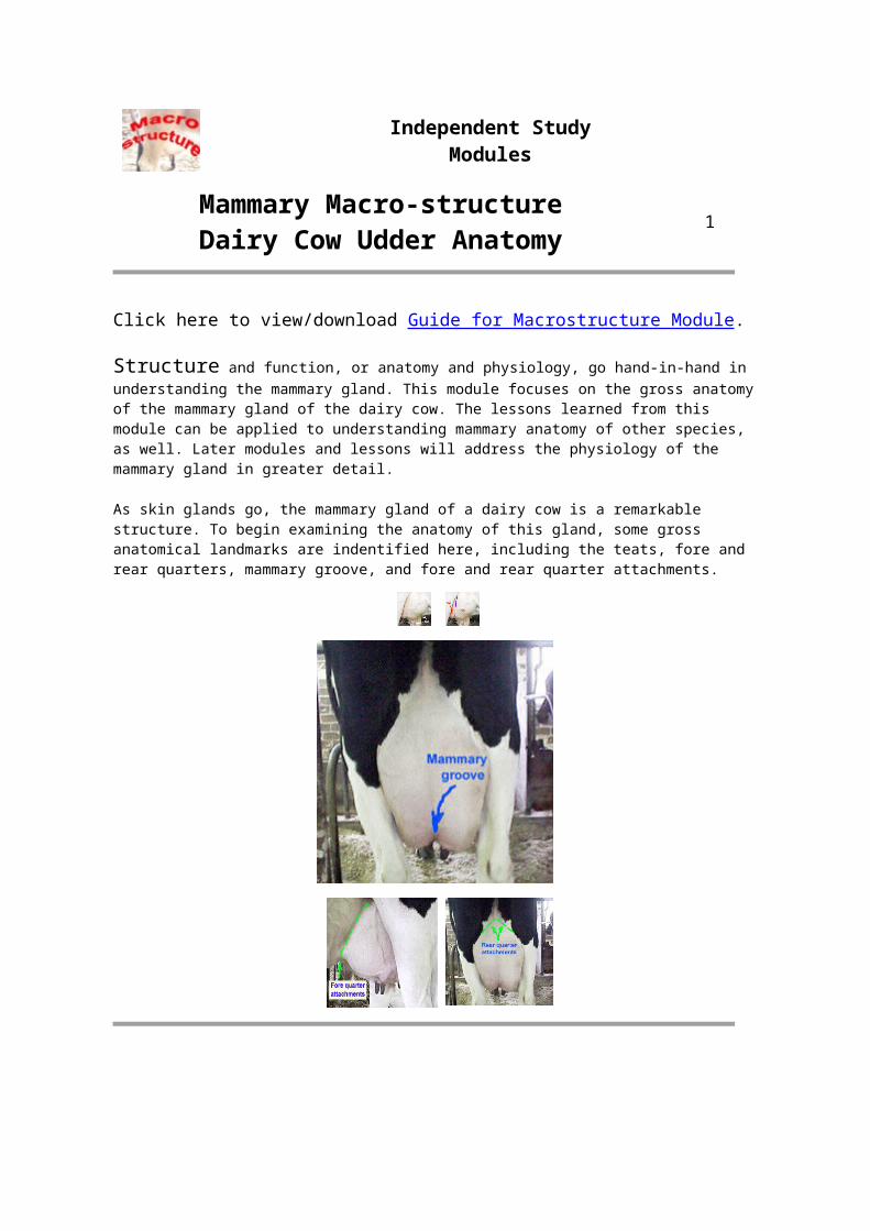
To achieve the functional capacity of the cow's mammary gland, a number of supporting systems must exist.
After reviewing each of these systems, which system do you think is most important to the lactation function of the cow's mammary gland?
Structure
Physical support of the udder
mass -Suspensory
system
On/off valve for intermittent
removal of milk -
Teat
Pathway for milk to travel from
the milk synthesis site to
the exit -Ducts & Cisterns
Means of actively
expelling milk from the udder
-Neural system
Continuous supply of
substrates for milk synthesis -Blood system
Internal secretory tissue -
Lobes/lobules/alveoli
Means of balancing the fluid
dynamics in the
tissue -Lymph system
Study Questio
Independent StudyModules
Mammary Macro-structureDairy Cow Udder Anatomy
3
Suspensory System - A strong udder suspensory system is required to maintain proper attachments of the gland to the body. Remember that the mammary gland is a skin gland, and is therefore external to the body cavity. As indicated earlier, an average Holstein cow easily may have 50 kg (>100 lbs) of weight hanging from her body when she walks into the milking parlor to be milked. The system of ligaments and other tissues which attach the udder to the cow are critical for successful lactation.
There are seven tissues that provide support for the udder:
1. Skin covering the gland is only of very minor support.2. Superficial fascia or Areolar subcutaneous tissue - This attaches the skin to
underlying the tissue. It, too is only of minor support for the cow's udder.
3. Coarse areolar or cordlike tissue - This tissue forms a loose bond between the dorsal surface of the front quarters and abdominal wall. Weakening of these causes the udder to break away from abdominal wall. This is part of what is referred to as the fore-quarter attachments when evaluating dairy cattle conformation. these are important for keeping the fore quarters closely attached to the body wall, but are not the major support of the udder.
4. Subpelvic tendon - This tendon not actually part of the suspensory apparatus, but gives rise to the superficial and the deep lateral suspensory ligaments. It is not a continuous tissue sheet but is attached to the pelvis at several points.
5. Superficial layers of lateral suspensory ligament - These are mostly composed of fibrous tissue (with some elastic tissue), arising from the subpelvic tendon. They extend downward and forward from the pubic are. When it reaches the udder it spreads out, continuing downward over the external udder surface beneath the skin and attaching to the areolar tissue.
6. Deep lateral suspensory ligament - The inner part of the lateral suspensory ligament also arises from the subpelvic tendon, but is thicker than the superficial layer, mostly fibrous tissue. It extends down over the udder and almost enveloping it. The ligament attaches to the convex lateral surfaces of the udder by numerous lamellae which pass into the gland and become continuous with the interstitial framework of the udder. Collectively, the lateral suspensory ligaments provide substantial support for the udder. The left and right lateral suspensory ligaments do not join under the bottom of the udder, and the fibrous nature of these ligaments means that they do not stretch as the gland fills with milk. So, the center of the udder tends to pull away from the body as the gland fills.
7. Median Suspensory Ligament - This is the most important part of the suspensory system in cattle. It is composed of two adjacent heavy yellow elastic sheets of tissue that arise from the abdominal wall and that attach to the medial flat surfaces of the two udder halves. The median suspensory ligament has great tensile strength. It is able to stretch somewhat as the gland fills with milk to allow for the increased weight of the gland. It is located at the center of gravity of the udder to give balanced suspension, so that even if rest of the
layers are cut away except for the median suspensory ligament, the gland stays balanced under the animal.
Suspensory System - The median suspensory ligament partially separates the left and right halves of the udder. Front and rear quarters are separated by a thin membrane and is not recognizable to the eye. There is NO internal crossover of the milk duct system of the quarters (glands). One way to demonstrate this is to infuse a dye into the teat and duct system of one quarter of an udder from a cow that has been culled from the herd and killed. Then when the udder is cut open, the dye will be seen only in the infused quarter and will not be seen in the other quarters.
Independent StudyModules
Mammary Macro-structureDairy Cow Udder Anatomy
4
Teats - (papilla mammae) The teat functions as the only exit for the secretion from the gland and the only means for the calf to receive milk. Usually, only one teat drains one gland. No hair, sweat glands or sebacious glands are found on the teats of the cow. Teat size and shape are independent of the size, shape or milk production of the udder. Average size for the fore teats is about 6.6 cm (2.6 in.) long and 2.9 cm (1.1 in.) in diameter, and for the rear teats is 5.2 cm (2.1 in.) long and 2.6 cm (1.0 in.) in diameter.
Supernumerary Teats - About 50% of all cows have extra teats, referred to as supernumerary teats. Some of these extra teats open into a "normal" gland, but many do not. Generally they are removed before 1 yr of age. A pseudo-teat has no streak canal, and therefore, no connection to the internal structures of the gland. Many animals of other species have supernumerary teats, including humans and pigs. The prevalence of these teats is low in humans, while many sows have extra, non-functional teats.
Streak canal - (ductus papillaris) Functions as the only orifice of the gland between internal milk secretory system and the external environment. The streak canal is the main barrier against intramammary infection. It is lined with a skin-like epidermis that forms the keratin material that has antibacterial properties. The streak canal is kept closed by sphincter muscles around the streak canal. Canal patency decreases and streak canal length increases with increasing lactation number.
When a cow is milked, the sphincter muscles relax allowing the orifice to open. The streak canal remains open for an hour or more after milking. This provides ready access of bacteria to the inside of the gland. Post-milking germicidal teat dips are designed to help minimize the chance of bacteria gaining access to the gland after milking. Keeping cows standing for a time after milking, such as providing access to
fresh feed, also helps minimize teat end contamination before the streak canal closes again. During the dry period (nonlactating period), the epidermal tissue lining the streak canal forms a keratin plug that effectively seals off the canal.
Furstenburg's rosette - These are mucosal folds of the streak canal lining at the internal end of the canal. It may fold over the canal opening due to pressure when the udder is full. It may be a major point of entry for leukocytes leaving the teat lining and entering into the teat cistern.
Cricoid rings - (Annular folds) Region at the proximal end of the teat cistern that marks the boundary between the teat cistern and the gland cistern. These are not always recognizable in the dissected gland.
Teat cistern - (Sinus papillaris) The cavity within the teat. It is continuous with the gland cistern. The teat cistern is lined with numerous longitudinal and circular folds in the mucosa, which form pockets on the inner lining of the teat. During milk letdown, the teat cistern fills with milk. It is this milk, and some of hte milk in the gland cistern just above the teta cistern, that is removed with each sucking action of the calf.
Independent StudyModules
Mammary Macro-structureDairy Cow Udder Anatomy
5
Ducts and Cisterns - While the cow's udder contains a large amount of secretory tissue responsible for synthesis of milk, it also contains a large proportion of ducts that are the tubing by which milk moves from the alveoli to the teat for milk removal. In addition, between the teat and the large ducts are open areas called cisterns. A cistern is a large cavity where milk can collect between milkings. Milk is synthesized in the microscopic alveoli. As it acumulates inthe alveolar lumen some milk oozes down into the smaller ducts and eventually into the large cisterns. This allows the cow to accumulate more milk in her udder between milkings or suckling by the calf than if she did not have these cisterns.
Gland Cisterns - (sinus lactiferus) Also called the udder cistern. It opens directly into the teat cistern. Occasionally a septum forms between teat and gland cisterns and the quarter may be blind. This can be corrected surgically. The cisterns function for milk storage (holds ~100-400 ml). The gland cistern varies greatly in size and shape. There are often pockets formed in the cistern at the end of the larger ducts.
Ultrasound image of cisternal ducts in a cow's udder. [Image courtesy of J Nagy, University of Illinois.]
Ducts are the tubes by which milk drains from the alveoli down to the gland cistern.
Interlobar or primary ducts drain multiple lobes. These are generally lined with two layers of non-secretory cells and have many myoepithelial cells.
Intralobar ducts are within a lobe and drain several regions of the lobe. Interlobular or secondary ducts drain multiple lobules. They are lined with one layer of secretory cells and surrounded by myoepithelial cells, and so participate in the oxytocin-induced milk ejection. Intralobular ducts are small ducts within a lobule Intercalary or tertiary ducts are the small ducts which exit from the alveolus. While this organized classification of ducts provides a basis for understanding the duct system of the gland, there is no uniformity in the system of duct branching.
Independent StudyModules
Mammary Macro-structureDairy Cow Udder Anatomy
6
Neural system - Several aspects of the neural system of the mammary gland ar important to mammary function:
1. Innervation inside of the udder is sparse compared with other tissues. 2. Sympathetic nerves are present in the tissue. These are the nerves that associate with the
arteries. They donot innervate the alveoli.
3. Sensory nerves are found in the teats and skin. These are critical for initiating the afferent pathway (neural pathway) of the milk ejection reflex.
4. There is no parasympathetic innervation to the gland. This is similar to other skin glands.
5. There is no innervation of the secretory system. Mypepithelial cells are not innervated. Myoepithelial cells do not contract in response to direct innervation, but rather contract in response to the blood-borne hormone, oxytocin.
6. Few nerves go to the interior of the udder. That means that performing a biopsy of the gland to collect tissue can be done with only local anesthetic administered to the skin.
Independent StudyModules
Mammary Macro-structureDairy Cow Udder Anatomy
7
Blood vascular system - The blood supply to the mammary gland is extremely important for mammary function! All of the milk precursors come from blood. On avg. 400 - 500 units of blood passes through the udder for each unit of milk synthesized by a high producing dairy cow; that is ~280 ml per sec.
High producing dairy goats have a lower (460:1) ratio of blood flow through the gland:milk produced, compared with low producers (1000:1). This means that the amount of blood flow through the mammary gland may by similar for the high and low producing goats, but the efficiency of extraction of the components from the blood while it passes through the udder very is important. This principle is probably similar for cows.
Total udder blood volume for lactating cows about 8% of total body blood volume, while for a non-lactating cow it is about 7.4%. There is a 2-6 fold increase in blood flow in the mammary gland starting 2-3 days prepartum. The decrease in production with advancing lactation is not due to decreased blood flow, rather it is due to the loss of secretory epithelial cells through a process programmed cell death (apoptosis).
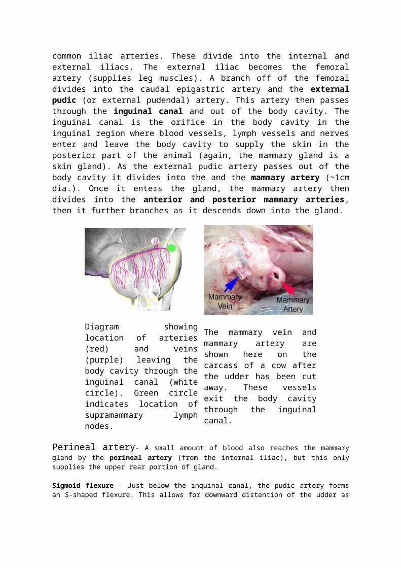
Arterial system - Blood leaves the heart and flows towards the rear of the cow by the abdominal aorta. When it reaches the pubic area of the animal the vessels are called the common iliac arteries. These divide into the internal and external iliacs. The external iliac becomes the femoral artery (supplies leg muscles). A branch off of the femoral divides into the caudal epigastric artery and the external pudic (or external pudendal) artery. This artery then passes through the inguinal canal and out of the body cavity. The inguinal canal is the orifice in the body cavity in the inguinal region where blood vessels, lymph vessels and nerves enter and leave the body cavity to supply the skin in the posterior part of the animal (again, the mammary gland is a skin gland). As the external pudic artery passes out of the body cavity it divides into the and the mammary artery (~1cm dia.). Once it enters the gland, the mammary artery then divides into the anterior and posterior mammary arteries, then it further branches as it descends down into the gland.
Diagram showing location of arteries (red) and veins (purple) leaving the body cavity through the inguinal canal (white circle). Green circle indicates location of supramammary lymph nodes.
The mammary vein and mammary artery are shown here on the carcass of a cow after the udder has been cut away. These vessels exit the body cavity through the inguinal canal.
Perineal artery- A small amount of blood also reaches the mammary gland by the perineal artery (from the internal iliac), but this only supplies the upper rear portion of gland.
Sigmoid flexure - Just below the inquinal canal, the pudic artery forms an S-shaped flexure. This allows for downward distention of the udder as it fills with milk, without stressing the blood vessels.
There is essentially no cross over of blood supply between udder halves (there are a few minor exceptions)
Venous system - Veins leave the mammary gland anti-parallel to the arteries. There are three veins on each side that carry blood away from the gland:
1. External pudic vein leaves the udder parallel to the external pudic artery (2-3 cm dia.).
2. Subcutaneous abdominal vein (milk vein) exits the gland at the anterior end of the front quarters and passes along abdominal wall (1-2.5 cm dia.). This is the large vein that is visible under the skin on the belly of he cow. It enters the body cavity at the xiphoid process via "milk wells", and eventually empties into vena cava.
3. Perineal vein leaves the rear of the gland parallel to the perineal artery (0.5 cm dia.). Carries less than 10% of blood leaving udder.
Venous circle - Formed by anastomoses between anterior and posterior mammary veins. Prevents pinching off of areas of venous outflow when the cow is lying down.
Independent StudyModules
Mammary Macro-structureDairy Cow Udder Anatomy
8
Lymph and Lymphatics - Many molecules of all sizes leave the capillaries but not all return to the venous drainage at the tissue level. Especially the larger molecules like proteins. These, along with cellular metabolites and some secretory products are in the interstitial (extracellular) spaces. If they stayed there, they would disrupt with the normal balance of osmotic pressure in the tissue, upsetting trans-capillary fluid exchange. Excess fluid (called extracellular fluid) would accumulate in the interstitial spaces.
Functions of lymphatics:
The extracellular fluids are drained from the tissue and conducted back to the circulatory system via the lymphatic network.
Also, the lymphatics contain concentrated areas of leukocytes (particularly lymphocytes and macrophages) in lymph nodes; these leukocytes can mount an immune response to bacteria and foreign material.
The lymphatic network serves to transport some things in the body (vitamin K, lipids absorbed in the intestine).
The lymphatic network originates in tissue spaces as very thin, closed endothelial tubes (lymphatic capillaries). These are analogous to blood capillaries, but are much more permeable, with little resistance to passage. They have no basement membrane. Lymph capillaries converge to form larger vessels. Lymph flow is unidirectional from the tissues through lymphatic vessels, eventually dumping lymph into the vena cava.
Lymph is a clear, colorless liquid with a composition similar to blood plasma. Changes in plasma composition will change lymph composition. Protein concentration of lymph is lower than in plasma, 1.5% vs. 6% for plasma. Specific proteins differ, for example albumin is a smaller molecular size than globulins and leaves the capillaries more readily than globulins, so the albumin : globulin ratio is 1.8 in plasma, 2.5 in lymph. Protein concentration in lymph varies inversely with rate of formation. Rate of filtration varies with the tissue. In liver and intestine, lymph has ~5% protein, compared with the extremities where lymph has ~0.5% protein).
Lymph flow rate is usually low. It is influenced primarily by the rate of lymph formation. For example, if blood capillary pressure is increased by arterial vasodilation or venous constriction, the flow rate of lymph increases. Also, the flow rate is affected by compression of lymphatics by contraction of neighboring musculature and by negative intrathoracic pressure (breathing). Valves in the lymph vessels prevent retrograde flow similar to those in veins.
Lymph flow through the mammary gland:
goat (lactating) 6.5-35 ml/hr or 150-840 ml/day cow (dry) 14-240 ml/hr or 68-5760 ml/day
cow (lactating) 1300 ml/hr or 31,200 ml/day or 31 kg/day
1.6 units lymph leave udder for every unit milk produced.
Edema is the excess accumulation of fluids in tissue spaces. This can retard normal exchange of nutrients and metabolites. Filtration of the extracellular fluid exceeds drainage. Anything that causes increased capillary pressure, such as decreased plasma protein, increased capillary permeability or lymphatic blockage, can result in swelling and congestion of the extravascular compartment.
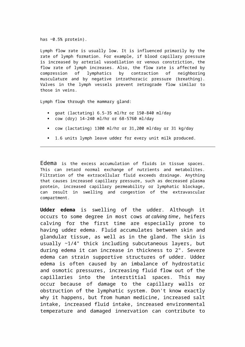
Udder edema is swelling of the udder. Although it occurs to some degree in most cows at calving time, heifers calving for the first time are especially prone to having udder edema. Fluid accumulates between skin and glandular tissue, as well as in the gland. The skin is usually ~1/4" thick including subcutaneous layers, but during edema it can increase in thickness to 2". Severe edema can strain supportive structures of udder. Udder edema is often caused by an imbalance of hydrostatic and osmotic pressures, increasing fluid flow out of the capillaries into the interstitial spaces. This may occur because of damage to the capillary walls or obstruction of the lymphatic system. Don't know exactly why it happens, but from human medicine, increased salt intake, increased fluid intake, increased environmental temperature and damaged innervation can contribute to edema.
This heifer had just calved and has substantial udder edema. Note the swelling under the belly.
Independent StudyModules
Mammary Macro-structureDairy Cow Udder Anatomy
9
Secretory tissue in the interior of the gland is made up of:
Connective tissue - This includes fibrous connective tissue of the parenchymal tissue and the fatty tissue of the fat pad. An example of fat pad is illustrated in the image below.
Secretory tissue - This is made up of secretory epithelial cells. These are the cells that produce milk during lactation. These also are part of the parenchymal tissue.
The relative amount of connective and secretory tissue varies from animal to animal.
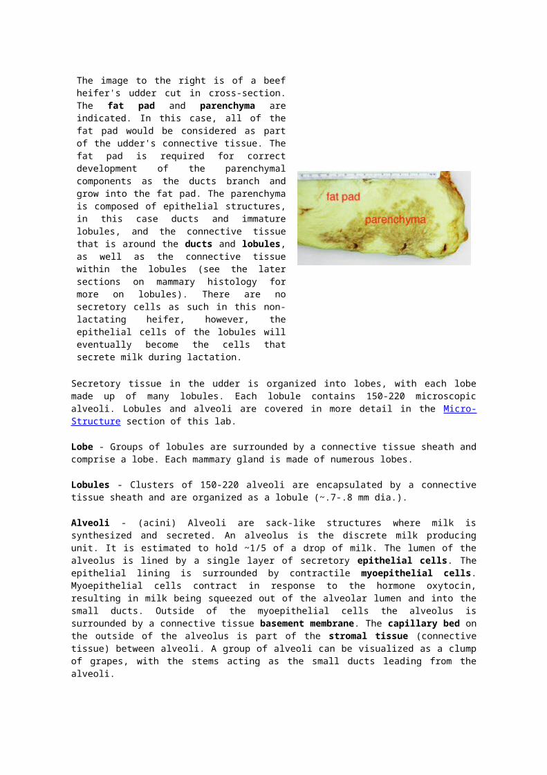
The image to the right is of a beef heifer's udder cut in cross-section. The fat pad and parenchyma are indicated. In this case, all of the fat pad would be considered as part of the udder's connective tissue. The fat pad is required for correct development of the parenchymal components as the ducts branch and grow into the fat pad. The parenchyma is composed of epithelial structures, in this case ducts and immature lobules, and the connective tissue that is around the ducts and lobules, as well as the connective tissue within the lobules (see the later sections on mammary histology for more on lobules). There are no secretory cells as such in this non-lactating heifer, however, the epithelial cells of the lobules will eventually become the cells that secrete milk during lactation.
Secretory tissue in the udder is organized into lobes, with each lobe made up of many lobules. Each lobule contains 150-220 microscopic alveoli. Lobules and alveoli are covered in more detail in the Micro-Structure section of this lab.
Lobe - Groups of lobules are surrounded by a connective tissue sheath and comprise a lobe. Each mammary gland is made of numerous lobes.
Lobules - Clusters of 150-220 alveoli are encapsulated by a connective tissue sheath and are organized as a lobule (~.7-.8 mm dia.).
Alveoli - (acini) Alveoli are sack-like structures where milk is synthesized and secreted. An alveolus is the discrete milk producing unit. It is estimated to hold ~1/5 of a drop of milk. The lumen of the alveolus is lined by a single layer of secretory epithelial cells.
The epithelial lining is surrounded by contractile myoepithelial cells. Myoepithelial cells contract in response to the hormone oxytocin, resulting in milk being squeezed out of the alveolar lumen and into the small ducts. Outside of the myoepithelial cells the alveolus is surrounded by a connective tissue basement membrane. The capillary bed on the outside of the alveolus is part of the stromal tissue (connective tissue) between alveoli. A group of alveoli can be visualized as a clump of grapes, with the stems acting as the small ducts leading from the alveoli.
Independent StudyModules
Mammary Macro-structureDairy Cow Udder Anatomy
10
Study Questions on macro-structure of the cow's mammary gland.
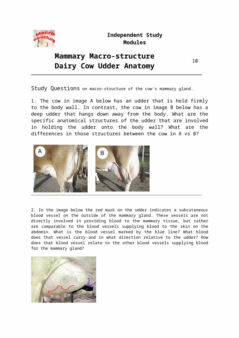
1. The cow in image A below has an udder that is held firmly to the body wall. In contrast, the cow in image B below has a deep udder that hangs down away from the body. What are the specific anatomical structures of the udder that are involved in holding the udder onto the body wall? What are the differences in those structures between the cow in A vs B?
2. In the image below the red mark on the udder indicates a subcutaneous blood vessel on the outside of the mammary gland. These vessels are not directly involved in providing blood to the mammary tissue, but rather are comparable to the blood vessels supplying blood to the skin on the abdomin. What is the blood vessel marked by the blue line? What blood does that vessel carry and in what direction relative to the udder? How does that blood vessel relate to the other blood vessels supplying blood for the mammary gland?
LactationPractice Quiz
PRINT THIS PAGE
This is a sample of a quiz on the Resource pages of the Lactation Cases Module similar to that on the Compass quizzes for this module. Print is out and use it to help you study the material. Be sure you know the material in the module BEFORE starting the Compass quiz!
1. In a lactating goat, why would the milk production from one udder half which is milked one time per day be lower than an udder half which is milked twice per day?
1. the 1X daily milked half has greater overall secretion rate of milk 2. the 2X daily milked half is taking all the nutrients away from the 1X daily milked half
3. the 2X daily milked half inhibits milk production of the 1X daily milked half
4. the 1X daily milked half has a lower overall secretion rate of milk
2. Myoepithelial cells can contract in response to which of the following?
1. oxytocin and vasopressin 2. prolactin only
3. vasopressin only
4. oxytocin and prolactin
5. acetylcholine only
3. Injection of bovine placental lactogen into lactating cows typically affects milk yield by a mechanism somewhat different from bovine somatotropin. What effect would injection of bovine placental lactogen have on milk yield in a mid-lactation dairy cow?
1. it would decrease milk yield 2. placental lactogen would inhibit the secretion of growth hormone
3. placental lactogen does not affect lactation in the cow
4. it would increase milk yield
4. Which of the following best describes the milk ejection reflex?
1. a reflex with a hormonal pathway
2. a reflex with a hormonal pathway and a blood borne pathway
3. mechanical stimulation of the gland
4. a reflex with a neural pathway and a hormonal pathway
5. When is the process of galactopoiesis important in the mammary gland?
1. during the initiation of lactation 2. after lactation has been initiated
3. before lactation has been initiated
6. Continuous infusion of cows with oxytocin will have what effect on milk yield?
1. milk yield will be unchanged 2. milk yield be decreased
3. milk yield will be increase
7. As milk is secreted from the mammary epithelial cells and accumulates in the alveolar lumen the rate of secretion slows. One of the factors that causes the epithelial cells to slow their rate of milk component secretion is which of the following?
1. excessive secretion of lactose 2. pressure buildup in the alveolus leads to locally decreased blood flow
3. pressure build-up in the lungs
4. nervous signals from the brain
8. Where and in what form is oxytocin stored in the pituitary?
1. oxytocin is stored in the herring bodies as oxytocin 2. oxytocin is stored in the hypothalamus as the oxytocin-neurophysin complex
3. oxytocin is stored in the herring bodies as casein granules
4. oxytocin is stored in the hypothalamus as neurophysin
5. oxytocin is stored in the herring bodies as the oxytocin-neurophysin complex
9. When changing from 2X per day milking to 3X per day milking of dairy cattle in early lactation there is an increase in milk yield of up to 25%. Approximately one-third of this increase is due to

increased overall milk secretion rate. To what is the remainder of the increased production attributed?
1. increased secretion of FIL 2. better feeding and management
3. decreased udder pressure
4. the increased milk yield only occurs in goats
10. The efferent pathway of the milk ejection reflex starts with which of the following?
1. mechanical stimulation of the teats 2. release of oxytocin into the blood
3. nerve impulses reaching the hypothalamus
4. degradation of oxytocin
5. synthesis of oxytocin