Embed Size (px)
Citation preview

MAGNETOM Harmony [Maestro Class]
The New Degree of Perfection


All tasks and applications within your
workflow are covered either with, or at
your system: from patient registration,
image acquisition, viewing, and post-
processing to filming as well as archiving.
The web-based patient record, for example,
provides quick access to important patient
information. As an alternative, you can
view lab results at the console without
time-consuming telephone calls to the
ward.
syngoSiemens is the leader in cross modality common sense!
syngo® is a comprehensive computer platform engineered
for medical imaging that runs on the majority of Siemens
medical products.
Different modalities use common intuitive icons to
initiate shared tasks such as patient registration, imaging,
3D reconstruction, etc.
The aim of Siemens MR is the perfection of care. To
create products and services that improve the quality of
life of all persons who come into contact with them.
We do this by caring for the health of the patient, caring
for the quality of the user’s work – and caring about the
owner’s profit.
MAGNETOM Family
The Perfection of Care

M a e s t r o C l a s s
is in
www.SiemensMedical.com
syngo – a console to feelcomfortable with
Task Cards – for structured workflow
Image Focus – images from all task
cards can be processed simultaneously
Automated Scan Programs – predefined
protocols account for most of your
daily routine, however, they may be
easily adapted
Flexible Parameters – offer more
control over parameters that affect
contrast and signal-to-noise ratio
syngo Scan Assistant – a standard
feature that immediately displays
conflicting parameters and provides
the technologist with a range of
acceptable values

innovative applicationsMaestro Class is setting standards!
Always a step ahead! MAGNETOM Harmony with Maestro Class addresses
the most complete range of applications from clinical standard to high-
end applications with a broad variety of tissue contrasts. Take, for example,
a comprehensive brain, spine and body exam with our revolutionary IPA™
coil concept, whole body MR angiography, spectroscopy, and more. Cost
effective and with high patient comfort.
increased speedMaestro Class saves time!
MAGNETOM Harmony with Maestro Class puts you in the driver’s seat of the
best balanced MR system. It combines ultra-fast imaging, cost-effectiveness
and powerful clinical performance. MAGNETOM Harmony makes this all
possible with one of the strongest gradient systems, latest imaging techniques,
high-speed computers, and high-quality RF technology that work together
perfectly.
intelligenceMaestro Class thinks with you!
MAGNETOM® Harmony with Maestro Class automates routine processes,
making them faster and simpler, giving you more time for the essentials, your
patient and diagnosis.
3
Siemens Medical Solutions: the Innovation Leader in MR Technology.Maestro Class – a new degree of perfection in magnetic resonance
imaging with a focus on intelligent technology, increased speed and innovative
applications.
< Siemens – the Leader in MR Technology.
Our top of the line hardware is based on decades of R&D experience
with homogeneous magnets and precise RF technology.
< Siemens stands for Innovative System Design.
Patient-friendly design, high-performance gradients and our ground-
breaking integrated coil concept, IPA, help you arrive at efficient and
comprehensive diagnoses.
< Siemens is Setting Standards with syngo.
We are the first medical solutions provider to offer a software
standard for different modalities. syngo stands for a common user interface
and a common operating system, offering true compatibility.

4
PACE Prospective Acquisition andCorrEction – Motion under control!1D PACE – Free breathing, the perfect
alternative to breath-hold scanning.
Detect respiratory motion – accept data
only during expiration – and view the
results, quickly and easily!
2D PACE – Improve selectivity and
precision in abdominal MRI. Auto-
matically align each multi breath-hold
– compensate for unwanted patient
movement – and obtain fast and
reliable diagnostic results.
Inline Technology – Processinginstead of Post-Processing
The complete exam is finished as
soon as image acquisition is completed.
Inline Technology uses an intelligent
on-the-fly feedback loop to control
scanning, reconstruction, and proces-
sing. This means motion is detected
and corrected in the acquired image,
providing you with excellent image
quality every single time.
Scanning
Reconstruction
Visualization
Post-Processing
Correction
Filter
Processing
Reporting
Conventional processes New Inline-Technology
Scanning
Reporting
Recon-struction,Visualizationand Post-Processing In
lineCorrection
Filter
Processing Plu
g-i
ns
Intelligentfeedback
telligenceM a e s t r o C l a s s
is in
Inline display of diaphragm positiondefine to accept data
Inline display of diaphragm positiondefine expiration phase to accept data

5
syngo Scan Assistant –Your MR consultant
Do you need to change one of the
MR parameters? The syngo Scan
Assistant visualizes the effect and
proposes different parameters in case
of conflict. This is the sure way
to consistently high image quality.
Maestro UI –Your cockpit for dynamic MRI
You are in the driver’s seat to opti-
mize your clinical workflow with ease.
Create a process, not just for data
acquisition, but for immediate access
to instant diagnostic results – cost-
effectively.
Simplify procedures with Inline
task cards, e.g. a complete stroke
protocol from anatomy, to diffusion,
perfusion and function.
Get a quick overview with Image
Stamps – your easy direct access to
what you want to see.
You want to apply exactly the
same imaging parameters used for
an existing image? Go ahead, use
Phoenix, our easy drag and drop
function. Insert the image into your
measurement queue, and let the
system extract all applicable para-
meters into the measurement card.
It‘s time to start the scan.
Contrast-enhanced MRA at yourfingertips!Simplify and automate standard
measurement procedures. Acquire
MRA data sets, pre- and post-contrast.
The data sets are subtracted auto-
matically. The MIP reconstruction is
available at the same time.
SuperMIP – automatically provides a
scout image across the entire region
of interest and lets you accurately plan
for additional procedures, allowing you
to spend more time with your patient.
Maestro Class thinks with you!
MAGNETOM Harmony Maestro Class
automates routine processes,
making them faster and simpler.
Thus you gain time for the
essentials, your patient and diagnosis.

6
Maestro Class saves time!
MAGNETOM Harmony with Maestro Class puts you in the driver’s seat of the
best balanced MR system. It combines ultra fast imaging, cost-effectiveness,
and powerful clinical performance. MAGNETOM Harmony makes this all possible
with one of the the strongest gradient systems, latest imaging techniques, high-
speed computers, and high-quality RF technology that work together perfectly.
PAT factor 2 is standard!With MAGNETOM Harmony a PAT
factor of 2 is standard. iPAT is fully
compatible with the unique Siemens
IPA coil concept.
What are the benefits of iPAT?
Increased patient comfort – shorter
breath-holds during abdominal
imaging result in shorter acquisition
times.
More diversified patient load –
comfortable, short breath-hold
times allow a greater range of
patients to be examined than ever
before.
Higher temporal resolution – in both
cardiac and abdominal MRI, e.g.
dynamic 3D VIBE liver imaging or
cine cardiac imaging.
Less blurring artifacts – shorter
measurement times ensure less
blurring anywhere in the body.
Improved diagnostic confidence –
shorter measurement times, higher
resolution and less distortion
artifacts result in higher image
quality with high reproducibility.
The solution to speed up youracquisition times – iPAT
iPAT with Siemens MAGNETOM
Harmony means integrated Parallel
Acquisition Techniques.
And the term speaks for itself:
Integrated feature
Integrated into the MAGNETOM
IPA philosophy (Integrated Pano-
ramic Array), our revolutionary
coil concept, which enables the use
up to 8 independent channels
Integrated auto-calibration
Combines the convenience of IPA
and IPP (Integrated Panoramic
Positioning) for many applications
iPAT is more than just a sequence.
iPAT with Siemens has the flexibility to
approach each clinical demand
differently. It allows for specific answers
to specific needs achieved at high
image quality in the shortest time
possible.
iPAT increases patient throughput as
well as patient comfort and delivers
excellent image quality.
creased speedM a e s t r o C l a s s
is in
without iPAT; 1:04 min, 512 with iPAT; 36 s, 512
without iPAT; 1:40 min, 512 with iPAT; 52 s, 512
7
without iPAT; 24 s with iPAT; 13 s
Siemens iPAT
ove
rall
exam
inat
ion
tim
e
Scan
Calibration
Scan
Calibration
Calibrationrequired eachtime the patientmoves
Initialcalibration
other PAT techniques
1 sec
1 sec
Scan
Scan
Computer power
MAGNETOM Harmony comes with a
dual Pentium 4 based host processor
and a single Pentium 4 based Pano-
ramic Recon Image processor, providing
ultimate reconstruction speeds of up
to 1299 images/sec (2562 FFT, 25% rec.
FoV).
Up to 5 data sets can be acquired,
processed, and reconstructed simul-
taneously. Our high performance com-
puter power allows real-time image
calculation parallel to scanning. The
result: higher patient throughput and
increased productivity.
8
High patient comfort, optimized workflow, and com-
plete anatomical coverage. Combine up to 4 coils of the
Integrated Panoramic Array (IPA) coil concept for opti-
mized image acquisition. Use the Integrated Panoramic
Positioning (IPP) with automatic table feed and remote
patient positioning for easy handling.
With a single coil set up you get excellent brain and
spine images as well as a complete abdominal study. All
this without the need to change coils or to reposition
the patient.
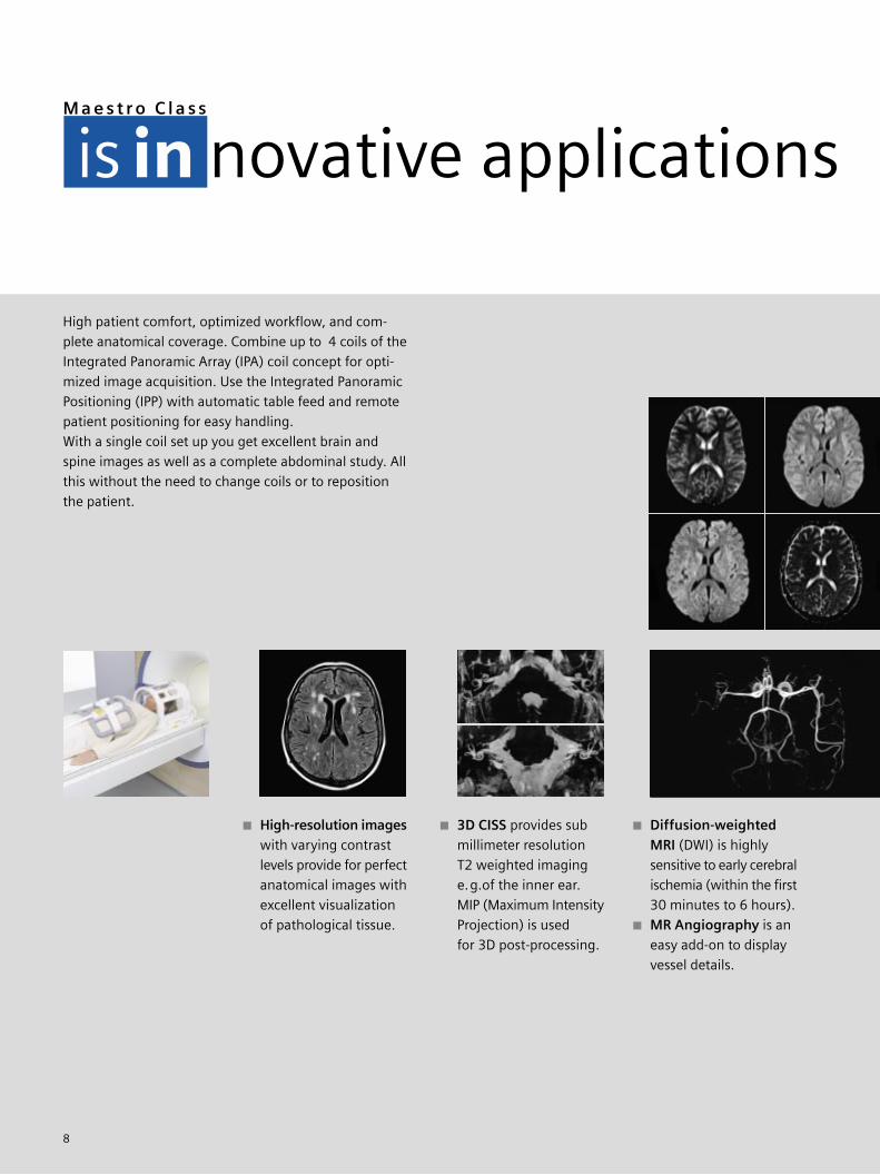
High-resolution imageswith varying contrast
levels provide for perfect
anatomical images with
excellent visualization
of pathological tissue.
3D CISS provides sub
millimeter resolution
T2 weighted imaging
e.g.of the inner ear.
MIP (Maximum Intensity
Projection) is used
for 3D post-processing.
novative applicationsM a e s t r o C l a s s
is in
Diffusion-weightedMRI (DWI) is highly
sensitive to early cerebral
ischemia (within the first
30 minutes to 6 hours).
MR Angiography is an
easy add-on to display
vessel details.

9
Maestro Class is setting standards!
Always a step ahead! MAGNETOM Harmony with Maestro Class addresses the most
complete range of applications from clinical standards to high-end applications with
a broad variety of tissue contrasts. Take, for example, a comprehensive brain, spine,
and body exam with our revolutionary IPA coil concept, whole body MR angiography,
spectroscopy, and more. Cost effectively and with high patient comfort.
Spine imaging with iPATCut your acquisition times in half
compared to conventional imaging
times using iPAT together with
MAGNETOM IPA coils.
MR MyelographyEasy add-on, non-invasive exami-
nation of the spinal cord to display
the nerve origin.
3D VIBE (Volumetric Interpolated
Breath-hold Examination) for 3D
dynamic imaging of the abdomen
with isotropic voxels. High spatial
resolution.
MIP for free! Click the MIP button
and obtain vessel information in
every phase of dynamic parenchy-
mal imaging without additional
contrast media.
MR CholangiographyNon-invasive pathological evalu-
ation of the biliary and pancreatic
system, presenting the ducts, milli-
meter-sized stones, and minimal
dilatations.
2D PACEMulti breath-hold 2D PACE with
HASTE for strong T2-weighted
imaging. You get all the information
you need about the liver, bowels,
fluid, lesions, as well as biliary and
pancreatic duct in no time at all.
with iPAT; 52 s, 512

10
Easy patient set-up with
highest patient comfort.
Diffusion-weighted MRI (DWI) in
combination with perfusion-
weighting may be used to predict
the severity of the stroke as well
as the level of expected recovery.
The ADC map (Apparent Diffusion
Coefficient) helps to estimate the
age of stroke lesions. Calculate ADC
maps automatically with Inline
Technology at the end of the scan.
Ultra fast imaging techniques such
as 3D segmented EPI and HASTE
provide excellent resolution and a
large variety of contrast with con-
tiguous slices and full coverage of
the brain.
novative applicationsM a e s t r o C l a s s
is in

11
Parameter map calculations, such
as Time-to-Peak or relative MTT
(Mean Transit Time) are performed
automatically using Inline Tech-
nology.
Maestro Class is setting standards!
Use the latest MAGNETOM innovations for advanced neurological MRI, such as
segmented EPI, diffusion, and perfusion, as well as functional MRI (BOLD).
Neuro MR provides high-contrast images, including the finest anatomic details
for diagnostic speed and confidence. Use IPA, IPP with automatic table feed
and remote patient handling, and increase patient throughput cost-effectively.
Without patient repositioning
you can acquire high resolution neck
images or a whole spine study.
MEDIC (Multi Echo Data Image Com-
bination) T2*-weighted imaging
with high signal-to-noise ratio.
Reduced flow artifacts.
Restore for increased T2 contrast in
less time, especially helpful in spine
imaging.
Spine Imaging
High resolution, a large variety of
contrasts and full coverage with
greatest possible patient comfort –
these are all standard features of
the MAGNETOM Harmony. Get a
whole spine 50 cm Field-of-View
study or look for details with smaller
FoVs.
MOSAIC images to display one acquired volume
at a time e.g. BOLD imaging

novative applicationsM a e s t r o C l a s s
is in
12
Always on the right track – image
stamps may be loaded into the
movie function, the post-processing
card, or the measurement queue.
Argus Flow Quantification: vessel
and valve flow results are quantified
and reported in DICOM format.
Morphology: Plan oblique ana-
tomical planes easily with 3 point
localization. Display your results
automatically with the Auto Movie
function.
Short scan times are necessary in
contrast-enhanced MRA. High
performance MAGNETOM Harmony
gradients allow the acquisition
of data with minimal sensitivity to
flow and motion artifacts.

13
The Body Coil, CP Head Array and
CP Flex Coil combination provide
vessel overviews and may be used
for post-operative exams.
Obtain maximum coverage by
combining Peripheral CP Angio Array
Coil, CP Body Flex Array, and CP
Spine Array Coil. This combination
is highly suitable for e.g. pre-
operative planning. Extend the FoV
even further using CP Body Array
Extender and Large FoV Adapter.
The scan program for the overall
examination is already programmed
using the Maestro User Interface.
Each measurement can be indivi-
dually adjusted to the anatomy of
the patient.
Inflow enhancement with Care
Bolus is done in real-time.
You decide when to start the scan.
Fast – reliable – easy-to-use. High speed MRA of high resolution is essential for
reliable diagnosis. The high performance gradients of the MAGNETOM Harmony
provide the shortest TR and TE parameters – a prerequisite for performing fast
and cost-effective MRA studies. All you have to do is inject contrast agent and go.
Easy and completely automatic – high-resolutionperipheral MRA from diaphragmatic level to distal vessels
Maestro Class Inline
Technology automates
the MIP (Maximum
Intensity Projection)
calculation for each
anatomical level.
Get the results while
the patient is still on
the table by using sub-
traction-on-the-fly.
Use the Body and CP Head Array Coil
or a multiple CP Array Coil set-up with
automatic table feed using Integrated
Panoramic Array (IPA) and Integrated
Panoramic Positioning (IPP). Your
optimized workflow is only a mouse
click away.
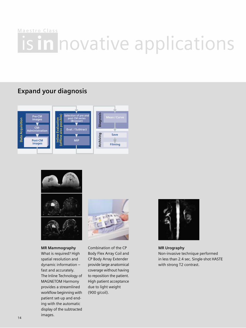
Expand your diagnosis
14
MR MammographyWhat is required? High
spatial resolution and
dynamic information –
fast and accurately.
The Inline Technology of
MAGNETOM Harmony
provides a streamlined
workflow beginning with
patient set-up and end-
ing with the automatic
display of the subtracted
images.
Arc
hiv
ing
Dat
a A
cqu
isit
ion
Inlin
e Ev
alu
atio
n(o
fflin
e al
so p
oss
ible
)
Dia
gn
osi
s
Filming
CM-Administration
Pre-CMimages
MIP
Mean / Curve
Save
Post-CMimages
Eval. / Subtract
Selection of pre andpost CM series
(Browser)
MR UrographyNon-invasive technique performed
in less than 2.4 sec. Single-shot HASTE
with strong T2 contrast.
Combination of the CP
Body Flex Array Coil and
CP Body Array Extender
provide large anatomical
coverage without having
to reposition the patient.
High patient acceptance
due to light weight
(900 g/coil).
novative applicationsM a e s t r o C l a s s
is in

15
Abundance in gradient strength leads to new faster imaging techniques
and shorter examination times. MAGNETOM Harmony with Maestro Class sets
a new milestone in the expanded use of MR, making it the gold standard
for answering many questions in routine clinical gastrointestinal problems.
Get two in one!Out of/In Phase FLASH
for fatty liver and adrenal
tumor imaging.
Out of Phase
In Phase
Get two in one!
Excellent image quality,
reduced artifacts, within
very short exam times
with respiratory gating.
TrueFISPSubsecond T2 imaging that elimi-
nates artifacts caused by motion. Short
acquisition times for high patient
comfort.
Patient-friendlyAcquire contrast-enhanced MRA
3D data sets in a 20 s breath-hold. Get
high image quality and avoid respi-
ratory motion artifacts.

16
Shoulder –High resolution and
excellent fat suppression.(All images on this page: Courtesy
of Uniontown Hospital, USA)
Elbow –Small joint imaging
with contiguous slices
and fat suppression;
1.5 mm slice thickness.
Wrist –Small FoV with
70 mm FOV for details.
Turbo Inversion pulse
visualizes fluid in the
joint while suppressing
fat signal; 512 matrix,
3 mm slice thickness.
Easy patient set-up
with highest comfort.
novative applicationsM a e s t r o C l a s s
is in

17
MR Orthopedic Imaging
Fast – precise – complete. High resolution, thin slices, and full anatomical coverage
are the focus in orthopedic imaging. MAGNETOM Harmony provides optimized
sequences with a large variety of contrasts to visualize joints in detail.
Knee –High resolution
(512 matrix for T1) and
contiguous slices for
excellent delineation of
the ligaments (3D DESS,
1.5 mm slice thickness)
CP coil technology
for small FoV and high
resolution imaging.
Ankle –High soft tissue con-
trast and high resolution
with and without fat
suppression;
3 mm slice thickness.

18
Spectroscopy – Automated and Comprehensive
Single Voxel Spectroscopy for proton
spectroscopy can be performed using
the CP Head Array for image acquisi-
tion. Automatic adjustment, measure-
ment and evaluation protocols permit
largely automatic spectroscopy
measurements.
Easy and efficient ”single-button”
spectroscopy, with spectra anno-
tation and quantification at the click
of a mouse. Keep your focus on the
clinical questions at hand.
Free slice positioning – just use the
mouse to tailor both slice position
and orientation to your patient.
Single voxel measurement with
Spin-Echo, FID and STEAM techni-
ques.
novative applicationsM a e s t r o C l a s s
is in
19
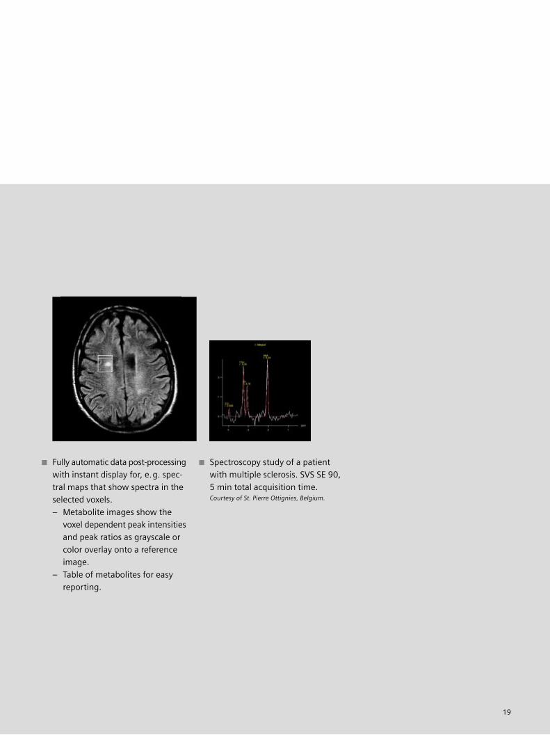
Fully automatic data post-processing
with instant display for, e.g. spec-
tral maps that show spectra in the
selected voxels.
– Metabolite images show the
voxel dependent peak intensities
and peak ratios as grayscale or
color overlay onto a reference
image.
– Table of metabolites for easy
reporting.
Spectroscopy study of a patient
with multiple sclerosis. SVS SE 90,
5 min total acquisition time.Courtesy of St. Pierre Ottignies, Belgium.

20
IPAThe Integrated Panoramic Array (IPA) coil concept is the unmatched revo-
lution in coil development as well as a major leap forward in productivity.
The underlying philosophy: combine various coil elements from different
coils to obtain optimal anatomical coverage including highest image quality.
Efficient – reduces the number of
coils and patient set-up times. Up
to 4 different coils may be connected
simultaneously.
Easy handling – coil elements
from various coils can be combined
for image acquisition.
High patient comfort – light-weight,
open design highly suitable for e.g.
cardiac or oncology patients.
High anatomic coverage – allows
for multiple exams covering the
largest field of view in the industry.
Integrated Panoramic Positioning
(IPP) and remote table feed support
step-by-step high-resolution imag-
ing of small regions. Table control
and coil elements are selected at
the main console.
High image quality due to whole
Body CP Array coil design.
MAGNETOM Harmony receives
signals from up to 16 CP coil
elements of 4 different coils.
CP Head+CP Neck+CP Spine=9 coil elements
CP Spine Array+
Peripheral CP Angio Array+
CP Body Flex Array=
16 coil elements
Peripheral CP Angio Array CoilShoulderArray Coil
Double LoopArray Coil
CP ExtremityCoil
CP ExtremityCoil
CP HeadArray Coil
CP BreastArray Coil
EndorectalCoil
CP Body Array Flex and/orExtender Coil
CP NeckArray Coil
Flex Coils
CP Spine Array Coil
21
EVOLVE
*In the event that upgrades require FDA
approval, Siemens cannot predict whether
or when the FDA will issue its approval.
Therefore, if regulatory clearance is obtained
and is applicable to this package, it will be
made available according to the terms of this
offer.
Within EVOLVE you can choose
between several options. You can
upgrade your system to the latest
generation or with the Harmony
EVOLVE Package™ schedule regular
hardware and software upgrades.
The financial alternative to expensive
new equipment is EVOLVE.
EVOLVE for MAGNETOM HarmonyWe offer complete packages suitable
for a number of specific applications.
These packages include dedicated
application software, coils and ex-
panded system performance.
EVOLVE elevates existing MAGNETOMsystems to Maestro ClassEVOLVE lets you upgrade your present
generation of MAGNETOM system to
Maestro Class performance quickly and
cost-effectively. This is certainly the
smart way to plan your future budget.
Your subscription to the future –the syngo EVOLVE PackageThe performance level of computer
chips doubles roughly every 18 months.
This means that todays leading pro-
cessors will be obsolete in a few years.
Similar time frames are valid for soft-
ware innovations.
Within the scope of the Siemens
Performance TOP maintenance
program your hard- and software is
upgraded regularly. You will get the
image processor and host computer
of your syngo-based system updated
twice over a period of six years.
New software versions will be made
available to you. The choice is yours.
Depending on your personal require-
ments you can select one of our specific
EVOLVE programs or the complete
syngo EVOLVE Package.

22
design
High patient comfort enhanced by
a comforting environ-ment. The
harmonized controls, display, and
colored ring as well as the gently
curved front panel invite a sense of
tranquility and ease.
New design using state-of-the-art
materials set a new precedent in system
design, far beyond known system
configurations. Choose the system that
fits your private practice or hospital.
Easy patient positioning with the
floating table. It can be lowered to just
45 cm (17.5 in) from the floor and
facilitates comfortable access since it
moves without a support column.
A detachable table allows you to set
up patients outside the exam room.
23
Short exam times –Higher patient throughputMAGNETOM Harmony translates into
an approximately 20% increase in daily
patient throughput. You are definitely
moving into the fast lane with such
functions as Inline Technology proces-
sing instead of post processing, auto-
matic routines, more reliable results as
well as optimal patient ease and system
handling.
syngo – Learn it once,know it for lifeYour staff costs will be effectively
reduced through syngo. This Siemens
wide software standard reduces long
learning phases. This allows you to
schedule staff members across moda-
lities and shifts, increasing both pro-
ductivity and flexibility.
efficiency
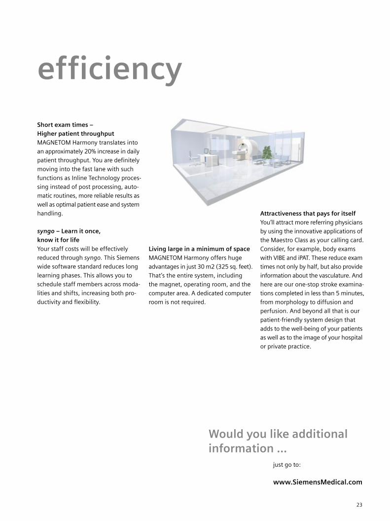
Living large in a minimum of spaceMAGNETOM Harmony offers huge
advantages in just 30 m2 (325 sq. feet).
That’s the entire system, including
the magnet, operating room, and the
computer area. A dedicated computer
room is not required.
just go to:
www.SiemensMedical.com
Would you like additionalinformation …
Attractiveness that pays for itselfYou’ll attract more referring physicians
by using the innovative applications of
the Maestro Class as your calling card.
Consider, for example, body exams
with VIBE and iPAT. These reduce exam
times not only by half, but also provide
information about the vasculature. And
here are our one-stop stroke examina-
tions completed in less than 5 minutes,
from morphology to diffusion and
perfusion. And beyond all that is our
patient-friendly system design that
adds to the well-being of your patients
as well as to the image of your hospital
or private practice.

Laser Radiationdo not stare into beam
PEAK POWER <3mWWAVE LENGTH 540-700nmCLASS II LASER PRODUCT
Acknowledgement:
Uniontown Hospital, USA
Praxis Dr. Sander, Charité, Berlin, Germany
Krankenhaus Fürth, Germany
Klinikum Mannheim, Germany
Institute Diagnostic Radiology (IDR)/Uni Erlangen, Germany
Drs. H. Elsner u. B. Marquardt/FDS
Aichiken Colony
Radiologie München-Solln, Germany
Praxis für Radiologie, Weiden, Germany
Please contact in the USA:Siemens Medical Solutions USA, Inc.51 Valley Stream ParkwayMalvern, PA 19355Tel.: 610-448-4500Fax: 610-488-2254
Please contact in Asia:Siemens Advanced Engineering Pte.Ltd.Medical DivisionAsean Business Centre2, Kallang SectorSingapore, 349277(+65) 841 35 28
Please contact in Japan:Siemens-AsahiMedical Technologies Ltd.Takanawa Park Tower 14th F20-14, Higashi-Gotanda 3-chomeShinagawa-kuTokyo 141-8641(03) 54 23 40 01
Order No. A91100-M2220-E866-3-7600
Printed in Germany
BKW 63198 WS 09024.
Siemens reserves the right to modify the design and
specifications contained herein without prior notice.
Please contact your local Siemens Sales representative
for the most current information.
Original images always lose a certain amount of detail
when reproduced.
This brochure refers to both standard and optional
features. Availability and packaging of options varies
by country and is subject to change without notice.
Some of the features described are not available for
commercial distribution in the U.S.
with syngo MR 2002B
The information about syngo MR 2002B is being
provided for planning purposes. The product is pending
510(k) review, and is not yet commercially available
in the U.S.
The information in this document contains general
descriptions of the technical options available, which
do not always have to be present in individual cases.
The required features should therefore be specified in
each individual case at the time of closing the contract.
Siemens AG, Medical SolutionsMagnetic ResonanceHenkestr. 127, D-91052 ErlangenGermanyTelephone: ++49 9131 84-0www.SiemensMedical.com


![[28] Magnetom Flash_28_Jan 2004](https://img.pdfslide.us/doc/110x75/55cf9d8b550346d033ae1705/28-magnetom-flash28jan-2004.jpg)

![[32] Magnetom Flash_32_apr 2006](https://img.pdfslide.us/doc/110x75/55cf9d8b550346d033ae170f/32-magnetom-flash32apr-2006.jpg)