Embed Size (px)
Citation preview

M i c r o s c o p y f r o m C a r l Z e i s s
LSM 5 PASCALThe Personal Laser Scanning Microscope
Focused on Your Success.
8-S. LSM 5 PASCAL Englisch 01.10.1999 15:42 Uhr Seite 3

xy
z
Detector
Pinholein ConfocalPlane
Beam Splitter
Scan Mirror
Objective
Laser
Specimen
Focal Plane
LSM 5 PASCALAn LSM of the Fifth Generation
Your work involves the acquisition of two-dimen-
sional and three-dimensional images? And you ex-
pect optimum quality along with high resolution?
You also need to record time series? You want to
acquire the image data in different orientations
and reconstruct at different viewing angles with-
out the need to remount your specimen? And you
especially expect to be able to perform quanti-
tative measurements of, for example, calcium
concentrations and pH-values, areas and surface
properties?
Then you should consider a very personal laser
scanning microscope:
The LSM 5 PASCAL – a laser scanning microscope
of the fifth generation which has been designed
for you or your team working in routine and
research.
The LSM 5 PASCAL is the ‘little brother’ of the
leading edge LSM 510 system.
‘Little’, however, is only descriptive of its ama-
zingly attractive price, as our engineers have made
no compromises to image quality, sensitivity and
flexibility!
More than 150 years of innovation in optics and
around 20 years of experience in all fields of laser
scanning microscopy combined with the ongoing
dialog with you, the users, make the LSM 5
PASCAL a rewarding long-term investment. An
investment which does not put a strain on your
budget and which can be expanded as your
applications requirements change or grow.
The LSM 5 PASCAL represents a compact system that uses confocal technique and the point scanning method to acquire 3 dimensional pictures of your specimen with the highest resolution,close to physical limitations.
8-S. LSM 5 PASCAL Englisch 01.10.1999 15:42 Uhr Seite 4

3
LSM 5 PASCALYours Personally
Maximum operating convenience...
Like all LSM units of the fifth generation, the hall-
mark of LSM 5 PASCAL is a fully motorized
scanning module. All moving components, such
as emission filter wheels, main and secondary
dichroic beam splitters, the pinhole and the
mechanical attenuators for each laser line, are
computer-controlled and take up the required po-
sition making the set-up error free and without
any need for cumbersome manual operation. A
special benefit in practice: the position can be
reset automatically at the press of a button.
...highest resolution...
In addition, the LSM 5 PASCAL provides image
sizes of up to 2,048x2,048 pixels, scan fields
of unrivaled size along with maximum linearity,
12-bit resolution per channel, high scan speeds,
high-sensitivity detectors (selected photomulti-
pliers) and short light paths from the specimen to
the detector – features which are unique in its
class.
...and great flexibility.
Special emphasis has been placed on the flexibi-
lity of the LSM 5 PASCAL. Six versions with one
or two confocal channels and combinations of
lasers in the blue and green spectrum and a choice
of five microscope stands are available for most
varied applications in the life and materials scien-
ces. The LSM 5 PASCAL can be converted from an
upright to an inverted microscope within minutes.
Confocal microscopy is no more than a flick of the
wrist away from conventional microscopy.
“The LSM 5 PASCAL grows withthe needs of your applications:
You can add a second fluores-cence channel as well as a trans-mitted light detector. Up to8 single emission filters perchannel and the six positiondichroic mirror wheels areexchangeable.
Furthermore you can chose froma variety of software options.”
3/1 Xanthidium cristatum (SAG 173.80),Immunofluorescence chloroplasts (red),storage vesicle (green) + DIC overlay(Lab for Experimental Phycology,Göttingen)
8-S. LSM 5 PASCAL Englisch 01.10.1999 15:42 Uhr Seite 5

3
8-S. LSM 5 PASCAL Englisch 01.10.1999 15:42 Uhr Seite 6

8-S. LSM 5 PASCAL Englisch 01.10.1999 15:42 Uhr Seite 7

LSM 5 PASCALProven Software on a New Platform
A system is always only as good as its user inter-
face. For this reason, the LSM 5 PASCAL uses
proven technology as its basis: the fast and reliable
software runs on a high-end PC under Windows
NT and is closely related to the tried and tested
software of the LSM 510. Just like the configura-
tion of the microscope and the scan module, the
software functionality can also be extended step
by step.
The synchronized control of scanners, data acqui-
sition and input/output signals by a digital signal
processor (DSP) allows extremely flexible scan
strategies.
Various scan functions are available for data
acquisition. They include multifluorescence ima-
ges free from cross talk and the scanning of any
number of regions of interest (ROI) of almost all
forms. The acquisition of 3D stacks combined with
time series supports the researcher studying pro-
cesses in living cells.
In addition to data acquisition, the software of the
LSM 5 PASCAL provides many 2D and 3D presen-
tation options, numerous on-line and off-line
measuring functions and a very convenient image
database for the management and the documen-
tation of the images.
The software controls the motorized system
components, i.e. the microscope, the scan and
the laser modules. The user-friendly and intuitive
user interface makes your daily work easier. As all
adjustable components are controlled by the scan
and laser module software, system configurations
once set can be reactivated by simply pressing a
button, time-consuming manual settings are now
a thing of the past. Macros can be used for re-
peating individual work processes.
6/1: “The Crop function: Fast and easy selectionof a new scan area”
OK cells, mitosis, eCFP indicatesPSD95 and Alexa546 shows Actin(Dr. Klöcker, Uni Konstanz)
6/2: “Defining and modifyingmultiple arbitrary RegionsOf Interest (ROI)”
Cells, MnSOD shown using Cy2,GFAP indicated by Cy3, 2kx2k pixels(Dr. Possel, Institute of MedicalNeurobiology, Magdeburg)
8-S. LSM 5 PASCAL Englisch 01.10.1999 15:43 Uhr Seite 8
7/1 7/2 7/3
7/4
7/5
7/6
7/7 7/8
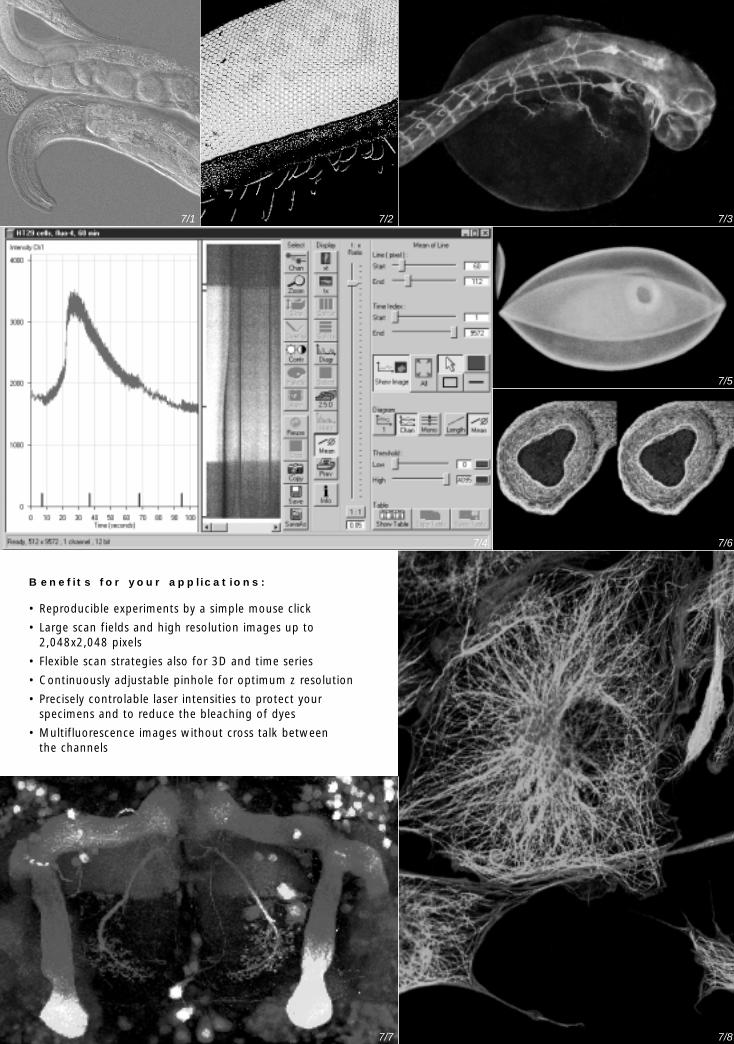
Benefits for your applications:
• Reproducible experiments by a simple mouse click
• Large scan fields and high resolution images up to2,048x2,048 pixels
• Flexible scan strategies also for 3D and time series
• Continuously adjustable pinhole for optimum z resolution
• Precisely controlable laser intensities to protect your specimens and to reduce the bleaching of dyes
• Multifluorescence images without cross talk between the channels
8-S. LSM 5 PASCAL Englisch 01.10.1999 15:43 Uhr Seite 1

Subject to change. 40-053 e/09.99
7/3
7/5
7/6
7/8
7/1 7/2 7/3
7/4
7/5
7/6
7/7 7/8
LSM 5 PASCALSpecification
Microscopes Upright: Axioplan 2 MOT, Axioskop 2 MOT, Axioskop 2 FS MOT,Inverted: Axiovert 100 M Side/BasePortAccessories like ICS objectives and filter cubes can be used
z Drives DC servo or step motor, smallest steps starting at 25 nmFine focusing stage HRZ 200, total range 200 µm, smallest step 10 nmXY stage Motorized XY scanning stage, smallest step 250 nm
Scan Module Two independent galvo scanning mirrorsScan resolution 1x4 to 2048x2048 pixels, user definableScan speed 2x10 levels, line frequencies from 4 up to 1300 Hz; 0.4 s per frame 512x512 pixelsScan zoom 0.7x to 8x, variable with steps of 0.1Scan rotation Any angle, variable with steps of 1°Scan field 18 mm diagonal in the primary image plane (with zoom factor 1x)Pinhole One pinhole with variable diameter size, adjustableDetection 1 or 2 confocal R/FL channels with built in highly sensitive photomultiplier tubes
1 external transmitted light channel (DIC capable) optionalDynamic range 12 bit per channel
Laser module Ar laser 488, 514 nm, 25 mW - HeNe laser 543 nm, 1 mW Laser lines (end of lifetime specifications)Attenuation Individual and variable intensity control of all lines
Electronic’s module Control circuitry for microscope, laser and scan moduleLSM 5 Control with built in high performance Digital Signal Processor (DSP)
Computer Well equipped High-end PC with ample RAM + hard disk space, many accessories, multi user operating system Windows NT 4.0
Monitor Ergonomic high contrast large screen monitor, 2nd monitor optional
Software Software for operating microscope, laser module and scan module;For image acquisition, display, processing and archiving, line and frame scan, 3D or/and timelapse recording, Multitracking and dual direction scanning, spline scanning, ROIs (region of interest), 3D projections, quantitative measurements, many options, morethan 20 export file formats (LSM, TIF, BMP, JPG, PCX, GIF, …)
7/1C. elegans with embryos,autofluorescence + DIC,(single frame out of time series),(Sample courtesy of Dr. Afaq, MBLWoods Hole)
7/2Fly eye (Musca domesticus), doublefluorescence, 3D reconstruction,(Dr. Lam, Dr. Jans, J. Curtin Schoolof Medical Research, Sydney)
7/3Zebrafish embryo, neurons (green),NCAM (red)(Dr. Marx, Dr. Bastmeyer, Uni Konstanz)
7/4HT29 cells loaded with Fluo-4, Line scan time series, 60 min(Dr. Nitschke, Uni Freiburg)
7/5Triticum spelta, auto fluorescence,maximum projection
7/6Fetal small intestine, double fluorescence,stereo projection(Dr. Hashimoto, Jikei University, Tokyo)
7/7Drosophila brain, neural circuit indicated with GFP(Dr. Ito, NI of Basic Biology Lab, Okasaki)
7/8Endothelial cells, Texas Red marks F-actin (red),BODIPY shows tubulin (green)multitracking scan without crosstalk of channels
Some of the components and software functions are optional.
For further details, please contact:
Prin
ted
on e
nviro
nmen
t-fr
iend
ly p
aper
,bl
each
ed w
ithou
t th
e us
e of
chl
orin
e.Carl Zeiss MicroscopyD-07740 JenaPhone: ++49-36 41/64 -1616Telefax: ++49-36 41/64 -3144E-mail: [email protected]: www.zeiss.de/micro
8-S. LSM 5 PASCAL Englisch 01.10.1999 15:43 Uhr Seite 2