Embed Size (px)
Citation preview
We make it visible.
M i c r o s c o p y f r o m C a r l Z e i s s
Vision Set in Motion
LSM 5 LIVE and LSM 5 LIVE DuoScanLaser Scanning Microscope
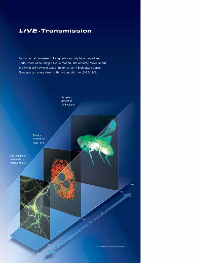
Fundamental processes in living cells can only be observed and
understood when imaged live in motion. The ultimate movie about
the living cell however was a dream so far in biological science.
Now you can come close to this vision with the LSM 5 LIVE.
LIVE -Transmission
Fast reaction ofnerve cells to external stimuli
Division of fertilized insect ova
Life cycle ofDrosophilaMelanogaster
Time constants of selected cellular processes
1µs
1ms
1s
103s
106s
Time
Confocal High-Speed Camera
Motion Studies in Detail 4
Accessing Living Cells
Data Production in Realtime 6
Detailed Motion Studies
Careful Imaging at Highest Speeds 8
PA-GFP, Dronpa and Kaede
Selective Activation of Fluorescent Proteins
with Violet Light 9
Physiological Measurements
Comprehensive Acquisition
and Analysis Options 10
FRAP, FLIP and FRET
Tracking Down Biological Molecules 12
Faster than Real Time
Development in 4 Dimensions 14
Specifications 16
System Overview 18
Content
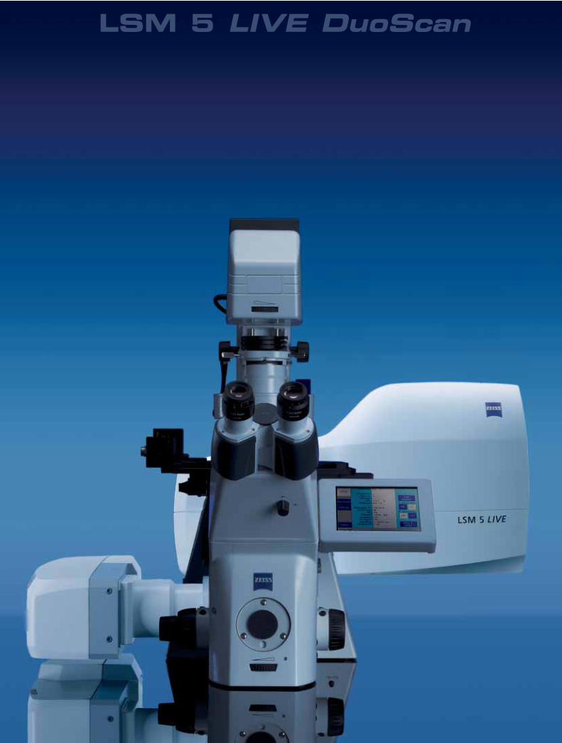
LSM 5 LIVE DuoScan
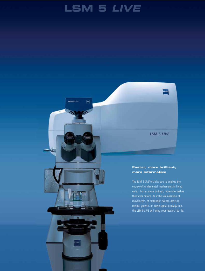
Faster, more brilliant,
more informative
The LSM 5 LIVE enables you to analyze the
course of fundamental mechanisms in living
cells – faster, more brilliant, more informative
than ever before. Be it the visualization of
movements, of metabolic events, develop-
mental growth, or nerve signal propagation,
the LSM 5 LIVE will bring your research to life.
LSM 5 LIVE
Confocal High-Speed Camera Motion Studies in Detail
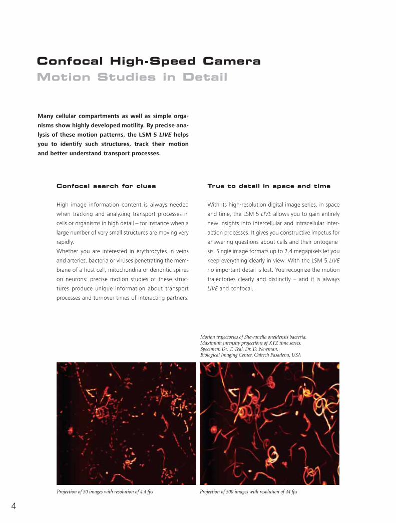
Many cellular compartments as well as simple orga-
nisms show highly developed motility. By precise ana-
lysis of these motion patterns, the LSM 5 LIVE helps
you to identify such structures, track their motion
and better understand transport processes.
Confocal search for clues
High image information content is always needed
when tracking and analyzing transport processes in
cells or organisms in high detail – for instance when a
large number of very small structures are moving very
rapidly.
Whether you are interested in erythrocytes in veins
and arteries, bacteria or viruses penetrating the mem-
brane of a host cell, mitochondria or dendritic spines
on neurons: precise motion studies of these struc-
tures produce unique information about transport
processes and turnover times of interacting partners.
True to detail in space and time
With its high-resolution digital image series, in space
and time, the LSM 5 LIVE allows you to gain entirely
new insights into intercellular and intracellular inter-
action processes. It gives you constructive impetus for
answering questions about cells and their ontogene-
sis. Single image formats up to 2.4 megapixels let you
keep everything clearly in view. With the LSM 5 LIVE
no important detail is lost. You recognize the motion
trajectories clearly and distinctly – and it is always
LIVE and confocal.
Motion trajectories of Shewanella oneidensis bacteria.Maximum intensity projections of XYZ time series.Specimen: Dr. T. Teal, Dr. D. Newman,Biological Imaging Center, Caltech Pasadena, USA
Projection of 50 images with resolution of 4.4 fps Projection of 500 images with resolution of 44 fps
Präparat: Färbung GFP, Objektiv C-Apochromat 63x/1.2 W.
4
5
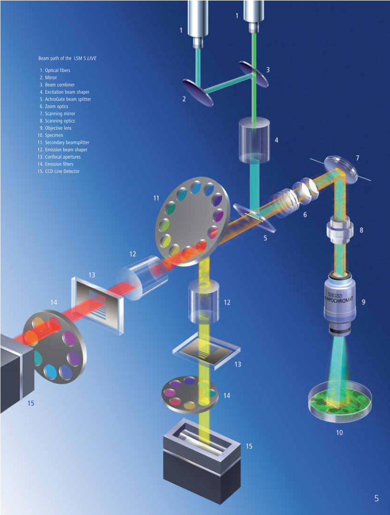
Beam path of the LSM 5 LIVE
1. Optical fibers 2. Mirror 3. Beam combiner 4. Excitation beam shaper 5. AchroGate beam splitter 6. Zoom optics 7. Scanning mirror 8. Scanning optics 9. Objective lens
10. Specimen 11. Secondary beamsplitter 12. Emission beam shaper 13. Confocal apertures 14. Emission filters15. CCD Line Detector
6
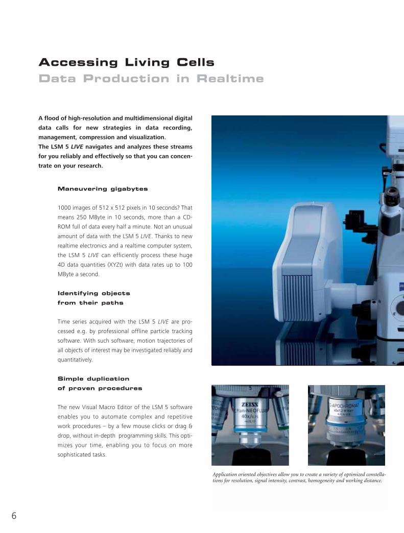
Accessing Living Cells Data Production in Realtime
A flood of high-resolution and multidimensional digital
data calls for new strategies in data recording,
management, compression and visualization.
The LSM 5 LIVE navigates and analyzes these streams
for you reliably and effectively so that you can concen-
trate on your research.
Maneuvering gigabytes
1000 images of 512 x 512 pixels in 10 seconds? That
means 250 MByte in 10 seconds, more than a CD-
ROM full of data every half a minute. Not an unusual
amount of data with the LSM 5 LIVE. Thanks to new
realtime electronics and a realtime computer system,
the LSM 5 LIVE can efficiently process these huge
4D data quantities (XYZt) with data rates up to 100
MByte a second.
Identifying objects
from their paths
Time series acquired with the LSM 5 LIVE are pro-
cessed e.g. by professional offline particle tracking
software. With such software, motion trajectories of
all objects of interest may be investigated reliably and
quantitatively.
Simple duplication
of proven procedures
The new Visual Macro Editor of the LSM 5 software
enables you to automate complex and repetitive
work procedures – by a few mouse clicks or drag &
drop, without in-depth programming skills. This opti-
mizes your time, enabling you to focus on more
sophisticated tasks.
Application oriented objectives allow you to create a variety of optimized constella-tions for resolution, signal intensity, contrast, homogeneity and working distance.
New high-performance objectives
C-Apochromat, LD C-Apochromat for confocal perfection with correction into NIR wavelengths.
LD LCI Plan-Apochromat, LCI Plan-Neofluar or sophisticated requirements in life cell imaging.
C-Plan-Apochromat, EC Plan-Neofluar for greater contrast on fixed specimens under glass
W Achroplan, W Plan-Apochromat for VIS-IR applications in physiology
Precise laser light play
Compact and long-lived solid-state lasers also put
thick or weakly fluorescent specimens in the right
light, limiting tissue damage. You can concentrate on
the emission of your specimen: the disturbing side
effects of conventional gas lasers like heat or sound
emissions are a topic of the past. Choose up to 4 lines
from the range of 405, 440, 488, 532, 561 and 635
nm lasers.
7
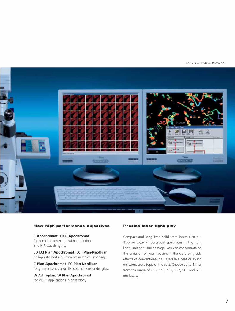
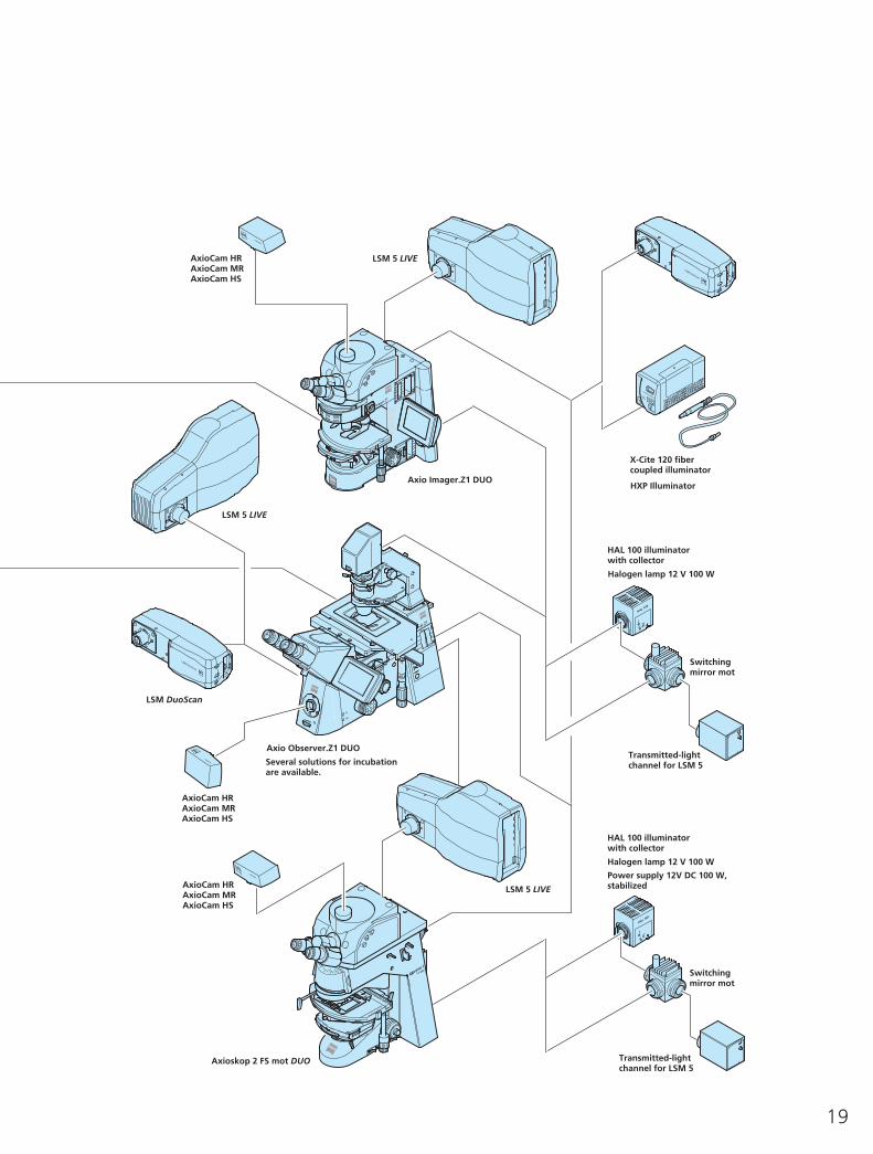
LSM 5 LIVE at Axio Observer.Z
Detailed Motion StudiesCareful Imaging at Highest Speeds
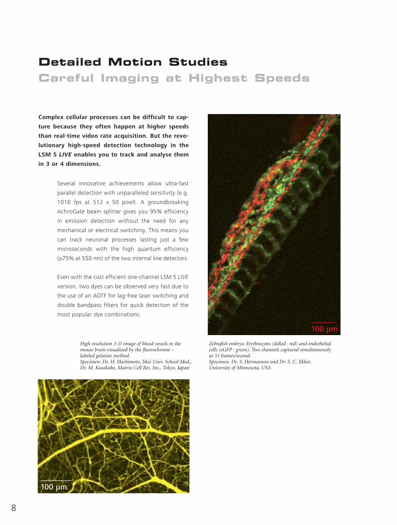
Complex cellular processes can be difficult to cap-
ture because they often happen at higher speeds
than real-time video rate acquisition. But the revo-
lutionary high-speed detection technology in the
LSM 5 LIVE enables you to track and analyse them
in 3 or 4 dimensions.
Several innovative achievements allow ultra-fast
parallel detection with unparalleled sensitivity (e.g.
1010 fps at 512 x 50 pixel). A groundbreaking
AchroGate beam splitter gives you 95% efficiency
in emission detection without the need for any
mechanical or electrical switching. This means you
can track neuronal processes lasting just a few
microseconds with the high quantum efficiency
(≥75% at 550 nm) of the two internal line detectors.
Even with the cost efficient one-channel LSM 5 LIVE
version, two dyes can be observed very fast due to
the use of an AOTF for lag-free laser switching and
double bandpass filters for quick detection of the
most popular dye combinations.
____________100 µm
_______100 µm
Zebrafish embryo. Erythrocytes (dsRed : red) and endothelialcells (eGFP : green). Two channels captured simultaneously at 33 frames/second.Specimen: Dr. S. Hermanson and Dr. S. C. Ekker,University of Minnesota, USA
High resolution 3-D image of blood vessels in themouse brain visualized by the fluorochrome – labeled gelatine method.Specimen: Dr. H. Hashimoto, Jikei Univ. School Med.,Dr. M. Kusakabe, Matrix Cell Res. Inc., Tokyo, Japan
8
PA-GFP, Dronpa and Kaede Selective Activation of FluorescentProteins with Violet Light
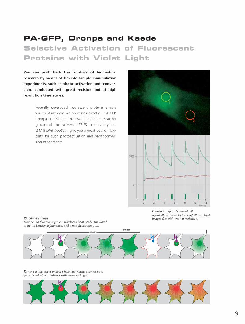
You can push back the frontiers of biomedical
research by means of flexible sample manipulation
experiments, such as photo-activation and -conver-
sion, conducted with great recision and at high
resolution time scales.
Recently developed fluorescent proteins enable
you to study dynamic processes directly – PA-GFP,
Dronpa and Kaede. The two independent scanner
groups of the universal ZEISS confocal system
LSM 5 LIVE DuoScan give you a great deal of flexi-
bility for such photoactivation and photoconver-
sion experiments.
Dronpa transfected cultured cell,repeatedly activated by pulses of 405 nm light,imaged fast with 488 nm excitation.
Kaede is a fluorescent protein whose fluorescence changes from green to red when irradiated with ultraviolet light.
DronpaPA-GFP
PA-GFP + DronpaDronpa is a fluorescent protein which can be optically stimulated to switch between a fluorescent and a non-fluorescent state.
1
2
9
10
Physiological MeasurementsComprehensive Acquisition and Analysis Options
The LSM 5 LIVE is the ideal workstation for obtain-
ing measurements that are perfectly matched to
the biological time scale as well as the spectral
properties of ion indicators and voltage sensitive
dyes. In addition, the ROI manipulation capability
of the LSM DuoScan point scanners ensures ex-
cellent precision for uncaging experiments.
The LSM 5 LIVE’s ultra-fast image acquisition capa-
bility makes it the ideal tool for observing dynamic
events, even at kilohertz resolution (e.g. 1010 fps
at 512x50 pixel). Even more important, this speed
is delivered in a true confocal system with simulta-
neous two-channel acquisition. Complemented by
a point scanner, the LSM 5 LIVE DuoScan gives you
the flexibility for uncaging and sample stimulation,
e.g. with UV light (351+364 nm). Apochromatic
dipping objectives such as the Plan Apochromat
20x/1.0 W or 63x/1.0 W are also available for
micro-manipulation.
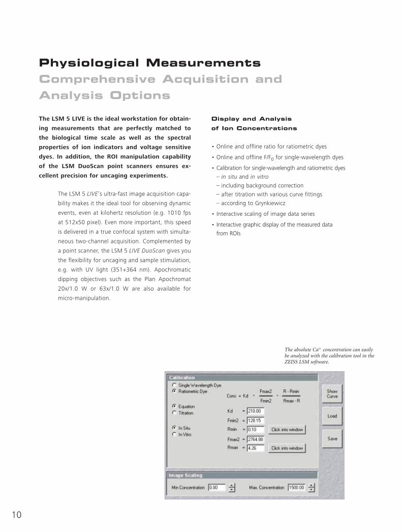
The absolute Ca2+ concentration can easily be analyzed with the calibration tool in the ZEISS LSM software.
Display and Analysis
of Ion Concentrations
• Online and offline ratio for ratiometric dyes
• Online and offline F/F0 for single-wavelength dyes
• Calibration for single-wavelength and ratiometric dyes
– in situ and in vitro
– including background correction
– after titration with various curve fittings
– according to Grynkiewicz
• Interactive scaling of image data series
• Interactive graphic display of the measured data
from ROIs
11
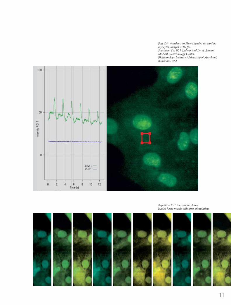
Fast Ca2+ transients in Fluo-4 loaded rat cardiacmyocytes, imaged at 80 fps.Specimen: Dr. W. J. Lederer and Dr. A. Ziman,Medical Biotechnology Center,Biotechnology Institute, Universtity of Maryland,Baltimore, USA
Repetitive Ca2+ increase in Fluo-4 loaded heart muscle cells after stimulation.
12
FRAP, FLIP and FRETTracking Down Biological Molecules
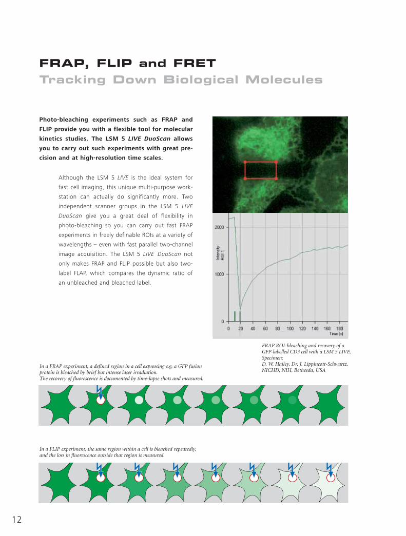
Photo-bleaching experiments such as FRAP and
FLIP provide you with a flexible tool for molecular
kinetics studies. The LSM 5 LIVE DuoScan allows
you to carry out such experiments with great pre-
cision and at high-resolution time scales.
Although the LSM 5 LIVE is the ideal system for
fast cell imaging, this unique multi-purpose work-
station can actually do significantly more. Two
independent scanner groups in the LSM 5 LIVE
DuoScan give you a great deal of flexibility in
photo-bleaching so you can carry out fast FRAP
experiments in freely definable ROIs at a variety of
wavelengths – even with fast parallel two-channel
image acquisition. The LSM 5 LIVE DuoScan not
only makes FRAP and FLIP possible but also two-
label FLAP, which compares the dynamic ratio of
an unbleached and bleached label.
In a FLIP experiment, the same region within a cell is bleached repeatedly,and the loss in fluorescence outside that region is measured.
In a FRAP experiment, a defined region in a cell expressing e.g. a GFP fusion protein is bleached by brief but intense laser irradiation.The recovery of fluorescence is documented by time-lapse shots and measured.
FRAP ROI-bleaching and recovery of a GFP-labelled CD3 cell with a LSM 5 LIVE.Specimen: D. W. Hailey, Dr. J. Lippincott-Schwartz,NICHD, NIH, Bethesda, USA
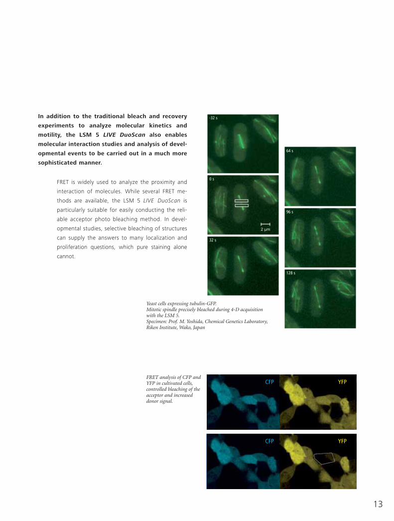
In addition to the traditional bleach and recovery
experiments to analyze molecular kinetics and
motility, the LSM 5 LIVE DuoScan also enables
molecular interaction studies and analysis of devel-
opmental events to be carried out in a much more
sophisticated manner.
FRET is widely used to analyze the proximity and
interaction of molecules. While several FRET me-
thods are available, the LSM 5 LIVE DuoScan is
particularly suitable for easily conducting the reli-
able acceptor photo bleaching method. In devel-
opmental studies, selective bleaching of structures
can supply the answers to many localization and
proliferation questions, which pure staining alone
cannot.
Yeast cells expressing tubulin-GFP.Mitotic spindle precisely bleached during 4-D acquisition with the LSM 5.Specimen: Prof. M. Yoshida, Chemical Genetics Laboratory,Riken Institute, Wako, Japan
CFP
-32 s
0 s
64 s
96 s
128 s
32 s
CFP
YFP
YFPFRET analysis of CFP and YFP in cultivated cells,controlled bleaching of the acceptor and increased donor signal.
13
14
Faster than Real Time Development in 4 Dimensions
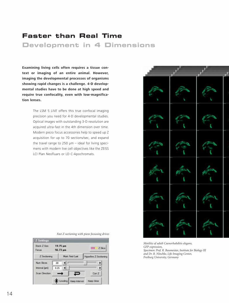
Examining living cells often requires a tissue con-
text or imaging of an entire animal. However,
imaging the developmental processes of organisms
showing rapid changes is a challenge. 4-D develop-
mental studies have to be done at high speed and
require true confocality, even with low-magnifica-
tion lenses.
The LSM 5 LIVE offers this true confocal imaging
precision you need for 4-D developmental studies.
Optical images with outstanding 3-D resolution are
acquired ultra-fast in the 4th dimension over time.
Modern piezo focus accessories help to speed up Z
acquisition for up to 70 sections/sec, and expand
the travel range to 250 µm – ideal for living speci-
mens with modern live cell objectives like the ZEISS
LCI Plan Neofluars or LD C-Apochromats.
Motility of adult Caenorhabditis elegans,GFP expression,Specimen: Prof. R. Baumeister, Institute for Biology III and Dr. R. Nitschke, Life Imaging Center,Freiburg University, Germany
Fast Z sectioning with piezo focussing drives
15
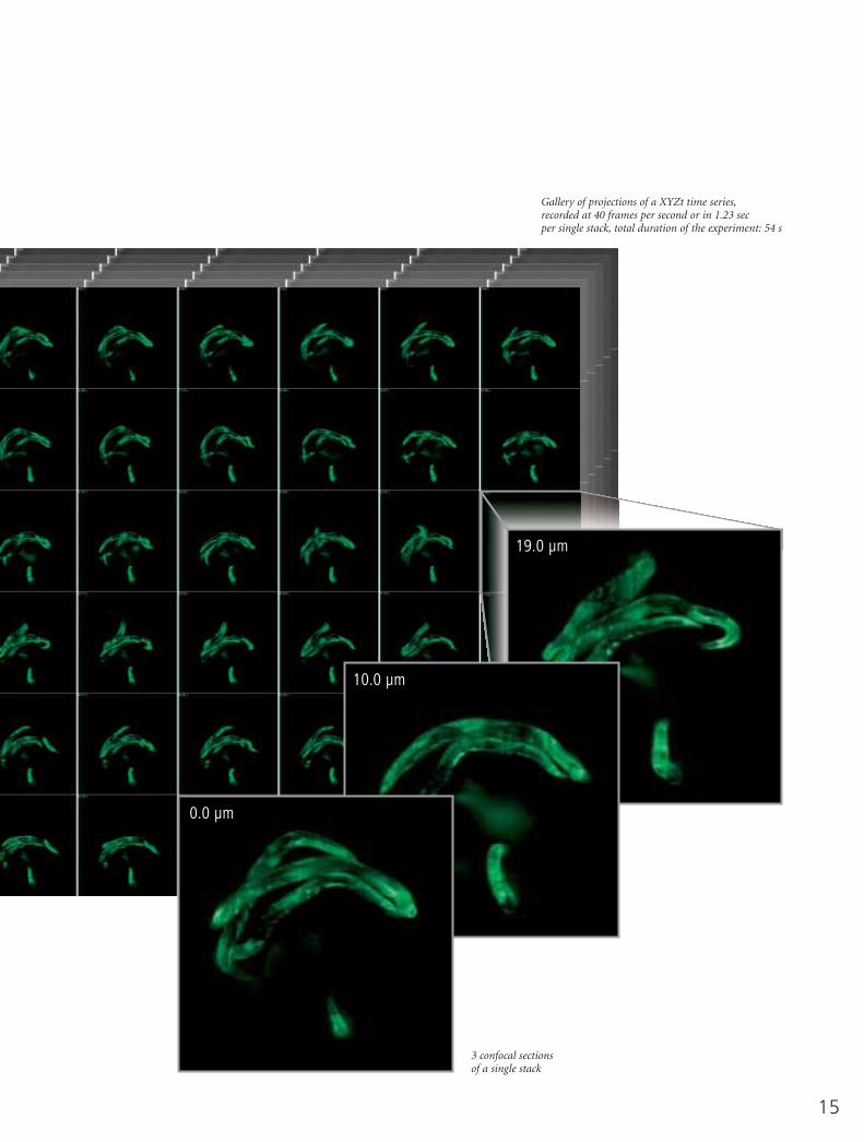
Gallery of projections of a XYZt time series,recorded at 40 frames per second or in 1.23 sec per single stack, total duration of the experiment: 54 s
3 confocal sectionsof a single stack
19.0 µm
10.0 µm
0.0 µm
16
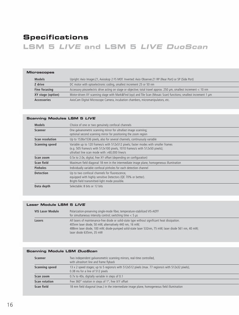
SpecificationsLSM 5 LIVE and LSM 5 LIVE DuoScan
Scanning Modules LSM 5 LIVE
Laser Module LSM 5 LIVE
Scanning Module LSM DuoScan
Microscopes
Models Upright: Axio Imager.Z1, Axioskop 2 FS MOT. Inverted: Axio Observer.Z1 RP (Rear Port) or SP (Side Port)
Z drive DC motor with optoelectronic coding, smallest increment 25 or 50 nm
Fine focusing Accessory piezoelectric drive acting on stage or objective; total travel approx. 250 µm, smallest increment < 10 nm
XY stage (option) Motor-driven XY scanning stage with Mark&Find (xyz) and Tile Scan (Mosaic Scan) functions; smallest increment 1 µm
Accessories AxioCam Digital Microscope Camera, incubation chambers, micromanipulators, etc.
Models Choice of one or two genuinely confocal channels
Scanner One galvanometric scanning mirror for ultrafast image scanning;optional second scanning mirror for positioning the zoom region
Scan resolution Up to 1536x1536 pixels, also for several channels, continuously variable
Scanning speed Variable up to 120 frames/s with 512x512 pixels; faster modes with smaller frames (e.g. 505 frames/s with 512x100 pixels, 1010 frames/s with 512x50 pixels);ultrafast line scan mode with >60,000 lines/s
Scan zoom 0.5x to 2.0x, digital, free XY offset (depending on configuration)
Scan field Maximum field diagonal 18 mm in the intermediate image plane, homogeneous illumination
Pinholes Individually variable confocal pinholes for each detection channel
Detection Up to two confocal channels for fluorescence,equipped with highly sensitive Detectors (QE 70% or better).Bright-field transmitted-light mode possible.
Data depth Selectable: 8 bits or 12 bits
VIS Laser Module Polarization-preserving single-mode fiber, temperature-stabilized VIS-AOTF for simultaneous intensity control; switching time < 5 µs
Lasers All lasers of maintenance-free diode or solid-state type without significant heat dissipation.405nm laser diode, 50 mW, alternatively 440 nm, 16 mW;488nm laser diode, 100 mW; diode-pumped solid-state laser 532nm, 75 mW; laser diode 561 nm, 40 mW;laser diode 635nm, 35 mW
Scanner Two independent galvanometric scanning mirrors, real-time controlled,with ultrashort line and frame flyback
Scanning speed 13 x 2 speed stages; up to 5 regions/s with 512x512 pixels (max. 77 regions/s with 512x32 pixels),0.38 ms for a line of 512 pixels
Scan zoom 0.7x to 40x, digitally variable in steps of 0.1
Scan rotation Free 360° rotation in steps of 1°, free X/Y offset
Scan field 18 mm field diagonal (max.) in the intermediate image plane, homogeneous field illumination
17
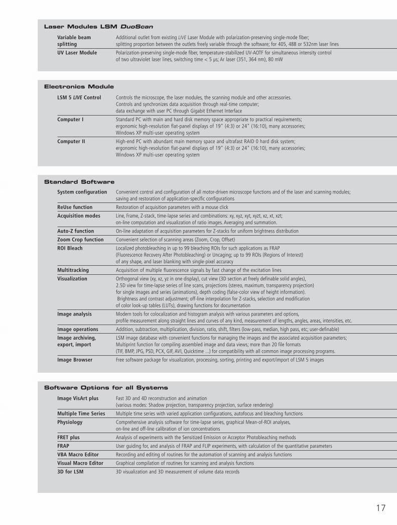
Standard Software
Software Options for all Systems
Laser Modules LSM DuoScan
Electronics Module
Variable beam Additional outlet from existing LIVE Laser Module with polarization-preserving single-mode fiber;splitting splitting proportion between the outlets freely variable through the software; for 405, 488 or 532nm laser lines
UV Laser Module Polarization-preserving single-mode fiber, temperature-stabilized UV-AOTF for simultaneous intensity control of two ultraviolet laser lines, switching time < 5 µs; Ar laser (351, 364 nm), 80 mW
LSM 5 LIVE Control Controls the microscope, the laser modules, the scanning module and other accessories.Controls and synchronizes data acquisition through real-time computer;data exchange with user PC through Gigabit Ethernet Interface
Computer I Standard PC with main and hard disk memory space appropriate to practical requirements;ergonomic high-resolution flat-panel displays of 19" (4:3) or 24" (16:10), many accessories;Windows XP multi-user operating system
Computer II High-end PC with abundant main memory space and ultrafast RAID 0 hard disk system;ergonomic high-resolution flat-panel displays of 19" (4:3) or 24" (16:10), many accessories;Windows XP multi-user operating system
System configuration Convenient control and configuration of all motor-driven microscope functions and of the laser and scanning modules;saving and restoration of application-specific configurations
ReUse function Restoration of acquisition parameters with a mouse click
Acquisition modes Line, Frame, Z-stack, time-lapse series and combinations: xy, xyz, xyt, xyzt, xz, xt, xzt;on-line computation and visualization of ratio images. Averaging and summation.
Auto-Z function On-line adaptation of acquisition parameters for Z-stacks for uniform brightness distribution
Zoom Crop function Convenient selection of scanning areas (Zoom, Crop, Offset)
ROI Bleach Localized photobleaching in up to 99 bleaching ROIs for such applications as FRAP (Fluorescence Recovery After Photobleaching) or Uncaging; up to 99 ROIs (Regions of Interest) of any shape, and laser blanking with single-pixel accuracy
Multitracking Acquisition of multiple fluorescence signals by fast change of the excitation lines
Visualization Orthogonal view (xy, xz, yz in one display), cut view (3D section at freely definable solid angles),2.5D view for time-lapse series of line scans, projections (stereo, maximum, transparency projection) for single images and series (animations), depth coding (false-color view of height information).Brightness and contrast adjustment; off-line interpolation for Z-stacks, selection and modification
of color look-up tables (LUTs), drawing functions for documentation
Image analysis Modern tools for colocalization and histogram analysis with various parameters and options,profile measurement along straight lines and curves of any kind, measurement of lengths, angles, areas, intensities, etc.
Image operations Addition, subtraction, multiplication, division, ratio, shift, filters (low-pass, median, high pass, etc; user-definable)
Image archiving, LSM image database with convenient functions for managing the images and the associated acquisition parameters;export, import Multiprint function for compiling assembled image and data views; more than 20 file formats
(TIF, BMP, JPG, PSD, PCX, GIF, AVI, Quicktime ...) for compatibility with all common image processing programs.
Image Browser Free software package for visualization, processing, sorting, printing and export/import of LSM 5 images
Image VisArt plus Fast 3D and 4D reconstruction and animation (various modes: Shadow projection, transparency projection, surface rendering)
Multiple Time Series Multiple time series with varied application configurations, autofocus and bleaching functions
Physiology Comprehensive analysis software for time-lapse series, graphical Mean-of-ROI analyses,on-line and off-line calibration of ion concentrations
FRET plus Analysis of experiments with the Sensitized Emission or Acceptor Photobleaching methods
FRAP User guiding for, and analysis of FRAP and FLIP experiments, with calculation of the quantitative parameters
VBA Macro Editor Recording and editing of routines for the automation of scanning and analysis functions
Visual Macro Editor Graphical compilation of routines for scanning and analysis functions
3D for LSM 3D visualization and 3D measurement of volume data records
18
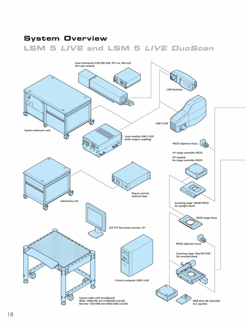
System OverviewLSM 5 LIVE and LSM 5 LIVE DuoScan
10
20
3040
50
60
70
8090
100
19

Carl Zeiss MicroImaging GmbH
www.zeiss.de/micro
07740 Jena, Germany
Phone: +49 3641 64 3400Fax: +49 3641 64 3144E-mail: [email protected]
45-0067 e/11.06
Subj
ect
to c
hang
e.
Prin
ted
on e
nviro
nmen
t-fr
iend
ly p
aper
,bl
each
ed w
ithou
t th
e us
e of
chl
orin
e.
Optical perfection, creative foresight and a
sure feeling for the technical challenges in the
life sciences: the basis for superior microscopy
concepts from Carl Zeiss. We have given a
name to our focus on the key method in re-
search into life: FluoresScience.
LSM 5 LIVEUS Patents: 6848825, 6888148
6947127, 60375836462345, 64864586941247