Embed Size (px)
Citation preview


Acute Lower Gastrointestinal Acute Lower Gastrointestinal BleedingBleeding
Essentials of Diagnosis:Essentials of Diagnosis: Hematochezia usually present. Hematochezia usually present. Ten percent of cases of Ten percent of cases of
hematochezia due to upper hematochezia due to upper gastrointestinal source. gastrointestinal source.
Evaluation with colonoscopy in Evaluation with colonoscopy in stable patients. stable patients.
Massive active bleeding calls for Massive active bleeding calls for evaluation with sigmoidoscopy, evaluation with sigmoidoscopy, upper endoscopy, angiography, or upper endoscopy, angiography, or nuclear bleeding scan. nuclear bleeding scan.

LGI hemorrhageLGI hemorrhage
Sites:Sites: Colon – 95-97%.Colon – 95-97%. Small bowel – 3-5%.Small bowel – 3-5%.
Only 15% of massive GI bleeding.Only 15% of massive GI bleeding. Finding the site:Finding the site:
Intermittent bleeding common.Intermittent bleeding common. Up to 42% have multiple sites.Up to 42% have multiple sites.

AetiologyAetiology


Causes of lower G.I. bleeding:Causes of lower G.I. bleeding:
1. Auto-Immune1. Auto-Immune Inflammatory bowel disease Inflammatory bowel disease
(I.B.D) eg. UC & CD(I.B.D) eg. UC & CD

2. Inflammatory2. Inflammatory Bacterial Bacterial Dysentry Dysentry Parasitic Parasitic Bilharzial Bilharzial ViralViral Solitary ulcer of the rectumSolitary ulcer of the rectum
3. Tumours3. Tumours Polyps Polyps Cancer caecumCancer caecum Cancer sigmoid Cancer sigmoid
4. Vascular4. Vascular AngiodysplasiaAngiodysplasia Ischaemic colitisIschaemic colitis PilesPiles

5.5. Meckel’s diverticulum and Meckel’s diverticulum and
Diverticular disease. Diverticular disease.
6. Anal Fissures.6. Anal Fissures.
NB:NB: Massive bleeding from lower Massive bleeding from lower G.I.T is rare.G.I.T is rare.
Lower Gastrointestinal BleedingLower Gastrointestinal Bleeding
Etiology:Etiology: Hemorrhoids and anal fissures:Hemorrhoids and anal fissures: Bleeding is typically small volume and Bleeding is typically small volume and
intermittent, with bright red blood on intermittent, with bright red blood on the surface of the stool. Occasionally the surface of the stool. Occasionally bleeding is severe. The diagnosis can bleeding is severe. The diagnosis can be confirmed on anoscopy and/or be confirmed on anoscopy and/or flexible sigmoidoscopy. Severe or flexible sigmoidoscopy. Severe or recurrent bleeding are indications for recurrent bleeding are indications for hemorrhoidal band ligation or hemorrhoidectomy. .
Anal fissures: may also bleed, but : may also bleed, but bleeding is usually minimal and is bleeding is usually minimal and is associated with anal discomfort. Fiber associated with anal discomfort. Fiber supplementation and laxatives are supplementation and laxatives are advised. advised.

HemmorrhoidsHemmorrhoids

Lower Gastrointestinal BleedingLower Gastrointestinal Bleeding
Etiology:Etiology:
Colonic diverticula: Colonic diverticula:
Local erosion into one of the Local erosion into one of the
arteries leads to brisk but usually arteries leads to brisk but usually
self-limited bleeding. Rarely self-limited bleeding. Rarely
bleeding is massive on bleeding is massive on
presentation, requiring emergent presentation, requiring emergent
diagnostic angiography followed diagnostic angiography followed
by intra-arterial infusion of by intra-arterial infusion of
vasopressin or segmental vasopressin or segmental
resection.resection.
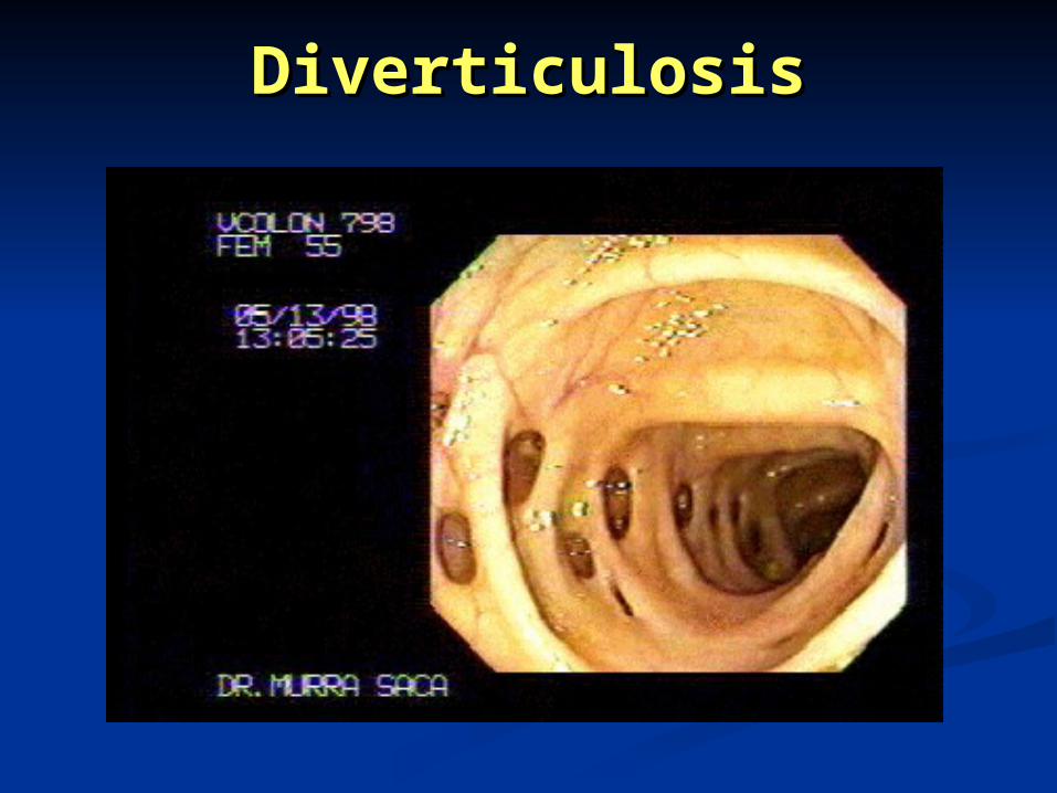
DiverticulosisDiverticulosis

Lower Gastrointestinal BleedingLower Gastrointestinal Bleeding
Etiology:Etiology:
Vascular anomalies:Vascular anomalies:
Sporadic and secondary Sporadic and secondary
angiodysplasia are a common angiodysplasia are a common
cause of bleeding from the small cause of bleeding from the small
bowel and colon. Vascular bowel and colon. Vascular
ectasias (or angiodysplasias) ectasias (or angiodysplasias)
occur throughout the upper and occur throughout the upper and
lower intestinal tracts and cause lower intestinal tracts and cause
painless bleeding ranging from painless bleeding ranging from
melena or hematochezia to occult melena or hematochezia to occult
blood loss.blood loss.

Lower Gastrointestinal BleedingLower Gastrointestinal Bleeding
Etiology:Etiology:
Colorectal neoplasm: Colorectal neoplasm:
Benign polyps and carcinoma are Benign polyps and carcinoma are
associated with chronic occult blood loss associated with chronic occult blood loss
or intermittent anorectal hematochezia. or intermittent anorectal hematochezia.
However, colonic neoplasms may cause However, colonic neoplasms may cause
up to 10% of acute lower up to 10% of acute lower
gastrointestinal hemorrhage. Although gastrointestinal hemorrhage. Although
colorectal cancer is most commonly colorectal cancer is most commonly
associated withassociated with occult blood loss rather rather
than overt bleeding, patients with than overt bleeding, patients with
rectosigmoid lesions may present with rectosigmoid lesions may present with
hematochezia. The diagnosis is readily hematochezia. The diagnosis is readily
made on endoscopy.made on endoscopy.

Colonic PolypsColonic Polyps

MalignancyMalignancy
Colon CarcinomaColon Carcinoma

Lower Gastrointestinal BleedingLower Gastrointestinal Bleeding
Etiology:Etiology: Infectious, inflammatory or ischemic Infectious, inflammatory or ischemic
colitis:colitis:
A variety of infectious, inflammatory and A variety of infectious, inflammatory and
ischemic colitides may present with ischemic colitides may present with
bloody diarrhea. The diagnosis of bloody diarrhea. The diagnosis of infectious colitis is usually confirmed by is usually confirmed by
stool culture or assay for Clostridium stool culture or assay for Clostridium
difficile toxin, but occasionally stool difficile toxin, but occasionally stool
studies are negative. Endoscopy is studies are negative. Endoscopy is
always indicated in the setting of always indicated in the setting of
possible inflammatory or ischemic possible inflammatory or ischemic
colitis, unless there is clinical evidence colitis, unless there is clinical evidence
for perforation. Again, mucosal biopsies for perforation. Again, mucosal biopsies
are usually diagnostic.are usually diagnostic.

Lower Gastrointestinal BleedingLower Gastrointestinal Bleeding
Etiology:Etiology:
-Inflammatory Bowel Disease:-Inflammatory Bowel Disease:
Patients with inflammatory bowel Patients with inflammatory bowel
disease (especially ulcerative colitis) disease (especially ulcerative colitis)
often have diarrhea with variable often have diarrhea with variable
amounts of hematochezia. Bleeding amounts of hematochezia. Bleeding
varies from occult blood loss to varies from occult blood loss to
recurrent hematochezia usually mixed recurrent hematochezia usually mixed
with stool. Symptoms of abdominal with stool. Symptoms of abdominal
pain, tenesmus, and urgency are often pain, tenesmus, and urgency are often
present.present.

Inflammatory Bowel DiseaseInflammatory Bowel Disease

Some distinguishing characteristics of ulcerative Some distinguishing characteristics of ulcerative colitis and Crohn’s diseasecolitis and Crohn’s disease::
CharacteristicsUlcerative ColitisCrohn's Disease
Rectal bleedingUsualSometimes
Abdominal massRareOften
Abdominal painSometimesOften
Perianal diseaseExtremely rare5-10%
Upper GI symptomsNeverOccasional
Cigarette smokingVery rareCommon
MalnutritionSometimesCommon
Low-grade feverSometimesOften
Rectal diseaseUsualSometimes
Continuous diseaseUsualRare

ContCont________________________.________________________.
CharacteristicsUlcerative ColitisCrohn's Disease
GranulomasNever10-30%
Crypt abscessesCommonRare
Discrete ulcersRareCommon
Aphthoid ulcersRareCommon
Cobblestone lesionsNeverCommon
Skip lesionsNo, except rarely in treated patients
Common
Ileal involvementRare, backwash ileitisUsual
FistulasNeverCommon
CancerRareVery rare
Microscopic skip lesionsNo, except rarely in treated patients
Common
Transmural inflammation
Only in fulminant diseaseCommon

Evaluation & Evaluation & ManagementManagement

Lower Gastrointestinal BleedingLower Gastrointestinal Bleeding
Evaluation & Management:Evaluation & Management: Initial stabilization, blood replacement.Initial stabilization, blood replacement.
The color of the stool helps distinguish The color of the stool helps distinguish upper from lower gastrointestinal upper from lower gastrointestinal bleeding, especially when observed by bleeding, especially when observed by the physician. Brown stools mixed or the physician. Brown stools mixed or streaked with blood predict a source in streaked with blood predict a source in the rectosigmoid or anus. Painless the rectosigmoid or anus. Painless large-volume bleeding usually suggests large-volume bleeding usually suggests diverticular bleeding or vascular diverticular bleeding or vascular ectasias. Bloody diarrhea associated ectasias. Bloody diarrhea associated with cramping abdominal pain, urgency, with cramping abdominal pain, urgency, or tenesmus is characteristic of or tenesmus is characteristic of inflammatory bowel disease, infectious inflammatory bowel disease, infectious colitis, or ischemic colitis.colitis, or ischemic colitis.

RESUSCITATION
Indications for transfusion Profuse bleeding Persistent hemodynamic instability
despite crystalloid resuscitation Symptomatic anemia (CP, SOB,
orthostasis with Hgb < 10) AMI or unstable angina with Hgb <
10

DiagnosisDiagnosis History History ExaminationExamination P.R for rectal lesions + cancer.P.R for rectal lesions + cancer. Proctoscopy for haemorrhoids (piles).Proctoscopy for haemorrhoids (piles). Sigmoidoscopy for I.B.DSigmoidoscopy for I.B.D Barium enema for mucosal lesionsBarium enema for mucosal lesions Colonoscopy for diagnosis + removal of Colonoscopy for diagnosis + removal of
polypspolyps Nuclear Bleeding Scans and Angiography for Nuclear Bleeding Scans and Angiography for
vascular lesionsvascular lesions Small Intestine Push Enteroscopy or Capsule Small Intestine Push Enteroscopy or Capsule
Imaging.Imaging.

Information about bleeding
Volume and frequency of bleeding Painful defecation? Relationship of bleeding to
defecation?
[before, during (mixed into faeces or coating surface?)
or after]
Associated abdominal pain? Colour of blood?

CLINICAL PRESENTATIONCLINICAL PRESENTATION
Presentation correlates not with Presentation correlates not with
location but with the rate of location but with the rate of
transit:transit: Hematemesis almost always UGI.Hematemesis almost always UGI. Hematochezia 3/4 patient with have a Hematochezia 3/4 patient with have a
LGI source.LGI source. Melena more likely UGI than LGI.Melena more likely UGI than LGI.
NGTL (+) highly suggestive of UGI.NGTL (+) highly suggestive of UGI.

CLINICAL PRESENTATIONCLINICAL PRESENTATION
Hematemesis:Hematemesis: Vomiting of blood; clots or coffee grounds.Vomiting of blood; clots or coffee grounds. Suggestive of an UGI source.Suggestive of an UGI source.
Hematochezia:Hematochezia: The passage of liquid blood or clots per The passage of liquid blood or clots per
rectum.rectum. 3/4 colonic source; 11% prox lig of Treitz.3/4 colonic source; 11% prox lig of Treitz.
Melena:Melena: Stools with altered blood that are black and Stools with altered blood that are black and
tarry and have a distinctive odor.tarry and have a distinctive odor. Suggestion of UGI source.Suggestion of UGI source. May cont for days p bleeding stops.May cont for days p bleeding stops.

TAGGED RBC SCANTAGGED RBC SCAN
AdvantagesAdvantages Safe, noninvasiveSafe, noninvasive Readily availableReadily available Detects slow Detects slow
bleedsbleeds at a rate of 0.1 to at a rate of 0.1 to
0.5 m/min 0.5 m/min more sensitive more sensitive
than angiographythan angiography 73% - 100%73% - 100%
DisadvantagesDisadvantages only localizes only localizes
bleeding to an bleeding to an area of the area of the abdomen, not SB abdomen, not SB vs LBvs LB
not therapeuticnot therapeutic less specific than less specific than
endoscopy and endoscopy and angiographyangiography

TAGGED RBC SCAN

Meckel’s DiverticulumMeckel’s Diverticulum
Cecal angiodysplasia Cecal angiodysplasia with extravasationwith extravasation
Small bowel ulcerationSmall bowel ulceration due to NSAIDSdue to NSAIDS

TAGGED RBC SCAN
Useful: To confirm bleeding. In planning angiography.
Not useful: Massive bleeds or critical illness.

COLONOSCOPYCOLONOSCOPY
AdvantagesAdvantages Potential for precise Potential for precise
localization:localization: diagnostic success 51% diagnostic success 51%
- 90%.- 90%.
Potential for rxPotential for rx therapeutic success therapeutic success
69%-100%.69%-100%.
Ability to collect Ability to collect
pathologic pathologic
specimens.specimens.
DisadvantagesDisadvantages Requires a Requires a
technically skilled technically skilled
endoscopist, not endoscopist, not
available at all available at all
centers.centers. Various rates of Various rates of
rebleeding rebleeding
depending on depending on
source.source. Post bleed transit.Post bleed transit.

Bleeding Bleeding diverticulosisdiverticulosis
Colonic Colonic angiodysplasiaangiodysplasia

COLONOSCOPY
Diagnostic procedure of choice when bleeding has stopped.
Many reports of good localization of bleed (74-85%) even with hematochezia.

ANGIOGRAPHYANGIOGRAPHY
Advantages:Advantages: anatomic localization anatomic localization
is accurate: is accurate: Specificity- 100%.Specificity- 100%. Sensitivity 47% (acute Sensitivity 47% (acute
bleeding), 30% bleeding), 30%
(recurrent (recurrent
hemorrhage).hemorrhage). 41-86% bleeds 41-86% bleeds
localized.localized.
Therapeutic Therapeutic
intervention.intervention.
DisadvantagesDisadvantages Requires active Requires active
bleeding > 0.5 bleeding > 0.5 cc/min.cc/min.
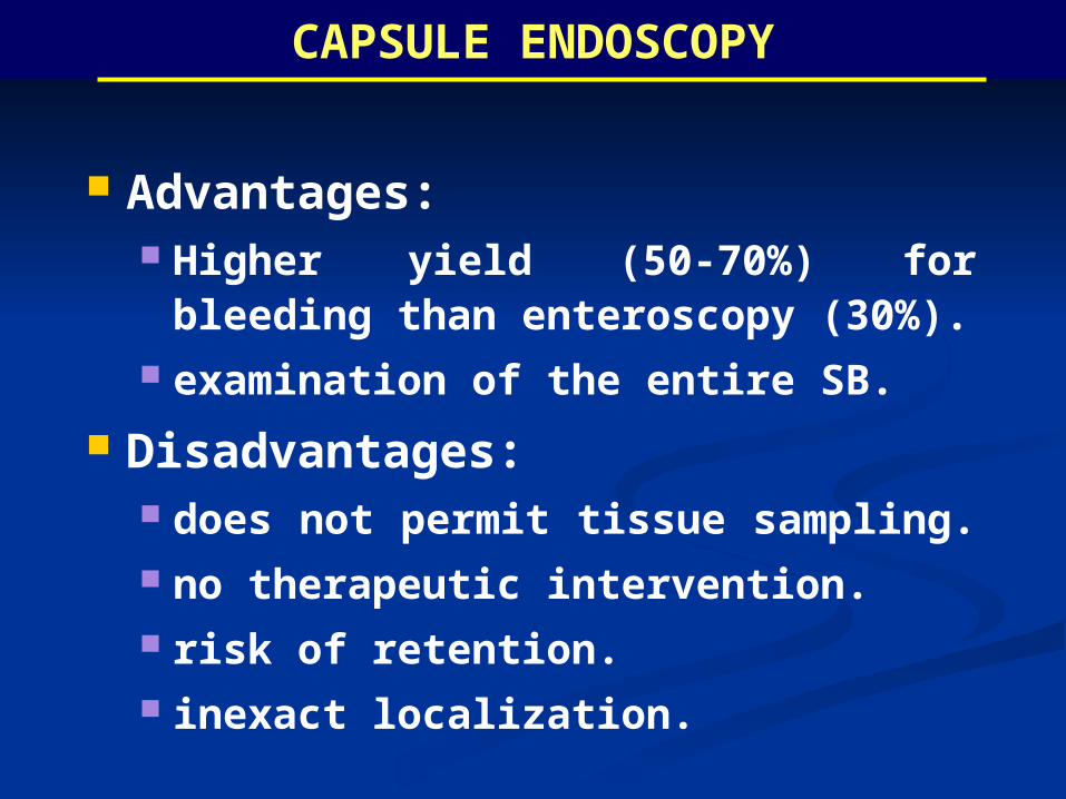
CAPSULE ENDOSCOPY
Advantages: Higher yield (50-70%) for
bleeding than enteroscopy (30%). examination of the entire SB.
Disadvantages: does not permit tissue sampling. no therapeutic intervention. risk of retention. inexact localization.

Indications for capsule endoscopy:
1-Iron deficiency anaemia when obscure gastrointestinal bleeding is suspected.
2-Diagnosis of early or suspected small bowel Crohn’s disease.
3-Detection of benign and malignant small intestinal tumours (e.g. polyps, GISTs, lymphoma).
4-Identification of medication related to small bowel injury (e.g. NSAID-induced enteropathy).
CAPSULE ENDOSCOPYCAPSULE ENDOSCOPY

Image Spectrum: PillCam Capsule Image Spectrum: PillCam Capsule EndoscopyEndoscopy
BleedingBleeding
Celiac DiseaseCeliac DiseaseTumorsTumors
Suspected Crohn’sSuspected Crohn’s


ManagementManagement Is individualized for every Is individualized for every
casecase

Acute Lower Gastrointestinal Acute Lower Gastrointestinal BleedingBleeding
Treatment:Treatment: Therapeutic ColonoscopyTherapeutic Colonoscopy
High-risk lesions may now be treated High-risk lesions may now be treated
endoscopically with epinephrine endoscopically with epinephrine
injection, cautery, or application of injection, cautery, or application of
metallic endoclips. metallic endoclips. Intra-arterial Vasopressin or Intra-arterial Vasopressin or
Embolization:Embolization: Surgical Treatment:Surgical Treatment:
With increasing experience with urgent With increasing experience with urgent
colonoscopy and angiographic colonoscopy and angiographic
embolization, the need for surgical embolization, the need for surgical
treatment is decreasing. treatment is decreasing.

VASOPRESSIN INFUSION
Causes reliable arteriolar vasoconstriction and bowel contraction, resulting in decreased blood flow.
36-100% will stop bleeding: >90% of patients with LGIB due to
diverticular disease or angiodysplasia. Rebleed rate 26-71%.
May be used to temporize bleed prior to surgical resection.
Avoid in pts with cardiac dz.

EMBOLIZATION
definitive means of controlling hemorrhage: Stops bleeding 67-100%. Rebleed 0-33%.
LGI compared to UGI tract has weaker blood supply: Supplied by end arterties. 5-21% post-embolic ischemia reported. 0-40% required emergent lap for
bleeding and/or ischemia.

INDICATIONS FOR SURGERY
Bleeding refractory to other therapies.
Hemodynamic instability. Re-bleeding after non-operative
treatment, esp if localized.

Summary of Treatment
Lower GI bleed
Small volume
Large volume
Investigate cause
Manage cause
Resuscitate
Bleeding stops
Bleeding persists
? Surgical intervention


Thank you
Thank you