Embed Size (px)
Citation preview
Objectives:
● Abdominal hernias: Inguinal hernias, Ventral hernias, Rare
external hernias, Internal hernias, Complications of hernias,
Management of complicated hernias.
Resources:
● 436 doctor’s slides.
● 435’ team work.
● Surgical recall.
● Davidson’s.
Done by: Mohammed khoja, Ashwaq Almajid, Jumana Alghtani. Leaders: Heba Alnasser, Jawaher Abanumy, Mohammed Habib, Mohammad Al-Mutlaq. Revised by: Basel Almeflh
Color index: Notes , Important , Extra , Davidson’s
Editing file Feedback
SURGICAL ANATOMYSURGICAL ANATOMY
Basic review: in BLACK are from the doctor’s slides
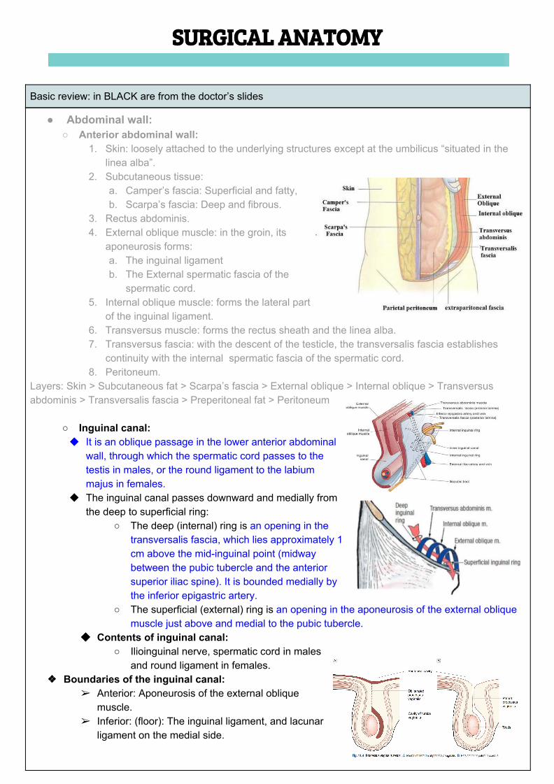
● Abdominal wall: ○ Anterior abdominal wall:
1. Skin: loosely attached to the underlying structures except at the umbilicus “situated in the linea alba”.
2. Subcutaneous tissue: a. Camper’s fascia: Superficial and fatty, b. Scarpa’s fascia: Deep and fibrous.
3. Rectus abdominis. 4. External oblique muscle: in the groin, its
aponeurosis forms: a. The inguinal ligament b. The External spermatic fascia of the
spermatic cord. 5. Internal oblique muscle: forms the lateral part
of the inguinal ligament. 6. Transversus muscle: forms the rectus sheath and the linea alba. 7. Transversus fascia: with the descent of the testicle, the transversalis fascia establishes
continuity with the internal spermatic fascia of the spermatic cord. 8. Peritoneum.
Layers: Skin > Subcutaneous fat > Scarpa’s fascia > External oblique > Internal oblique > Transversus abdominis > Transversalis fascia > Preperitoneal fat > Peritoneum
○ Inguinal canal: ◆ It is an oblique passage in the lower anterior abdominal
wall, through which the spermatic cord passes to the testis in males, or the round ligament to the labium majus in females.
◆ The inguinal canal passes downward and medially from the deep to superficial ring:
○ The deep (internal) ring is an opening in the transversalis fascia, which lies approximately 1 cm above the mid-inguinal point (midway between the pubic tubercle and the anterior superior iliac spine). It is bounded medially by the inferior epigastric artery.
○ The superficial (external) ring is an opening in the aponeurosis of the external oblique muscle just above and medial to the pubic tubercle.
◆◆ Contents of inguinal canal: ○ Ilioinguinal nerve, spermatic cord in males
and round ligament in females. ❖❖ Boundaries of the inguinal canal:
➢ Anterior: Aponeurosis of the external oblique muscle.
➢ Inferior: (floor): The inguinal ligament, and lacunar ligament on the medial side.
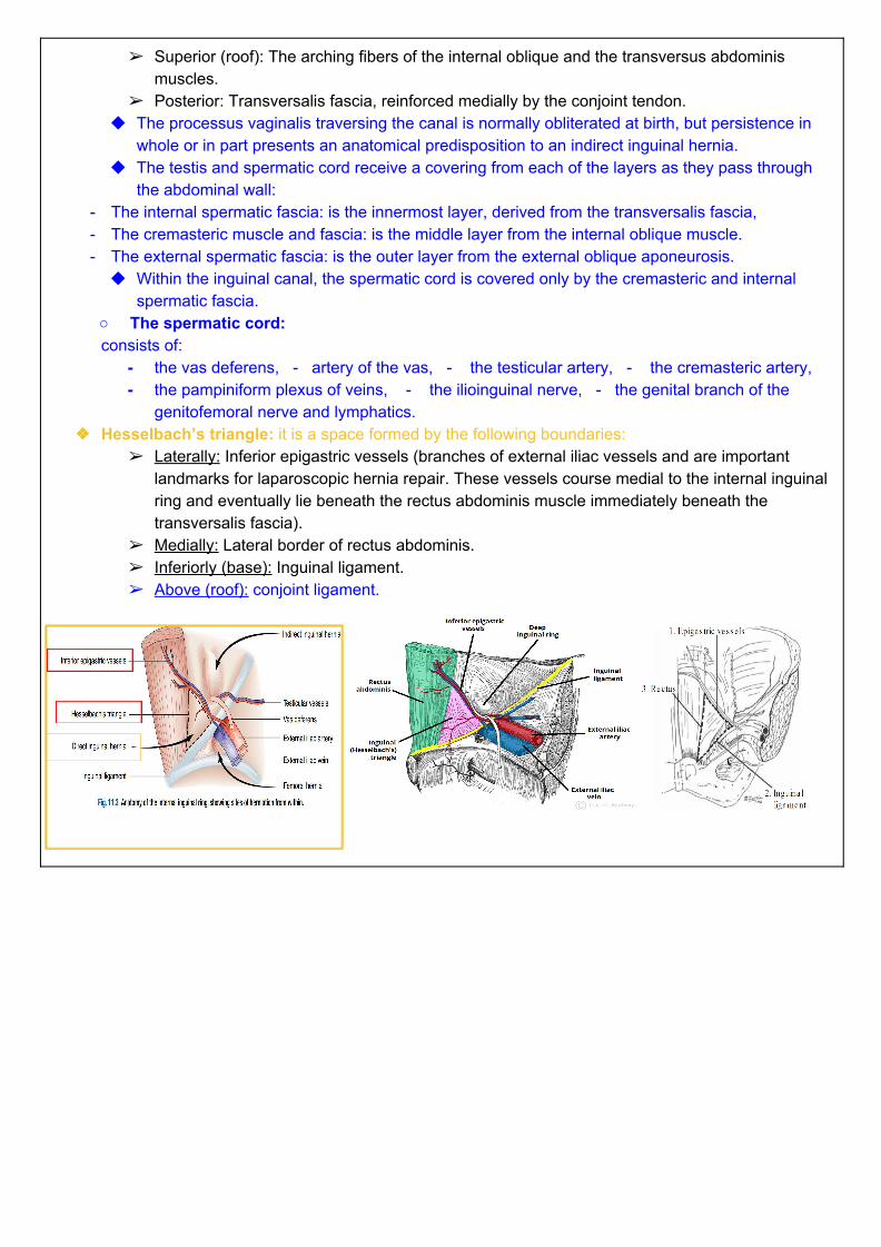
➢ Superior (roof): The arching fibers of the internal oblique and the transversus abdominis muscles.
➢ Posterior: Transversalis fascia, reinforced medially by the conjoint tendon. ◆ The processus vaginalis traversing the canal is normally obliterated at birth, but persistence in
whole or in part presents an anatomical predisposition to an indirect inguinal hernia. ◆ The testis and spermatic cord receive a covering from each of the layers as they pass through
the abdominal wall: - The internal spermatic fascia: is the innermost layer, derived from the transversalis fascia, - The cremasteric muscle and fascia: is the middle layer from the internal oblique muscle. - The external spermatic fascia: is the outer layer from the external oblique aponeurosis.
◆ Within the inguinal canal, the spermatic cord is covered only by the cremasteric and internal spermatic fascia.
○ The spermatic cord: consists of:
- the vas deferens, - artery of the vas, - the testicular artery, - the cremasteric artery, - the pampiniform plexus of veins, - the ilioinguinal nerve, - the genital branch of the
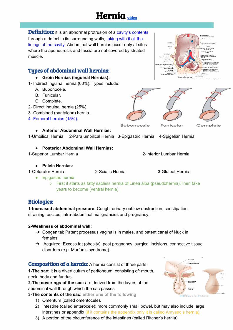
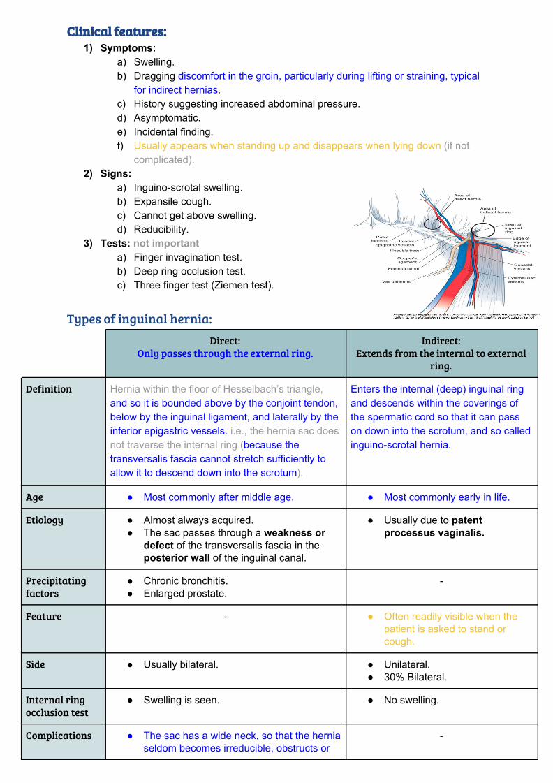
genitofemoral nerve and lymphatics. ❖❖ Hesselbach’s triangle: it is a space formed by the following boundaries:
➢ Laterally: Inferior epigastric vessels (branches of external iliac vessels and are important landmarks for laparoscopic hernia repair. These vessels course medial to the internal inguinal ring and eventually lie beneath the rectus abdominis muscle immediately beneath the transversalis fascia).
➢ Medially: Lateral border of rectus abdominis. ➢ Inferiorly (base): Inguinal ligament. ➢ Above (roof): conjoint ligament.
Hernia Hernia videovideo
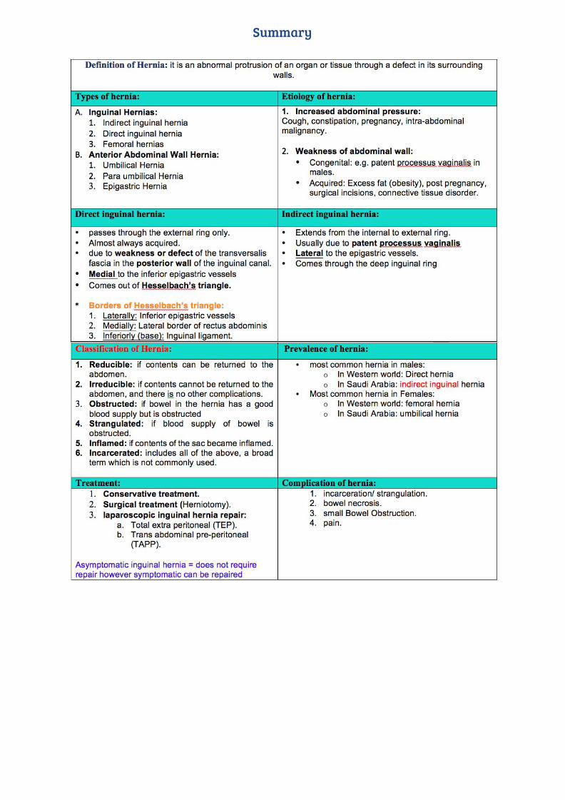
Definition:Definition: it is an abnormal protrusion of a cavity’s contents through a defect in its surrounding walls, taking with it all the linings of the cavity. Abdominal wall hernias occur only at sites where the aponeurosis and fascia are not covered by striated muscle. Types of abdominal wall hernias:Types of abdominal wall hernias:
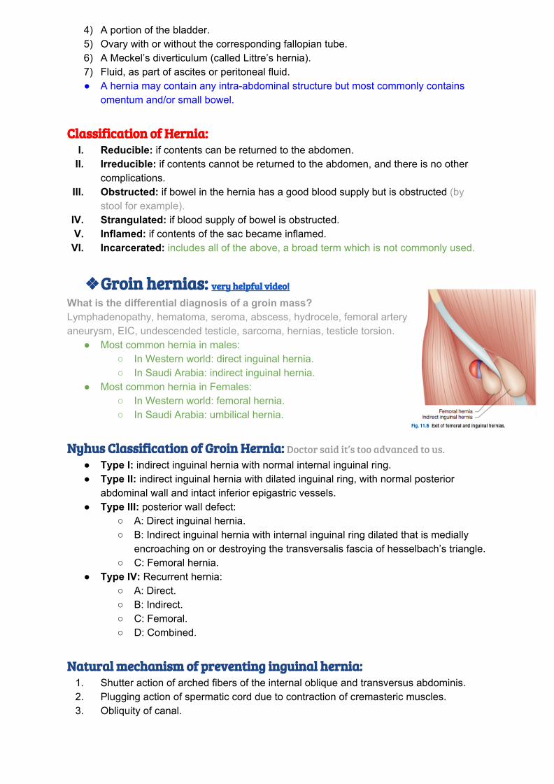
● Groin Hernias (Inguinal Hernias): 1- Indirect inguinal hernia (60%): Types include:
A. Bubonocele. B. Funicular. C. Complete.
2- Direct inguinal hernia (25%). 3- Combined (pantaloon) hernia. 4- Femoral hernias (15%).
● Anterior Abdominal Wall Hernias: 1-Umbilical Hernia 2-Para umbilical Hernia 3-Epigastric Hernia 4-Spigelian Hernia
● Posterior Abdominal Wall Hernias: 1-Superior Lumbar Hernia 2-Inferior Lumbar Hernia
● Pelvic Hernias: 1-Obturator Hernia 2-Sciatic Hernia 3-Gluteal Hernia
● Epigastric hernia: ○ First it starts as fatty sacless hernia of Linea alba (pseudohernia),Then take
years to become (ventral hernia) Etiologies:Etiologies: 1-Increased abdominal pressure: Cough, urinary outflow obstruction, constipation,straining, ascites, intra-abdominal malignancies and pregnancy. 2-Weakness of abdominal wall:
➔ Congenital: Patent processus vaginalis in males, and patent canal of Nuck in females.
➔ Acquired: Excess fat (obesity), post pregnancy, surgical incisions, connective tissue disorders (e.g. Marfan’s syndrome).
Composition of a hernia:Composition of a hernia: A hernia consist of three parts: 1-The sac: it is a diverticulum of peritoneum, consisting of: mouth, neck, body and fundus. 2-The coverings of the sac: are derived from the layers of the abdominal wall through which the sac passes. 3-The contents of the sac: either one of the following
1) Omentum (called omentocele). 2) Intestine (called enterocele): more commonly small bowel, but may also include large
intestines or appendix (if it contains the appendix only it is called Amyand’s hernia). 3) A portion of the circumference of the intestines (called Ritcher’s hernia).
4) A portion of the bladder. 5) Ovary with or without the corresponding fallopian tube. 6) A Meckel’s diverticulum (called Littre’s hernia). 7) Fluid, as part of ascites or peritoneal fluid. ● A hernia may contain any intra-abdominal structure but most commonly contains
omentum and/or small bowel. Classification of Hernia:Classification of Hernia:
I. Reducible: if contents can be returned to the abdomen. II. Irreducible: if contents cannot be returned to the abdomen, and there is no other
complications. III. Obstructed: if bowel in the hernia has a good blood supply but is obstructed (by
stool for example). IV. Strangulated: if blood supply of bowel is obstructed. V. Inflamed: if contents of the sac became inflamed. VI. Incarcerated: includes all of the above, a broad term which is not commonly used.
❖❖Groin hernias: Groin hernias: very helpful video!very helpful video!
What is the differential diagnosis of a groin mass? Lymphadenopathy, hematoma, seroma, abscess, hydrocele, femoral artery aneurysm, EIC, undescended testicle, sarcoma, hernias, testicle torsion.
● Most common hernia in males: ○ In Western world: direct inguinal hernia. ○ In Saudi Arabia: indirect inguinal hernia.
● Most common hernia in Females: ○ In Western world: femoral hernia. ○ In Saudi Arabia: umbilical hernia.
Nyhus Classification of Groin Hernia:Nyhus Classification of Groin Hernia: Doctor said it’s too advanced to us.
● Type I: indirect inguinal hernia with normal internal inguinal ring. ● Type II: indirect inguinal hernia with dilated inguinal ring, with normal posterior
abdominal wall and intact inferior epigastric vessels. ● Type III: posterior wall defect:
○ A: Direct inguinal hernia. ○ B: Indirect inguinal hernia with internal inguinal ring dilated that is medially
encroaching on or destroying the transversalis fascia of hesselbach’s triangle. ○ C: Femoral hernia.
● Type IV: Recurrent hernia: ○ A: Direct. ○ B: Indirect. ○ C: Femoral. ○ D: Combined.
Natural mechanism of preventing inguinal hernia:Natural mechanism of preventing inguinal hernia:
1. Shutter action of arched fibers of the internal oblique and transversus abdominis. 2. Plugging action of spermatic cord due to contraction of cremasteric muscles. 3. Obliquity of canal.
Clinical features:Clinical features: 1) Symptoms:
a) Swelling. b) Dragging discomfort in the groin, particularly during lifting or straining, typical
for indirect hernias. c) History suggesting increased abdominal pressure. d) Asymptomatic. e) Incidental finding. f) Usually appears when standing up and disappears when lying down (if not
complicated). 2) Signs:
a) Inguino-scrotal swelling. b) Expansile cough. c) Cannot get above swelling. d) Reducibility.
3) Tests: not important a) Finger invagination test. b) Deep ring occlusion test. c) Three finger test (Ziemen test).
Types of inguinal hernia:
Direct: Only passes through the external ring.
Indirect: Extends from the internal to external
ring.
Definition Hernia within the floor of Hesselbach’s triangle, and so it is bounded above by the conjoint tendon, below by the inguinal ligament, and laterally by the inferior epigastric vessels. i.e., the hernia sac does not traverse the internal ring (because the transversalis fascia cannot stretch sufficiently to allow it to descend down into the scrotum).
Enters the internal (deep) inguinal ring and descends within the coverings of the spermatic cord so that it can pass on down into the scrotum, and so called inguino-scrotal hernia.
Age ● Most commonly after middle age. ● Most commonly early in life.
Etiology ● Almost always acquired. ● The sac passes through a weakness or
defect of the transversalis fascia in the posterior wall of the inguinal canal.
● Usually due to patent processus vaginalis.
Precipitating factors
● Chronic bronchitis. ● Enlarged prostate.
-
Feature - ● Often readily visible when the patient is asked to stand or cough.
Side ● Usually bilateral. ● Unilateral. ● 30% Bilateral.
Internal ring occlusion test
● Swelling is seen. ● No swelling.
Complications ● The sac has a wide neck, so that the hernia seldom becomes irreducible, obstructs or
-
strangulates.
Relations ● Sac is posterior to the spermatic cord. ● Medial to the inferior epigastric vessels.
● Sac is anterolateral to the cord. ● Lateral to the epigastric vessels.
Direction of the sac
● Comes out of Hesselbach’s triangle. ● Comes through the deep inguinal ring.
Treatment of uncomplicated inguinal hernias:Treatment of uncomplicated inguinal hernias:
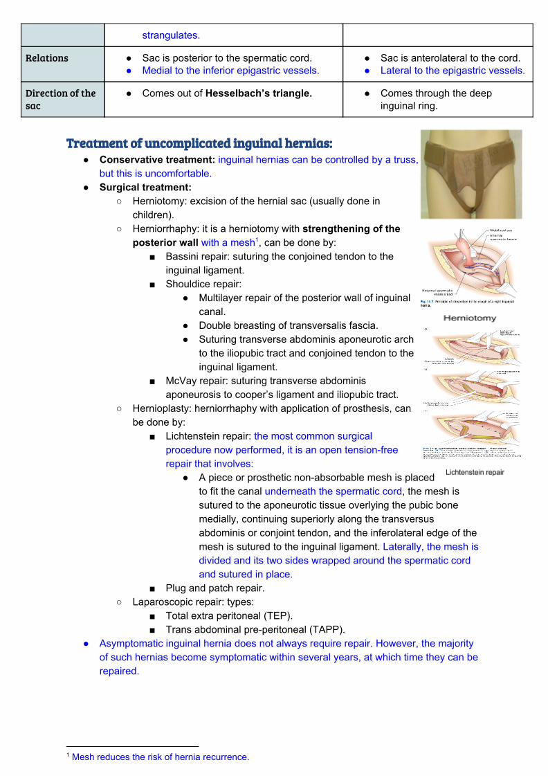
● Conservative treatment: inguinal hernias can be controlled by a truss, but this is uncomfortable.
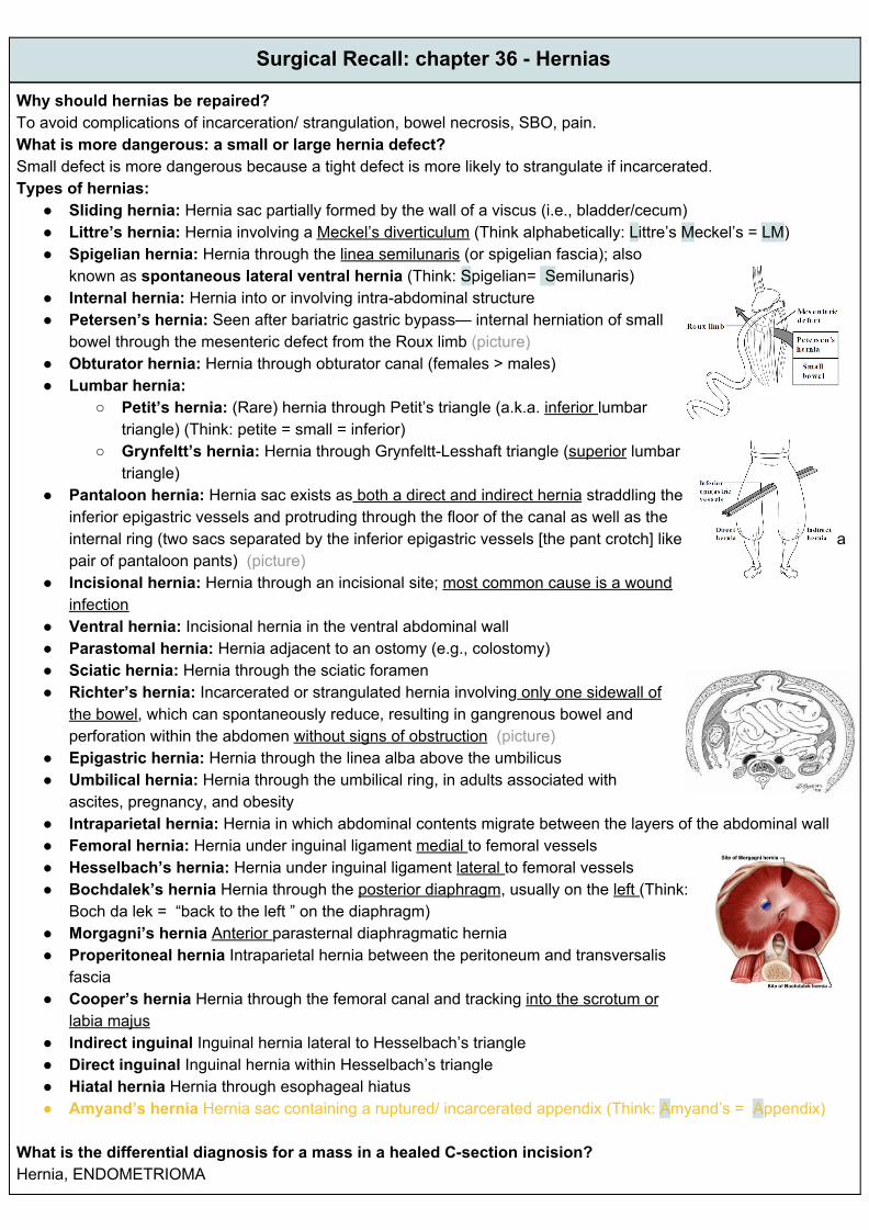
● Surgical treatment: ○ Herniotomy: excision of the hernial sac (usually done in
children). ○ Herniorrhaphy: it is a herniotomy with strengthening of the
posterior wall with a mesh , can be done by: 1
■ Bassini repair: suturing the conjoined tendon to the inguinal ligament.
■ Shouldice repair: ● Multilayer repair of the posterior wall of inguinal
canal. ● Double breasting of transversalis fascia. ● Suturing transverse abdominis aponeurotic arch
to the iliopubic tract and conjoined tendon to the inguinal ligament.
■ McVay repair: suturing transverse abdominis aponeurosis to cooper’s ligament and iliopubic tract.
○ Hernioplasty: herniorrhaphy with application of prosthesis, can be done by:
■ Lichtenstein repair: the most common surgical procedure now performed, it is an open tension-free repair that involves:
● A piece or prosthetic non-absorbable mesh is placed to fit the canal underneath the spermatic cord, the mesh is sutured to the aponeurotic tissue overlying the pubic bone medially, continuing superiorly along the transversus abdominis or conjoint tendon, and the inferolateral edge of the mesh is sutured to the inguinal ligament. Laterally, the mesh is divided and its two sides wrapped around the spermatic cord and sutured in place.
■ Plug and patch repair. ○ Laparoscopic repair: types:
■ Total extra peritoneal (TEP). ■ Trans abdominal pre-peritoneal (TAPP).
● Asymptomatic inguinal hernia does not always require repair. However, the majority of such hernias become symptomatic within several years, at which time they can be repaired.
1 Mesh reduces the risk of hernia recurrence.
Surgical Recall: chapter 36 - Hernias
Why should hernias be repaired? To avoid complications of incarceration/ strangulation, bowel necrosis, SBO, pain. What is more dangerous: a small or large hernia defect? Small defect is more dangerous because a tight defect is more likely to strangulate if incarcerated. Types of hernias:
● Sliding hernia: Hernia sac partially formed by the wall of a viscus (i.e., bladder/cecum) ● Littre’s hernia: Hernia involving a Meckel’s diverticulum (Think alphabetically: Littre’s Meckel’s = LM) ● Spigelian hernia: Hernia through the linea semilunaris (or spigelian fascia); also
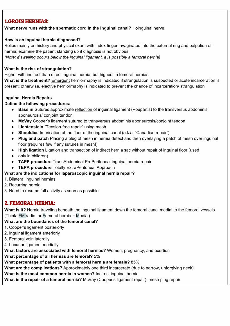
known as spontaneous lateral ventral hernia (Think: Spigelian= Semilunaris) ● Internal hernia: Hernia into or involving intra-abdominal structure ● Petersen’s hernia: Seen after bariatric gastric bypass— internal herniation of small
bowel through the mesenteric defect from the Roux limb (picture) ● Obturator hernia: Hernia through obturator canal (females > males) ● Lumbar hernia:
○ Petit’s hernia: (Rare) hernia through Petit’s triangle (a.k.a. inferior lumbar triangle) (Think: petite = small = inferior)
○ Grynfeltt’s hernia: Hernia through Grynfeltt-Lesshaft triangle (superior lumbar triangle)
● Pantaloon hernia: Hernia sac exists as both a direct and indirect hernia straddling the inferior epigastric vessels and protruding through the floor of the canal as well as the internal ring (two sacs separated by the inferior epigastric vessels [the pant crotch] like a pair of pantaloon pants) (picture)
● Incisional hernia: Hernia through an incisional site; most common cause is a wound infection
● Ventral hernia: Incisional hernia in the ventral abdominal wall ● Parastomal hernia: Hernia adjacent to an ostomy (e.g., colostomy) ● Sciatic hernia: Hernia through the sciatic foramen ● Richter’s hernia: Incarcerated or strangulated hernia involving only one sidewall of
the bowel, which can spontaneously reduce, resulting in gangrenous bowel and perforation within the abdomen without signs of obstruction (picture)
● Epigastric hernia: Hernia through the linea alba above the umbilicus ● Umbilical hernia: Hernia through the umbilical ring, in adults associated with
ascites, pregnancy, and obesity ● Intraparietal hernia: Hernia in which abdominal contents migrate between the layers of the abdominal wall ● Femoral hernia: Hernia under inguinal ligament medial to femoral vessels ● Hesselbach’s hernia: Hernia under inguinal ligament lateral to femoral vessels ● Bochdalek’s hernia Hernia through the posterior diaphragm, usually on the left (Think:
Boch da lek = “back to the left ” on the diaphragm) ● Morgagni’s hernia Anterior parasternal diaphragmatic hernia ● Properitoneal hernia Intraparietal hernia between the peritoneum and transversalis
fascia ● Cooper’s hernia Hernia through the femoral canal and tracking into the scrotum or
labia majus ● Indirect inguinal Inguinal hernia lateral to Hesselbach’s triangle ● Direct inguinal Inguinal hernia within Hesselbach’s triangle ● Hiatal hernia Hernia through esophageal hiatus ● Amyand’s hernia Hernia sac containing a ruptured/ incarcerated appendix (Think: Amyand’s = Appendix)
What is the differential diagnosis for a mass in a healed C-section incision? Hernia, ENDOMETRIOMA
1.GROIN HERNIAS:1.GROIN HERNIAS: What nerve runs with the spermatic cord in the inguinal canal? Ilioinguinal nerve How is an inguinal hernia diagnosed? Relies mainly on history and physical exam with index finger invaginated into the external ring and palpation of hernia; examine the patient standing up if diagnosis is not obvious. (Note: if swelling occurs below the inguinal ligament, it is possibly a femoral hernia) What is the risk of strangulation? Higher with indirect than direct inguinal hernia, but highest in femoral hernias What is the treatment? Emergent herniorrhaphy is indicated if strangulation is suspected or acute incarceration is present; otherwise, elective herniorrhaphy is indicated to prevent the chance of incarceration/ strangulation Inguinal Hernia Repairs Define the following procedures:
● Bassini Sutures approximate reflection of inguinal ligament (Poupart’s) to the transversus abdominis aponeurosis/ conjoint tendon
● McVay Cooper’s ligament sutured to transversus abdominis aponeurosis/conjoint tendon ● Lichtenstein “Tension-free repair” using mesh ● Shouldice Imbrication of the floor of the inguinal canal (a.k.a. “Canadian repair”) ● Plug and patch Placing a plug of mesh in hernia defect and then overlaying a patch of mesh over inguinal
floor (requires few if any sutures in mesh!) ● High ligation Ligation and transection of indirect hernia sac without repair of inguinal floor (used ● only in children) ● TAPP procedure TransAbdominal PrePeritoneal inguinal hernia repair ● TEPA procedure Totally ExtraPeritoneal Approach
What are the indications for laparoscopic inguinal hernia repair? 1. Bilateral inguinal hernias 2. Recurring hernia 3. Need to resume full activity as soon as possible 2. FEMORAL HERNIA:2. FEMORAL HERNIA: What is it? Hernia traveling beneath the inguinal ligament down the femoral canal medial to the femoral vessels (Think: FM radio, or Femoral hernia = Medial) What are the boundaries of the femoral canal? 1. Cooper’s ligament posteriorly 2. Inguinal ligament anteriorly 3. Femoral vein laterally 4. Lacunar ligament medially What factors are associated with femoral hernias? Women, pregnancy, and exertion What percentage of all hernias are femoral? 5% What percentage of patients with a femoral hernia are female? 85%! What are the complications? Approximately one third incarcerate (due to narrow, unforgiving neck) What is the most common hernia in women? Indirect inguinal hernia. What is the repair of a femoral hernia? McVay (Cooper’s ligament repair), mesh plug repair
Summary
Questions
1- Which of the following hernias passes from hesselbach’s triangle ?
a) Femoral hernias b) Direct hernias c) Indirect hernias d) Combined
2- Which of the following hernias contains a ruptured appendix ?
a) Hesselbach’s hernia b) Cooper’s hernia c) Amyand’s hernia d) Morgagni hernia
3- Which of the following hernias has the highest risk of strangulation ?
a) Indirect hernias b) Direct hernias c) Femoral hernias d) Combined
4- Which of the following hernia repair procedures involve suturing of the transverse abdominis aponeurotic arch to the illiopubic tract and conjoined tendon of inguinal ligamint ?
a) Bassini repair b) MCvay repair c) Lichtenstein repair d) Shouldice repair
5- Which of the following hernias can be detected by asking the pt. to cough ?
a) Femoral hernias b) Direct hernias c) Indirect hernias d) Combined inguinal hernia
6- An indirect hernia is due to:
a) Chronic cough b) Previous surgical incision c) Weakness of transversalis fascia d) Persistent processus vaginalis
Answers: 1- B , 2- C , 3- C , 4- D , 5-C , 6-D
Good luck!