Embed Size (px)
Citation preview
LAB 12 – The Cardiovascular System and its Role in Human Health
Introduction Anatomy is the study of the structure of cells, tissues and organs of organisms. Physiology is the study of how they function. In his monumental book, On the Origin of Species by means of Natural Selection, Charles Darwin explained that the blueprints animals inherit from their parents play the major role in the formation of structure. Organisms that have a structure whose function provides increased survivability and/or reproductive success will have their blueprints (genetic alleles) present in higher proportions in future generations. Thus, through the process of mutation and genetic recombination, novel structures and functions arise in populations and through natural selection evolve over time. In this way the overall form and function of a species changes over the course of many, many generations. If you look around the laboratory at all of your classmates, you will observe that, although we all share common structures, the exact nature of those structures differ in a measurable way. Each person’s hair is different, height is different, body size is different – and there are even differences in characteristics we cannot easily observe (e.g. size of the stomach, function of liver enzymes, blood type). It is also important to note that environmental factors, factors that are non-genetic, can dramatically influence the characteristics of an organism. For example, if one identical twin is raised eating a healthy, nutritious diet and the other develops under conditions of starvation, each twin will have dramatically different physical characteristics. In this lab, we will focus on the relationship between anatomy (structure) and physiology (function) by examining the cardiovascular system. You will work from the macroscopic level (body and organ) to the microscopic level (tissues and cells). We will also see how environmental factors, such as diet and exercise, can influence the integrity of the cardiovascular system and thus, a person’s health. NOTE: Your professor may have asked you to track your diet for one to three days so you can actually see how you eat. Proper diet is the first factor you can control in order to decrease your risk of getting cardiovascular disease. Use the Diet Tracker Sheet at the end of this lab to monitor how you eat.
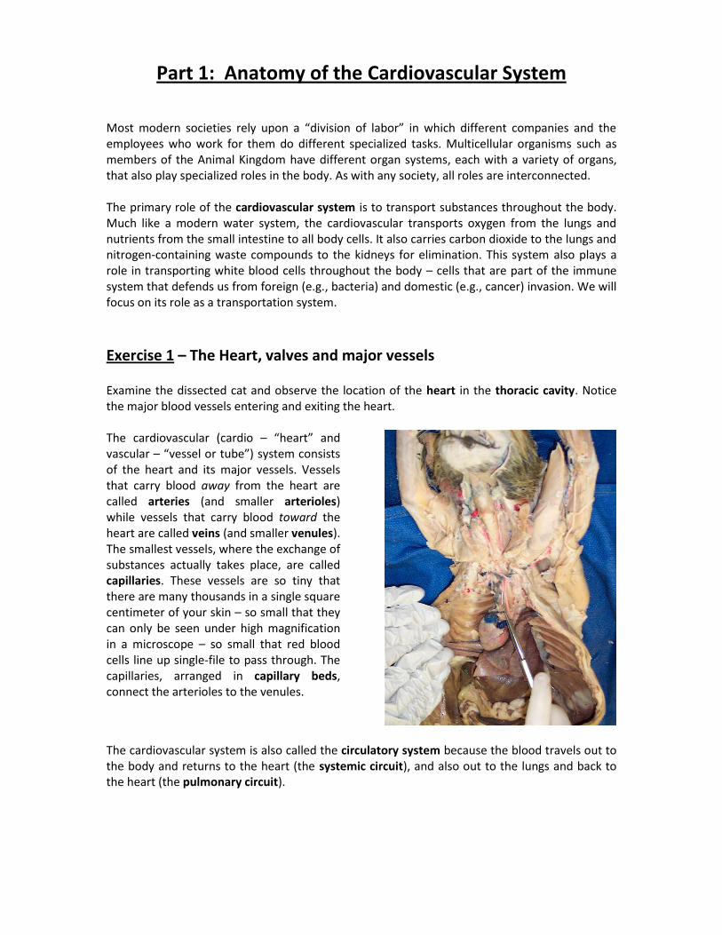
Part 1: Anatomy of the Cardiovascular System Most modern societies rely upon a “division of labor” in which different companies and the employees who work for them do different specialized tasks. Multicellular organisms such as members of the Animal Kingdom have different organ systems, each with a variety of organs, that also play specialized roles in the body. As with any society, all roles are interconnected. The primary role of the cardiovascular system is to transport substances throughout the body. Much like a modern water system, the cardiovascular transports oxygen from the lungs and nutrients from the small intestine to all body cells. It also carries carbon dioxide to the lungs and nitrogen-containing waste compounds to the kidneys for elimination. This system also plays a role in transporting white blood cells throughout the body – cells that are part of the immune system that defends us from foreign (e.g., bacteria) and domestic (e.g., cancer) invasion. We will focus on its role as a transportation system.
Exercise 1 – The Heart, valves and major vessels Examine the dissected cat and observe the location of the heart in the thoracic cavity. Notice the major blood vessels entering and exiting the heart. The cardiovascular (cardio – “heart” and vascular – “vessel or tube”) system consists of the heart and its major vessels. Vessels that carry blood away from the heart are called arteries (and smaller arterioles) while vessels that carry blood toward the heart are called veins (and smaller venules). The smallest vessels, where the exchange of substances actually takes place, are called capillaries. These vessels are so tiny that there are many thousands in a single square centimeter of your skin – so small that they can only be seen under high magnification in a microscope – so small that red blood cells line up single-file to pass through. The capillaries, arranged in capillary beds, connect the arterioles to the venules.
The cardiovascular system is also called the circulatory system because the blood travels out to the body and returns to the heart (the systemic circuit), and also out to the lungs and back to the heart (the pulmonary circuit).
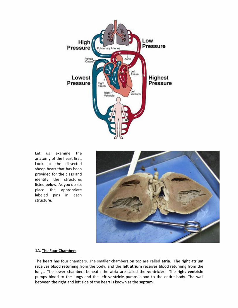
Let us examine the anatomy of the heart first. Look at the dissected sheep heart that has been provided for the class and identify the structures listed below. As you do so, place the appropriate labeled pins in each structure.
1A. The Four Chambers The heart has four chambers. The smaller chambers on top are called atria. The right atrium receives blood returning from the body, and the left atrium receives blood returning from the lungs. The lower chambers beneath the atria are called the ventricles. The right ventricle pumps blood to the lungs and the left ventricle pumps blood to the entire body. The wall between the right and left side of the heart is known as the septum.
Question: Which ventricle of the heart has to pump harder? The right side (to the lungs) or the left side (to the body)? Notice how much thicker the wall of the left ventricle is compared to the right ventricle. Where does the left ventricle send its blood?
1. Use the labeled pins to correctly identify the four chambers and the septum.
1B. The Valves The right and left atrioventricular (AV) valves separate the atria and the ventricles. They both have parachute-like doors called cusps that prevent blood from moving back into the atria when the ventricles contract, forcing the blood to the lungs and body. The right AV valve is commonly referred to as the tricuspid valve while the left AV valve is commonly called the bicuspid valve or mitral valve. (Why are they named tricuspid and bicuspid?) Also notice that the cusps have parachute-like strings attached to them called the chordae tendineae.
1. Use the correct labeled pins to identify the AV valves of the heart.
More difficult valves to locate are the semilunar valves located in the aorta and the pulmonary artery. The purpose of these valves is to prevent blood from moving in a backward direction when the ventricles of the heart relax. People who have defective valves from anatomical defects or the consequences of poor diet (as we shall see) must use extra energy to pump blood through the body. This is comparable to shoveling sand from the ground into a truck, only to have the sand slide back onto the ground.
1C. The Major Vessels of the Heart There are four major vessels of the heart. The vessels that carry blood from the body back to the heart are the:
superior and inferior vena cava – return blood from the body back to the heart
pulmonary veins – return blood from the lungs back to the heart
pulmonary arteries or pulmonary trunk – moving blood from the heart to the lungs
aorta – moving blood from the heart to all major arteries supplying the body
1. On your worksheet, label the chambers, valves and major vessels on the heart picture provided.
1D. Structure of Arteries and Veins Arteries and veins are the pipes through which the blood moves throughout the body. Blood leaves the heart via the aorta and then gradually moves through the major arteries of the body, which branch into smaller arteries, which branch into smaller arterioles and eventually into the capillary beds where the exchange of oxygen, carbon dioxide, nutrients and wastes occurs. There are thousands of capillaries in a single square centimeter of your skin. They are extremely tiny.
After passing through the capillary beds, blood enters the small venules of the body which eventually lead to veins, which then merge into larger veins, ultimately reaching the superior and inferior vena cava and on to the right atrium of the heart.
1. Get a prepared slide of a cross section of an artery and a vein. Use your microscope and focus in
on these vessels. You will observe that arteries and veins are located right next to each other.
2. Moving from the inside of the vessel toward the outside of the vessel, you will notice three different layers or “tunicas” of a blood vessel. The innermost layer is called the tunica interna. The middle layer is called the tunica media. This layer contains a type of muscle tissue called smooth muscle. If you look carefully, you will notice that this layer is thicker in the artery than in the vein. (Why do you think this layer is thicker in the arteries than in the veins? Hint: in which type of vessel is blood pressure higher?) The outermost layer of a blood vessel is called the tunica externa.
3. Draw a picture of a cross section of an artery and a vein on your worksheet.
One of the major causes of cardiovascular disease is arteriosclerosis (“artery “hardening”). This clogging of the arteries causes reduced blood flow to parts of the body, a condition called ischemia. When something causes a complete blockage of an artery or arteriole in the body it is called a thrombus (“clot”). As we will see later in this lab, the primary causes of arteriosclerosis are poor diet, lack of exercise and smoking.
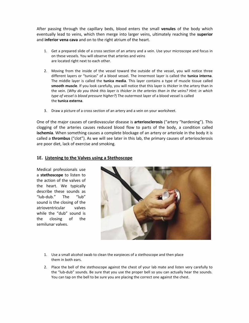
1E. Listening to the Valves using a Stethoscope Medical professionals use a stethoscope to listen to the action of the valves of the heart. We typically describe these sounds as “lub-dub.” The “lub” sound is the closing of the atrioventricular valves while the “dub” sound is the closing of the semilunar valves.
1. Use a small alcohol swab to clean the earpieces of a stethoscope and then place them in both ears.
2. Place the bell of the stethoscope against the chest of your lab mate and listen very carefully to the “lub-dub” sounds. Be sure that you use the proper bell so you can actually hear the sounds. You can tap on the bell to be sure you are placing the correct one against the chest.

1F. Cardiac Muscle Tissue There are three types of muscle tissue in the body: skeletal muscle, smooth muscle and cardiac muscle. These tissues are composed of cells that have the ability to contract and generate force. You will notice that skeletal muscle cells, which are found in muscles you use voluntarily (like your biceps muscle), are cylindrically shaped and have striations – little lines. In contrast, cardiac muscle cells are arranged in patches that look like a quilt.
1. Use your microscope and the prepared slides of skeletal muscle and cardiac muscle to observe
the different structures of these two tissues.
2. Draw a picture of these muscle tissues on your worksheet.
Cardiac muscle cells are commonly referred to as the myocardium (“muscle” “heart”). When an artery or arteriole supplying the heart itself with blood gets blocked, the result is a heart attack, or myocardial infarction (infarction means “cell death”). Heart attacks are the leading cause of death in the United States today, accounting for one-fourth of all deaths.
Part 2: Physiology of the Cardiovascular System
The heart works as a pump to push blood to the lungs and to the body via the pulmonary trunk and the aorta. If you are a blood cell, the pathway would be:
from body >> superior or inferior vena cava >> right atrium >> (right AV valve) >>
right ventricle >> pulmonary trunk >> lungs >> pulmonary veins >>
left atrium >> (left AV valve) >> left ventricle >> aorta >> to body
However, most people do not realize how the chambers of the heart actually pump:
Blood enters the atria of the heart first, filling up the atria and then the ventricles, like pouring water into a cup.
Then the atria contract from the top down, almost simultaneously, forcing the blood into the ventricles, causing them to expand and become completely filled.
Lastly, the ventricles of the heart contract from the bottom up, forcing the blood into the pulmonary trunk and aorta.
Exercise 2 – Measuring Pulse and Blood Pressure
2A. Measuring Pulse What causes the atria to start this cardiac cycle? In the wall of the right atrium is a special group of cells called pacemaker cells that get electrically excited at regular intervals. For most people
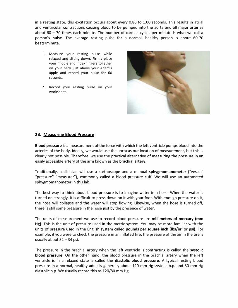
in a resting state, this excitation occurs about every 0.86 to 1.00 seconds. This results in atrial and ventricular contractions causing blood to be pumped into the aorta and all major arteries about 60 – 70 times each minute. The number of cardiac cycles per minute is what we call a person’s pulse. The average resting pulse for a normal, healthy person is about 60-70 beats/minute.
1. Measure your resting pulse while
relaxed and sitting down. Firmly place your middle and index fingers together on your neck just above your Adam’s apple and record your pulse for 60 seconds.
2. Record your resting pulse on your worksheet.
2B. Measuring Blood Pressure Blood pressure is a measurement of the force with which the left ventricle pumps blood into the arteries of the body. Ideally, we would use the aorta as our location of measurement, but this is clearly not possible. Therefore, we use the practical alternative of measuring the pressure in an easily accessible artery of the arm known as the brachial artery. Traditionally, a clinician will use a stethoscope and a manual sphygmomanometer (“vessel” “pressure” “measurer”), commonly called a blood pressure cuff. We will use an automated sphygmomanometer in this lab. The best way to think about blood pressure is to imagine water in a hose. When the water is turned on strongly, it is difficult to press down on it with your foot. With enough pressure on it, the hose will collapse and the water will stop flowing. Likewise, when the hose is turned off, there is still some pressure in the hose just by the presence of water. The units of measurement we use to record blood pressure are millimeters of mercury (mm Hg). This is the unit of pressure used in the metric system. You may be more familiar with the units of pressure used in the English system called pounds per square inch (lbs/in2 or psi). For example, if you were to check the pressure in an inflated tire, the pressure of the air in the tire is usually about 32 – 34 psi. The pressure in the brachial artery when the left ventricle is contracting is called the systolic blood pressure. On the other hand, the blood pressure in the brachial artery when the left ventricle is in a relaxed state is called the diastolic blood pressure. A typical resting blood pressure in a normal, healthy adult is generally about 120 mm Hg systolic b.p. and 80 mm Hg diastolic b.p. We usually record this as 120/80 mm Hg.
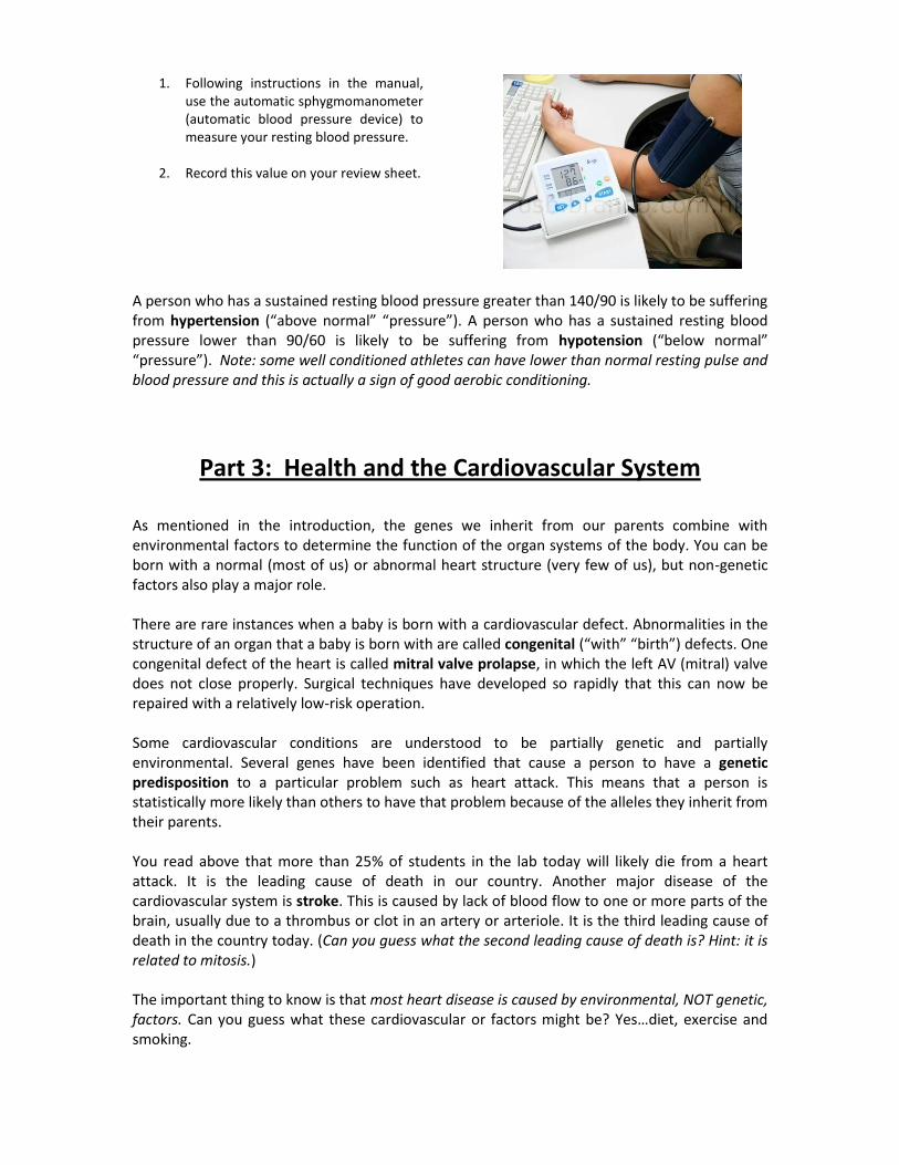
1. Following instructions in the manual, use the automatic sphygmomanometer (automatic blood pressure device) to measure your resting blood pressure.
2. Record this value on your review sheet.
A person who has a sustained resting blood pressure greater than 140/90 is likely to be suffering from hypertension (“above normal” “pressure”). A person who has a sustained resting blood pressure lower than 90/60 is likely to be suffering from hypotension (“below normal” “pressure”). Note: some well conditioned athletes can have lower than normal resting pulse and blood pressure and this is actually a sign of good aerobic conditioning.
Part 3: Health and the Cardiovascular System
As mentioned in the introduction, the genes we inherit from our parents combine with environmental factors to determine the function of the organ systems of the body. You can be born with a normal (most of us) or abnormal heart structure (very few of us), but non-genetic factors also play a major role. There are rare instances when a baby is born with a cardiovascular defect. Abnormalities in the structure of an organ that a baby is born with are called congenital (“with” “birth”) defects. One congenital defect of the heart is called mitral valve prolapse, in which the left AV (mitral) valve does not close properly. Surgical techniques have developed so rapidly that this can now be repaired with a relatively low-risk operation. Some cardiovascular conditions are understood to be partially genetic and partially environmental. Several genes have been identified that cause a person to have a genetic predisposition to a particular problem such as heart attack. This means that a person is statistically more likely than others to have that problem because of the alleles they inherit from their parents. You read above that more than 25% of students in the lab today will likely die from a heart attack. It is the leading cause of death in our country. Another major disease of the cardiovascular system is stroke. This is caused by lack of blood flow to one or more parts of the brain, usually due to a thrombus or clot in an artery or arteriole. It is the third leading cause of death in the country today. (Can you guess what the second leading cause of death is? Hint: it is related to mitosis.) The important thing to know is that most heart disease is caused by environmental, NOT genetic, factors. Can you guess what these cardiovascular or factors might be? Yes…diet, exercise and smoking.
As noted above, arteriosclerosis is the leading cause of cardiovascular disease. The major environmental factors in this disease process are also poor diet, lack of exercise and smoking. In addition, these same factors play the major role in the development of the metabolic disorder called Type II Diabetes or Adult Onset Diabetes. This is a disease in which the body cannot properly regulate the amount of glucose in the blood. Scientists project that one of every three people below the age of 30 in the United States will have a form of this disorder in the next 20 years. It has become a public health ticking time bomb. (Type I Diabetes, known as Juvenile Onset Diabetes, is a congenital condition.) Type II diabetes and obesity are increasing so rapidly that many scientists anticipate that, for the first the first time in our country’s history, younger generations will have a lower life expectancy than the generations before them.
The best way to reduce the likelihood of having a heart attack or stroke, or suffering
from Type II Diabetes is to eat a balanced diet, exercise moderately and do not smoke. How can you measure the health of your cardiovascular system today? Let us see.
Exercise 3 – Active Pulse and Recovery Rate 3A. Resting vs. Post-Activity Pulse Record all measurements on your review sheet. Read all of the directions below before starting the exercise.
1. Measure your resting pulse.
2. Go outside the lab and walk quickly up and down the stairs for a total of 5 cycles.
3. Immediately after walking the stairs, measure your post-exercise pulse.
4. Measure it again at one minute intervals up to four minutes post-exercise.
How many minutes did it take for your pulse to return to the resting pulse rate? The amount of time it takes for your pulse to return to normal after activity is called your recovery rate. The healthier your cardiovascular system is, the faster your recovery should be. Athletes such as runners and swimmers often use this measurement as an indication of aerobic fitness.
3B. Resting vs. Post-Activity Blood Pressure Record all measurements on your review sheet. Read all of the steps below before you start this activity.
1. Measure your resting blood pressure.
2. Go outside the lab and walk quickly up and down the stairs for a total of 5 cycles.
3. Immediately after walking the stairs, measure your post-exercise blood pressure.
4. Measure it again at one minute intervals up to four minutes post-exercise.
A person who is in an aerobically fit state should have their blood pressure return close to the resting state after the last measurement.
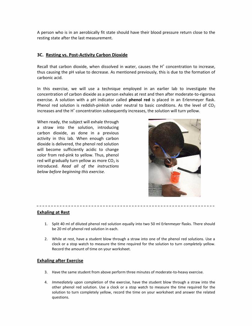
3C. Resting vs. Post-Activity Carbon Dioxide Recall that carbon dioxide, when dissolved in water, causes the H+ concentration to increase, thus causing the pH value to decrease. As mentioned previously, this is due to the formation of carbonic acid. In this exercise, we will use a technique employed in an earlier lab to investigate the concentration of carbon dioxide as a person exhales at rest and then after moderate-to-rigorous exercise. A solution with a pH indicator called phenol red is placed in an Erlenmeyer flask. Phenol red solution is reddish-pinkish under neutral to basic conditions. As the level of CO2 increases and the H+ concentration subsequently increases, the solution will turn yellow. When ready, the subject will exhale through a straw into the solution, introducing carbon dioxide, as done in a previous activity in this lab. When enough carbon dioxide is delivered, the phenol red solution will become sufficiently acidic to change color from red-pink to yellow. Thus, phenol red will gradually turn yellow as more CO2 is introduced. Read all of the instructions below before beginning this exercise.
Exhaling at Rest
1. Split 40 ml of diluted phenol red solution equally into two 50 ml Erlenmeyer flasks. There should be 20 ml of phenol red solution in each.
2. While at rest, have a student blow through a straw into one of the phenol red solutions. Use a
clock or a stop watch to measure the time required for the solution to turn completely yellow. Record the amount of time on your worksheet.
Exhaling after Exercise
3. Have the same student from above perform three minutes of moderate-to-heavy exercise.
4. Immediately upon completion of the exercise, have the student blow through a straw into the other phenol red solution. Use a clock or a stop watch to measure the time required for the solution to turn completely yellow, record the time on your worksheet and answer the related questions.
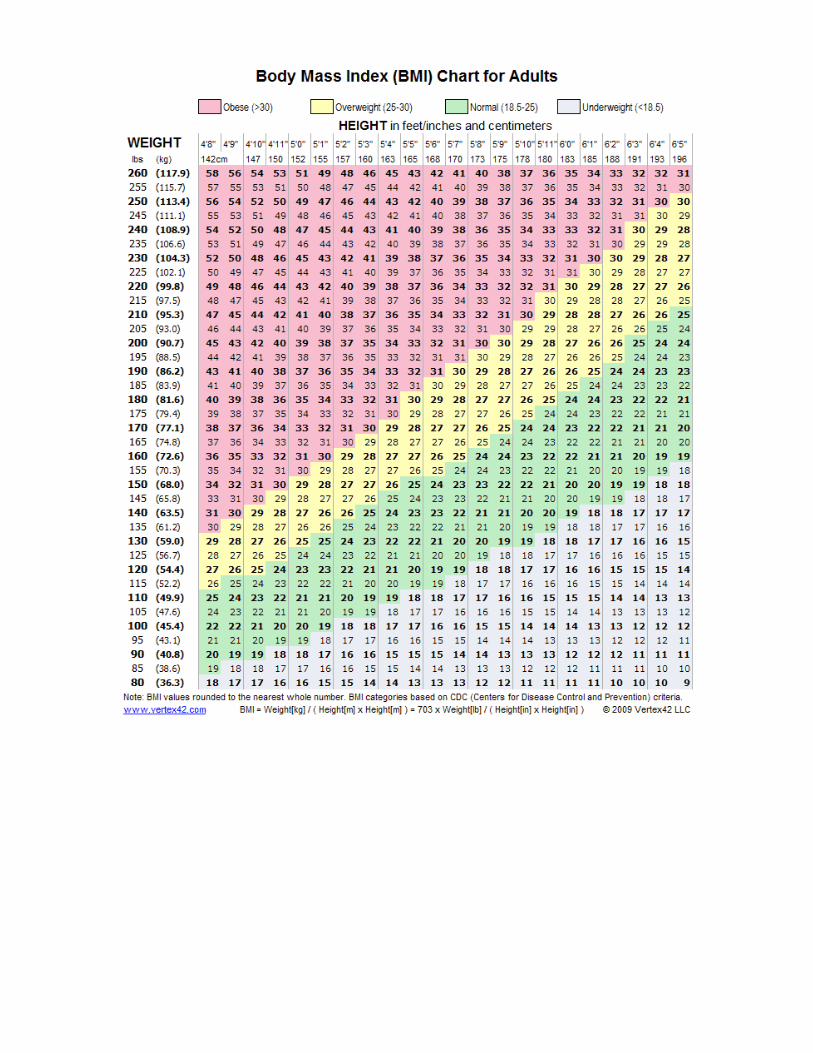
3D. Body-Mass Index (BMI) Although it is not perfect, many clinicians and dieticians like to use the Body-Mass Index (BMI) as an indicator of whether or not a person is at a healthy body weight. This index is a number that represents the ratio of a person’s weight measured in kilograms divided by height measured in meters (weight (kg) / height (m)). To determine your BMI, examine the chart provided. Notice the following categories:
BMI General Description
9 – 18 underweight
19- 25 normal
26 – 30 overweight
31 – 58 obese
NOTE: The BMI is an estimate of healthy body weight. People have different body compositions in terms of the amount of muscle and other factors. However, it is true that many people in our country have unhealthy body weights due to poor diet and lack of exercise.
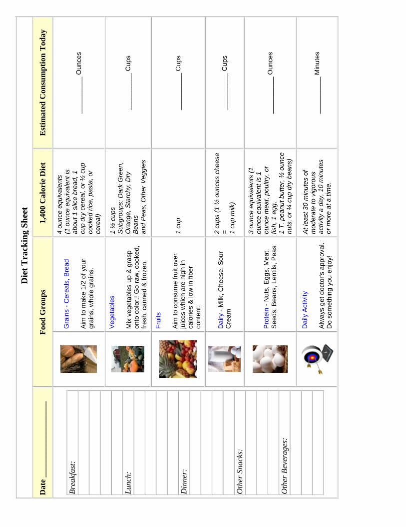
3D. Tracking Your Diet for 3 Days As part of this laboratory, your instructor may direct you to use the Diet Tracker Sheets to monitor your diet for 3 or more days. As they say, “You are what you eat.”
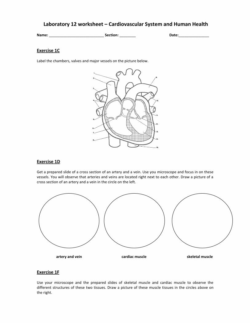
Laboratory 12 worksheet – Cardiovascular System and Human Health Name: ___________________________ Section: ________ Date:_______________
Exercise 1C Label the chambers, valves and major vessels on the picture below.
Exercise 1D Get a prepared slide of a cross section of an artery and a vein. Use you microscope and focus in on these vessels. You will observe that arteries and veins are located right next to each other. Draw a picture of a cross section of an artery and a vein in the circle on the left.
artery and vein cardiac muscle skeletal muscle
Exercise 1F Use your microscope and the prepared slides of skeletal muscle and cardiac muscle to observe the different structures of these two tissues. Draw a picture of these muscle tissues in the circles above on the right.
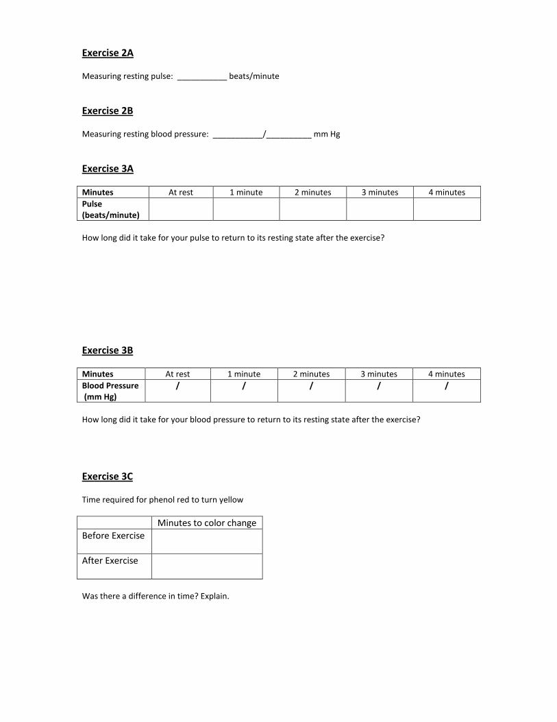
Exercise 2A Measuring resting pulse: ___________ beats/minute
Exercise 2B Measuring resting blood pressure: ___________/__________ mm Hg
Exercise 3A
Minutes At rest 1 minute 2 minutes 3 minutes 4 minutes
Pulse (beats/minute)
How long did it take for your pulse to return to its resting state after the exercise?
Exercise 3B
Minutes At rest 1 minute 2 minutes 3 minutes 4 minutes
Blood Pressure (mm Hg)
/ / / / /
How long did it take for your blood pressure to return to its resting state after the exercise?
Exercise 3C Time required for phenol red to turn yellow
Minutes to color change
Before Exercise
After Exercise
Was there a difference in time? Explain.
Die
t T
rack
ing
Sh
eet
Date
__________
_____
____
F
ood
Gro
up
s 1,4
00 C
alo
rie
Die
t E
stim
ate
d C
on
sum
pti
on
Tod
ay
Bre
akf
ast
:
Lun
ch:
Din
ner
:
Oth
er S
nack
s:
Oth
er B
ever
ages
:
Gra
ins -
Cere
als
, B
read
Aim
to m
ake 1
/2 o
f your
gra
ins,
who
le g
rain
s.
4 o
unce e
quiv
ale
nts
(1
ounce e
quiv
ale
nt
is
abou
t 1 s
lice b
read,
1
cup d
ry c
ere
al, o
r ½
cup
cooked r
ice,
pasta
, or
cere
al)
____
___
___
Ounces
Veg
eta
ble
s
Mix
vege
tab
les u
p &
gra
sp
onto
co
lor.
! G
o r
aw
, co
oked,
fresh, canned &
fro
ze
n.
1 ½
cu
ps
Sub
gro
ups:
Dark
Gre
en,
Ora
nge,
Sta
rchy, D
ry
Bea
ns
and P
eas, O
ther
Veg
gie
s
____
___
___
Cu
ps
Fru
its
Aim
to c
onsum
e f
ruit o
ver
juic
es w
hic
h a
re h
igh in
calo
ries &
lo
w in f
iber
conte
nt.
1 c
up
____
___
___
Cu
ps
Dair
y -
Milk
, C
heese,
So
ur
Cre
am
2 c
ups (
1 ½
ou
nces c
heese
=
1 c
up m
ilk)
____
___
___
Cu
ps
Pro
tein
- N
uts
, E
ggs, M
eat,
S
ee
ds, B
eans,
Lentils
, P
ea
s
3 o
unce e
quiv
ale
nts
(1
ounce e
qu
ivale
nt
is 1
ounce m
eat,
poultry
, or
fish, 1
eg
g,
1 T
. pe
anu
t bu
tter,
½ o
unce
nuts
, or
¼ c
up d
ry b
ea
ns)
____
___
___
Ounces
Daily
Activity
Alw
ays g
et d
octo
r's a
ppro
val.
Do s
om
eth
ing y
ou e
njo
y!
At le
ast 3
0 m
inute
s o
f m
odera
te t
o v
igoro
us
activity a
day, 1
0 m
inute
s
or
more
at a t
ime.
____
___
___
Min
ute
s

Die
t T
rack
ing
Sh
eet
Date
__________
_____
____
F
ood
Gro
up
s 1,4
00 C
alo
rie
Die
t E
stim
ate
d C
on
sum
pti
on
Tod
ay
Bre
akf
ast
:
Lun
ch:
Din
ner
:
Oth
er S
nack
s:
Oth
er B
ever
ages
:
Gra
ins -
Cere
als
, B
read
Aim
to m
ake 1
/2 o
f your
gra
ins,
who
le g
rain
s.
4 o
unce e
quiv
ale
nts
(1
ounce e
quiv
ale
nt
is
abou
t 1 s
lice b
read,
1
cup d
ry c
ere
al, o
r ½
cup
cooked r
ice,
pasta
, or
cere
al)
____
___
___
Ounces
Veg
eta
ble
s
Mix
vege
tab
les u
p &
gra
sp
onto
co
lor.
! G
o r
aw
, co
oked,
fresh, canned &
fro
ze
n.
1 ½
cu
ps
Sub
gro
ups:
Dark
Gre
en,
Ora
nge,
Sta
rchy, D
ry
Bea
ns
and P
eas, O
ther
Veg
gie
s
____
___
___
Cu
ps
Fru
its
Aim
to c
onsum
e f
ruit o
ver
juic
es w
hic
h a
re h
igh in
calo
ries &
lo
w in f
iber
conte
nt.
1 c
up
____
___
___
Cu
ps
Dair
y -
Milk
, C
heese,
So
ur
Cre
am
2 c
ups (
1 ½
ou
nces c
heese
=
1 c
up m
ilk)
____
___
___
Cu
ps
Pro
tein
- N
uts
, E
ggs, M
eat,
S
ee
ds, B
eans,
Lentils
, P
ea
s
3 o
unce e
quiv
ale
nts
(1
ounce e
qu
ivale
nt
is 1
ounce m
eat,
poultry
, or
fish, 1
eg
g,
1 T
. pe
anu
t bu
tter,
½ o
unce
nuts
, or
¼ c
up d
ry b
ea
ns)
____
___
___
Ounces
Daily
Activity
Alw
ays g
et d
octo
r's a
ppro
val.
Do s
om
eth
ing y
ou e
njo
y!
At le
ast 3
0 m
inute
s o
f m
odera
te t
o v
igoro
us
activity a
day, 1
0 m
inute
s
or
more
at a t
ime.
____
___
___
Min
ute
s