Embed Size (px)
DESCRIPTION
bio
Citation preview

GASTROINTESTINAL SYSTEM
DEPARTEMEN ANATOMI FK USU

GASTROINTESTINAL SYSTEM :ORAL CAVITY
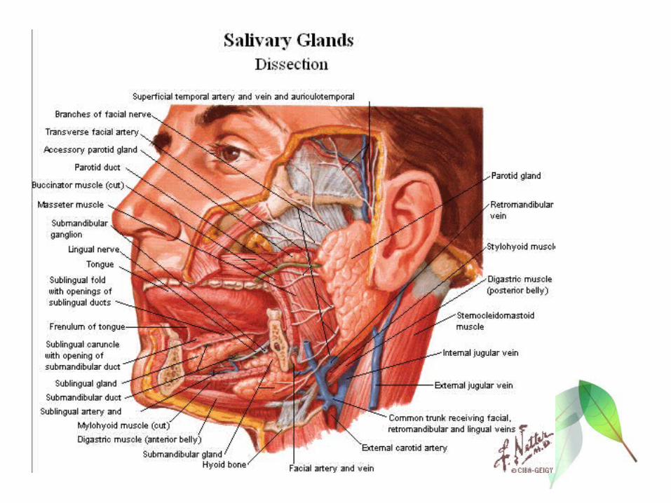
SALIVARY GLAND
OESOPHAGUS
PHARYNX
STOMACH
SMAL INTESTINE
LARGE INTESTINA
HEPAR
PANCREAS

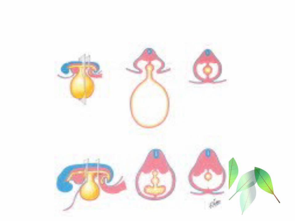
• 4 th Weeks: created somit • GIS is origin from endoderm layer → anterior
gut, mid gut & posterior gut• Anterior gut → mouth,oesophagus, stomach,
liver, pancreas• Mid gut → duodenum, small intestine, colon
transversum 2/3 proximal• Posterior gut → colon transversum 1/3 distal to
rectum

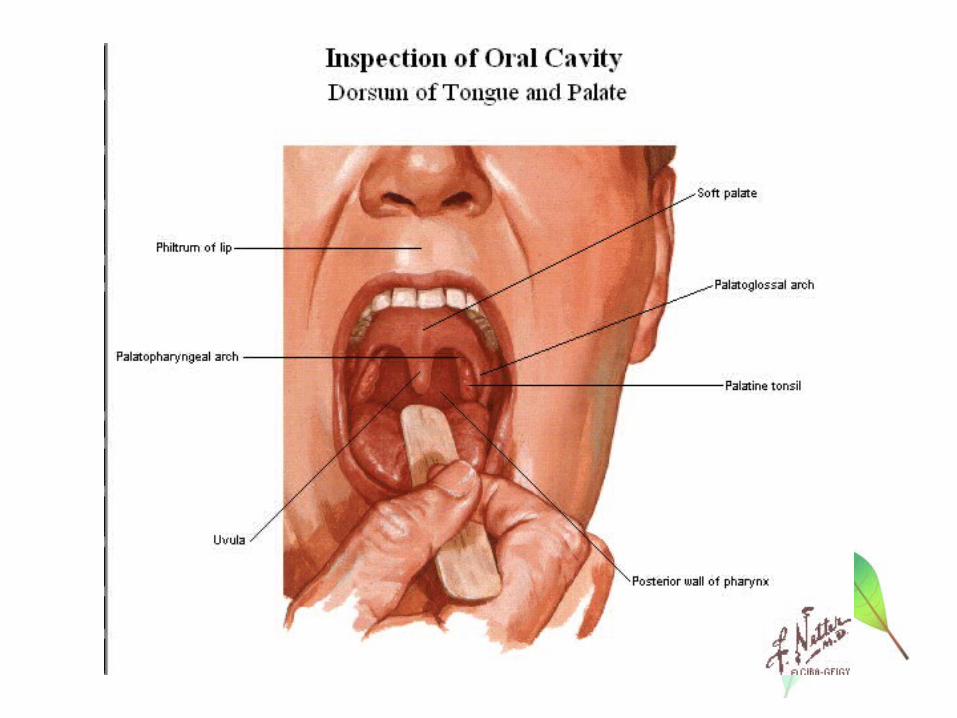
ORAL CAVITY ( CAVUM ORIS)
Oral cavity is the beginning of the digestive tube In infront it is bounded by the mucous membrane of the
lips Lateral : the cheeks Above : palate Below : tongue and mucous membrane

Roof of the mouth cavity is formed by the palate (palatum)
The substratum of the palate is bony infront (palatum durum)
In the posterior part it is muscular (palatum molle)

THE TEETH ( DENTES)The teeth appaear in two succesive series called dentition
1. First dentition : 20 milk teeth
2. Second dentition : 32 permanent teeth
PERMANENT TEETH:
Permanent teeth are present in the upper and lower jaws , eight on each side
- 2 incisor teeth
- 1 canine tooth
- 2 premolar teeth
- 3 molar teeth

Teeth are firmly implanted in the jaw and are surrounded by the gingival mucosa.
Each tooth consists of :• Dentine ( substantia eburnea)
forms the main mass and give the tooth its form
• Enamel (substantia adamantina)
covers the free part of the tooth superficially
• Cement (substantia ossea)
The parts hidden in the bone and gum


According to form of the tooth are distinguished :Crown (corona dentis)
thickest part projecting free into the mouth cavity
Neck (collum dentis)
Root (radix dentis)
longest portion of the tooth


Within the dentine lies cavity wich resembles the external form of the tooth :
- pulp cavity ( cavum dentis)
rich in blood vessels and nerve
- root canal (canalis radicis dentis)
- apex radicis dentis
- foramen apicis dentis

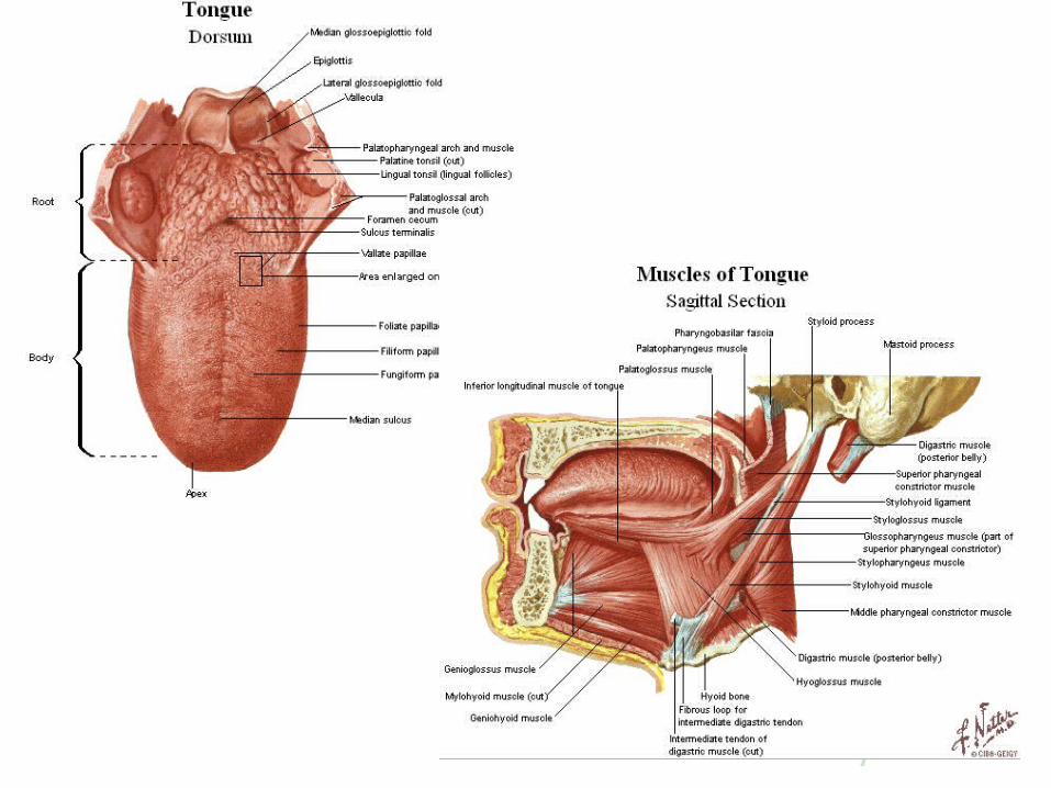
THE TONGUE ( LINGUA)The tongue is a flat, oblong body consisting mainly of muscle
and is fastened below to the floor of the mouth
Part of tongue :
1. Radix
2. Corpus
3. Apex

Mucous membran of the tongue is covered by papillae linguales :
Papillae filiformes Papillae fungiformes Papillae vallatae Papillae foliatae


OESOPHAGUS
D
A muscular tube, length averages 25 cm Begins as the continuation of the pharynx behind the
cartilage cricoidea Run downward through the spatium mediastinale
posterior passes through the hiatus oesophagus diapraghmatis iinto abdominal cavity
Consist : pars thoracalis and pars abdominalis


MUSCLES OF THE ABDOMEN• M. Obliquus externus abdominis• M.Pyramidalis• M.Obliquus internus abdominis• M.Transversus abdominis• M.Rectus abdominis
- vagina m.reectus abdominis
- inscriptiones tendineae
Linea alba : is a connective tissue strip wich extend in the median plane between the m. rectus abdominis from the proc.xyphoideus to the sympisis pubis

Linea semilunaris : paralel fiber, run transversely forward and go over into a tendon plate along a line wich is concave medianward
In the lower third of m.obliquus abdoinis internus, all the fiber run in front of m.rectus abdominis, the lower margin is curved and concave

LIGAMENTUM INGUINAL (POUPARTI)
Is a pewrful, flatly rounded tendon, wich extends from the spina iliaca anterior superior to the tuberculum pubicum , and can be palpated through the skin as a hard cord
CANALIS INGUINALIS
Is a cylindrical space filled up completely by the ductus deferens (male) / ligamentum teres uteri (female) with the accompaning vessels
the canal has two openings : - annulus inguinalis abdominalis
- annulus inguinalis subcutaneus

INNERVASION MOTORIK ABDOMINAL MUSCLES• M.Obliquus abdominis externus : - Nn.Intercostalis 5 – 12
- Nn.Ilio Hypogastricus
- Nn. Ilio Inguinalis
• M. Obliquus abdominis internus : - Nn.Intercostalis 5 – 12
- Nn.Ilio Hypogastricus
- Nn. Ilio Inguinalis
• M. Transversus abdominis : - Nn.Intercostalis 10 – 12
- Nn.Ilio Hypogastricus
- Nn. Ilio Inguinalis

INNERVASION SENSORIS SKIN AND ABDOMINAL WALL• Innervasion anterior , lateral and posterio- lateral
abdominal wall ramus cutaneous Nn.Intercostalis 7 – 11
• Innervasion postero – medial abdominal wallRamus posterior Nervus Spinalis Th 10 – 11
Vascularisation Abdominal Wall• Arteri-vena Epigastrica superior• Arteri – vena Epigastrica inferior• Arteri – vena Circumflexia Iliaca superficialis• Arteri – vena Circumflexia Iliaca profundus

PERITONEUM
PARIETALE
VISCERALE
The smooth glistening serouse membraneWhich covers the walls and the viscera of abdominal and pelvic cavities

Peritoneum Paritale
Part of peritoneum wich covers the walls cavities , the large vascular and the nerve thrunk attached to them as well as some parts of the urinary and sexual apparatus
Peritoneum Viscerale
The part of peritoneum wich is firmly attached to the surface of the freely movable portion of digestive tube , the pancreas, liver and spleen
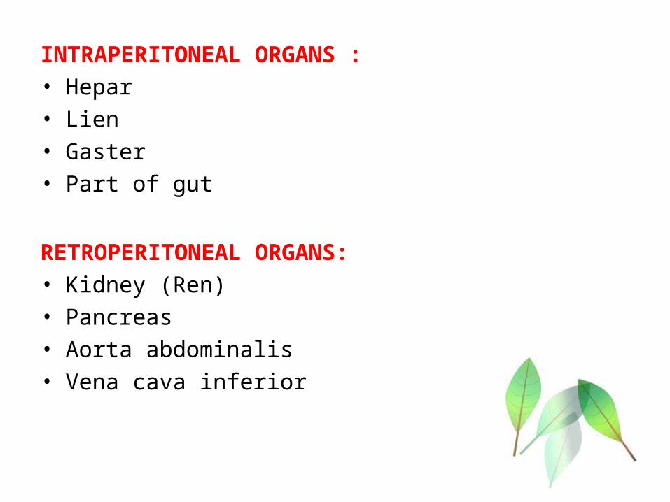
INTRAPERITONEAL ORGANS :• Hepar• Lien• Gaster• Part of gut
RETROPERITONEAL ORGANS:• Kidney (Ren)• Pancreas• Aorta abdominalis• Vena cava inferior

MESENTERIUM :
Large fold of peritoneum , arises from posterior wall of the abdomen at flexura duodenojejunalis . The strarting point is radix mesentery
OMENTUM MAJUS
Part of peritoneum viscerale wich is hanging downward from curvatura major of the stomach like an apron in front of the colon transversum and small intestine.
Contain large accumulations of fat

Omentum majus

BURSA OMENTALIS
A slit-like cavity surrounded on all sides by peritoneum, wich is connected with the general cavum peritonei at one spot only.
FORAMEN EPIPLOICUM (WINSLOWI)
Bounded infront by the lig.hepatodudenale, above by processus caudatus of the liver, behind by the v.cava inferior and below by flexura duodeni superior

REGION OF ANTERIOR ABDOMINAL WALL
1. Hypochondrium dekster
2. Epigastrium
3. Hypochondrium sinister
4. Lumbalis dekster
5. Umbilikalis
6. Lumbalis sinister
7. Iliaca dekster
8. Hypogastrium
9. Iliaca sinister

1 2 3
4 5 6
7 8 9

PROYEKSION OF INTRA ABDOMINAL ORGANS
Lien : on latero-posterior of costae 9 – 10 Hepar : on anterior right body wall, from right
costae 6th to the last arch costae Gaster (Stomach) : from epigastrium regio to
umbilikalis regio

STOMACH (VENTRICULUS , GASTER)
A saccular dilatation of the alimentary canal conecting above with oesophagus below with the duodenum
The form of stomach depends particulary on the volume of its contents and on the position of the body
when empty contracted
when filled fundus and corpus distend
Position : the main portion of the stomach lies on the left side of the body

PARTS OF STOMACH
Cardia,
Fundus ventriculi
Corpus ventriculi
Curvatura ventriculi major
Curvatura ventriculi minor
Pars pyloricum pylorus
Muscularis of the stomach:• Outer layer : longitudinal muscle fibres• Second layer : circular muscle fibers• Deepest layer : oblique muscle fibre with numerous fold
Plica mucosae

Position stomach :
At the upper end of the curvatura minor, oesophagus enter the wall of stomach by the spincter cardia
At the end ventriculi continued into pars pylorica, approximately cylindrical in shape separated from duodenum by a ring-formed constriction (spincter pylori)
The fundus is bounded directly above by the cupola diaphragm, to the left and behind by the fascies gastrica of the spleen


Vascularisation : • ARTERI GASTRICA SIN/DEX• ARTERI GASTROEPIPLOICA SIN/DEX• ARTERI GASTRICA BREVIS

INTESTINUM
SMALL INTESTINEINTESTINUM TENUE
LARGE IINTESTINEINTESTINUM CRASSUM

INTESTINUM TENUE
DUODENUM JEJENUM ILEUM
From orificium pilorycum to ileocaecal junction

DUODENUM The first portion of the small intestine which is directly and
firmly attached to the posterior abdominal wall It forms a spirally curved ring, open to the left and above In the concavity of which the pancreas is inserted Consist : - pars superior (shortest portion)
- pars descendens papilla vateri
- pars inferior It lies to the right and in front of the pars lumbalis of
diaphragm, of the v.portae, a.hepatica and ductus choledocus, behind and below the lobus quadratus of the liver
Bends around markedly to the right and forward to go over into the intestinum jejenum flexura duodenojejunalis

The flexuraduodenojejunalis is firmly attached to the diaphragm by the m.suspensorium duodeni (Lig. Treitz )
In the mucosa present a longitudinal fold (plica longitudinalis duodeni) and small projection upon which open the ductus choledocus and ductus pancreaticus ( papilla Vateri)

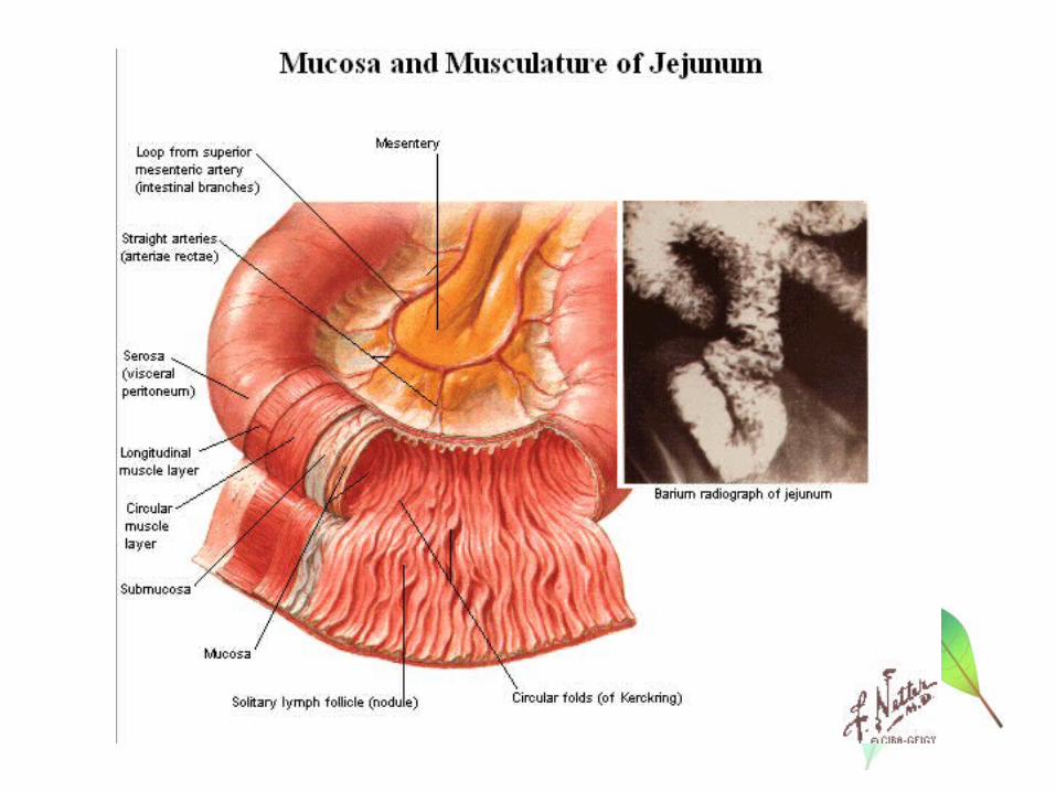

JEJENUM AND ILEUM
Infront and lateralward jejenum and ileum is covered by the great omentum (omentum majus)
The mucous membrane presents the special structur : Plica circularis (Kerckring) :
- the constant fold
- In jejenum , they are longer and higher than in ileum
Lymph node
- in jejenum, they are usually single (noduli lymphatici solitary )
- in ileum, they are numerous and higher and in part are crowded together , Peyer Patcher
(noduli lymphatici agregati)

LARGE INTESTINE ( INTESTINUM CRASSUM)
Following upon the small intestine, its begins as the intestinum caecum in the fossa iliaca dextra
Part of intestinum crassum :
Caecum
Appendix vermiformis
Colon ascendens
Colon transversum
Colon descendens
Colon sigmoideum
Rectum

The large intestine is caracterized, its surface is not smoothly cylindrical, but presents a nodular appearance due to the three rows of irregular, flask like projection haustra
These rows of haustra are separated from one another by three bands- like strips of the longitudinal muscle
taenia coli (taenia libera, taenia omentalis and taenia mesocolica)
Along the whole large intestine developed lobe-like fatty
appendices epipolica
The mucosa have a large sicke-shaped fold
plica semilunares

CAECUM
The blind-sac-like portion of the large intestine situated below the opening of the ileum into the large intestine
At the upper limit of the caecum , in the posterior part of the left wall lies the opening of the small intestine within the valvula coli.
Tere arise two high folds formed by the wall of the sall and large intestine : - labium superius of the valvula coli
- labium inferius of the valvula coli
In the formation of this the terminal portion of the small intestine is invaginated into the large intestine cavity

APPENDIX VERMIFORMIS Anarrow , generally cylindrical, hollow, blind ending of the
caecum. Length average 8,5 cm
COLON ASCENDENS• Begins at the frenula valvulae coli as the direct continuation
of caecum
COLON TRANSVERSUM
Runs out to the left and somewhat upward from the flexura coli dextra over the meedian plane in a curve convex forward and at the same time downward and goes over into the colon dscendens

COLON DESCENDENS
A descending limb of colon
COLON SIGMOIDEUM
Terminal portion of colon , hangs as a loop generally down into the cavity of small pelvis and goes over in front of the sacrum into the rectum
RECTUM
Terminal portion of the digestive tube which extends from the colon sigmoideum through the inferior wall of the small pelvis to the inferior opeing (anus)


LIVER• A large , reddish brown gland , almost 2 kg in weight, which
is situated for the most part to right of median plane in the regio hypochondrica dextra and regio epigastrica.
• A small part in regio hypochondrica sinistra• Have four lobus : - lobis hepatis dexter
- lobus hepatis sinister
- lobus quadratus
- lobus caudatus• Ductus hepaticus : the excretory duct of the liver , begins in
the porta hepatis by the union of the right and a left branch• Porta hepatis consist : - v porta
- a. hepatica
- ductus choledocus


LIVER VASCULARITATION
Aorta Abdominalis Arteri Coeliaca
Arteri Hepatica Prorii Porta Hepatis A. HEPATICA LOBUS DEXTER ( Lobus
Caudatus & Quadratus ) ARTERI HEPATICA LOBUS SINISTER ARTERI CYSTICA ( Vesica Fellae )

VESICA FELLEA (GALL BLADER)
Is an oblong , pear-shaped sac , which is fastened by loose connective tissue in the fossa vesica fellea of the liver
The ductus cysticus leaves the blader and then join with ductus hepaticus


PANCREAS• Lies tranversely in front of the posterior wall of the
abdominal cavity• It presents for examination : caput, corpus and cauda• The caput pancreatis fills up the concavity of the pars
descendens and pars inferior duodenum• The anterior and inferior surfaces are covered by
peritoneum and the posterior surface is free from it• The ductus of pancreas ( ductus pancreaticus wirsungi and
ductus pancreaticus santorini) is opening in the duodenum

PANCREAS
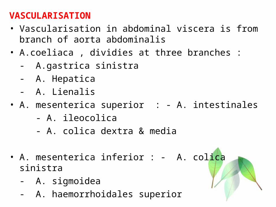
VASCULARISATION• Vascularisation in abdominal viscera is from branch of aorta
abdominalis• A.coeliaca , dividies at three branches :
- A.gastrica sinistra
- A. Hepatica
- A. Lienalis• A. mesenterica superior : - A. intestinales
- A. ileocolica
- A. colica dextra & media
• A. mesenterica inferior : - A. colica sinistra
- A. sigmoidea
- A. haemorrhoidales superior

• A. Iliaca communis• A.Hypogastrica
Vena
Vena portae : collects the blood from the digestive tube