Embed Size (px)
Citation preview

King’s Research Portal
DOI:10.1002/hep.28265
Document VersionPublisher's PDF, also known as Version of record
Link to publication record in King's Research Portal
Citation for published version (APA):Abu-Hayyeh, S., Ovadia, C., Lieu, T., Jensen, D. D., Chambers, J., Dixon, P. H., ... Williamson, C. (2015).Prognostic and mechanistic potential of progesterone sulfates in intrahepatic cholestasis of pregnancy andpruritus gravidarum. Hepatology. 10.1002/hep.28265
Citing this paperPlease note that where the full-text provided on King's Research Portal is the Author Accepted Manuscript or Post-Print version this maydiffer from the final Published version. If citing, it is advised that you check and use the publisher's definitive version for pagination,volume/issue, and date of publication details. And where the final published version is provided on the Research Portal, if citing you areagain advised to check the publisher's website for any subsequent corrections.
General rightsCopyright and moral rights for the publications made accessible in the Research Portal are retained by the authors and/or other copyrightowners and it is a condition of accessing publications that users recognize and abide by the legal requirements associated with these rights.
•Users may download and print one copy of any publication from the Research Portal for the purpose of private study or research.•You may not further distribute the material or use it for any profit-making activity or commercial gain•You may freely distribute the URL identifying the publication in the Research Portal
Take down policyIf you believe that this document breaches copyright please contact [email protected] providing details, and we will remove access tothe work immediately and investigate your claim.
Download date: 18. Feb. 2017

Prognostic and Mechanistic Potential of ProgesteroneSulfates in Intrahepatic Cholestasis of Pregnancy and
Pruritus GravidarumShadi Abu-Hayyeh,1* Caroline Ovadia,1* TinaMarie Lieu,2 Dane D. Jensen,2 Jenny Chambers,1,3
Peter H. Dixon,1,3 Anita L€ovgren-Sandblom,4 Ruth Bolier,5 Dagmar Tolenaars,5 Andreas E. Kremer,5,6
Argyro Syngelaki,7 Muna Noori,3 David Williams,8 Jose J.G. Marin,9 Maria J. Monte,9 Kypros H. Nicolaides,7
Ulrich Beuers,5 Ronald Oude-Elferink,5 Paul T. Seed,1 Lucy Chappell,1 Hanns-Ulrich Marschall,10
Nigel W. Bunnett,2,11 and Catherine Williamson1,3
A challenge in obstetrics is to distinguish pathological symptoms from those associatedwith normal changes of pregnancy, typified by the need to differentiate whether gesta-tional pruritus of the skin is an early symptom of intrahepatic cholestasis of pregnancy(ICP) or due to benign pruritus gravidarum. ICP is characterized by raised serum bileacids and complicated by spontaneous preterm labor and stillbirth. A biomarker forICP would be invaluable for early diagnosis and treatment and to enable its differentia-tion from other maternal diseases. Three progesterone sulfate compounds, whose con-centrations have not previously been studied, were newly synthesized and assayed in theserum of three groups of ICP patients and found to be significantly higher in ICP at 9-15 weeks of gestation and prior to symptom onset (group 1 cases/samples: ICP n 5 35/80, uncomplicated pregnancy 5 29/100), demonstrating that all three progesterone sul-fates are prognostic for ICP. Concentrations of progesterone sulfates were associatedwith itch severity and, in combination with autotaxin, distinguished pregnant womenwith itch that would subsequently develop ICP from pruritus gravidarum (group 2: ICPn 5 41, pruritus gravidarum n 5 14). In a third group of first-trimester samples allprogesterone sulfates were significantly elevated in serum from low-risk asymptomaticwomen who subsequently developed ICP (ICP/uncomplicated pregnancy n 5 54/51).Finally, we show mechanistically that progesterone sulfates mediate itch by evoking aTgr5-dependent scratch response in mice. Conclusion: Our discovery that sulfated pro-gesterone metabolites are a prognostic indicator for ICP will help predict onset of ICPand distinguish it from benign pruritus gravidarum, enabling targeted obstetric care toa high-risk population. Delineation of a progesterone sulfate-TGR5 pruritus axis identi-fies a therapeutic target for itch management in ICP. (HEPATOLOGY 2015; 00:000–000)
Amajor challenge for obstetricians is to distin-
guish serious disorders associated with increasedmaternal and fetal mortality from low-risk gesta-
tional changes. Currently, the presenting symptoms ofmany obstetric syndromes are nonspecific with few earlybiomarkers of serious maternal disease. We aimed toaddress this problem for intrahepatic cholestasis of preg-nancy (ICP), the commonest liver-specific disorder ofpregnancy.1 ICP is complicated by spontaneous preterm
labor, fetal distress, and intrauterine death.1,2 Early rec-ognition of ICP is important to enable prompt treat-ment and appropriate pregnancy surveillance. Thepresenting symptom of ICP is pruritus (skin), and diag-nosis is confirmed by demonstration of raised totalserum bile acids. However, maternal pruritus withouthepatic impairment or dermatological disorder (i.e.,pruritus gravidarum [PG]) affects up to 25% of preg-nant women,3,4 while ICP is much less common. It has
Abbreviations: cAMP, cyclic adenosine monophosphate; CAMYEL, cAMP sensor using YFP-Epac-RLuc; cDNA, complementary DNA; CI, confidence interval;FXR, farnesoid X receptor; ICP, intrahepatic cholestasis of pregnancy; KO, knockout; MeOH, methanol; OR, odds ratio; PG, pruritus gravidarum; PM2DiS, 5a-pregnan-3a,-20a-diol-3,20-disulfate; PM3DiS, 5b-pregnan-3a,-20a-diol-3,20-disulfate; PM3S, 5b-pregnan-3a,-20a-diol-3-sulfate; UDCA, ursodeoxycholicacid; WT, wild type
1

a variable geographic prevalence: In the United King-dom ICP affects 0.7% of pregnant women but is twiceas common in women of Indian or Pakistani origin,5
while in Chile it affects up to 4% of pregnant women.6
The etiology of gestational pruritus (both benign andin ICP) is not established. Several endogenous com-pounds have been proposed as biochemical mediators ofpruritus in ICP, including lysophosphatidic acid, a neu-ronal activator that can act as a pruritogen, the forma-tion of which is catalyzed by the enzyme autotaxin.7,8
These molecules are raised in the serum of women withICP after disease onset.8 The secondary bile acids deoxy-cholic acid and lithocholic acid can activate the Gprotein-coupled receptor TGR5 on sensory nerves tostimulate release of itch-selective neuropeptides in thespinal cord and evoke a Tgr5-dependent itch responsein mice.9 These results indicate that bile acids mayinduce pruritus but require further evaluation in ICP assecondary bile acids are not typically raised in the condi-tion and concentrations of total maternal serum bileacids do not correlate with pruritus severity.10 Althoughstudies of urine samples from ICP cases implicate pro-gesterone sulfates as pruritogens,11 the precise structuresof the compounds and their capacity to cause pruritusremain to be determined.
Sulfated progesterone metabolites contribute to theetiology of ICP; they are partial agonists of the bile acidreceptor farnesoid X receptor (FXR)12 and competitivelyinhibit hepatic bile acid uptake13 and efflux,14 resultingin cholestasis and hypercholanemia.12 Serum concentra-tions of progesterone sulfates are elevated in women
with ICP at 35-41 weeks of gestation,12,15 typically afterdiagnosis. We hypothesized that progesterone sulfatesare raised in early pregnancy prior to the onset of ICPand thus are potential early biomarkers that can distin-guish ICP from benign PG. We also hypothesized thatthey signal through TGR5 to mediate pruritus.
This study used three groups of ICP cases and preg-nant controls to establish whether progesterone sulfatesare biomarker candidates for ICP diagnosis prior tobiochemical derangement and to evaluate their roleand potential mechanism of action as pruritogensusing in vitro and in vivo approaches. Our resultsreveal a key role for the progesterone sulfate-TGR5axis in ICP.
Materials and Methods
Study Approval. This study conformed to the1975 Declaration of Helsinki guidelines; permissionwas obtained from the ethics committees of Hammer-smith Hospitals NHS Trust, London (97/5197 and 08/H0707/21), and King’s College Hospitals NHS Trust,London (03WH06). Written informed consent wasreceived from participants prior to inclusion in thestudy. Murine studies were approved by the MonashUniversity Animal Ethics Committee.
Human Serum Samples. Serial blood sampleswere collected from three prospectively recruited groupsof women with ICP, PG, or controls with uncompli-cated pregnancies at intervals dependent upon gestationand patient attendance. Sample preparation was as
From the 1Women’s Health Academic Centre, King’s College London, London, United Kingdom; 2Monash Institute of Pharmaceutical Sciences and AustralianResearch Council Centre of Excellence in Convergent Bio-Nano Science and Technology, Monash University, Parkville, Victoria, Australia; 3Institute of Reproduc-tive and Developmental Biology, Imperial College London, London, United Kingdom; 4Department of Clinical Chemistry, Karolinska University Hospital Hud-dinge, Stockholm, Sweden; 5Tytgat Institute for Liver and Intestinal Research, Academic Medical Centre, Amsterdam, The Netherlands; 6Department of Medicine1, Friedrich-Alexander-University of Erlangen-Nuremberg, Erlangen, Germany; 7Harris Birthright Research Centre for Fetal Medicine, King’s College Hospital,London, United Kingdom; 8Institute for Women’s Health, University College London Hospitals, London, United Kingdom; 9Laboratory of Experimental Hepatologyand Drug Targeting (HEVEFARM), Biomedical Research Institute of Salamanca (IBSAL), University of Salamanca, National Institute for the Study of Liver andGastrointestinal Diseases (CIBERehd), Salamanca, Spain; 10Institute of Medicine, Department of Molecular and Clinical Medicine, University of Gothenburg,Gothenburg, Sweden; 11Department of Pharmacology, University of Melbourne, Parkville, Victoria, Australia
Received July 15, 2015; accepted September 28, 2015.Additional Supporting Information may be found at onlinelibrary.wiley.com/doi/10.1002/hep.28265/suppinfo.*These authors contributed equally to this work.Supported by the Wellcome Trust (grant P30874); the National Institute of Health Research Biomedical Research Centre at Guy’s and St Thomas NHS Founda-
tion Trust and King’s College London, National Health and Medical Research Council (grants 63303, 1049682, 1031886); the Australian Research Council; andMonash University. The views expressed are those of the author(s) and not necessarily those of the NHS, NIHR or the Department of Health.
Address reprint requests to: Catherine Williamson, M.D., F.R.C.P., Maternal and Fetal Disease Group, Hodgkin Building, Guy’s Campus, London, SE1 1UL,UK. Email: [email protected]; tel: 144(0)2078486350.
Copyright VC 2015 The Authors. HEPATOLOGY published by Wiley Periodicals, Inc., on behalf of the American Association for the Study of Liver Diseases. This is anopen access article under the terms of the Creative Commons Attribution License, which permits use, distribution and reproduction in any medium, provided theoriginal work is properly cited.
View this article online at wileyonlinelibrary.com.DOI 10.1002/hep.28265Potential conflict of interest: Dr. Bunnett received grants from Takeda.
2 ABU-HAYYEH, OVADIA, ET AL. HEPATOLOGY, Month 2015

described.16 Three separate patient groups were used, toensure that results could be replicated.
Group 1 comprised 64 women: 35 opportunisticallyrecruited “high-risk” ICP cases with a history of choles-tasis in a previous pregnancy and 29 with uncompli-cated pregnancies. Women with ICP commencedursodeoxycholic acid (UDCA) treatment per personaland practitioner preference following diagnosis (sevenwere untreated, six were treated with UDCA, and 22were recruited untreated and subsequently UDCA-treated). Group 2 (the pruritus group) comprised 55women with skin pruritus in pregnancy, 41 of whomhad pregnancies complicated by ICP (23 with previousICP) and 14 of whom had normal pregnancies (ninewith previous ICP); of the women who subsequentlydeveloped ICP, 14 provided serum samples prior to theonset of hypercholanemia (raised bile acids) but afterthe onset of pruritus. Women in groups 1 and 2 wererecruited while undergoing antenatal care at the tertiaryhospitals of Imperial College London or through theICP Support charity. Cases were recruited between 2007and 2014 and selected to include all cases where longitu-dinal samples were available; 20% of ICP cases were ter-tiary referrals (of this group 71% were referred fromspecialists in different UK regions and 29% werereferred from the UK charity ICP Support).
To evaluate whether progesterone sulfate concentra-tions reduce after delivery, we identified postnatal serumsamples from a subgroup of 12 ICP cases (due to thelimited number of postnatal samples collected) andcompared the concentration of progesterone sulfateswith the third-trimester serum sample.
Group 3 comprised 105 asymptomatic women at 11-14 weeks’ gestation, 54 of whom later developed ICPand 51 of whom subsequently had normal pregnancies.Women were recruited at aneuploidy screening at King’sCollege Hospital, serum samples were taken, and clini-cal follow-up by a research midwife identified womenwho developed ICP; the next sample taken from awoman with a normal pregnancy was then used as acontrol (serum analyses were incomplete due to techni-cal error resulting in exclusion of three women with nor-mal pregnancies).
All cases of ICP were confirmed by demonstration ofserum bile acids �10 lmol/L, and some cases also hadraised liver transaminases in association with pruritusand no additional identifiable cause for their liver dys-function. Exclusion criteria were other causes of hepaticdysfunction, including preeclampsia; hemolysis, elevatedliver enzymes, and low platelets (HELLP) syndrome;acute fatty liver of pregnancy; primary biliary cirrhosis;active viral hepatitis; any ultrasound abnormality that
may result in biliary obstruction; and multifetalpregnancy.
Biophysical Profiling of Participants. Details ofthe participants’ relevant previous medical history, fam-ily history, ethnicity, results of investigations, pregnancy,and delivery were taken throughout their attendance.Birth weight centile was calculated according to gesta-tional age and weight at delivery17 using GROW soft-ware (http://www.gestation.net/cc/about.htm). At thetime of serum sampling, patients with pruritus used ahorizontal visual analogue score18 (0-100 mm) to quan-tify in millimeters the worst itch symptoms experiencedover the previous 24 hours. The marked point wasmeasured, and the distance in millimeters from 0 (noitch) was converted to an itch score from 0 to 100. Theitch analogue score quantified severity of pruritus butdid not specify the physical location and extent of theitch.
Prior to analysis, participants were grouped accordingto retrospective assessment of their diagnosis of ICP atany point during the pregnancy.
Serum Bile Acid and Progesterone Sulfate Analysisby High-Performance Liquid Chromatography-Tandem Mass Spectrometry. Internal standards (100ng of d4-glycholic acid, d4-glycochenodeoxycholic acid,d4-glycodeoxycholic acid, d4-glyco-UDCA, d4-glycoli-thocholic acid, d4-UDCA, d4-lithocholic acid (all fromQmx Laboratories, Essex, UK), d5-cholic acid (TorontoResearch Chemicals, Toronto, Canada), and d4-taurocholic acid (TLC PharmaChem, Vaughan, Can-ada), dissolved in 40 lL methanol [MeOH]) wereadded to 100 lL of serum and vortexed. Acetonitrile(800 lL) was added to precipitate proteins. After vortex-ing and centrifugation, the supernatant was dried in astream of nitrogen and then first taken up in 125 lLMeOH, followed by 125 lL of an aqueous solutioncontaining 40% MeOH, 0.02% formic acid, and 10mmol/L ammonium acetate. Before injection 75 lL ofthe sample was transferred to new vials and 80 lL of thefollowing mix was added: three parts of MeOH and onepart of an aqueous solution containing 40% MeOH,0.02% formic acid, and 10 mmol/L ammonium acetate.
Ten microliters of this mixture was analyzed on ahigh-performance liquid chromatography Alliance 2695system coupled to a Xevo TQ mass spectrometer(Waters, Manchester, UK) using a SunFire C18 (4.6 3
100 mm, 3.5 lm) column (Waters) and gradient elutionwith 0.01% formic acid and 5 mmol/L ammonium ace-tate in water along with 0.01% formic acid 1 5 mmol/L ammonium acetate in MeOH as the mobile phase.Cone voltage was 60 V and collision energy 18 eV forunconjugated bile acids, 60 V and 29-43 eV for glycine
HEPATOLOGY, Vol. 00, No. 00, 2015 ABU-HAYYEH, OVADIA, ET AL. 3
conjugates, and 88 V and 56-65 eV for taurine conju-gates, respectively. Analytes were detected using selectedion monitoring and quantified by internal standardmethods. The desolvation temperature was 6508C, andthe source temperature was 1508C. Selected reactionmonitoring was used with dwell times of 100 ms. Analy-tes were quantified using deuterized internal standardsexcept for progesterone sulfates for which d4-glyco-UDCA was used. Results were calculated as response(area analyte/areainternal std). Retention times and responsecurves of bile acids listed (Supporting Table S1) wereevaluated from reference compounds obtained fromSigma; 5b-pregnan-3b-ol,20-one,3-sulfate (pregnan-diol-3-sulfate), 5a-pregnan-3a-ol,20-one,3-sulfate (allo-pregnandiol-3-sulfate), and 5a-pregnan-3b-ol,20-one,3-sulfate (epiallopregnandiol-3-sulfate) were obtainedfrom Steraloids, USA; 5b-pregnan-3a,20a-diol-3-sulfate, 5b-pregnan-3a,20a-diol-disulfate, and 5a-pregnan-3a,20a-diol-disulfate were from Sai Advan-tium, India. 5a-Pregnan-3b,20a-diol-disulfate was ten-tatively identified as the remaining isomer from itsretention times and mass spectrum. 5b-Pregnan-3a,20a-diol-disulfate and 5a-pregnan-3a,20a-diol-disulfate coeluted at all of the conditions tested. Usingthis system, we observed less than 10% intra-assay vari-ability when rerunning the same sample. These assayswere performed in the Department of Molecular andClinical Medicine, University of Gothenburg, Gothen-burg, Sweden.
Measurement of Serum Autotaxin Activity.Autotaxin activity was measured as described.7 Serumwas incubated with 1 mmol/L lysophosphatidylcholine14:0, 500 mmol/L NaCl, 5 mmol/L MgCl2, 100mmol/L Tris (pH 9.0), and 0.05% Triton X-100 for 60minutes at 37oC. Liberated choline was detected usingcholine oxidase (2 U/mL), horseradish peroxidase (1.6U/mL), and homovanillic acid, with emitted fluores-cence recorded using a NOVOstar analyzer. Using thissystem, we observed less than 10% inter-assay andintra-assay variance. The autotaxin assay was per-formed at the Academic Medical Centre, Amsterdam,The Netherlands.
Cyclic Adenosine Monophosphate BioluminescenceResonance Energy Transfer CAMYEL Assay. Thebioluminescence resonance energy transfer CAMYEL(cAMP sensor using YFP-Epac-RLuc) cyclic adenosinemonophosphate (cAMP) sensor permits quantificationof intracellular cAMP concentrations with high sensitiv-ity and a broad dynamic range19 and has been used pre-viously to measure TGR5 signaling in cells.20 HEK293cells that stably express the TGR5 receptor were gener-ated using the FLP-In system (Invitrogen). The charac-
terization of these cells has been described.21 HEK293and HEK-HA-TGR5 cells (4 3 106 per 10-cm plate)were transfected with 4 lg of complementary DNA(cDNA) encoding the CAMYEL sensor. Cells weretransfected using polyethylenimine with a 6:1 polyethy-lenimine:cDNA ratio in 500 lL of 0.15 M NaCl. Thepolyethylenimine:cDNA mixture was added to the cellsin the 10-cm plate, and cells were incubated overnightin 5% CO2 at 378C in Dulbecco’s modified Eagle’smedium supplemented with 10% fetal bovine serum.Cells were washed in phosphate-buffered saline andincubated in 1 mL of versene for 10 minutes. Cells weresuspended in Dulbecco’s modified Eagle’s medium and10% fetal bovine serum, plated onto poly-D-lysine-treated 96-well plates, and incubated overnight in 5%CO2 at 378C. To test cAMP production, cells werewashed in prewarmed Hank’s balanced salt solution andthen incubated in 80 lL Hank’s balanced salt solutionfor 30 minutes at 378C. Coelenterazine (NanoLightTechnology, Pinetop, AZ; 10 lL of 10 lmol/L inHank’s balanced salt solution) was added, and cells wereincubated in the dark for 10 minutes at 378C. Lumines-cence for RLuc8 (480 nm) and YFP (530 nm) wasmeasured using a microplate reader (PHERAstarOmega; BMG Labtech, Mornington, Australia). A 2-minute baseline was established before addition of theagonists. cAMP production was measured for 10minutes following addition of the agonists, forskolin(10 lmol/L), or vehicle. Baseline and vehicle controlvalues were subtracted, and the bioluminescence reso-nance energy transfer signal was normalized as a per-centage of the forskolin response. This assay wasperformed at Monash University, Parkville, Australia.
Scratching Behavior. Scratching behavior wasstudied in mice (C57BL/6 (wild-type [WT]), Tgr5knockout [KO], male and female, 6-10 weeks) asdescribed.9 The fur at the base of the neck was shaved,and mice were placed in individual cylinders on a glassshelf. Mice were acclimatized to the experimental room,restraint apparatus, and investigator for 2-hour periodson 2 successive days before experiments. After acclimati-zation, 20 lL of 100 lmol/L 5b-pregnan-3a-20a-diol-sulfate (PM3S) or vehicle (1% dimethyl sulfoxide) wasinjected intradermally at the nape of the neck (vehicleWT n 5 4, PM3S WT n 5 5, PM3S Tgr5-KO n 5 4).Hind limb scratching to the injection site was video-recorded for 120 minutes. Two observers unaware oftest agents or genotypes quantified scratching behavior.One scratch was defined as lifting the hind limb to theinjection site and then placing the paw on the floor,regardless of the number of strokes. If counts differed bymore than three scratches over a 30-minute period, both
4 ABU-HAYYEH, OVADIA, ET AL. HEPATOLOGY, Month 2015

observers reevaluated the record. Results are expressed asscratching events during 60 minutes of observation.
Transactivation Assays. Huh7 cells seeded into96-well plates were transfected with 10.4 ng plasmidcircular DNA (pcDNA)-retinoid X receptor, 10.4 ngpcDNA-FXRa2/pcDNA3.1 together with 10.4 ng and40 ng pGL3-IBAP-Luc and pcDNA3.1-green fluores-cent protein using Fugene 6 transfection reagent (Prom-ega) at a 3:1 ratio. Twenty-four hours later, cells werewashed and treated with 0 or 50 lM compound 6 0.5lM GW4064 (Sigma-Aldrich). After 24 hours, greenfluorescent protein activity was measured (internal con-trol for normalization) followed by the addition ofSteadylite plus (PerkinElmer) to determine luciferaseactivity, both of which were measured in a PheraStar FS(BMG) plate reader. Transfection experiments were per-formed three times, and the results are shown as meanvalues of triplicates and standard deviations.
Statistics. For group 1, log transformations of datawere undertaken and results are presented as ratios ofthe geometric mean values between groups and overtime. Results were corrected for multiple measures andmultiple markers being analyzed. Interval regression wasused for each assay.
Trend tests were performed by analyzing the random-effects interval regression on the logged concentrations,with interactions between patient groups and lineareffects of time.
For group 2, patient demographic group results andvisual analogue itch scores were compared using theMann-Whitney U test, and serial serum concentrationsof progesterone metabolites using unpaired Student ttest (Prism 6; Graphpad Software Inc.). Progesteronemetabolite concentrations were log-transformed priorto analysis due to nonnormally distributed data. Lon-gitudinal comparisons between disease groups of pruri-tus scores with biochemical markers were performedusing Stata software (version 11; StataCorp, CollegeStation, TX). Confounding based on multiple meas-ures and gestational effects was accounted for, and sub-sequent linear and logistic regression analyses wereperformed.
For the HEK-HA-TGR5 cAMP assays and murinescratching assays, results are expressed as mean 6 stand-ard error of the mean. Data were compared statisticallyusing Graphpad Prism 6 for multiple groups analysis ofvariance and Tukey-Kramer post hoc test. P < 0.05 wasconsidered significant.
Table 1. Clinical and Demographic Characteristics of ICP, PG and Control Cases in Two Groups Used to Evaluate SulfatedProgesterone Metabolites as Biomarkers
Characteristic
Group 1 Group 2
ICP (n 5 35) Control (n 5 29) P ICP (n 5 41) PG (n 5 14) P
Age (years, 6 SD) 33.4 6 4.6 30.8 6 4.8 0.02 32.7 6 4.3 35.6 6 4.3 0.01
Ethnic group, number (%)
White 22 (63) 27 (93) 0.02 25 (61) 11 (79) NS
Black 4 (11) 0 NS 4 (10) 0 NS
Asian 8 (23) 1 (3) NS 9 (22) 1 (7) NS
Other 1 (3) 1 (3) NS 3 (7) 2 (14) NS
Previous pregnancies �24 weeks, number (%)
0 0 26 (90) <0.01 11 (27) 2 (14) NS
1 20 (57) 1 (3) <0.01 18 (44) 9 (64) NS
�2 15 (43) 1 (3) <0.01 11 (27) 2 (14) NS
Unknown 0 1 (3) NS 1 (2) 1 (7) NS
Gestational age at diagnosis (weeks 6 SD) 2911 6 613 n/a 3010 6 712 n/a
Severity of ICP, number (%)
Total bile acids 5 10-39.9 lmol/L 13 (37) n/a 16 (39) n/a
Total bile acids �40 lmol/L 22 (63) n/a 25 (61) n/a
Mean serum ALT, IU/L 6 SD 88.3 6 131.2 n/a 145.3 6 197.2 29.4 6 30.6
Onset of labor, number (%)
Spontaneous 5 (14) 16 (55) <0.01 7 (17) 4 (29) NS
Induced 14 (40) 8 (28) NS 24 (59) 3 (21) 0.02
Prelabor cesarean section 13 (37) 2 (7) 0.02 8 (20) 6 (43) NS
Unknown 3 (9) 3 (10) NS 2 (5) 1 (7) NS
Gestational age at delivery (weeks 6 SD) 3710 6 114 3916 6 112 <0.01 3710 6 113 3812 6 012 <0.01
Preterm delivery <37/40, number (%) 12 (34) 0 <0.01 14 (34) 2 (14) NS
Birth weight, kg 6 SD 3.1 6 0.4 3.5 6 0.4 <0.01 3.1 6 0.4 3.1 6 0.6 NS
Birth weight centile, number 6 SD 67 6 28 49 6 32 0.01 72 6 25 47 6 35 0.01
Mean serum ALT values based on levels detected in the first sample obtained from each ICP case. P value shown where a comparison resulted in statistical sig-
nificance. Values are given as means, unless otherwise stated.
Abbreviations: ALT, alanine transaminase; n/a, not applicable; NS, not significant.
HEPATOLOGY, Vol. 00, No. 00, 2015 ABU-HAYYEH, OVADIA, ET AL. 5

Results
Progesterone Sulfates Are Prognostic Indicators ofICP. To establish whether progesterone sulfates canpredict women at risk of ICP in early pregnancy beforesymptom onset, a group of ICP cases and uncompli-cated pregnancy controls was used to establish gesta-tional profiles of three sulfated progesterone metabolites(group 1; Table 1). We obtained the following progester-one sulfate standards, which were previously implicatedin ICP based on analysis of gas chromatograhic/massspectrometric spectra22,23 and all of which were synthe-sized de novo: 5a-pregnan-3a,-20a-diol-3,20-disulfate(PM2DiS), 5b-pregnan-3a,-20a-diol-3-sulfate (PM3S),and 5b-pregnan-3a,-20a-diol-3,20-disulfate (PM3DiS)(Supporting Fig. S1).
A comparison of geometric means across all gesta-tional weeks for PM2DiS, PM3S, and PM3DiS
revealed, respectively, 4.5-fold, 2.0-fold, and 12.2-foldsignificant increases in serum concentrations inuntreated ICP cases compared to pregnant controls (P< 0.001) (Fig. 1). Importantly, concentrations ofPM2DiS, PM3S, and PM3DiS were supraphysiologi-cally raised compared to normal pregnant controls by10.6-fold, 1.7-fold, and 24.3-fold, respectively, at weeks9-15 (P < 0.05), when 91% of these participants wereasymptomatic, indicating their potential as predictivebiomarkers for ICP. Concentrations of PM3S in ICPsteadily increased at a constant rate from 9 to 41 weeks,whereas concentrations of PM3DiS and PM2DiSincreased steeply from 24 to 41 weeks for the ICP groupcompared to controls (P < 0.05) (Fig. 1).
UDCA treatment improves maternal pruritus andbiochemical derangements in ICP. UDCA signifi-cantly reduced PM2DiS and PM3DiS concentrationsrelative to untreated ICP women throughout the lasttrimester of pregnancy (P < 0.05). A trend analysisshowed a significant change in the trend of PM3DiSwith UDCA treatment in the third trimester of ICPcompared to the untreated ICP group (P < 0.05),becoming similar to the pregnant control group trend(Fig. 1).
Fig. 1. Gestational serum profiles of PM2DiS, PM3S, and PM3DiSin group 1. Panels A, B, and C show the mean concentrations ofPM2DiS, PM3S, and PM3DiS, respectively, for serum samples obtainedat different gestational time points from women with uncomplicatedpregnancies (control, closed squares), untreated ICP (closed circles),and UDCA-treated ICP (closed triangles). Error bars represent 6standard error of the mean. P values for gestational week categorycomparison of untreated ICP versus controls were determined by Stu-dent t test.
Table 2. Maternal Concentrations of Progesterone Sulfatesin the Last Serum Sample Prior to Parturition and in
Subsequent Postnatal Serum Samples in ICP
Case
Gestational
Day/Postnatal Day
PM3S
(lmol/L)
PM3DiS
(lmol/L)
PM2DiS
(lmol/L)
1 GD 266 8.03 0.62 1.48
PN16 2.24 0.09 0.49
2 GD 241 2.75 1.28 1.27
PN166 0.00 0.00 0.00
3 GD 256 12.44 1.88 3.89
PN134 0.03 0.01 0.00
4 GD 255 7.22 2.19 3.30
PN112 0.04 0.03 0.09
5 GD 232 21.07 6.71 3.31
PN121 0.00 0.00 0.00
6 GD 269 2.28 0.52 1.25
PN140 0.00 0.00 0.00
7 GD 237 47.36 11.81 1.81
PN11 19.99 9.45 0.94
PN142 0.00 0.00 0.00
8 GD 261 9.85 4.76 5.94
PN140 0.02 0.00 0.00
9 GD 248 11.10 14.41 8.97
PN142 0.02 0.00 0.00
10 GD 252 20.04 5.31 4.90
PN113 3.74 3.44 1.72
11 GD 268 23.72 3.88 4.28
PN156 0.06 0.00 0.07
12 GD 255 15.64 5.60 6.04
PN11 9.29 5.15 6.00
Abbreviations: GD, gestational day; PN, postnatal.
6 ABU-HAYYEH, OVADIA, ET AL. HEPATOLOGY, Month 2015

Progesterone Sulfate Concentrations RapidlyResolve in ICP Serum Following Parturition. Toestablish whether progesterone sulfate concentrationspersist following parturition in ICP, concentrations ofprogesterone metabolites in the last sample in ICPcases prior to parturition and postnatal samples col-lected thereafter were assayed in a subgroup ofpatients. Concentrations of PM2DiS, PM3S, andPM3DiS decreased rapidly following birth and nor-malized to almost undetectable levels as early as 12days postpartum (Table 2).
Progesterone Sulfates Are Associated With Severityof Itch in ICP and Can Predict Its SubsequentOnset. To assess the involvement of progesterone sul-fates in pruritus, we investigated the relationshipbetween pruritus severity and serum concentrations ofprogesterone metabolites in women with pregnancy-associated pruritus (Table 1). Serum PM2DiS, PM3S,and PM3DiS concentrations all differentiated womenwith ICP from PG (P < 0.05) (Table 3). Serum concen-trations of PM3S (odds ratio [OR] 5 6.1, 95% confi-dence interval [CI] 0.6-11.5, P < 0.05) and autotaxinactivity (OR 5 1.4, 95% CI 0.3-2.4, P < 0.05) weresignificantly associated with itch severity in ICP.
To determine whether progesterone sulfates couldpredict subsequent ICP, logistic regression was per-formed on PM2DiS, PM3S, and PM3DiS concentra-tions and autotaxin activity, using the first serum samplefrom women at presentation with pruritus and normalserum biochemistry (Table 4). PM2DiS and PM3DiSdifferentiated between the women who would subse-quently develop ICP (P < 0.05, OR 5 2.8, 95% CI1.5-5.2, and OR 5 2.5, 95% CI 1.2-5.4, respectively).
To refine a prediction algorithm, we evaluatedwhether a combination of markers could more reliablypredict disease. PM2DiS, PM3DiS, and autotaxin incombination resulted in an improved area under thereceiver operating characteristic (ROC) curve of 0.91(95% CI 0.80-1.00) in contrast to autotaxin (0.73, 95%CI 0.52-0.94), PM2DiS (0.72, 95% CI 0.52-0.92), or
PM3DiS (0.74, 95% CI 0.55-0.94) alone (Fig. 2A).Plotting this combination as a predictive score for thefirst serum sample from women presenting with PGenabled clear differentiation between those who wouldsubsequently develop ICP and those who continue tohave benign PG (Fig. 2B).
Progesterone Sulfates Are Supraphysiologically Raisedin Early Gestation in Low-Risk ICP Cases. We eval-uated this predictive algorithm in a third group ofasymptomatic pregnant women who gave serum sam-ples at 11-14 gestational weeks for a study of serum bio-markers to predict adverse pregnancy outcome (Table5). Fifty-four women from this group developed ICP inlater pregnancy, and their progesterone sulfate concen-trations were compared to those of 51 women withuncomplicated pregnancies. PM2DiS, PM3S, andPM3DiS concentrations were significantly raised inwomen with subsequent ICP (Table 5). Autotaxin didnot predict ICP at this early gestation (area under thecurve 5 0.55, 95% CI 0.43-0.66), while PM3DiS andPM2DiS in combination showed some predictive ability(area under the curve 5 0.68, 95% CI 0.58-0.78) (Sup-porting Fig. S2).
Progesterone Sulfates Signal Through TGR5 toMediate Itch. Activation of the G protein-coupledreceptor Tgr5 elicits an itch response in mice.9 Wetherefore hypothesized that progesterone metabolitesassociated with itch in ICP can activate TGR5 in vitro.HEK cells stably transfected with TGR5 or empty vec-tor control cells were transfected with the CAMYEL
Table 4. Autotaxin, PM2DiS, and PM3DiS All Have the Abilityto Predict ICP When Measured at the Time of Onset of
Gestational Pruritus
ICP Marker OR of Future ICP (95% CI) P Area Under ROC Curve
PM3S 1.70 (0.97-3.01) 0.07 0.45 (0.23-0.68)
PM3DiS 2.77 (1.48-5.19) <0.01 0.74 (0.55-0.94)
PM2DiS 2.54 (1.18-5.44) 0.02 0.72 (0.52-0.92)
Autotaxin 2.22 (0.99-4.86) 0.07 0.73 (0.52-0.94)
Abbreviation: ROC, receiver operating characteristic.
Table 3. Associations Between Biochemical Markers and Pruritus Scores and Their Ability to Differentiate ICP From PG
Biomarker
Association With Pruritus
ICP PG Ability to Identify ICP
Change in VAS (95% CI) P Change in VAS (95% CI) P OR (95% CI) P
PM3S 6.1 (0.6-11.5) 0.03 0.9 (24.6–6.3) NS 1.7 (1.1-2.4) 0.01
PM3DiS 2.2 (21.6–6) NS 22.5 (26.2–1.2) NS 2.1 (1.4-3.4) <0.01
PM2DiS 20.3 (25.3–4.6) NS 28.0 (214.9–1.2) 0.03 1.7 (1.2-2.5) 0.01
Autotaxin 1.4 (0.3-2.4) 0.01 2.0 (20.1–4.1) NS 2.3 (2.1-2.6) <0.01
Linear regression results showing the effect of doubling biochemical markers and change in visual analogue score for ICP and PG and ORs for developing ICP. P
value shown where a comparison resulted in statistical significance.
Abbreviations: NS, not significant; VAS, visual analogue score.
HEPATOLOGY, Vol. 00, No. 00, 2015 ABU-HAYYEH, OVADIA, ET AL. 7

Table 5. Maternal Characteristics of Pregnancies Assessed in a Group of Low-Risk Women Taken in the First Trimester ofPregnancy and Their Pregnancy Outcomes, With Levels of Serum Sulfated Progesterone Metabolites in These
First-Trimester Samples
Characteristic/Marker
Group 3
ICP (n 5 54) Control (n 5 51) P
Age, years 6 SD 32 6 5.4 31 6 5.2 NS
Ethnic group, number (%)
White 42 (78) 35 (69) NS
Black 6 (11) 11 (22) NS
Asian 5 (9) 3 (6) NS
Other 1 (2) 2 (4) NS
Previous pregnancies �24 weeks, number (%)
0 28 (52) 26 (51) NS
1 22 (41) 15 (29) NS
�2 4 (7) 10 (20) NS
Onset of labor, number (%)
Spontaneous 12 (22) 46 (90) <0.01
Induced 34 (63) 3 (6) <0.01
Prelabor cesarean section 8 (15) 2 (4) NS
Gestational age at delivery, weeks 6 SD 3813 6 111 4011 6 111 <0.01
Preterm delivery <37/40, number (%) 2 (4) 0 NS
Birth weight, kg 6 SD 3.3 6 0.5 3.4 6 0.3 NS
Birth weight centile, number 6 SD 60 6 30 42 6 23 <0.01
Stillbirth, number (%) 0 0
Progesterone metabolite, lmol/L mean 6 SEM
PM3S 1.4 6 0.1 1.1 6 0.1 0.02
PM3DiS 0.3 6 0 0.2 6 0 <0.01
PM2DiS 2.8 6 0.3 1.9 6 0.2 <0.01
Total bile acids, lmol/L mean 6 SEM 3.9 6 0.4 4 6 0.5 NS
Autotaxin activity, nmol/ml/min 6 SEM 12.9 6 1.2 11.6 6 1.1 NS
P value shown where a comparison resulted in statistical significance. Values given as means, unless otherwise stated.
Abbreviations: NS, not significant; SD, standard deviation; SEM, standard error of mean.
Fig. 2. Progesterone sulfates and autotaxin can predict subsequent onset of ICP in pregnant women with pruritus. The receiver operatingcurves (A) improved toward an optimal area under the curve of 1.0 when biomarkers were evaluated in combination: PM2DiS 1 PM3DiS (com-plete line), autotaxin (dashed line), and PM2DiS 1 PM3DiS 1 autotaxin (dotted and dashed line). (B) A combined predictive score (PM2DiS1 PM3DiS 1 autotaxin) of greater than 0.25 for individual samples plotted against the gestational day of sampling reliably predicted all ICPcases. Women who developed ICP (n 5 14, closed circles) and PG (n 5 14, open triangles) were reliably distinguished by this score; dashedline represents demarcation between the two groups. Abbreviation: AUC, area under the curve.
8 ABU-HAYYEH, OVADIA, ET AL. HEPATOLOGY, Month 2015
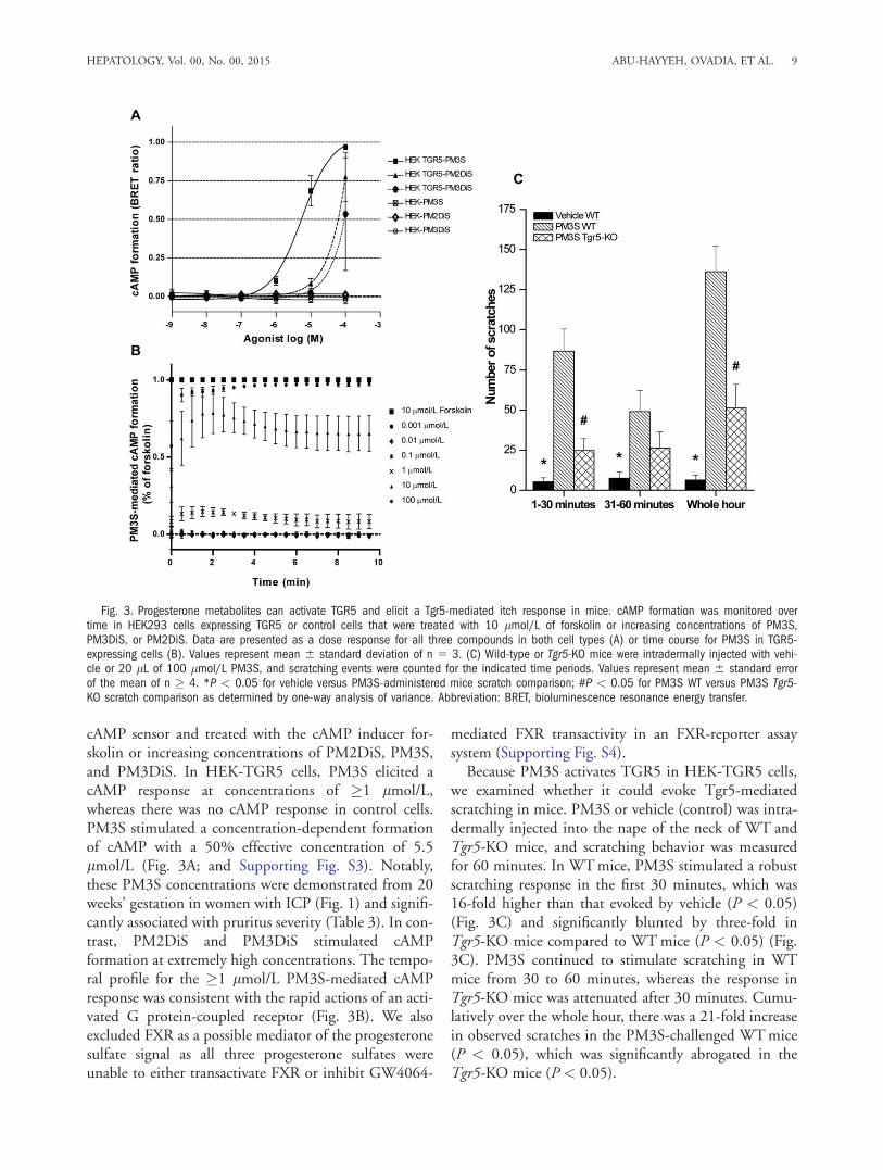
cAMP sensor and treated with the cAMP inducer for-skolin or increasing concentrations of PM2DiS, PM3S,and PM3DiS. In HEK-TGR5 cells, PM3S elicited acAMP response at concentrations of �1 lmol/L,whereas there was no cAMP response in control cells.PM3S stimulated a concentration-dependent formationof cAMP with a 50% effective concentration of 5.5lmol/L (Fig. 3A; and Supporting Fig. S3). Notably,these PM3S concentrations were demonstrated from 20weeks’ gestation in women with ICP (Fig. 1) and signifi-cantly associated with pruritus severity (Table 3). In con-trast, PM2DiS and PM3DiS stimulated cAMPformation at extremely high concentrations. The tempo-ral profile for the �1 lmol/L PM3S-mediated cAMPresponse was consistent with the rapid actions of an acti-vated G protein-coupled receptor (Fig. 3B). We alsoexcluded FXR as a possible mediator of the progesteronesulfate signal as all three progesterone sulfates wereunable to either transactivate FXR or inhibit GW4064-
mediated FXR transactivity in an FXR-reporter assaysystem (Supporting Fig. S4).
Because PM3S activates TGR5 in HEK-TGR5 cells,we examined whether it could evoke Tgr5-mediatedscratching in mice. PM3S or vehicle (control) was intra-dermally injected into the nape of the neck of WT andTgr5-KO mice, and scratching behavior was measuredfor 60 minutes. In WT mice, PM3S stimulated a robustscratching response in the first 30 minutes, which was16-fold higher than that evoked by vehicle (P < 0.05)(Fig. 3C) and significantly blunted by three-fold inTgr5-KO mice compared to WT mice (P < 0.05) (Fig.3C). PM3S continued to stimulate scratching in WTmice from 30 to 60 minutes, whereas the response inTgr5-KO mice was attenuated after 30 minutes. Cumu-latively over the whole hour, there was a 21-fold increasein observed scratches in the PM3S-challenged WT mice(P < 0.05), which was significantly abrogated in theTgr5-KO mice (P < 0.05).
Fig. 3. Progesterone metabolites can activate TGR5 and elicit a Tgr5-mediated itch response in mice. cAMP formation was monitored overtime in HEK293 cells expressing TGR5 or control cells that were treated with 10 lmol/L of forskolin or increasing concentrations of PM3S,PM3DiS, or PM2DiS. Data are presented as a dose response for all three compounds in both cell types (A) or time course for PM3S in TGR5-expressing cells (B). Values represent mean 6 standard deviation of n 5 3. (C) Wild-type or Tgr5-KO mice were intradermally injected with vehi-cle or 20 lL of 100 lmol/L PM3S, and scratching events were counted for the indicated time periods. Values represent mean 6 standard errorof the mean of n � 4. *P < 0.05 for vehicle versus PM3S-administered mice scratch comparison; #P < 0.05 for PM3S WT versus PM3S Tgr5-KO scratch comparison as determined by one-way analysis of variance. Abbreviation: BRET, bioluminescence resonance energy transfer.
HEPATOLOGY, Vol. 00, No. 00, 2015 ABU-HAYYEH, OVADIA, ET AL. 9

Discussion
Our results show that the sulfated progesteronemetabolites PM2DiS, PM3S, and PM3DiS are prognos-tic for ICP as their concentrations are elevated duringearly gestation when patients are asymptomatic. Fur-thermore, UDCA treatment reduces the ICP-associatedelevation of disulfated progesterone metabolites. Inter-estingly, concentrations of progesterone sulfates decreaserapidly following birth, consistent with clinical reportsof rapid resolution of pruritus in ICP.24 PM3S concen-trations were associated with the pruritus of ICP, whileall three progesterone sulfates were able to differentiatebetween women with pruritus in pregnancy secondaryto ICP and those with benign PG. CombiningPM2DiS, PM3DiS, and autotaxin activity enabled pre-diction of women who would subsequently develop ICPwhen they first started itching in pregnancy, prior to ele-vation in bile acids. Furthermore, concentrations ofPM3S consistent with ICP were capable of mediatingcAMP release in a TGR5-dependent manner andresulted in a scratch response that was reduced in Tgr5-KO mice.
This study has shown that PM3S is a likely pruritogenin ICP as concentrations consistent with ICP can activateTGR5 and mediate a Tgr5-dependent itch. Although thisresult is based on a mouse model, Keitel et al. have alsoshown that progesterone sulfates can modulate the activityof TGR5 in other human tissues.25 We demonstratedthat autotaxin and progesterone sulfates are associatedwith pruritus in ICP and PG, and it is likely thatautotaxin-mediated elevations in lysophosphatidic acidcause itch through a distinct mechanism from that of pro-gesterone sulfate-induced pruritus.
The demonstration that PM2DiS, PM3S, andPM3DiS are significantly raised in maternal serum priorto disease onset indicates that women with ICP arelikely to have an underlying abnormality in phase 2metabolism (conjugation) of progesterone or phase 3(biliary excretion) of progesterone sulfates.23 As the pro-gesterone sulfates that are supraphysiologically raised inICP are agonists of the bile acid receptor TGR5, it ispossible that they impact additional downstream gesta-tional metabolic pathways mediated by this receptor.26
These results have the potential to provide insights intostrategies to treat other cholestatic disorders complicatedby itch, e.g., primary biliary sclerosis, primary sclerosingcholangitis, and drug-induced liver injury. They arelikely to also have a global impact as ICP is commonerin women of South Asian and South American origin.5,6
At present there are no biomarkers for ICP in clinicaluse. The potential use of the predictive score to establish
whether pregnant women with pruritus will developICP is enticing and should be evaluated in future pro-spective, well-powered studies. This is important as thepatient groups in the current study were all managed ina single specialist center and this may have introducedpopulation bias. If the results are confirmed in differentpopulations, a feasible extension to this study would beto assay concentrations of urinary progesterone sulfates(Glantz et al.11) to identify a predictive score that can beused in early pregnancy to establish whether a womanwith pruritus will develop this high-risk disease. Thiscould have wider clinical application with the develop-ment of high-throughput urinary assays for progesteronesulfates or similar laboratory tests for serum levels ofprogesterone sulfates and autotaxin. This will enableobstetricians to refer women for hospital care in a high-risk setting or alternatively to reassure them that theirpruritus is unlikely to have pathological consequences.
In conclusion, this study describes the mechanism ofaction of pruritogens that are prognostic for ICP, whichhas the potential to enable obstetricians to diagnose ICP,a common metabolic disorder of pregnancy, prior toonset of symptoms or biochemical derangements.
Acknowledgment: We thank Dr. Graeme Hogarthfor assistance with chemical structures and Mr. Bilalfor helpful discussion.
References
1. Williamson C, Geenes V. Intrahepatic cholestasis of pregnancy. Obstet
Gynecol 2014;124:120-133.2. Glantz A, Marschall HU, Mattsson LA. Intrahepatic cholestasis of
pregnancy: relationships between bile acid levels and fetal complication
rates. HEPATOLOGY 2004;40:467-474.3. Geenes V, Chappell LC, Seed PT, Steer PJ, Knight M, Williamson C.
Association of severe intrahepatic cholestasis of pregnancy with adverse
pregnancy outcomes: a prospective population-based case-control study.
HEPATOLOGY 2014;59:1482-1491.4. Kenyon AP, Tribe RM, Nelson-Piercy C, Girling JC, Williamson C,
Seed PT, et al. Pruritus in pregnancy: a study of anatomical distribu-
tion and prevalence in relation to the development of obstetric choles-
tasis. Obstet Med 2010;3:25-29.5. Abedin P, Weaver JB, Egginton E. Intrahepatic cholestasis of preg-
nancy: prevalence and ethnic distribution. Ethn Health 1999;4:35-37.6. Reyes H. Sex hormones and bile acids in intrahepatic cholestasis of
pregnancy. HEPATOLOGY 2008;47:376-379.7. Kremer AE, Martens JJ, Kulik W, Rueff F, Kuiper EM, van Buuren
HR, et al. Lysophosphatidic acid is a potential mediator of cholestatic
pruritus. Gastroenterology 2010;139:1008-1018.8. Kremer AE, Bolier R, Dixon PH, Geenes V, Chambers J, Tolenaars D,
et al. Autotaxin activity has a high accuracy to diagnose intrahepatic
cholestasis of pregnancy. J Hepatol 2015;62:897-904.9. Alemi F, Kwon E, Poole DP, Lieu T, Lyo V, Cattaruzza F, et al. The
TGR5 receptor mediates bile acid-induced itch and analgesia. J Clin
Invest 2013;123:1513-1530.
10 ABU-HAYYEH, OVADIA, ET AL. HEPATOLOGY, Month 2015

10. Heikkinen J, Maentausta O, Ylostalo P, Janne O. Serum bile acid levels in
intrahepatic cholestasis of pregnancy during treatment with phenobarbital
or cholestyramine. Eur J Obstet Gynecol Reprod Biol 1982;14:153-162.11. Glantz A, Reilly SJ, Benthin L, Lammert F, Mattsson LA, Marschall
HU. Intrahepatic cholestasis of pregnancy: amelioration of pruritus by
UDCA is associated with decreased progesterone disulphates in urine.
HEPATOLOGY 2008;47:544-551.12. Abu-Hayyeh S, Papacleovoulou G, Lovgren-Sandblom A, Tahir M,
Oduwole O, Jamaludin NA, et al. Intrahepatic cholestasis of pregnancy
levels of sulfated progesterone metabolites inhibit farnesoid X receptor
resulting in a cholestatic phenotype. HEPATOLOGY 2013;57:716-726.13. Abu-Hayyeh S, Martinez-Becerra P, Sheikh Abdul Kadir SH, Selden C,
Romero MR, Rees M, et al. Inhibition of Na1-taurocholate co-
transporting polypeptide-mediated bile acid transport by cholestatic sul-
fated progesterone metabolites. J Biol Chem 2010;285:16504-16512.14. Vallejo M, Briz O, Serrano MA, Monte MJ, Marin JJ. Potential role of
trans-inhibition of the bile salt export pump by progesterone metabo-
lites in the etiopathogenesis of intrahepatic cholestasis of pregnancy.
J Hepatol 2006;44:1150-1157.15. Meng LJ, Reyes H, Palma J, Hernandez I, Ribalta J, Sjovall J. Profiles
of bile acids and progesterone metabolites in the urine and serum of
women with intrahepatic cholestasis of pregnancy. J Hepatol 1997;27:
346-357.16. Geenes V, Lovgren-Sandblom A, Benthin L, Lawrance D, Chambers J,
Gurung V, et al. The reversed feto-maternal bile acid gradient in intra-
hepatic cholestasis of pregnancy is corrected by ursodeoxycholic acid.
PLoS One 2014;9:e83828.17. Moser K, Stanfield KM, Leon DA. Birthweight and gestational age by
ethnic group, England and Wales 2005: introducing new data on
births. Health Stat Q 2008;(39):22-55.18. Reich A, Heisig M, Phan NQ, Taneda K, Takamori K, Takeuchi S,
et al. Visual analogue scale: evaluation of the instrument for the assess-
ment of pruritus. Acta Derm Venereol 2012;92:497-501.19. Jiang LI, Collins J, Davis R, Lin KM, DeCamp D, Roach T, et al. Use
of a cAMP BRET sensor to characterize a novel regulation of cAMP by
the sphingosine 1-phosphate/G13 pathway. J Biol Chem 2007;282:10576-10584.
20. Jensen DD, Godfrey CB, Niklas C, Canals M, Kocan M, Poole DP,et al. The bile acid receptor TGR5 does not interact with beta-arrestinsor traffic to endosomes but transmits sustained signals from plasmamembrane rafts. J Biol Chem 2013;288:22942-22960.
21. Poole DP, Godfrey C, Cattaruzza F, Cottrell GS, Kirkland JG, PelayoJC, et al. Expression and function of the bile acid receptor GpBAR1(TGR5) in the murine enteric nervous system. NeurogastroenterolMotil 2010;22:814-818.
22. Sjovall J, Sjovall K. Steroid sulphates in plasma from pregnant womenwith pruritus and elevated plasma bile acid levels. Ann Clin Res 1970;2:321-337.
23. Reyes H, Sjovall J. Bile acids and progesterone metabolites in intrahe-patic cholestasis of pregnancy. Ann Med 2000;32:94-106.
24. Chappell LC, Gurung V, Seed PT, Chambers J, Williamson C,Thornton JG. Ursodeoxycholic acid versus placebo, and early termdelivery versus expectant management, in women with intrahepaticcholestasis of pregnancy: semifactorial randomised clinical trial. BMJ2012;344:e3799.
25. Keitel V, Spomer L, Marin JJ, Williamson C, Geenes V, Kubitz R,et al. Effect of maternal cholestasis on TGR5 expression in human andrat placenta at term. Placenta 2013;34:810-816.
26. Watanabe M, Houten SM, Mataki C, Christoffolete MA, Kim BW,Sato H, et al. Bile acids induce energy expenditure by promoting intra-cellular thyroid hormone activation. Nature 2006;439:484-489.
Author names in bold designate shared co-firstauthorship.
Supporting Information
Additional Supporting Information may be found atonlinelibrary.wiley.com/doi/10.1002/hep.28265/suppinfo.
HEPATOLOGY, Vol. 00, No. 00, 2015 ABU-HAYYEH, OVADIA, ET AL. 11