Embed Size (px)
Citation preview
Zygomatic Implants Placed Using the ZygomaticAnatomy-Guided Approach versus the ClassicalTechnique: A Proposed System to ReportRhinosinusitis DiagnosisCarlos Aparicio, MD, DDS, MSc, MSc, DLT;* Carolina Manresa, DDS, MSc;† Karen Francisco, DDS;‡
Arnau Aparicio, DDS, MSc;§ Jonas Nunes, DDS, PhD, MSc;¶ Pedro Claros, MD, MSc, MSc, PhD;**
Josep M. Potau, MD, PhD††
ABSTRACT
Purpose: The first aim of this study is to compare the outcomes in rehabilitating the atrophic maxilla using zygomaticimplants (ZIs) and regular implants (RIs) using the classical zygomatic technique (CZT) versus the zygomatic anatomy-guided approach (ZAGA). The second goal of this paper is to propose a standardized system to report rhinosinusitisdiagnosis.
Materials and Methods: Twenty-two consecutive zygomatic patients operated on from 1998 to 2002 and 80 consecutivezygomatic patients operated on from 2004 to October 2009 were selected. All included patients were in a maintenanceprogram. Survival rates (SRs) of ZI and RI were recorded. Implants were individually tested using Periotest® (Periotestvalue [PTv], Siemens AG, Bensheim, UK). Sinus health was radiographically and clinically assessed according to Lund-Mackay system and Lanza and Kennedy survey recommended by Task Force on Rhinosinusitis for research outcomes. Asatisfaction questionnaire (Oral Health Impact Profile for assessing health-related quality of life in Edentulous adults) anddifferent anatomical measurements were also performed.
Results: No significant differences (p = .602) were observed with respect to SR between the two groups (95.12% vs 96.79%).Significant differences (p = .000) were found comparing measurements of ZI head distance to the alveolar crest(5.12 1 2.38 mm vs 2.92 1 2.30 mm). With the CZT, more palatal emergence of ZI was observed. PTv gave significantlygreater stability for the CZT compared with the ZAGA group in both measurements (-4.38 1 1.75 vs -2.49 1 4.31, p = .000;-4.94 1 1.46 vs -3.11 1 5.06, p = .000). Lund-Mackay score was significantly lower for the ZAGA group (2.38 1 3.86 vs0.56 1 1.26, p = .042). Statistically significant difference (p = .047) regarding the percentage of patients with no signs orsymptoms of rhinosinusitis (Lanza and Kennedy test negative and Lund-Mackay score zero) was observed between groups(54.55% vs 76.25%, p = .047).
Conclusions: Both procedures had similar clinical outcomes with respect to implant survival. The ZAGA concept is able toimmediately rehabilitate the severely atrophic maxillae, minimizing the risk of maxillary sinus-associated pathology.Moreover, less bulky, more comfortable, and easy to clean prostheses are achieved.
KEY WORDS: atrophic maxilla, immediate load, rhinosinusitis diagnosis report, zygomatic anatomy-guided approach,zygomatic implants
*Private practice, Clínica Aparicio-Plénido, Barcelona, Spain; †assis-tant professor, Department of Comprehensive Dentistry, School ofDentistry, University of Barcelona, Barcelona, Spain, and privatepractice, Clínica Aparicio-Plénido, Barcelona, Spain; ‡private prac-tice, Clínica Aparicio-Plénido, Barcelona, Spain; §assistant professor,Department of Prosthodontics, School of Dentistry, University ofLouisiana, New Orleans, LA, USA; ¶private practice, Plenido QualityDental Group, Barcelona, Spain; **private practice, ENT, ClínicaClaros, Barcelona, Spain; ††professor, Department of Anatomy,School of Medicine, University of Barcelona, Barcelona, Spain
Reprint requests: Dr. Carlos Aparicio, Clínica Aparicio, RondaGeneral Mitre 72-74, 08017 Barcelona, Spain; e-mail: [email protected]
http://www.clinicaaparicio.com
© 2013 Wiley Periodicals, Inc.
DOI 10.1111/cid.12047
1
INTRODUCTION
The prosthetic rehabilitation of patients with insuffi-
cient maxillary bone for placement and integration
of dental implants constitutes a therapeutic challenge.1
Many different approaches using bone grafts have been
presented in the literature, and the choice of a method
is dictated by the severity of the resorption and its
effect on facial morphology.2–4 Zygomatic implants
(ZIs) were introduced for the prosthetic rehabilitation
of patients with extensive defects of the maxilla caused
by tumor resections, trauma, and congenital condi-
tions. During the last two decades, ZIs have proven to
be an effective option in the management of the atro-
phic edentulous maxilla, as well as for maxillectomy
defects.5 The technique has enabled sufficient rehabili-
tation of these patients, with restored function and
improved aesthetics. Subsequently, the ZI indications
have been widened to include routine cases as well.6,7
Yet, despite the fact that ZIs have been used for more
than two decades, there are no randomized controlled
trials evaluating their clinical effectiveness in relation to
alternative means for rehabilitating patients with atro-
phic edentulous maxillae.8
The original Brånemark protocol9 made use of an
intrasinus path for the implant body. This often resulted
in a nonoptimal osteotomy from an implant stability
point of view because all available bone in the area was
not utilized for anchorage. Moreover, in patients with
pronounced buccal concavities on the lateral aspect of
the maxillary sinus, the use of an intrasinus path results
in excessive angulation in the palatal emergence of the
implant head. This often results in a bulky dental bridge
at the palatal aspect followed by patient discomfort and
complaints about difficulty with hygiene procedures. In
addition, problems related to the penetration of the
maxillary sinus through thin palate bone have also been
reported.10–16
Several proposals for changes and simplifications of
the original Brånemark technique have been made. The
possibility of using an extrasinusal approach for the
placement of ZIs has been proposed by different
authors11,15,17–22 in patients with extreme buccal concavi-
ties in the maxillary sinus areas. With this novel
approach, no initial window or slot is opened at the
lateral wall of the maxillary sinus. The extrasinusal tech-
nique allows placement of the implant head at or near
the top of the residual crest, which results in a more
normal extension of the bridge framework. The concept
of zygomatic anatomy-guided approach (ZAGA) was
described by Aparicio23,24 as a refinement of the extrasi-
nusal technique. The concept applies not only to
patients with extreme buccal concavities but also to all
the maxillary anatomies from the flat maxillary wall to
the very concave or atrophied maxillae. The placement
of the ZI is guided by an anatomically and prosthetically
driven approach. This is achieved by understanding the
possibility of finding not only interindividual anatomy
differences but also intraindividual ones. By following
specific prosthetic, biomechanical, and anatomical
factors, the establishment of the intraoral entrance point
depends on the vertical and horizontal resorption of the
alveolar/basal process and on the anterior maxillary wall
curvature. As a result, a classification for the zygomatic
patient has been proposed by establishing the relation-
ship of the zygomatic buttress/alveolar crest complex to
the various anatomy-guided ZI pathways (ZAGA). The
possibility23 of placing ZIs with part or all of their body
out of the maxillary sinus could result in fewer sinus
complications because of the following: (i) a smaller
part of the implant is inside the sinus; (ii) implants are
placed more crestally, with less possibility of oroantral
communication. Moreover, the understanding of the
ZAGA concept would help the clinician to better use the
available crestal bone allowing for the following: (i)
bone integration also at the implant neck and body level
in most of the ZAGA types and (ii) better soft tissue
control to predictably cover the ZI in comparison with
an exclusively extra-maxillary technique.
Sinusitis in patients with ZIs should be diagnosed in
the same way as sinusitis in conventional patients, with
some particularities. However, in dental literature, there
is no consensus on how to report rhinosinusitis diagno-
sis. According to the ear, nose, and throat (ENT) litera-
ture,10 two or more symptoms must be present to make
the diagnosis of chronic rhinosinusitis (CRS). A defini-
tive diagnosis of CRS requires objective confirmation of
disease either by nasal endoscopy or sinus computerized
tomography (CT) scanning because objective documen-
tation of mucosal inflammation is required. Typical
findings on sinus CT scans include sinus ostial narrow-
ing or obstruction, sinus mucosal thickening or opacifi-
cation, and, less commonly, air-fluid levels in the
sinuses. In most of the studies, using ZIs, the term used
to describe the sinus pathology is sinusitis, without clari-
fying the type, the associated signs and symptoms, or
2 Clinical Implant Dentistry and Related Research, Volume *, Number *, 2013
whether a CT scan or endoscopy was performed to
confirm the diagnosis. For these reasons, it is not pos-
sible to determine sufficient useful detail of the sinusitis
described.
The first aim of this study is to compare long-term
outcomes (survival rate [SR], implant stability, sinus
conditions, prostheses design, soft tissue sealing, etc.) in
a cohort group of patients treated with the classical
zygomatic technique (CZT) versus another cohort
group of patients treated with the ZAGA. The second
goal of this paper is to propose a standardized system to
report rhinosinusitis diagnosis.
MATERIALS AND METHODS
The study was conducted in accordance with the ethical
principles originated in the Declaration of Helsinki. It
has been reported according to the Strengthening the
Reporting of Observational Studies in Epidemiology
statement (http://www.strobe-statement.org/). This
clinical study was approved by an independent ethical
committee (School of Medicine, University of Barce-
lona). All patients received thorough explanations and
signed a written informed consent prior to participating
in the study. An independent investigator (K.F.) fully
explained the nature of the study, along with the aims,
methods, potential hazards, and discomfort that partici-
pation might entail. The patient was given the opportu-
nity to read and ask questions about the patient
information leaflet prior to signing the informed
consent form to enroll in the survey. The clinical part of
this comparison study was conducted in a single center
(Clinica Aparicio, Barcelona, Spain). For the assessment
of sinus health, an independent otolaryngologist (P.C.)
was enrolled in the study. Albrektsson and Isidor’s25
implant success criteria were used to evaluate the
implant condition.
Patient Selection
Twenty-two consecutive patients with severely atrophic
edentulous maxillae restored with zygomatic and
regular implants (RIs) following the CZT, who partici-
pated in a previous study,26 with at least 10 years of
follow-up, were included in the study as control group. A
cohort group of 80 consecutive patients treated with
implants according to the ZAGA surgical and prosthetic
principles and undergoing a periodic maintenance
program were included in the study as the test group.
The surgery period was January 2004 to October 2009,
with a mean follow-up of 4.62 years. All patients
included in the test group had at least 3 years of pros-
thetic follow-up and we were able to compare a presur-
gical to a final CT.
All patients were contacted for a final radiological
and clinical evaluation and were invited to answer
two specific questionnaires to assess implant and sinus
health status and their degree of satisfaction regarding
the treatment received. The methodology used to evalu-
ate the results was the same as the one used in a retro-
spective study to evaluate the 10-year follow-up of the
original technique.26
The following inclusion criteria were applied:
• Age 3 18 years.
• Absence of relevant medical conditions contraindi-
cating surgical interventions.
• A residual alveolar crest less than 4 mm in width
and height, immediately distal to the canine
eminence.
• Enough residual alveolar crest to place 7-mm-long
implants on the anterior maxillae was required
for CZT. The latter was not a ZAGA inclusion
criterion because in cases where this amount
of residual bone was not present, four ZIs were
placed instead.
• In the case of partial edentulism, the possibility of
establishing a tripodization by placing a minimum
of three implants per quadrant (one zygomatic plus
two regulars) was required.
• Willingness to enroll in a regular maintenance
program.
Patients were excluded on the basis of the following:
• Erratic compliance with the maintenance program.
The classification of patients in the two groups was
carried out on the basis of the ZI surgical technique:
ZCT versus ZAGA principles.
Implant Placement
A total of eight hundred fifty-seven titanium implants
were placed by a single surgeon (C.A.) in the maxillary
bone. Six hundred sixty were RIs with lengths from 7 to
18 mm and diameters from 3.3 to 4 mm. A total of one
hundred ninety-seven ZIs, machined surface (Nobel
Biocare AB, Göteborg, Sweden), with lengths from 30 to
50 mm were positioned.
Classical versus Zygomatic Anatomy-Guided Approach 3
• Classical approach (November 1998–June 2002):
one hundred thirty-one RIs (Nobel Biocare AB)
were placed, 94 had machined titanium surface, and
37 belonged to mkIII TiUnite surface type. Fifty-five
of the RIs were anchored in the residual bone at the
canine areas, 42 were intentionally anchored in the
subnasal crest penetrating the nasal cavity, after
raising the nasal floor, and five were placed in the
anterior nasal spine. Twenty-nine RIs were placed in
the pterygoid process of the sphenoid bone and the
pyramidal process of the palatine bone. All 41 ZIs
were placed according to the original technique,
described elsewhere,9,27 in the zygomatic bone fol-
lowing an intrasinusal path that was controlled with
the help of a maxillary window. All the ZIs, except
for two, achieved good primary stability at insertion
time. A two-stage procedure with 5 to 6 months of
healing between placement and abutment connec-
tion was used. One week after surgery, sutures were
removed and patients were controlled monthly in
follow-up appointments both to assess the soft
tissue health and to adjust the provisional prosthe-
sis. Twenty to 27 weeks later, healing abutments
were screwed in Nobel Biocare AB in a second-stage
surgery and, after soft tissue healing, the healing
abutments were substituted for standard abutments
(Nobel Biocare AB).
• Transition period (August 2002–December 2003):
this period was considered a transition between the
two techniques. Patients that underwent surgery in
this transitional period are not included in this
study.
• ZAGA (January 2004–October 2009): a total of five
hundred twenty-nine RIs were placed, five hundred
five were TiUnite surface (Nobel Biocare AB), and
24 were Full Osseotite® surface (3·I Biomed, Barce-
lona, Spain). One hundred ninety-two RIs were
anchored in the anterior alveolar crest and one
hundred one in the pterygoid process of the sphe-
noid bone and the pyramidal process of the palatine
bone. A total of one hundred fifty-seven ZIs were
placed according to the modified surgical protocol
described in 2008 in which ZIs were placed with
an anatomy-guided approach.17 According to the
anatomy of buccal concavity of maxilla and the ZI
pathway, patients were divided into five categories23
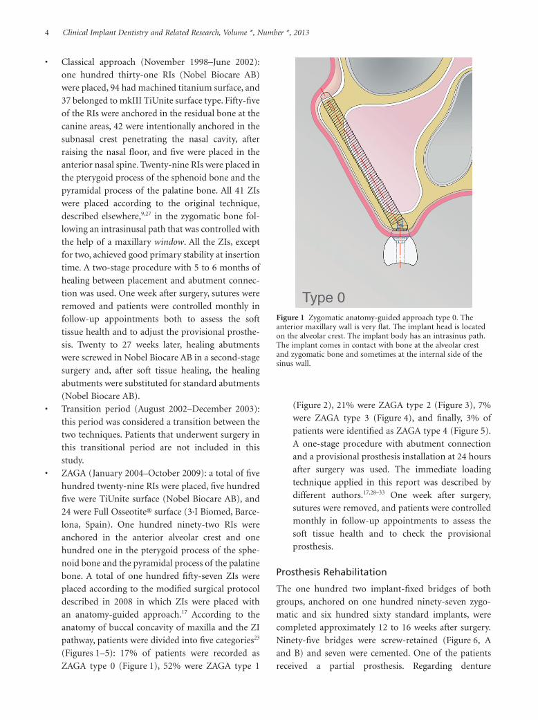
(Figures 1–5): 17% of patients were recorded as
ZAGA type 0 (Figure 1), 52% were ZAGA type 1
(Figure 2), 21% were ZAGA type 2 (Figure 3), 7%
were ZAGA type 3 (Figure 4), and finally, 3% of
patients were identified as ZAGA type 4 (Figure 5).
A one-stage procedure with abutment connection
and a provisional prosthesis installation at 24 hours
after surgery was used. The immediate loading
technique applied in this report was described by
different authors.17,28–33 One week after surgery,
sutures were removed, and patients were controlled
monthly in follow-up appointments to assess the
soft tissue health and to check the provisional
prosthesis.
Prosthesis Rehabilitation
The one hundred two implant-fixed bridges of both
groups, anchored on one hundred ninety-seven zygo-
matic and six hundred sixty standard implants, were
completed approximately 12 to 16 weeks after surgery.
Ninety-five bridges were screw-retained (Figure 6, A
and B) and seven were cemented. One of the patients
received a partial prosthesis. Regarding denture
Figure 1 Zygomatic anatomy-guided approach type 0. Theanterior maxillary wall is very flat. The implant head is locatedon the alveolar crest. The implant body has an intrasinus path.The implant comes in contact with bone at the alveolar crestand zygomatic bone and sometimes at the internal side of thesinus wall.
4 Clinical Implant Dentistry and Related Research, Volume *, Number *, 2013
material, 76 dental prostheses were metal-resin full-arch
design and 26 were metal-porcelain bridges.
Clinical Examination
For the subjects who experienced implant loss, data
related to the causes and the timing of explantation were
collected.
Periotest (PT) Measurements. Implant stability was
measured individually using the Periotest® device
(Siemens AG, Bensheim, UK) according to Olive and
Aparicio.34 Measurements were made on the day of
bridge delivery and annually thereafter. Patients who
were not wearing a screw-retained prosthesis were
excluded. The aim of the measurements was to compare
the PT values obtained before prosthesis placement and
the stability of the same implants after definitive pros-
thesis delivery throughout the follow-up period.
Sinus Symptomatology (Questionnaire for Research Out-
comes). A patient questionnaire (Table 1) to identify the
presence of sinusitis symptoms, as specified by the Task
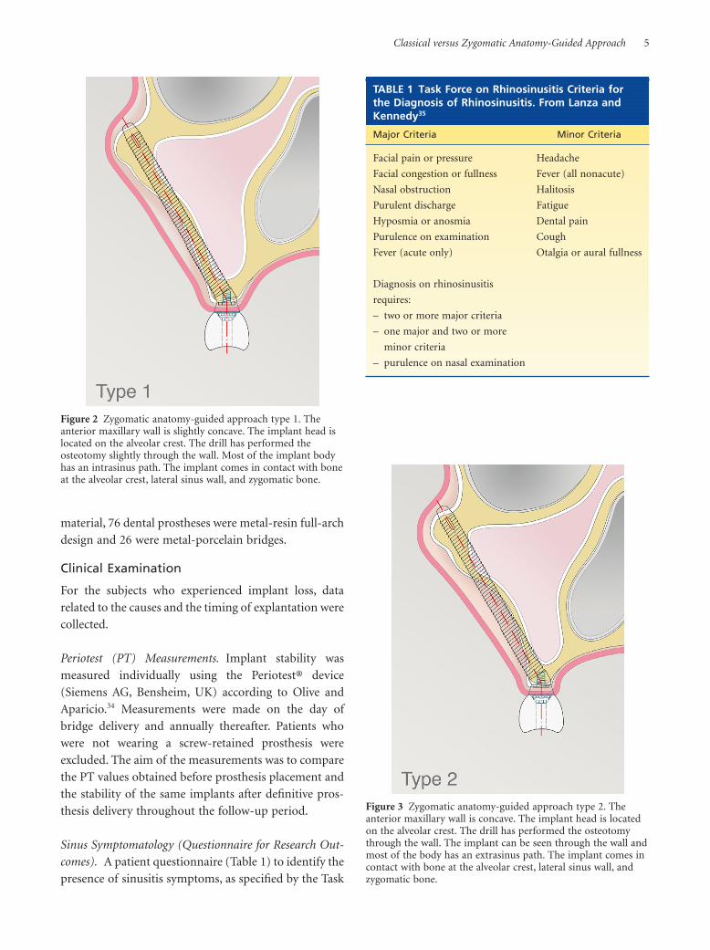
Figure 2 Zygomatic anatomy-guided approach type 1. Theanterior maxillary wall is slightly concave. The implant head islocated on the alveolar crest. The drill has performed theosteotomy slightly through the wall. Most of the implant bodyhas an intrasinus path. The implant comes in contact with boneat the alveolar crest, lateral sinus wall, and zygomatic bone.
Figure 3 Zygomatic anatomy-guided approach type 2. Theanterior maxillary wall is concave. The implant head is locatedon the alveolar crest. The drill has performed the osteotomythrough the wall. The implant can be seen through the wall andmost of the body has an extrasinus path. The implant comes incontact with bone at the alveolar crest, lateral sinus wall, andzygomatic bone.
TABLE 1 Task Force on Rhinosinusitis Criteria forthe Diagnosis of Rhinosinusitis. From Lanza andKennedy35
Major Criteria Minor Criteria
Facial pain or pressure Headache
Facial congestion or fullness Fever (all nonacute)
Nasal obstruction Halitosis
Purulent discharge Fatigue
Hyposmia or anosmia Dental pain
Purulence on examination Cough
Fever (acute only) Otalgia or aural fullness
Diagnosis on rhinosinusitis
requires:
– two or more major criteria
– one major and two or more
minor criteria
– purulence on nasal examination
Classical versus Zygomatic Anatomy-Guided Approach 5
Force on Rhinosinusitis (TFR) diagnostic criteria,35,36
was given to each patient at the final examination. Each
symptom question is answered by “yes” or “no.” Diag-
nosis of sinusitis requires a “yes” answer in two or more
major criteria, one major and two or more minor crite-
ria, or purulence on nasal examination.
Radiographic Examination
Lund-Mackay (L-M) Score. Each Cone Beam Comput-
erized Tomography (CBCT) scan was scored by an inde-
pendent otolaryngological researcher (P.C.) using the
L-M staging system, a validated scoring system recom-
mended by the TFR for research outcomes37–39 (Table 2
and Figure 7). The test includes six regions: anterior
ethmoid, posterior ethmoid, maxillary, frontal, sphe-
noid, and ostiomeatal complex. Each region is given a
score of 0, 1, or 2, 0 representing normality no opacifi-
cation, 1 partial opacification, and 2 total opacification.
Osteomeatal complex can only be scored 0 or 2. Total
scores range from 0 to 24. For purposes of this study, a
normal or “negative” scan was defined as any scan with a
L-M score of 0. Any scan with a score >0 was considered
an abnormal or “positive” scan.
Anatomical Measurements. CBCT scans (Kodak 9500
Cone Beam 3D System, Rochester, NY, USA) were per-
formed on the one hundred two patients and analyzed
by two independent researchers: an otorhinolaryngo-
logist (P.C.) and a fellow clinical researcher (K.F.).
Images in the coronal and horizontal axial planes were
obtained for each of the ZIs studied. Special emphasis
was devoted to assessing and storing the status of both
right and left osteomeatal complex permeability and on
the axial plane to be able to relate the ZI head position
to the bone crest on each side (Figure 8, A and B). Four
anatomical measurements (numbered 1–4 in Table 3)
were performed to assess the following: (i) the height of
the alveolar ridge at the location of the head of the ZI
(measurement 2 minus 1) and (ii) the position of the
head of the ZI with regard to the center of the crest of
the alveolar ridge in the horizontal axial dimension
(measurement 4 minus 3). A positive value on this
implant head position to the alveolar ridge relationship
indicates a palatal position of the implant, whereas a
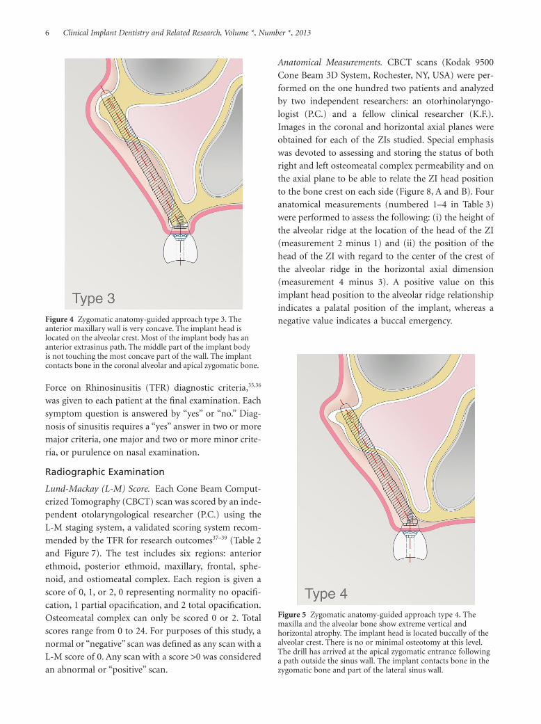
negative value indicates a buccal emergency.Figure 4 Zygomatic anatomy-guided approach type 3. Theanterior maxillary wall is very concave. The implant head islocated on the alveolar crest. Most of the implant body has ananterior extrasinus path. The middle part of the implant bodyis not touching the most concave part of the wall. The implantcontacts bone in the coronal alveolar and apical zygomatic bone.
Figure 5 Zygomatic anatomy-guided approach type 4. Themaxilla and the alveolar bone show extreme vertical andhorizontal atrophy. The implant head is located buccally of thealveolar crest. There is no or minimal osteotomy at this level.The drill has arrived at the apical zygomatic entrance followinga path outside the sinus wall. The implant contacts bone in thezygomatic bone and part of the lateral sinus wall.
6 Clinical Implant Dentistry and Related Research, Volume *, Number *, 2013
Satisfaction QuestionnaireThe satisfaction level and the masticatory capacity were
evaluated by means of the Oral Health Impact Profile for
assessing health-related quality of life in Edentulous
adults (OHIP-EDENT).40 Patients answered questions
regarding their ability or lack of ability to comminute
hard and soft foods relating it to the discomfort and
instability of the dentures, their perception of satisfac-
tion in relation to the aesthetics, pleasure when eating,
level of comfort, and self-assurance. Patients answered
nine questions about their dentures, the answer scale
ranging from 0 to 440,41 (Table 4). The highest scores
represent the worst satisfaction levels and the lowest
scores represent the best satisfaction levels. The
maximum score is 36. Results were translated into per-
centage values of satisfaction, 0% representing worst
possible satisfaction level and 100% representing best
possible satisfaction level.
A B
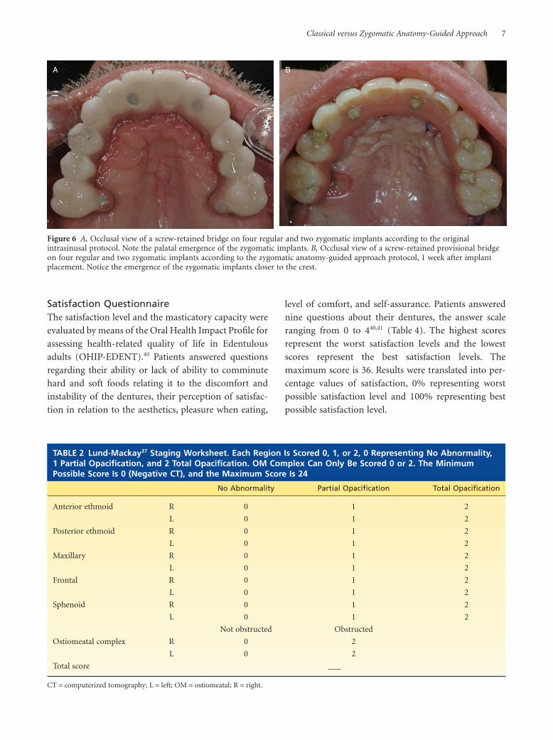
Figure 6 A, Occlusal view of a screw-retained bridge on four regular and two zygomatic implants according to the originalintrasinusal protocol. Note the palatal emergence of the zygomatic implants. B, Occlusal view of a screw-retained provisional bridgeon four regular and two zygomatic implants according to the zygomatic anatomy-guided approach protocol, 1 week after implantplacement. Notice the emergence of the zygomatic implants closer to the crest.
TABLE 2 Lund-Mackay37 Staging Worksheet. Each Region Is Scored 0, 1, or 2, 0 Representing No Abnormality,1 Partial Opacification, and 2 Total Opacification. OM Complex Can Only Be Scored 0 or 2. The MinimumPossible Score Is 0 (Negative CT), and the Maximum Score Is 24
No Abnormality Partial Opacification Total Opacification
Anterior ethmoid R 0 1 2
L 0 1 2
Posterior ethmoid R 0 1 2
L 0 1 2
Maxillary R 0 1 2
L 0 1 2
Frontal R 0 1 2
L 0 1 2
Sphenoid R 0 1 2
L 0 1 2
Not obstructed Obstructed
Ostiomeatal complex R 0 2
L 0 2
Total score ___
CT = computerized tomography; L = left; OM = ostiomeatal; R = right.
Classical versus Zygomatic Anatomy-Guided Approach 7
Data Analysis
The heterogeneity between the two groups with respect
to gender, smoking, and rhinosinusitis criteria was
assessed using the Mann-Whitney test. A p-value <.05
was considered to indicate a statistically significant
difference.
The SR was calculated and compared between the
different groups using the Kaplan-Meier analysis.
The intergroup comparison of age, horizontal and
vertical CBCT measurements, L-M score, and PT values
was performed using the unpaired t-test. A p-value <.05
was considered to indicate a statistically significant
difference.
The data analysis was performed using a commer-
cially available statistical software package. All statistical
analyses were performed using SPSS 13.0 software (SPSS
Inc., Chicago, IL, USA).
RESULTS
At the time of implant placement, no statistical differ-
ences (p > .05) were identified between groups with
respect to gender and smoking. However, statistically
significant differences (p = .001) were found with
respect to age (53.81 1 10.97 years vs 63.10 1 9.00 years)
(Table 5).
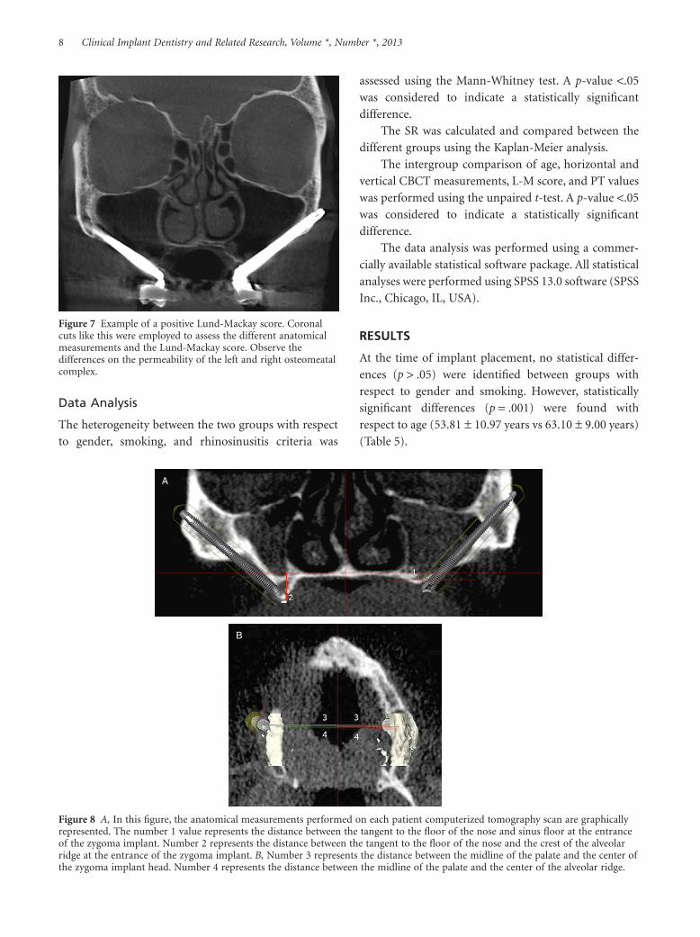
Figure 7 Example of a positive Lund-Mackay score. Coronalcuts like this were employed to assess the different anatomicalmeasurements and the Lund-Mackay score. Observe thedifferences on the permeability of the left and right osteomeatalcomplex.
A
B
Figure 8 A, In this figure, the anatomical measurements performed on each patient computerized tomography scan are graphicallyrepresented. The number 1 value represents the distance between the tangent to the floor of the nose and sinus floor at the entranceof the zygoma implant. Number 2 represents the distance between the tangent to the floor of the nose and the crest of the alveolarridge at the entrance of the zygoma implant. B, Number 3 represents the distance between the midline of the palate and the center ofthe zygoma implant head. Number 4 represents the distance between the midline of the palate and the center of the alveolar ridge.
8 Clinical Implant Dentistry and Related Research, Volume *, Number *, 2013
With the classical technique, three implants, mecha-
nized surface, failed. Two failed between implant instal-
lation and prosthesis placement; one after 3 years of
function. No failures of RIs were observed on the ZAGA
group. Although none of the ZIs were removed because
of disosseointegration, during the follow-up period,
seven ZIs were considered failures, yielding a total SR of
96.95%. No early failures were observed. In the classical
technique group, two ZIs were cut 10 years after place-
ment through the surgical maxillary window and
partially removed (both in the same patient, a heavy
smoker) due to extreme peri-implant infection with
complete dissolution of the palatal bone. In the ZAGA
group, one ZI fractured, and four zygoma implants (in
the same patient) had clear clinical mobility at the
crestal portion of the implant. The final SR for ZIs on
control and test groups was 95.12% and 97.44%, respec-
tively. However, no statistically significant differences
(p = .602) were observed between the SRs of the two
groups (Tables 6 and 7).
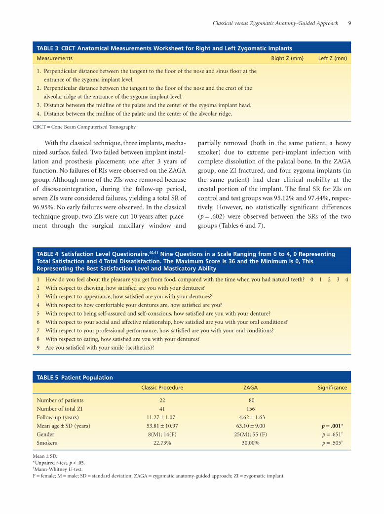
TABLE 4 Satisfaction Level Questionaire.40,41 Nine Questions in a Scale Ranging from 0 to 4, 0 RepresentingTotal Satisfaction and 4 Total Dissatisfaction. The Maximum Score Is 36 and the Minimum Is 0, ThisRepresenting the Best Satisfaction Level and Masticatory Ability
1 How do you feel about the pleasure you get from food, compared with the time when you had natural teeth? 0 1 2 3 4
2 With respect to chewing, how satisfied are you with your dentures?
3 With respect to appearance, how satisfied are you with your dentures?
4 With respect to how comfortable your dentures are, how satisfied are you?
5 With respect to being self-assured and self-conscious, how satisfied are you with your denture?
6 With respect to your social and affective relationship, how satisfied are you with your oral conditions?
7 With respect to your professional performance, how satisfied are you with your oral conditions?
8 With respect to eating, how satisfied are you with your dentures?
9 Are you satisfied with your smile (aesthetics)?
TABLE 5 Patient Population
Classic Procedure ZAGA Significance
Number of patients 22 80
Number of total ZI 41 156
Follow-up (years) 11.27 1 1.07 4.62 1 1.63
Mean age 1 SD (years) 53.81 1 10.97 63.10 1 9.00 p = .001*
Gender 8(M); 14(F) 25(M); 55 (F) p = .651†
Smokers 22.73% 30.00% p = .505†
Mean 1 SD.*Unpaired t-test, p < .05.†Mann-Whitney U-test.F = female; M = male; SD = standard deviation; ZAGA = zygomatic anatomy-guided approach; ZI = zygomatic implant.
TABLE 3 CBCT Anatomical Measurements Worksheet for Right and Left Zygomatic Implants
Measurements Right Z (mm) Left Z (mm)
1. Perpendicular distance between the tangent to the floor of the nose and sinus floor at the
entrance of the zygoma implant level.
2. Perpendicular distance between the tangent to the floor of the nose and the crest of the
alveolar ridge at the entrance of the zygoma implant level.
3. Distance between the midline of the palate and the center of the zygoma implant head.
4. Distance between the midline of the palate and the center of the alveolar ridge.
CBCT = Cone Beam Computerized Tomography.
Classical versus Zygomatic Anatomy-Guided Approach 9
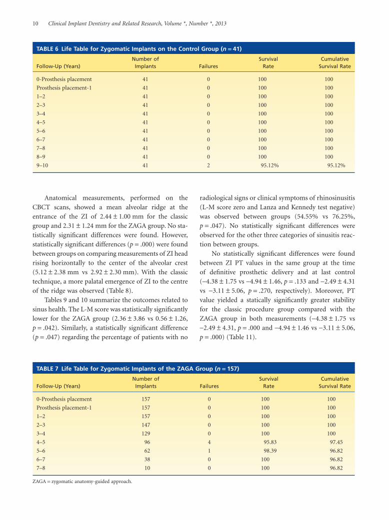
Anatomical measurements, performed on the
CBCT scans, showed a mean alveolar ridge at the
entrance of the ZI of 2.44 1 1.00 mm for the classic
group and 2.31 1 1.24 mm for the ZAGA group. No sta-
tistically significant differences were found. However,
statistically significant differences (p = .000) were found
between groups on comparing measurements of ZI head
rising horizontally to the center of the alveolar crest
(5.12 1 2.38 mm vs 2.92 1 2.30 mm). With the classic
technique, a more palatal emergence of ZI to the centre
of the ridge was observed (Table 8).
Tables 9 and 10 summarize the outcomes related to
sinus health. The L-M score was statistically significantly
lower for the ZAGA group (2.36 1 3.86 vs 0.56 1 1.26,
p = .042). Similarly, a statistically significant difference
(p = .047) regarding the percentage of patients with no
radiological signs or clinical symptoms of rhinosinusitis
(L-M score zero and Lanza and Kennedy test negative)
was observed between groups (54.55% vs 76.25%,
p = .047). No statistically significant differences were
observed for the other three categories of sinusitis reac-
tion between groups.
No statistically significant differences were found
between ZI PT values in the same group at the time
of definitive prosthetic delivery and at last control
(-4.38 1 1.75 vs -4.94 1 1.46, p = .133 and -2.49 1 4.31
vs -3.11 1 5.06, p = .270, respectively). Moreover, PT
value yielded a statically significantly greater stability
for the classic procedure group compared with the
ZAGA group in both measurements (-4.38 1 1.75 vs
-2.49 1 4.31, p = .000 and -4.94 1 1.46 vs -3.11 1 5.06,
p = .000) (Table 11).
TABLE 6 Life Table for Zygomatic Implants on the Control Group (n = 41)
Follow-Up (Years)Number ofImplants Failures
SurvivalRate
CumulativeSurvival Rate
0-Prosthesis placement 41 0 100 100
Prosthesis placement-1 41 0 100 100
1–2 41 0 100 100
2–3 41 0 100 100
3–4 41 0 100 100
4–5 41 0 100 100
5–6 41 0 100 100
6–7 41 0 100 100
7–8 41 0 100 100
8–9 41 0 100 100
9–10 41 2 95.12% 95.12%
TABLE 7 Life Table for Zygomatic Implants of the ZAGA Group (n = 157)
Follow-Up (Years)Number ofImplants Failures
SurvivalRate
CumulativeSurvival Rate
0-Prosthesis placement 157 0 100 100
Prosthesis placement-1 157 0 100 100
1–2 157 0 100 100
2–3 147 0 100 100
3–4 129 0 100 100
4–5 96 4 95.83 97.45
5–6 62 1 98.39 96.82
6–7 38 0 100 96.82
7–8 10 0 100 96.82
ZAGA = zygomatic anatomy-guided approach.
10 Clinical Implant Dentistry and Related Research, Volume *, Number *, 2013
Few mechanical and biological problems were
observed during the follow-up period. Some of these
problems were the following: loosening and fracture of
prosthetic ZI screws, loosening of the abutments, frac-
ture of ceramic prosthetic teeth, and fracture of resin
prostheses. Records of these prosthetic complications
and other biological complications are collected on
Tables 12 and 13.
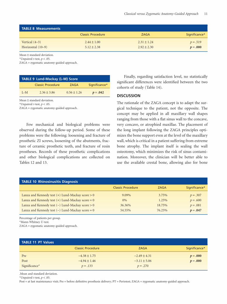
Finally, regarding satisfaction level, no statistically
significant differences were identified between the two
cohorts of study (Table 14).
DISCUSSION
The rationale of the ZAGA concept is to adapt the sur-
gical technique to the patient, not the opposite. The
concept may be applied in all maxillary wall shapes
ranging from those with a flat sinus wall to the concave,
very concave, or atrophied maxillae. The placement of
the long implant following the ZAGA principles opti-
mizes the bone support even at the level of the maxillary
wall, which is critical in a patient suffering from extreme
bone atrophy. The implant itself is sealing the wall
osteotomy, which minimizes the risk of sinus contami-
nation. Moreover, the clinician will be better able to
use the available crestal bone, allowing also for bone
TABLE 8 Measurements
Classic Procedure ZAGA Significance*
Vertical (4–3) 2.44 1 1.00 2.31 1 1.24 p = .519
Horizontal (10–9) 5.12 1 2.38 2.92 1 2.30 p = .000
Mean 1 standard deviation.*Unpaired t-test, p < .05.ZAGA = zygomatic anatomy-guided approach.
TABLE 9 Lund-Mackay (L-M) Score
Classic Procedure ZAGA Significance*
L-M 2.36 1 3.86 0.56 1 1.26 p = .042
Mean 1 standard deviation.*Unpaired t-test, p < .05.ZAGA = zygomatic anatomy-guided approach.
TABLE 10 Rhinosinusitis Diagnosis
Classic Procedure ZAGA Significance*
Lanza and Kennedy test (+) Lund-Mackay score > 0 9.09% 3.75% p = .307
Lanza and Kennedy test (+) Lund-Mackay score = 0 0% 1.25% p = .600
Lanza and Kennedy test (-) Lund-Mackay score > 0 36.36% 18.75% p = .081
Lanza and Kennedy test (-) Lund-Mackay score = 0 54.55% 76.25% p = .047
Percentage of patients per group.*Mann-Whitney U-test.ZAGA = zygomatic anatomy-guided approach.
TABLE 11 PT Values
Classic Procedure ZAGA Significance*
Pre -4.38 1 1.75 -2.49 1 4.31 p = .000
Post -4.94 1 1.46 -3.11 1 5.06 p = .000
Significance* p = .133 p = .270
.Mean and standard deviation.*Unpaired t-test, p < .05.Post = at last maintenance visit; Pre = before definitive prosthesis delivery; PT = Periotest; ZAGA = zygomatic anatomy-guided approach.
Classical versus Zygomatic Anatomy-Guided Approach 11

TAB
LE12
Nu
mb
ero
fC
om
plic
atio
ns
du
rin
gth
e10
-Yea
rFo
llow
-Up
Peri
od
of
the
Cla
ssic
alPr
oce
du
re.
Failu
res
of
Imp
lan
tsA
reN
ot
Incl
ud
ed.
Nin
ety
Perc
ent
of
Bio
log
ical
Late
Co
mp
licat
ion
sB
elo
ng
toTw
oPa
tien
tsan
d74
%o
fPr
ost
het
icC
om
plic
atio
ns
Occ
urr
edin
Five
Pati
ents
Follo
w-U
p(Y
ears
)0-
Pro
sth
esis
Plac
emen
tPr
ost
hes
isPl
acem
ent-
11–
22–
33–
44–
55–
66–
77–
88–
99–
1010
–11
Tota
l
Bio
logi
cal
Faci
alh
emat
oma/
edem
a6
6
Lip
lace
rati
on5
5
Ch
eek
and/
orpa
ran
asal
pare
sth
esia
(tem
pora
ry)
66
Supp
ura
tion
ofre
gula
rim
plan
t1
1
Acu
te/c
hro
nic
sin
usi
tis
11
11
11
6
Oro
sin
usa
lcom
mu
nic
atio
n(p
erim
plan
t)1
23
Mec
han
ical
Frac
ture
coat
ing
mat
eria
l:ac
rylic
11
11
4
Frac
ture
coat
ing
mat
eria
l:po
rcel
ain
21
21
36
41
23
25
Frac
ture
ofm
etal
fram
ewor
k1
12
Frac
ture
scre
ws
11
11
26
Loos
enin
gof
scre
ws
orab
utm
ent
11
21
22
9
TAB
LE13
Nu
mb
ero
fC
om
plic
atio
ns
du
rin
gth
eFo
llow
-Up
Peri
od
of
the
ZAG
AG
rou
p.
Failu
res
of
Imp
lan
tsA
reN
ot
Incl
ud
ed
Follo
w-U
p(Y
ears
)0-
Pro
sth
esis
Plac
emen
tPr
ost
hes
isPl
acem
ent-
11–
22–
33–
44–
55–
66–
77–
88–
99–
1010
–11
Tota
l
Bio
logi
cal
Faci
alh
emat
oma/
edem
a1
00
00
00
00
00
01
Lip
lace
rati
on0
00
00
00
00
00
00
Ch
eek
and/
orpa
ran
asal
pare
sth
esia
(tem
pora
ry)
10
00
00
00
00
00
0
Supp
ura
tion
ofre
gula
rim
plan
t2
02
10
00
00
00
05
Acu
te/c
hro
nic
sin
usi
tis
00
10
20
00
00
00
3
Oro
sin
usa
lcom
mu
nic
atio
n(p
erim
plan
t)2
00
00
00
00
00
02
Mec
han
ical
Frac
ture
coat
ing
mat
eria
l:ac
rylic
15
721
1111
121
20
00
65
Frac
ture
coat
ing
mat
eria
l:po
rcel
ain
00
01
10
00
00
00
2
Frac
ture
ofm
etal
fram
ewor
k0
00
00
00
00
00
00
Frac
ture
scre
ws
00
12
21
10
00
00
7
Loos
enin
gof
scre
ws
orab
utm
ent
05
55
11
01
00
00
16
ZA
GA
=zy
gom
atic
anat
omy-
guid
edap
proa
ch.
12 Clinical Implant Dentistry and Related Research, Volume *, Number *, 2013
integration at the implant body and neck level in most of
the ZAGA types. As a result, implants are placed more
crestally, with less possibility of oroantral communica-
tion. As well, the understanding of the ZAGA concept
helps to better soft tissue control to predictably cover the
ZI in comparison with an exclusively extra-maxillary
technique.
In the last decade, a large number of publications
have focused on ZI success and long-term survival to
demonstrate that the procedure is a predictable solu-
tion for rehabilitation of atrophic maxilla. The out-
comes of the present retrospective study showed that
ZI’s SR in both groups is in agreement with data from
previous reports,9,12,14–16,18–20,26,29–34,42–53 independent of
the technique used. It seems that a low percentage of
patients with ZIs will develop rhinosinusitis. Interest-
ingly, the rate is not very different from the sinusitis
rate in the general population or the sinusitis rate asso-
ciated with sinus grafts.10 Only few authors12–14,54 have
reported on biological and mechanical complications,
the most common being the sinus infection. The
relationship between the implant and the maxillary
sinus structures remains controversial: zygoma
implants could potentially cause inflammatory prob-
lems or infections at the level of the maxillary antrum.
Oroantral communication is not necessarily related to
an infectious process because often soft tissue is able to
seal the interface around the implant head. Neverthe-
less, there is little evidence-based data concerning the
relationship of the zygoma implants and the sinus
cavity. Few studies have analyzed sinus reactions to
zygoma implants.10
Currently, there is no consensus on how to report
rhinosinus status. Usually, there is no systematic evalu-
ation of the status of the sinus after placement of ZIs,
either clinically or radiologically. There are few clinical
studies evaluating the evolution of sinus reactions
and sinus health from the clinical and/or radiological
perspective after placement of ZIs. Plain radiographs
are not useful for study of the sinus cavity or osti-
omeatal complex, and CT scans are not routinely
obtained. As yet there are only three published studies,
performed by Nakai and colleagues,55 Davó and col-
leagues,56 and Aparicio and colleagues26 using a CT scan
after ZI placement.
The diagnosis and treatment (by ENT departments)
must follow the current recommendations for the treat-
ment of rhinosinusitis in conventional patients. Because
there is no consensus in the reporting of the diagnosis of
a sinusal pathology associated with the placement of ZIs,
the authors of this study are proposing the use of the
clinical and radiological scores recommended by the
TFR as a standardized system to report on the diagnosis
of rhinosinusitis associate with implant placement.
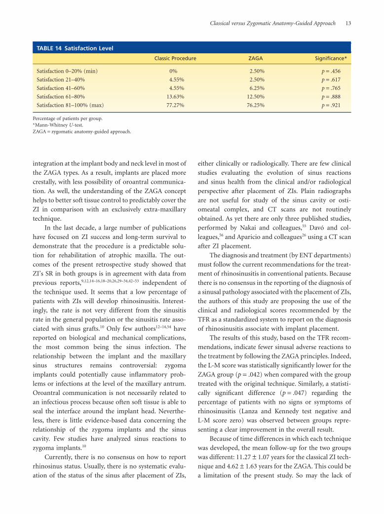
The results of this study, based on the TFR recom-
mendations, indicate fewer sinusal adverse reactions to
the treatment by following the ZAGA principles. Indeed,
the L-M score was statistically significantly lower for the
ZAGA group (p = .042) when compared with the group
treated with the original technique. Similarly, a statisti-
cally significant difference (p = .047) regarding the
percentage of patients with no signs or symptoms of
rhinosinusitis (Lanza and Kennedy test negative and
L-M score zero) was observed between groups repre-
senting a clear improvement in the overall result.
Because of time differences in which each technique
was developed, the mean follow-up for the two groups
was different: 11.27 1 1.07 years for the classical ZI tech-
nique and 4.62 1 1.63 years for the ZAGA. This could be
a limitation of the present study. So may the lack of
TABLE 14 Satisfaction Level
Classic Procedure ZAGA Significance*
Satisfaction 0–20% (min) 0% 2.50% p = .456
Satisfaction 21–40% 4.55% 2.50% p = .617
Satisfaction 41–60% 4.55% 6.25% p = .765
Satisfaction 61–80% 13.63% 12.50% p = .888
Satisfaction 81–100% (max) 77.27% 76.25% p = .921
Percentage of patients per group.*Mann-Whitney U-test.ZAGA = zygomatic anatomy-guided approach.
Classical versus Zygomatic Anatomy-Guided Approach 13
data regarding presurgical L-M score (CTBC) and sinus
symptomatology of the classic approach group. Due to
justified time-related reasons, it was not possible to
compare intragroup (before and after ZI placement) and
intergroup (presurgical data, classical vs modified tech-
nique) sinus health.
Some authors9,15,16,18–20,30–34,44–46,48–53,57,58 have
pointed out variations on the incidence of sinusitis
being related to different factors such as implant design
(the internal thread of the abutment chamber is com-
municated to the external surface, allowing an eventual
pass of bacteria vs a closed design) and the type of
implant surface used (rough vs machined). However,
there are no data validating those findings individually
as influencing in the development of sinusal complica-
tions. Another factor that has been claimed as impor-
tant in decreasing the incidence of sinusal disease is
the time of implant connection/loading (immediate
vs delayed). From the available data,10 sinusitis rates
for the classic two-stage protocol are approximately
6.6%; for immediate function protocols, this is appro-
ximately 2.8%, and if both protocols are considered
together, the rate is approximately 5.5%. However, the
new surgical approaches, with less of the implant in
the sinus and the placement of the implant head
more crestally, also include the immediate loading of
the implants as a part of the protocol. This may create
confusion regarding the specific importance of a single
factor, that is, surgical technique versus the load
moment.
The main drawback of the original zygoma implant
technique when using an intrasinus approach is the
palatal emergence of the implant head, which is often
the case because of the desire to maintain the implant
body within the boundaries of the maxillary sinus. This
commonly results in a bulky dental, fixed denture at the
palatal aspect. When using the original surgical proto-
col, authors have reported on speech alterations and
problems in maintaining correct hygiene of the dental
prostheses, largely because of the palatine emergence of
the head of the zygoma implant.15,55,59,60 The use of the
modified ZAGA allowed a substantial reduction in the
mean distance from the zygoma implant to the central
part of the residual crest: from 5.12 mm (SD 2.38) with
the original technique to a mean value of 2.92 mm (SD
2.30) with ZAGA. The more favorable emergence posi-
tion obtained by following the ZAGA principles can
minimize these complications.
CONCLUSIONS
Within the limits of this study, the outcomes demon-
strated that both procedures – the classical and the
ZAGA – were found with similar positive clinical
outcome with respect to implant survival. The ZAGA
concept is able to immediately rehabilitate the severely
atrophic maxillae, minimizing the risk of maxillary
sinus-associated pathology in comparison with the clas-
sical ZI surgical technique. Moreover, less bulky, more
comfortable, and easy to clean prostheses are achieved.
ACKNOWLEDGMENT
The authors would like to thank the precise help of Dr
Martín Rios with the statistical analysis.
REFERENCES
1. Lekholm U, Zarb GA. Patient selection and preparation.
In: Brånemark P-I, Zarb GA, Albrektsson T, eds. Tissue
integrated prostheses: osseointegration in clinical dentistry.
Chicago, IL: Quintessence, 1985:199–209.
2. Sjöström M, Sennerby L, Nilson H, Lundgren S. Reconstruc-
tion of the atrophic edentulous maxilla with free iliac crest
grafts and implants: a 3-year report of a prospective clinical
study. Clin Implant Dent Relat Res 2007; 9:46–59.
3. Nyström E, Lundgren S, Gunne J, Nilson H. Interpositional
bone grafting and Le Fort I osteotomy for reconstruction of
the atrophic edentulous maxilla. A two-stage technique. Int J
Oral Maxillofac Surg 1997; 26:423–427.
4. Lundgren S, Nyström E, Nilson H, Gunne J, Lindhagen O.
Bone grafting to the maxillary sinuses, nasal floor and
anterior maxilla in the atrophic edentulous maxilla. A
two stage technique. Int J Oral Maxillofac Surg 1997; 26:428–
434.
5. Higuchi KW. Minimization in oral implant rehabilitation: a
patient-centered ethics-based approach in zygomatic
implants. In: Aparicio C, ed. Zygomatic implants. The
anatomy-guided approach. Berlin: Ed. Quintessence,
2012:1–4.
6. Weischer T, Schettler D, Ch M. Titanium implants in the
zygoma as retaining elements after hemimaxillectomy. Int J
Oral Maxillofac Implants 1997; 12:211–221.
7. Higuchi KW. The zygomaticus fixture: an alternative
approach for implant anchorage in the posterior maxilla.
Ann R Australas Coll Dent Surg 2000; 15:23–33.
8. Esposito M, Worthington HV, Coulthard P. Interventions
for replacing missing teeth: dental implants in zygomatic
bone for the rehabilitation of the severely deficient edentu-
lous maxilla. Cochrane Database Syst Rev 2005; (4):
CD004151.
9. Brånemark PI, Grondahl K, Ohrnell LO, et al. Zygoma
fixture in the management of advanced atrophy of the
14 Clinical Implant Dentistry and Related Research, Volume *, Number *, 2013
maxilla: technique and long-term results. Scand J Plast
Reconstr Surg Hand Surg 2004; 38:70–85.
10. Davo R. Sinus reactions to zygomatic implants. In: Aparicio
C, ed. Zygomatic implants. The anatomy-guided approach.
Berlin: Ed. Quintessence, 2012:59–78.
11. Aparicio C, Ouazzani W, Aparicio A, et al. Extra-sinus zygo-
matic implants: three year experience from a new surgical
approach for patients with pronounced buccal concavities
for the edentulous maxilla. Clin Implant Dent Relat Res
2010; 12:55–61. Epub 2008 Dec 3.
12. Bedrossian E. Rehabilitation of the edentulous maxilla with
the zygoma concept: a 7-year prospective study. Int J Oral
Maxillofac Implants 2010; 25:1213–1221.
13. Al-Nawas B, Wegener J, Bender C, Wagner W. Clinical soft
tissue parameters of the zygomatic implant. J Clin Period-
ontol 2004; 31:497–500.
14. Becktor JP, Isaksson S, Abrahamsson O, Sennerby L. Evalu-
ation of 31 zygomatic implants and 74 regular dental
implants used in 16 patients for prosthetic reconstruction of
the atrophic maxilla with cross-arch fixed bridges. Clin
Implant Dent Relat Res 2005; 7:159–165.
15. Boyes-Varley JG, Howes DG, Lownie JF, Blackbeard GA.
Surgical modifications to the Branemark zygomaticus proto-
col in the treatment of the severely resorbed maxilla: a clinical
report. Int J Oral Maxillofac Implants 2003; 18:232–237.
16. Farzad P, Andersson L, Gunnarsson S, Johansson B. Reha-
bilitation of severely resorbed maxillae with zygomatic
implants: an evaluation of implant stability, tissue condi-
tions, and patients opinion before and after treatment. Int J
Oral Maxillofac Implants 2006; 21:399–404.
17. Aparicio C, Ouazzani W, Hatano N. The use of zygomatic
implants for prosthetic rehabilitation of the severely
resorbed maxilla. Periodontol 2000 2008; 47:162–171.
18. Aparicio C, Ouazzani W, Garcia R, Arevalo X, Muela R,
Fortes V. A prospective clinical study on titanium implants
in the zygomatic arch for prosthetic rehabilitation of the
atrophic edentulous maxilla with a follow-up of 6 months to
5 years. Clin Implant Dent Relat Res 2006; 8:114–122.
19. Maló P, Nobre Mde A, Lopes I. A new approach to rehabili-
tate the severely atrophic maxilla using extramaxillary
anchored implants in immediate function: a pilot study. J
Prosthet Dent 2008; 100:354–366.
20. Peñarrocha M, Garcıa B, Martı E, Boronat A. Rehabilitation
of severely atrophic maxillae with fixed implant-supported
prostheses using zygomatic implants placed using the sinus
slot technique: clinical report on a series of 21 patients. Int J
Oral Maxillofac Implants 2007; 22:645–650.
21. Stella JP, Warner MR. Sinus slot technique for simplification
and improved orientation of zygomaticus dental implants. A
technical note. Int J Oral Maxillofac Implants 2000; 15:889–
893.
22. Tan WC, Lang NP, Zwahlen M, Pjetursson BE. A systematic
review of the success of sinus floor elevation and survival of
implants inserted in combination with sinus floor elevation.
Part II: transalveolar technique. J Clin Periodontol 2008; 35
(Suppl 8):241–254. doi: 10.1111/j.1600-051X.2008.01273.x.
23. Aparicio C. A proposed classification for zygomatic implant
patient based on the zygoma anatomy guided approach
(ZAGA): a cross-sectional survey. Eur J Oral Implantol 2011;
4:269–275.
24. Aparicio C. The zygoma anatomy guided approach (ZAGA).
In: Aparicio C, ed. Zygomatic implants. The anatomy-guided
approach. Berlin: Ed. Quintessence, 2012;113–136.
25. Albrektsson T, Isidor F. Criteria for success and failure of an
implant system. Consensus report. In: Lang NP, Karring T,
eds. Proceedings of the 1st European workshop on period-
ontology. Chicago, IL: Quintessence, 1994:243–244.
26. Aparicio C, Manresa C, Francisco K, et al. The long term use
of zygomatic implants: a ten years clinical and radiographic
report. Clin Implant Dent Relat Res 2012. DOI: 10.1111/
cid.12007.
27. The zygomaticus fixture: clinical procedures. Göteborg,
Sweden: Nobel Biocare, 1998.
28. Aparicio C, Ouazzani W, Aparicio A, et al. Immediate/early
loading of zygomatic implants: clinical experiences after 2 to
5 years of follow-up. Clin Implant Dent Relat Res 2010;
12(Suppl 1):77–82.
29. Bedrossian E, Rangert B, Stumpel L, Indresano T. Immediate
function with the zygomatic implant: a graftless solution for
the patient with mild to advanced atrophy of the maxilla. Int
J Oral Maxillofac Implants 2006; 21:937–942.
30. Chow J, Hui E, Lee PK, Li W. Zygomatic implants protocol
for immediate occlusal loading: a preliminary report. J Oral
Maxillofac Surg 2006; 64:804–811.
31. Davó R, Malevez C, Rojas J, Rodríguez J, Regolf J. Clinical
outcome of 42 patients treated with 81 immediately loaded
zygomatic implants: a 12-to-42 month retrospective study.
Eur J Oral Implantol 2008; 1:141–150.
32. Davo C, Malevez C, Rojas J. Immediate function in the atro-
phic maxilla using zygoma implants: a preliminary study. J
Prosthet Dent 2007; 97:S44–S51.
33. Duarte LR, Filho HN, Francischone CE, Peredo LG,
Branemark PI. The establishment of a protocol for the total
rehabilitation of atrophic maxillae employing four zygo-
matic fixtures in an immediate loading system a 30-month
clinical and radiographic follow-up. Clin Implant Dent Relat
Res 2007; 9:186–196.
34. Olive J, Aparicio C. The Periotest method as a measure of
osseointegrated oral implant stability. Int J Oral Maxillofac
Implants 1990; 5:390–400.
35. Lanza DC, Kennedy DW. Adult rhinosinusitis defined.
Otolaryngol Head Neck Surg 1997; 117:s1–s7.
36. Hwang PH, Irwin SB, Griest SE, Caro JE, Nesbit GM. Radio-
logic correlates of symptom-based diagnostic criteria for
chronic rhinosinusitis. Otolaryngol Head Neck Surg 2003;
128:489–496.
Classical versus Zygomatic Anatomy-Guided Approach 15
37. Lund VJ, Mackay IS. Staging in rhinosinusitis. Rhinology
1993; 31:183–184.
38. Metson R, Gliklich RE, Stankiewicz JA, et al. Comparison of
sinus computed tomography staging systems. Otolaryngol
Head Neck Surg 1997; 117:372–379.
39. Oluwole M, Russell N, Tan L, et al. A comparison of com-
puterized tomographic staging systems in chronic sinusitis.
Clin Otolaryngol 1996; 21:91–95.
40. Allen F, Locker D. A modified short version of the oral
health impact profile for assessing health-related quality of
life in edentulous adults. Int J Prosthodont 2002; 15:446–
450.
41. Pocztaruk RL, Frasca LCF, Rivaldo EG, et al. Satisfaction
level and masticatory capacity in edentulous patients with
conventional dentures and implant-retained overdentures.
Braz J Oral Sci 2006; 5:1232–1238.
42. Bedrossian E, Stumpel L III, Beckely ML, Indresano T.
The zygomatic implant: preliminary data on treatment of
severely resorbed maxillae. A clinical report. Int J Oral Max-
illofac Implants 2002; 17:861–865.
43. Parel SM, Brånemark PI, Ohrnell LO, Svensson B. Remote
implant anchorage for the rehabilitation of maxillary
defects. J Prosthet Dent 2001; 86:377–381.
44. Vrielinck L, Politis C, Schepers S, Pauwels M, Naert I. Image
based planning and clinical validation of zygoma and ptery-
goid implant placement in patients with severe bone atrophy
using customized drill guides. Preliminary results from a
prospective clinical follow-up study. Int J Oral Maxillofac
Surg 2003; 32:7–14.
45. Malevez C, Abarca M, Durdu F, Daelemans P. Clinical
outcome of 103 consecutive zygomatic implants: a 6–48
months follow-up study. Clin Oral Implants Res 2004;
15:18–22.
46. Hirsch JM, Ohrnell LO, Henry PJ, et al. A clinical evaluation
of the zygoma fixture: one year of follow-up at 16 clinics. J
Oral Maxillofac Surg 2004; 62(Suppl 2):22–29.
47. Peñarrocha M, Uribe R, García B, Martí E. Zygomatic
implants using the sinus slot technique: clinical report of a
patient series. Int J Oral Maxillofac Implants 2005; 20:788–
792.
48. Ahlgren F, Storksen K, Tornes K. A study of 25 zygomatic
dental implants with 11 to 49 months follow-up after
loading. Int J Oral Maxillofac Implants 2006; 21:421–425.
49. Balshi SF, Wolfinger GJ, Balshi TJ. A retrospective analysis of
110 zygomatic implants in a single-stage immediate loading
protocol. Int J Oral Maxillofac Implants 2009; 24:335–341.
50. Davo R. Zygomatic implants placed with a two-stage proce-
dure: a 5-year retrospective study. Eur J Oral Implantol 2009;
2:115–124.
51. Davo R, Pons O, Rojas J, Carpio E. Immediate function of
four zygomatic implants: a 1-year report of a prospective
study. Eur J Oral Implantol 2010; 3:323–334.
52. Stiévenart M, Malevez C. Rehabilitation of totally atrophied
maxilla by means of four zygomatic implants and fixed pros-
thesis: a 6–40-month follow-up. Int J Oral Maxillofac Surg
2010; 39:358–363.
53. Migliorança RM, Coppedê A, Dias Rezende RC, de Mayo T.
Restoration of the edentulous maxilla using extrasinus
zygomatic implants combined with anterior conventional
implants: a retrospective study. Int J Oral Maxillofac
Implants 2011; 26:665–672.
54. Dykewicz MS, Hamilos DL. Rhinitis and sinusitis. J Allergy
Clin Immunol 2010; 125:S103–S115.
55. Nakai H, Okazaki Y, Ueda M. Clinical application of zygo-
matic implants for rehabilitation of the severely resorbed
maxilla: a clinical report. Int J Oral Maxillofac Implants
2003; 18:566–570.
56. Davó R, Malevez C, López-Orellana C, Pastor-Bevia F,
Rojas J. Sinus reactions to immediately loaded zygomatic
implants: a clinical and radiological study. Eur J Oral
Implantol 2008; 1:53–60.
57. Mozzati M, Monfrin SB, Pedretti G, Schierano G, Bassi F.
Immediate loading of maxillary fixed prostheses retained by
zygomatic and conventional implants: 24-month prelimi-
nary data for a series of clinical case reports. Int J Oral
Maxillofac Implants 2008; 23:308–314.
58. Zwahlen RA, Grätz KW, Oechslin CK, Studer SP. Survival
rate of zygomatic implants in atrophic or partially resected
maxillae prior to functional loading: a retrospective clinical
report. Int J Oral Maxillofac Implants 2006; 21:413–420.
59. Bothur S, Garsten M. Initial speech problems in patients
treated with multiple zygomatic implants. Int J Oral Maxil-
lofac Implants 2010; 25:379–384.
60. Petrovic A. Speech sound distortions caused by changes
in complete denture morphology. J Oral Rehabil 1985;
12:69–79.
16 Clinical Implant Dentistry and Related Research, Volume *, Number *, 2013