Embed Size (px)
Citation preview

The neural bases of complex tool usein humansScott H. Johnson-Frey
Center for Cognitive Neuroscience, Dartmouth College, 6162 Moore Hall, Hanover, NH 03755-3569, USA
The behaviors involved in complex human tool use cut
across boundaries traditionally drawn between social,
cognitive, perceptual and motor processes. Longstand-
ing neuropsychological evidence suggests a distinction
between brain systems responsible for representing:
(1) semantic knowledge about familiar tools and their
uses, and (2) the acquired skills necessary for perform-
ing these actions. Contemporary findings in functional
neuroimaging support and refine this distinction by
revealing the distributed neural systems that support
these processes and the conditions under which they
interact. Together, these findings indicate that beha-
viors associated with complex tool use arise from
functionally specialized networks involving temporal,
parietal and frontal areas within the left cerebral
hemisphere.
Although many animals use simple tools to extend theirphysical capabilities, humans are unique in havingestablished a culture in which the manufacture and useof complex tools is a universal feature. In contrast to thesimple tools used by other species (e.g. sticks for reaching,rocks for pounding), we create complex artifacts (axes,spoons, pencils) that reflect a deep understanding of thephysics of our bodies, surrounding objects, and the uniquedemands of the external environments in which we live [1].To our knowledge, we are the only species for whom theseartifacts and the skills associated with their usage arerefined over successive generations and actively taught toour offspring, that is, transmitted culturally [2].
A fundamental question in human evolution concernsthe relationship between phylogenetic changes in thebrain and the development of hominid tool manufactureand use [3]. Yet this question has received surprisinglylittle attention in mainstream cognitive and neuroscienceresearch. Until very recently, our understanding of thefunctional architecture of complex tool use came exclu-sively from investigations of behavioral impairmentsresulting from brain damage. With increasing access tonon-invasive functional neuroimaging and a growingconcern for studying complex real-world actions, theliterature on tool use is undergoing rapid expansion.Results of this work provide an opportunity to evaluatehypotheses generated from patient-based studies inhealthy populations and to seek convergence across
methods that have their own, often complementary,strengths and weaknesses.
Early and enduring insights from case studies of brain
injury
Until very recently, our understanding of the brainmechanisms involved in representing complex tools andtheir usage came exclusively from studies of brain-injuredpatients suffering from apraxia – a disorder of learned,voluntary actions, or skills. Over a century ago, severalEuropean neurologists recognized that brain injurycould selectively disrupt various processes necessary forskillful behaviors, including tool use [4,5]. Their obser-vations began a tradition of apraxia research in behavioralneurology and neuropsychology that has yielded severalimportant insights into how the brain represents knowl-edge about familiar tools and their uses.
Distinguishing between conceptual and production
systems
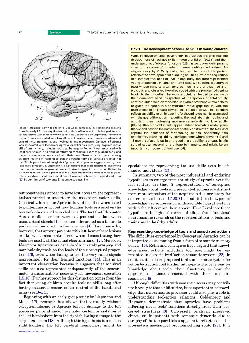
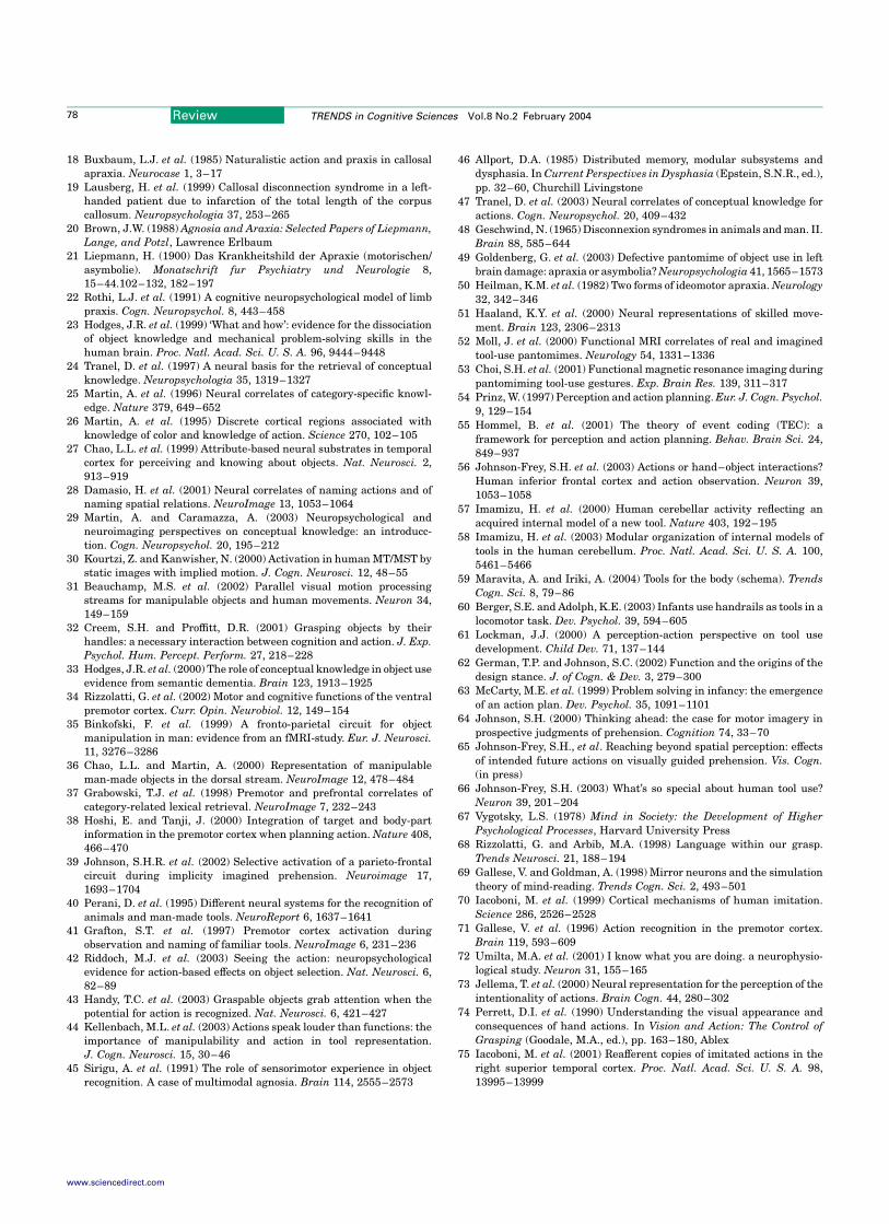
From as early as Morlass in 1928, it has been noted thatbrain damage could selectively impair conceptual knowl-edge about tools versus the skills necessary for theirdexterous usage (cited in [6]). A schematic summarizingwhat was known about locations of brain lesions associ-ated with conceptual versus production difficulties duringthe early 20th century is shown in Figure 1.
When asked to pantomime, or in some cases explicitlydemonstrate, how a familiar tool is used, patients withconceptual level difficulties often make ‘errors of content’in which actions are performed skillfully but out of context.For instance, Ochipa and colleagues report a patient whoattempted to brush his teeth with a comb and eat with atoothbrush. This is not due to a failure of object recognition(agnosia) because the individual could identify the objectsby name [7]. Content errors indicate that representationsnecessary for performing tool-use skills are separable fromsemantic knowledge concerning the relationships betweentools and their associated functions [8]. The terms‘Ideational’ and ‘Conceptual’ Apraxia have been used torefer to this disorder with the latter pertaining specificallyto this semantic component [9]. Although lesion data arenot entirely unequivocal, these semantic deficits areassociated frequently with damage to the left hemisphereat the intersection of the temporal-parietal-occipitalcortices [10].
The reverse dissociation also occurs: Ideomotor Apraxicsretain knowledge of tools’ functions and associated actions,
Corresponding author: Scott H. Johnson-Frey([email protected]).
Review TRENDS in Cognitive Sciences Vol.8 No.2 February 2004
www.sciencedirect.com 1364-6613/$ - see front matter q 2003 Elsevier Ltd. All rights reserved. doi:10.1016/j.tics.2003.12.002
but nonetheless appear to have lost access to the represen-tations needed to undertake the associated motor skills.Classically, Ideomotor Apraxicshave difficultieswhenaskedto pantomime or act out how familiar tools are used on thebasis of either visual or verbal cues. The fact that IdeomotorApraxics often perform worse at pantomime than whenusing actual objects [11], is often interpreted as a failure toperform volitional actions from memory [4]. It is noteworthy,however, that apraxic patients with left-hemisphere lesionsare known to also make errors when demonstrating howtools are used with the actual objects in hand [12]. Moreover,Ideomotor Apraxics are capable of accurately grasping andmanipulating tools on the basis of their perceptual proper-ties [13], even when failing to use the very same objectsappropriately for their learned functions [14]. This is animportant observation because it suggests that acquiredskills are also represented independently of the sensori-motor transformations necessary for movement execution[15,16]. Further support for this distinction comes from thefact that young children acquire tool-use skills long afterhaving mastered sensori-motor control of the hands andarms (see Box 1).
Beginning with an early group study by Liepmann andMaas [17], research has shown that virtually withoutexception Ideomotor Apraxia follows damage to the leftposterior parietal and/or premotor cortex, or isolation ofthe left hemisphere from the right following damage to thecorpus callosum [18]. Although this hypothesis is based onright-handers, the left cerebral hemisphere might be
specialized for representing tool-use skills even in left-handed individuals [19].
In summary, two of the most influential and enduringhypotheses to emerge from the study of apraxia over thelast century are that: (i) representations of conceptualknowledge about tools and associated actions are distinctfrom representations of the acquired skills necessary fordexterous tool use [17,20,21], and (ii) both types ofknowledge are represented in dissociable neural systemswithin the left cerebral hemisphere. Here I evaluate thesehypotheses in light of current findings from functionalneuroimaging research on the representations of tools andtheir uses in healthy adults.
Representing knowledge of tools and associated actions
The difficulties experienced by Conceptual Apraxics can beinterpreted as stemming from a form of semantic memorydeficit [10]. Rothi and colleagues have argued that knowl-edge about actions, including tool use, might be rep-resented in a specialized ‘action semantic system’ [22]. Inaddition, it has been proposed that the semantic system foraction be fractionated further into separate subsystems forknowledge about tools, their functions, or how theappropriate actions associated with their uses aresequenced [8].
Although difficulties with semantic access may contrib-ute heavily to these difficulties, it is important to acknowl-edge that non-semantic processes could also play a role inunderstanding tool-action relations. Goldenberg andHagmann demonstrate that apraxics have problemsinferring novel tools’ functions directly from their per-ceived structures [6]. Conversely, relatively preservedobject use in patients with semantic dementia due toatrophy of the temporal lobes appears to reflect use of thisalternative mechanical problem-solving route [23]. It is
Figure 1. Regions known to affect tool use when damaged. This schematic drawing
from the early 20th century illustrates locations of brain lesions in left parietal cor-
tex associated with three forms of apraxia as understood by Liepmann. Damage to
Region 1 was associated with Limb-Kinetic Apraxia arising from a disturbance of
sensori-motor transformations involved in limb movements. Damage in Region 2
was associated with Ideomotor Apraxia, or difficulties producing acquired motor
skills from memory, including tool use. Damage to Region 3 was associated with
Ideational Apraxia, or difficulties retrieving conceptual knowledge about tools and
the action sequences associated with their uses. There is partial overlap among
adjacent regions in recognition that the various forms of apraxia are often not
manifest in pure form. Although this figure would appear to suggest a strong loca-
lizationist perspective, Liepmann did not believe that representations underlying
tool use, or praxis in general, are exclusive to specific brain sites. Rather he
believed that they were a product of the whole brain with posterior regions poss-
ibly supporting visual representations of planned actions [5]. Reproduced from
[20] by permission of Lawrence Erlbaum Associates, Inc.
Box 1. The development of tool-use skills in young children
Work in developmental psychology has yielded insights into the
development of tool-use skills in young children [60,61] and their
understanding of objects’ functions [62] that could provide important
clues to the nature of underlying neurocognitive mechanisms. An
elegant study by McCarty and colleagues illustrates the important
role that the development of planning abilities play in the acquisition
of a complex tool-use skill [63]. In one study, the authors presented
young children (9-, 14-, and 19-month-olds) with spoons loaded with
food whose handles alternately pointed in the direction of 3 or
9 o’clock, and observed how they coped with the problem of getting
food into their mouths. The youngest children tended to reach with
their dominant hand irrespective of the spoon’s orientation. By
contrast, older children tended to use whichever hand allowed them
to grasp the spoon in a comfortable radial grip; that is, with the
thumb-side of the hand toward the spoon’s bowl. This solution
reflects an ability to anticipate the forthcoming demands associated
with the goal of the action (i.e. getting the food into their mouths) and
adjusting their tool-using movements accordingly. Like adults
[64,65], 18-month-old infants appear able to formulate motor plans
that extend beyond the immediate spatial constraints of the task, and
capture the demands of forthcoming actions. Apparently, this
anticipatory planning ability develops somewhere between 9 and
18 months of age. It has been argued that the ability to engage in this
sort of causal reasoning is unique to humans, and might be an
important component of tool use [66].
Review TRENDS in Cognitive Sciences Vol.8 No.2 February 200472
www.sciencedirect.com
conceivable that functional neuroimaging could be helpfulin exploring the relationship between mechanismsinvolved in these semantic and non-semantic processes.With respect to the former, insights into the representationof knowledge about tools and their usage can be found inthe related literature on semantic memory.
Left posterior temporal cortex and tool identification
Whereas Conceptual Apraxics can accurately name andidentify tools, Tranel and colleagues report patients whoare particularly impaired at tool naming. Like manyConceptual Apraxics, these patients have lesions thatoverlap maximally near the intersection of the parietal,occipital and temporal cortices in the left cerebral hemi-sphere [24]. One interpretation of these data is that thisregion of the left hemisphere computes distinct represen-tations for naming versus other types of semantic knowl-edge associated with tools.
Neuroimaging studies in healthy adults specificallyimplicate posterior left temporal cortex in tool identifi-cation. Martin and colleagues [25] found that namingtools selectively activates posterior left middle temporalgyrus (MTG), an area that is also engaged when subjectsgenerate action words [26], or answer questions abouttools [27]. Likewise, Damasio and colleagues reportactivation in this region when subjects identify actionsor spatial relations performed with versus without atool [28].
The relationship between category-specific namingdeficits, localized patterns of brain activity, and thefunctional architecture of conceptual representations isunresolved [29]. Are these activations reflecting thecategory ‘tool’ per se, or some more elemental propertycommon to all members of this category? One account ofleft MTG activity is that this region is coding perceptualproperties associated with tools [28]. But, what specificallyis different about tools as compared to other artifacts? Onthe basis of its proximity to motion processing centers(putative V5/MT) and its selectivity for ‘manipulable’versus ‘non-manipulable’ artifacts, it has been suggestedthat activations in left MTG might be involved inrepresenting non-biological motions associated with tooluse [27]. The idea that naming a tool could drive theseareas seems reasonable given that putative V5/MT can beactivated by static images that imply action [30]. Recently,Beauchamp and colleagues demonstrated that posteriorMTG is indeed selectively activated when subjects observethe non-biological motion of tools versus the biologicalmotion of human forms [31]. A similar logic has beenapplied to category-specific activations associated withprocessing visually presented tools in bilateral, medialfusiform gyrus. It has been suggested that these areasmight be involved in the representation of tools’shapes [27].
The distinction between Conceptual and IdeomotorApraxia, discussed above, suggests that semantic infor-mation about tools and the representations necessary forthe production of tool-use skills are constructed infunctionally dissociable systems. Nevertheless, behavioralstudies of healthy adults [32] and individuals withsemantic dementia [33] demonstrate that conceptual
representations influence the production of tool-use skills.An important revelation from functional neuroimagingstudies, not predicted from studies of brain-injuredpatients, is that such interactions may come about throughautomatic activation of action representations in premotorand/or parietal areas when semantic information concern-ing familiar tools is accessed.
Left frontal and parietal cortices and action knowledge
In addition to temporal cortex, functional neuroimagingstudies consistently demonstrate that identification oftools and actions activates frontal and parietal areas nottypically associated with recognition or semantic access.Activation of left inferior frontal cortex – a regionassociated with visuomotor transformations for graspingand manipulating objects in both macaques [34] andhumans [35] – is observed during tool naming [25,36] andviewing [36], whereas a larger region including, left middlefrontal gyrus (GFm) is activated when identifying theactions with which tools are associated [37]. Similarly,Perani and colleagues also observed activation in leftdorsal premotor cortex – an area involved in visuomotortransformations for reaching in macaques [38] andhumans [39] – during a same/different tool recognitiontask [40]. Grafton, et al. also found left dorsal premotoractivity when tools are viewed passively, whereas namingthe uses of observed tools additionally recruited leftventral premotor cortex and the supplementary motorarea [41]. These passive viewing effects are consistent withbehavioral observations showing that tools can preferen-tially capture visual selective attention [42]. Recentfindings suggest that selective responses to tools indorsal premotor cortex only occur when this capturetakes place [43].
The consistent activation of left ventral and/or dorsalpremotor cortex during tool naming and/or observation isconsistent with automatic engagement of mechanismsinvolved in the planning of grasping and reachingmovements, respectively. Yet, as detailed below, there isconsiderable evidence to indicate that specific tool-useskills are represented in left posterior parietal cortex(PPC). Although less common, activations in this posteriorregion are also noted when subjects name tools [25,36] andthe actions with which they are associated [28]. Onepossible reason why these sites are not reported more oftenin tasks involving tool perception is that they might only beactivated when subjects are explicitly required to retrievesemantic information concerning tool-use actions [44].Kellenbach et al. observed that visually presented toolsengage left ventral premotor cortex and posterior left MTGregardless of whether the retrieval task demands judg-ments about their functions or associated actions. In otherwords, responses in these areas are automatically evokedby the mere observation of familiar tools. Conversely,activations in left PPC are only observed when subjectsexplicitly retrieve actions associated with tools (Figure 2).
Neuroimaging data suggesting automatic activation offrontal and/or parietal areas involved in representingactions during perceptual tasks has implications forinterpreting existing findings in the neuropsychologyliterature as well. For instance, Sirigu et al. report an
Review TRENDS in Cognitive Sciences Vol.8 No.2 February 2004 73
www.sciencedirect.com
agnosic patient with bilateral temporal lobe lesions whohad considerable difficulty identifying the functions oftools or the contexts in which they would typically be used.Nevertheless, he was capable of manipulating these itemsskillfully in a fashion appropriate with their usage [45].This case might be interpreted as evidence for a non-semantic route between structural descriptions of objectsconstructed in earlier visual centers directly to parieto-frontal action representations. It is also possible thatactivation of semantic representations, insufficient forexplicit recognition, is still capable of inducing automaticactivation of action representations.
It is worth considering an alternative to the automaticactivation of action representations during semantic tasksinvolving tools and/or actions. Perhaps activation of leftparietal and premotor sites during semantic tasks invol-ving tools indicates that they too play a role in represent-ing conceptual information associated with these objects.More precisely, semantic information about tools might bedistributed among several regions of the left hemispherethat are active at the time of encoding [46]. This seemsreasonable if one assumes that visual properties of tools(involving left MTG) are likely to be acquired during activemanipulation (involving sensori-motor regions of leftparietal and premotor cortices). Conceivably, functional
neuroimaging studies could be developed to distinguishbetween these two alternatives.
Overall, the neuroimaging results are generally con-sistent with a recent lesion analysis showing that patientswith selective impairments performing non-verbal con-ceptual judgments about actions, including tool use,have maximal lesion overlap in a network of left-hemi-sphere regions including posterior left MTG, as well aspremotor/prefrontal and parietal cortices [47].
Representing acquired tool-use skills
As early as 1905 (Liepmann, [17]), it was known thatdamage to the left PPC could affect the ability to produceskills associated with tools (Figure 1). In the interveningcentury there have been a variety of attempts to explainthis fact [4]. One class of theories posit that IdeomotorApraxia reflects damage to a more general faculty uniqueto the left hemisphere, such as the ability to constructsymbolic representations (i.e. asymbolia) [48], or to formactions on the basis of objects’ perceptual properties [49].A second class argues that the posterior left hemisphere isthe locus for representations of acquired tool-use skills.Specifically, Heilman and colleagues implicate the supra-marginal gyrus, or Brodmann Area (BA) 40, of the leftinferior parietal lobule. According to this view, Ideomotor
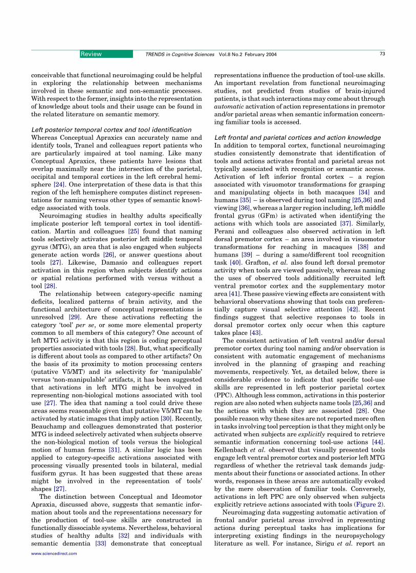
Figure 2. Attention mediated activation of the left inferior parietal lobule during semantic retrieval involving manipulable objects, i.e. tools. In contrast to left MTG and
inferior frontal cortex that activate in response to the mere observation of tools, left BA40 (green arrows) and a location in the anterior intraparietal sulcus (blue arrows)
show a marked sensitivity to the type of retrieval task. When performing action judgments in response to manipulable objects (MA), this region is more active than when
making function judgments about manipulable objects (MF), function judgments about non-manipuable objects (NMF), or in a control condition that involved simply
observing tools (C). The histogram illustrates the relative strength of the response within the point marked by the red dot within the parietal region of interest (white cir-
cles). Reproduced from [44] by permission of The MIT Press.
Review TRENDS in Cognitive Sciences Vol.8 No.2 February 200474
www.sciencedirect.com
Apraxia resulting from damage to this region can thereforebe understood as resulting from degradation of thesemotor memories [50].
Left parieto-frontal mechanisms and tool-use skills
A recent MRI-based lesion analysis reports that incomparison with left-hemisphere injured patients withoutapraxia, Ideomotor Apraxics present with maximal lesionoverlap within and adjacent to the left intraparietalsulcus – including BA7, angular (BA 39) and supramar-ginal (BA 40) gyri – and/or the left middle frontal gyrus(GFm) [51]. To account for such observations, andbehavioral differences between parietal and frontal-lesioned patients, Heilman and colleagues propose thatthese regions play different roles in representing tool-useskills [50]. Consistent with the idea that skill represen-tations are damaged, patients with lesions including theleft BA 40 have difficulty performing manual actions anddiscriminating ‘good’ versus ‘bad’ instances of observedactions. Patients with frontal lesions also perform poorlyat production, but have no difficulties with actiondiscrimination.
According to this view, intact discrimination indicatesthat patients with frontal lesions retain intact skillrepresentations, yet have difficulty accessing this infor-mation for purposes of action production. This interpret-ation assumes that the same representations are involvedin action production and recognition (see Box 2). Thisproposed distinction between the roles of frontal (retrieval)and parietal (representation) mechanisms in tool-useskills is well-suited for evaluation with functional neuro-imaging techniques. However, in contrast to the sizeableliterature on conceptual-level representations, few studieshave used these methods to investigate mechanismsinvolved in representing and producing tool-use skills.Undoubtedly, the constraints of current neuroimagingtechniques have contributed to this situation. Headmovements, especially those correlated with task perform-ance, can compromise the validity of fMRI results, and theworkspace for limb movements is often highly constrained.Available studies to date have focused on pantomime,mental imagery, action observation, and the acquisition ofskills associated with novel tools.
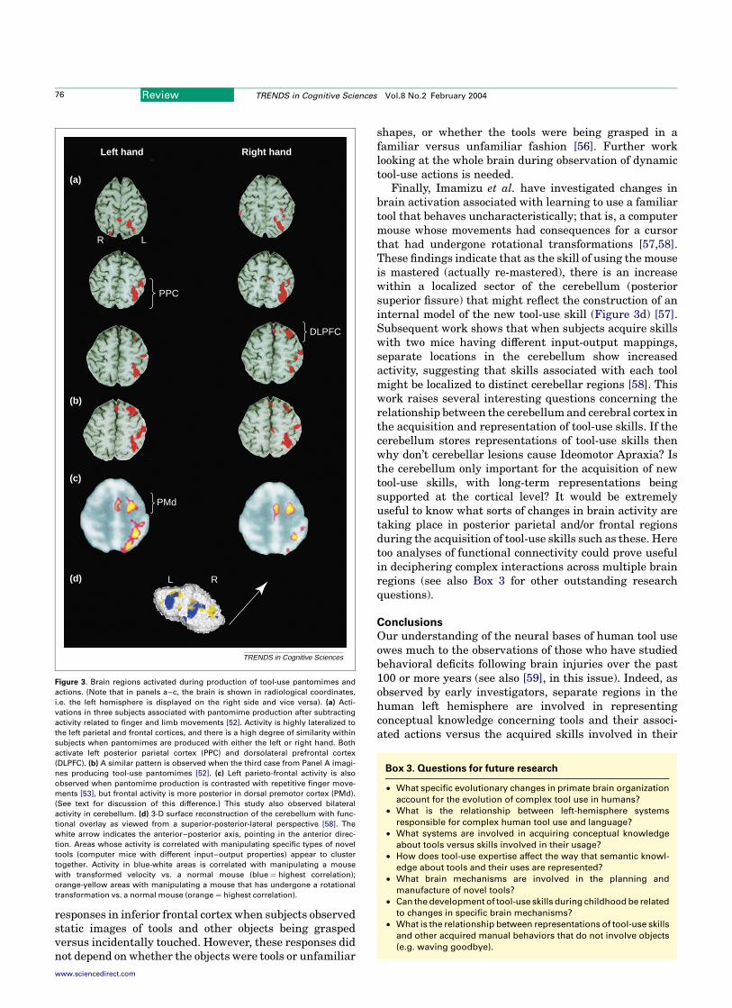
Consistent with the lesion-analysis data of IdeomotorApraxics discussed above, Figure 3 illustrates that whenactivations associated with complex yet meaninglessfinger and limb movements are removed, pantomiming(Figure 3a) or imagining (3b) tool-use gestures with eitherhand activates left PPC in and around the intraparietalsulcus [52]. A similar pattern is also present when tool-usepantomimes involving either hand are contrasted withrepetitive finger movements (Figure 3c) [53]. However,neither investigation observed activations in left GFm, aswould be expected given the lesion-analysis data. Instead,they report left dorsolateral prefrontal [52] or dorsalpremotor cortex [53] activations. One reason for theseinconsistencies could have to do with experimental design.Both studies used block paradigms that assume thatactivations related to pantomime execution could beremoved by subtracting data from non-gestural motor-control tasks. A limitation of this strategy is that any brain
areas active in both the pantomime and motor-controlconditions are eliminated. This could possibly be the fate ofleft GFm. The fact that the two studies differ from oneanother in terms of the left frontal areas they do find mightreflect differences in the demands of their respectivecontrol tasks. Studies with more sophisticated experimen-tal designs are needed before any strong conclusions can beadvanced regarding the source(s) of differences betweenthe lesion and neuroimaging data with regard to areas ofleft frontal cortex involved in representing tool-use skills.A crucial next step will involve using event-related and/orparametric designs to address outstanding issues likethis one. For instance, is it possible to test Heilman et al.’shypothesis about the differential roles of parietal andfrontal mechanisms in skill representations? Likewise,studies of functional connectivity might enable neuroima-gers to determine how and under what conditions brainregions in this left-lateralized network interact.
Another approach is to examine how brain areasrespond to observation of tool-use actions. The validity ofthis approach for studying representations of skill dependson whether or not the same mechanisms contribute bothto action comprehension and production, the so-called‘Common-Coding Hypothesis’ [54,55]. By way of illus-tration, Johnson-Frey and colleagues found bilateral
Box 2. Toward a neural basis for the cultural transmission
of tool-use skills
An important component of skill acquisition is the social exchange
that occurs between people as new behaviors are taught and learned
[67]. Recently, there has been considerable interest in identifying
brain mechanisms that might enable us to form a link between
observing others’ actions and performing similar behaviors our-
selves [68,69]. Much of this work is motivated by observations of
mirror neurons in macaque inferior frontal cortex (area F5c) that
respond either when the animal produces a given action or observes
the experimenter performing a comparable behavior. Neuroimaging
studies demonstrate similar responses in human left inferior frontal
cortex (BA44), which might be a homologue of macaque area F5 [70].
With respect to tool use, it is important to note that these mirror cells’
responses appear to depend on the animal observing [71] or inferring
[72] specific interactions between the hand and target object. In other
words, these neurons might represent observed goals of hand
actions. Recent evidence indicates that human inferior frontal
cortex also distinguishes between the goals inherent in observed
hand–object interactions [56].
Inferior frontal cortex receives inputs from regions of the inferior
parietal lobule implicated in the representation of object grasping
and manipulation (anterior intraparietal cortex), and tool-use skills
(supramarginal gyrus, or BA40; see Figure 4 in main text), as well as
the superior temporal sulcus (STS). Perrett and colleagues describe
cells in macaque anterior STS that code specific observed limb
movements, and are sensitive to the direction of the actors’ attention
[73]. Like cells in F5c, some of these units are also sensitive to hand–
object interactions [74]. Likewise, Iacoboni and colleagues recently
presented evidence that a region in the human STS is involved in
matching observed actions with those being produced during
imitation [75]. Together these sources of evidence suggest the
existence of a distributed representational system for bridging
between the perception and production of action [54,55]. This circuit
has characteristics that could serve as a critical mechanism for the
cultural transmission of skills including tool use in humans through
observational learning and/or imitation. The relationship between
this system and those involved in representing semantic knowledge
about tools and tool-use skills is a topic in need of investigation.
Review TRENDS in Cognitive Sciences Vol.8 No.2 February 2004 75
www.sciencedirect.com
responses in inferior frontal cortex when subjects observedstatic images of tools and other objects being graspedversus incidentally touched. However, these responses didnot depend on whether the objects were tools or unfamiliar
shapes, or whether the tools were being grasped in afamiliar versus unfamiliar fashion [56]. Further worklooking at the whole brain during observation of dynamictool-use actions is needed.
Finally, Imamizu et al. have investigated changes inbrain activation associated with learning to use a familiartool that behaves uncharacteristically; that is, a computermouse whose movements had consequences for a cursorthat had undergone rotational transformations [57,58].These findings indicate that as the skill of using the mouseis mastered (actually re-mastered), there is an increasewithin a localized sector of the cerebellum (posteriorsuperior fissure) that might reflect the construction of aninternal model of the new tool-use skill (Figure 3d) [57].Subsequent work shows that when subjects acquire skillswith two mice having different input-output mappings,separate locations in the cerebellum show increasedactivity, suggesting that skills associated with each toolmight be localized to distinct cerebellar regions [58]. Thiswork raises several interesting questions concerning therelationship between the cerebellum and cerebral cortex inthe acquisition and representation of tool-use skills. If thecerebellum stores representations of tool-use skills thenwhy don’t cerebellar lesions cause Ideomotor Apraxia? Isthe cerebellum only important for the acquisition of newtool-use skills, with long-term representations beingsupported at the cortical level? It would be extremelyuseful to know what sorts of changes in brain activity aretaking place in posterior parietal and/or frontal regionsduring the acquisition of tool-use skills such as these. Heretoo analyses of functional connectivity could prove usefulin deciphering complex interactions across multiple brainregions (see also Box 3 for other outstanding researchquestions).
Conclusions
Our understanding of the neural bases of human tool useowes much to the observations of those who have studiedbehavioral deficits following brain injuries over the past100 or more years (see also [59], in this issue). Indeed, asobserved by early investigators, separate regions in thehuman left hemisphere are involved in representingconceptual knowledge concerning tools and their associ-ated actions versus the acquired skills involved in their
Figure 3. Brain regions activated during production of tool-use pantomimes and
actions. (Note that in panels a–c, the brain is shown in radiological coordinates,
i.e. the left hemisphere is displayed on the right side and vice versa). (a) Acti-
vations in three subjects associated with pantomime production after subtracting
activity related to finger and limb movements [52]. Activity is highly lateralized to
the left parietal and frontal cortices, and there is a high degree of similarity within
subjects when pantomimes are produced with either the left or right hand. Both
activate left posterior parietal cortex (PPC) and dorsolateral prefrontal cortex
(DLPFC). (b) A similar pattern is observed when the third case from Panel A imagi-
nes producing tool-use pantomimes [52]. (c) Left parieto-frontal activity is also
observed when pantomime production is contrasted with repetitive finger move-
ments [53], but frontal activity is more posterior in dorsal premotor cortex (PMd).
(See text for discussion of this difference.) This study also observed bilateral
activity in cerebellum. (d) 3-D surface reconstruction of the cerebellum with func-
tional overlay as viewed from a superior-posterior-lateral perspective [58]. The
white arrow indicates the anterior–posterior axis, pointing in the anterior direc-
tion. Areas whose activity is correlated with manipulating specific types of novel
tools (computer mice with different input–output properties) appear to cluster
together. Activity in blue-white areas is correlated with manipulating a mouse
with transformed velocity vs. a normal mouse (blue ¼ highest correlation);
orange-yellow areas with manipulating a mouse that has undergone a rotational
transformation vs. a normal mouse (orange ¼ highest correlation).
TRENDS in Cognitive Sciences
(a)
(b)
(c)
(d)
Left hand Right hand
R L
PPC
DLPFC
PMd
L R
Box 3. Questions for future research
† What specific evolutionary changes in primate brain organization
account for the evolution of complex tool use in humans?
† What is the relationship between left-hemisphere systems
responsible for complex human tool use and language?
† What systems are involved in acquiring conceptual knowledge
about tools versus skills involved in their usage?
† How does tool-use expertise affect the way that semantic knowl-
edge about tools and their uses are represented?
† What brain mechanisms are involved in the planning and
manufacture of novel tools?
† Can the development of tool-use skills during childhood be related
to changes in specific brain mechanisms?
† What is the relationship between representations of tool-use skills
and other acquired manual behaviors that do not involve objects
(e.g. waving goodbye).
Review TRENDS in Cognitive Sciences Vol.8 No.2 February 200476
www.sciencedirect.com
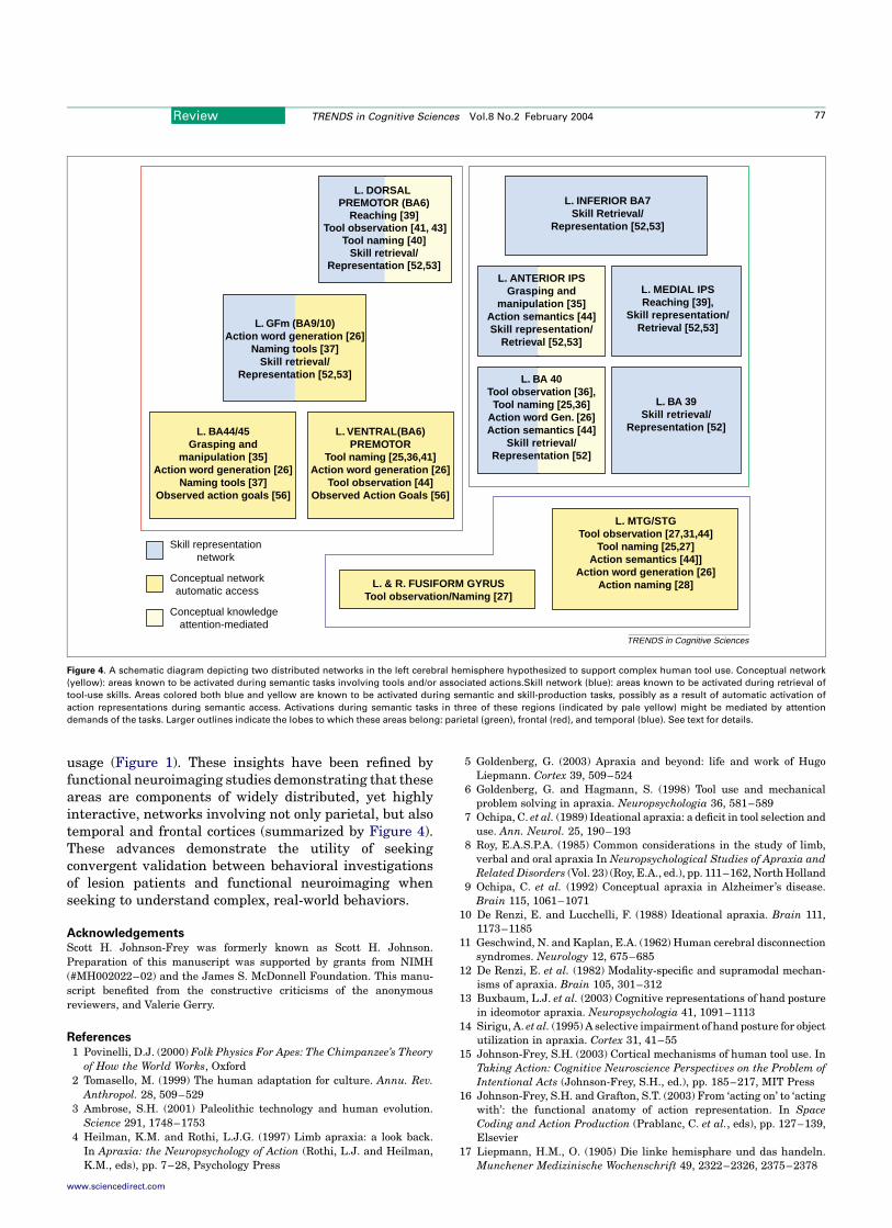
usage (Figure 1). These insights have been refined byfunctional neuroimaging studies demonstrating that theseareas are components of widely distributed, yet highlyinteractive, networks involving not only parietal, but alsotemporal and frontal cortices (summarized by Figure 4).These advances demonstrate the utility of seekingconvergent validation between behavioral investigationsof lesion patients and functional neuroimaging whenseeking to understand complex, real-world behaviors.
AcknowledgementsScott H. Johnson-Frey was formerly known as Scott H. Johnson.Preparation of this manuscript was supported by grants from NIMH(#MH002022–02) and the James S. McDonnell Foundation. This manu-script benefited from the constructive criticisms of the anonymousreviewers, and Valerie Gerry.
References
1 Povinelli, D.J. (2000) Folk Physics For Apes: The Chimpanzee’s Theory
of How the World Works, Oxford2 Tomasello, M. (1999) The human adaptation for culture. Annu. Rev.
Anthropol. 28, 509–5293 Ambrose, S.H. (2001) Paleolithic technology and human evolution.
Science 291, 1748–17534 Heilman, K.M. and Rothi, L.J.G. (1997) Limb apraxia: a look back.
In Apraxia: the Neuropsychology of Action (Rothi, L.J. and Heilman,K.M., eds), pp. 7–28, Psychology Press
5 Goldenberg, G. (2003) Apraxia and beyond: life and work of HugoLiepmann. Cortex 39, 509–524
6 Goldenberg, G. and Hagmann, S. (1998) Tool use and mechanicalproblem solving in apraxia. Neuropsychologia 36, 581–589
7 Ochipa, C. et al. (1989) Ideational apraxia: a deficit in tool selection anduse. Ann. Neurol. 25, 190–193
8 Roy, E.A.S.P.A. (1985) Common considerations in the study of limb,verbal and oral apraxia In Neuropsychological Studies of Apraxia andRelated Disorders (Vol. 23) (Roy, E.A., ed.), pp. 111–162, North Holland
9 Ochipa, C. et al. (1992) Conceptual apraxia in Alzheimer’s disease.Brain 115, 1061–1071
10 De Renzi, E. and Lucchelli, F. (1988) Ideational apraxia. Brain 111,1173–1185
11 Geschwind, N. and Kaplan, E.A. (1962) Human cerebral disconnectionsyndromes. Neurology 12, 675–685
12 De Renzi, E. et al. (1982) Modality-specific and supramodal mechan-isms of apraxia. Brain 105, 301–312
13 Buxbaum, L.J. et al. (2003) Cognitive representations of hand posturein ideomotor apraxia. Neuropsychologia 41, 1091–1113
14 Sirigu, A. et al. (1995) A selective impairment of hand posture for objectutilization in apraxia. Cortex 31, 41–55
15 Johnson-Frey, S.H. (2003) Cortical mechanisms of human tool use. InTaking Action: Cognitive Neuroscience Perspectives on the Problem ofIntentional Acts (Johnson-Frey, S.H., ed.), pp. 185–217, MIT Press
16 Johnson-Frey, S.H. and Grafton, S.T. (2003) From ‘acting on’ to ‘actingwith’: the functional anatomy of action representation. In SpaceCoding and Action Production (Prablanc, C. et al., eds), pp. 127–139,Elsevier
17 Liepmann, H.M., O. (1905) Die linke hemisphare und das handeln.Munchener Medizinische Wochenschrift 49, 2322–2326, 2375–2378
Figure 4. A schematic diagram depicting two distributed networks in the left cerebral hemisphere hypothesized to support complex human tool use. Conceptual network
(yellow): areas known to be activated during semantic tasks involving tools and/or associated actions.Skill network (blue): areas known to be activated during retrieval of
tool-use skills. Areas colored both blue and yellow are known to be activated during semantic and skill-production tasks, possibly as a result of automatic activation of
action representations during semantic access. Activations during semantic tasks in three of these regions (indicated by pale yellow) might be mediated by attention
demands of the tasks. Larger outlines indicate the lobes to which these areas belong: parietal (green), frontal (red), and temporal (blue). See text for details.
TRENDS in Cognitive Sciences
Reaching [39] Tool observation [41, 43]
Tool naming [40]
L. DORSAL PREMOTOR (BA6)
Skill retrieval/Representation [52,53]
L. INFERIOR BA7Skill Retrieval/
Representation [52,53]
L. GFm (BA9/10)Action word generation [26]
Naming tools [37]Skill retrieval/
Representation [52,53]
L. BA44/45Grasping and
manipulation [35]Action word generation [26]
Naming tools [37]Observed action goals [56]
L. VENTRAL(BA6)PREMOTOR
Tool naming [25,36,41]Action word generation [26]
Tool observation [44]Observed Action Goals [56]
L. ANTERIOR IPSGrasping and
manipulation [35]Action semantics [44]Skill representation/
Retrieval [52,53]
L. MEDIAL IPSReaching [39],
Skill representation/Retrieval [52,53]
L. BA 39Skill retrieval/
Representation [52]
L. BA 40Tool observation [36],Tool naming [25,36]
Action word Gen. [26]Action semantics [44]
Skill retrieval/Representation [52]
L. MTG/STGTool observation [27,31,44]
Tool naming [25,27]Action semantics [44]]
Action word generation [26]Action naming [28]L. & R. FUSIFORM GYRUS
Tool observation/Naming [27]
Skill representationnetwork
Conceptual networkautomatic access
Conceptual knowledgeattention-mediated
Review TRENDS in Cognitive Sciences Vol.8 No.2 February 2004 77
www.sciencedirect.com
18 Buxbaum, L.J. et al. (1985) Naturalistic action and praxis in callosalapraxia. Neurocase 1, 3–17
19 Lausberg, H. et al. (1999) Callosal disconnection syndrome in a left-handed patient due to infarction of the total length of the corpuscallosum. Neuropsychologia 37, 253–265
20 Brown, J.W. (1988) Agnosia and Araxia: Selected Papers of Liepmann,Lange, and Potzl, Lawrence Erlbaum
21 Liepmann, H. (1900) Das Krankheitshild der Apraxie (motorischen/asymbolie). Monatschrift fur Psychiatry und Neurologie 8,15–44.102–132, 182–197
22 Rothi, L.J. et al. (1991) A cognitive neuropsychological model of limbpraxis. Cogn. Neuropsychol. 8, 443–458
23 Hodges, J.R. et al. (1999) ‘What and how’: evidence for the dissociationof object knowledge and mechanical problem-solving skills in thehuman brain. Proc. Natl. Acad. Sci. U. S. A. 96, 9444–9448
24 Tranel, D. et al. (1997) A neural basis for the retrieval of conceptualknowledge. Neuropsychologia 35, 1319–1327
25 Martin, A. et al. (1996) Neural correlates of category-specific knowl-edge. Nature 379, 649–652
26 Martin, A. et al. (1995) Discrete cortical regions associated withknowledge of color and knowledge of action. Science 270, 102–105
27 Chao, L.L. et al. (1999) Attribute-based neural substrates in temporalcortex for perceiving and knowing about objects. Nat. Neurosci. 2,913–919
28 Damasio, H. et al. (2001) Neural correlates of naming actions and ofnaming spatial relations. NeuroImage 13, 1053–1064
29 Martin, A. and Caramazza, A. (2003) Neuropsychological andneuroimaging perspectives on conceptual knowledge: an introducc-tion. Cogn. Neuropsychol. 20, 195–212
30 Kourtzi, Z. and Kanwisher, N. (2000) Activation in human MT/MST bystatic images with implied motion. J. Cogn. Neurosci. 12, 48–55
31 Beauchamp, M.S. et al. (2002) Parallel visual motion processingstreams for manipulable objects and human movements. Neuron 34,149–159
32 Creem, S.H. and Proffitt, D.R. (2001) Grasping objects by theirhandles: a necessary interaction between cognition and action. J. Exp.Psychol. Hum. Percept. Perform. 27, 218–228
33 Hodges, J.R. et al. (2000) The role of conceptual knowledge in object useevidence from semantic dementia. Brain 123, 1913–1925
34 Rizzolatti, G. et al. (2002) Motor and cognitive functions of the ventralpremotor cortex. Curr. Opin. Neurobiol. 12, 149–154
35 Binkofski, F. et al. (1999) A fronto-parietal circuit for objectmanipulation in man: evidence from an fMRI-study. Eur. J. Neurosci.11, 3276–3286
36 Chao, L.L. and Martin, A. (2000) Representation of manipulableman-made objects in the dorsal stream. NeuroImage 12, 478–484
37 Grabowski, T.J. et al. (1998) Premotor and prefrontal correlates ofcategory-related lexical retrieval. NeuroImage 7, 232–243
38 Hoshi, E. and Tanji, J. (2000) Integration of target and body-partinformation in the premotor cortex when planning action. Nature 408,466–470
39 Johnson, S.H.R. et al. (2002) Selective activation of a parieto-frontalcircuit during implicity imagined prehension. Neuroimage 17,1693–1704
40 Perani, D. et al. (1995) Different neural systems for the recognition ofanimals and man-made tools. NeuroReport 6, 1637–1641
41 Grafton, S.T. et al. (1997) Premotor cortex activation duringobservation and naming of familiar tools. NeuroImage 6, 231–236
42 Riddoch, M.J. et al. (2003) Seeing the action: neuropsychologicalevidence for action-based effects on object selection. Nat. Neurosci. 6,82–89
43 Handy, T.C. et al. (2003) Graspable objects grab attention when thepotential for action is recognized. Nat. Neurosci. 6, 421–427
44 Kellenbach, M.L. et al. (2003) Actions speak louder than functions: theimportance of manipulability and action in tool representation.J. Cogn. Neurosci. 15, 30–46
45 Sirigu, A. et al. (1991) The role of sensorimotor experience in objectrecognition. A case of multimodal agnosia. Brain 114, 2555–2573
46 Allport, D.A. (1985) Distributed memory, modular subsystems anddysphasia. In Current Perspectives in Dysphasia (Epstein, S.N.R., ed.),pp. 32–60, Churchill Livingstone
47 Tranel, D. et al. (2003) Neural correlates of conceptual knowledge foractions. Cogn. Neuropsychol. 20, 409–432
48 Geschwind, N. (1965) Disconnexion syndromes in animals and man. II.Brain 88, 585–644
49 Goldenberg, G. et al. (2003) Defective pantomime of object use in leftbrain damage: apraxia or asymbolia? Neuropsychologia 41, 1565–1573
50 Heilman, K.M. et al. (1982) Two forms of ideomotor apraxia. Neurology32, 342–346
51 Haaland, K.Y. et al. (2000) Neural representations of skilled move-ment. Brain 123, 2306–2313
52 Moll, J. et al. (2000) Functional MRI correlates of real and imaginedtool-use pantomimes. Neurology 54, 1331–1336
53 Choi, S.H. et al. (2001) Functional magnetic resonance imaging duringpantomiming tool-use gestures. Exp. Brain Res. 139, 311–317
54 Prinz, W. (1997) Perception and action planning. Eur. J. Cogn. Psychol.9, 129–154
55 Hommel, B. et al. (2001) The theory of event coding (TEC): aframework for perception and action planning. Behav. Brain Sci. 24,849–937
56 Johnson-Frey, S.H. et al. (2003) Actions or hand–object interactions?Human inferior frontal cortex and action observation. Neuron 39,1053–1058
57 Imamizu, H. et al. (2000) Human cerebellar activity reflecting anacquired internal model of a new tool. Nature 403, 192–195
58 Imamizu, H. et al. (2003) Modular organization of internal models oftools in the human cerebellum. Proc. Natl. Acad. Sci. U. S. A. 100,5461–5466
59 Maravita, A. and Iriki, A. (2004) Tools for the body (schema). TrendsCogn. Sci. 8, 79–86
60 Berger, S.E. and Adolph, K.E. (2003) Infants use handrails as tools in alocomotor task. Dev. Psychol. 39, 594–605
61 Lockman, J.J. (2000) A perception-action perspective on tool usedevelopment. Child Dev. 71, 137–144
62 German, T.P. and Johnson, S.C. (2002) Function and the origins of thedesign stance. J. of Cogn. & Dev. 3, 279–300
63 McCarty, M.E. et al. (1999) Problem solving in infancy: the emergenceof an action plan. Dev. Psychol. 35, 1091–1101
64 Johnson, S.H. (2000) Thinking ahead: the case for motor imagery inprospective judgments of prehension. Cognition 74, 33–70
65 Johnson-Frey, S.H., et al. Reaching beyond spatial perception: effectsof intended future actions on visually guided prehension. Vis. Cogn.(in press)
66 Johnson-Frey, S.H. (2003) What’s so special about human tool use?Neuron 39, 201–204
67 Vygotsky, L.S. (1978) Mind in Society: the Development of HigherPsychological Processes, Harvard University Press
68 Rizzolatti, G. and Arbib, M.A. (1998) Language within our grasp.Trends Neurosci. 21, 188–194
69 Gallese, V. and Goldman, A. (1998) Mirror neurons and the simulationtheory of mind-reading. Trends Cogn. Sci. 2, 493–501
70 Iacoboni, M. et al. (1999) Cortical mechanisms of human imitation.Science 286, 2526–2528
71 Gallese, V. et al. (1996) Action recognition in the premotor cortex.Brain 119, 593–609
72 Umilta, M.A. et al. (2001) I know what you are doing. a neurophysio-logical study. Neuron 31, 155–165
73 Jellema, T. et al. (2000) Neural representation for the perception of theintentionality of actions. Brain Cogn. 44, 280–302
74 Perrett, D.I. et al. (1990) Understanding the visual appearance andconsequences of hand actions. In Vision and Action: The Control ofGrasping (Goodale, M.A., ed.), pp. 163–180, Ablex
75 Iacoboni, M. et al. (2001) Reafferent copies of imitated actions in theright superior temporal cortex. Proc. Natl. Acad. Sci. U. S. A. 98,13995–13999
Review TRENDS in Cognitive Sciences Vol.8 No.2 February 200478
www.sciencedirect.com