Embed Size (px)
Citation preview
On the Nature and Evolution of the Neural Bases ofHuman LanguagePHILIP LIEBERMAN
Department of Cognitive and Linguistic Sciences and Department of Anthropology, Brown University, Providence,Rhode Island 02912-1978
KEY WORDS brain; neurophysiology; neural circuits; evolution; language
ABSTRACT The traditional theory equating the brainbases of language with Broca’s and Wernicke’s neocorticalareas is wrong. Neural circuits linking activity in anatom-ically segregated populations of neurons in subcorticalstructures and the neocortex throughout the human brainregulate complex behaviors such as walking, talking, andcomprehending the meaning of sentences. When we hearor read a word, neural structures involved in the percep-tion or real-world associations of the word are activated aswell as posterior cortical regions adjacent to Wernicke’sarea. Many areas of the neocortex and subcortical struc-tures support the cortical-striatal-cortical circuits thatconfer complex syntactic ability, speech production, and alarge vocabulary. However, many of these structures alsoform part of the neural circuits regulating other aspects ofbehavior. For example, the basal ganglia, which regulatemotor control, are also crucial elements in the circuits thatconfer human linguistic ability and abstract reasoning.The cerebellum, traditionally associated with motor con-trol, is active in motor learning. The basal ganglia are alsokey elements in reward-based learning. Data from studiesof Broca’s aphasia, Parkinson’s disease, hypoxia, focalbrain damage, and a genetically transmitted brain anom-aly (the putative “language gene,” family KE), and fromcomparative studies of the brains and behavior of otherspecies, demonstrate that the basal ganglia sequence the
discrete elements that constitute a complete motor act,syntactic process, or thought process. Imaging studies ofintact human subjects and electrophysiologic and tracerstudies of the brains and behavior of other species confirmthese findings. As Dobzansky put it, “Nothing in biologymakes sense except in the light of evolution” (cited inMayr, 1982). That applies with as much force to the hu-man brain and the neural bases of language as it does tothe human foot or jaw. The converse follows: the mark ofevolution on the brains of human beings and other speciesprovides insight into the evolution of the brain bases ofhuman language. The neural substrate that regulatedmotor control in the common ancestor of apes and humansmost likely was modified to enhance cognitive and linguis-tic ability. Speech communication played a central role inthis process. However, the process that ultimately re-sulted in the human brain may have started when ourearliest hominid ancestors began to walk. Am J PhysAnthropol 45:36–62, 2002. © 2002 Wiley-Liss, Inc.
TABLE OF CONTENTS
Brain-Behavior Models ............................................................................................................................................. 37The data base ......................................................................................................................................................... 37The traditional locationist model ......................................................................................................................... 37Circuit models ........................................................................................................................................................ 38Aphasia ................................................................................................................................................................... 40
Cortical-Striatal-Cortical Circuits ............................................................................................................................ 40Probable basal ganglia operations ....................................................................................................................... 42
Sequencing .......................................................................................................................................................... 42Reward-based learning ...................................................................................................................................... 42
Linguistic and Cognitive Consequences of Subcortical Brain Damage ................................................................ 43Speech ..................................................................................................................................................................... 43Syntax ..................................................................................................................................................................... 44Motor and cognitive set-shifting ........................................................................................................................... 45Cerebellum ............................................................................................................................................................. 46
The “Language Gene” and Universal Grammar ..................................................................................................... 46Cortex ......................................................................................................................................................................... 47
Grant Sponsor: National Space Biomedical Research Institute.
DOI 10.1002/ajpa.10171Published online in Wiley InterScience (www.interscience.wiley.
com).
YEARBOOK OF PHYSICAL ANTHROPOLOGY 45:36–62 (2002)
© 2002 WILEY-LISS, INC.

Verbal working memory: Broca’s and Wernicke’s areas .................................................................................... 47Dynamic neural systems ....................................................................................................................................... 48The brain’s dictionary ........................................................................................................................................... 49Cortical plasticity .................................................................................................................................................. 50Acquisition of plans for motor control, cognition, and language ....................................................................... 50Brain lateralization and language ....................................................................................................................... 50
Summary: Neural Bases of Language ..................................................................................................................... 51On the Evolution of the Brain Bases of Language ................................................................................................. 51
Lexical ability ........................................................................................................................................................ 52Syntax ..................................................................................................................................................................... 52Brain size ............................................................................................................................................................... 52Fossil evidence for lateralization: Broca’s and Wernicke’s areas ...................................................................... 52Gestural language ................................................................................................................................................. 53Social factors .......................................................................................................................................................... 53
Speech and Complex Syntax .................................................................................................................................... 53Speech ..................................................................................................................................................................... 53The selective advantages of human speech ......................................................................................................... 54The supralaryngeal vocal tract ............................................................................................................................. 54
When Did Human Speech, Complex Syntax, and the Lexicon Evolve? ............................................................... 56Complex syntax ..................................................................................................................................................... 56Walking .................................................................................................................................................................. 57The archaeological record ..................................................................................................................................... 57
Concluding Comments .............................................................................................................................................. 57Literature Cited ........................................................................................................................................................ 58
BRAIN-BEHAVIOR MODELS
The data base
The major problem that has vexed studies of theevolution of language is that human beings are pres-ently the single living species to possess complexlinguistic ability. The archaic hominids who mayhave possessed intermediate stages of linguisticability are extinct. While evidence from genetics andcomparative anatomy shows that living apes appearto retain many of the features that characterized thecommon ancestor that they shared with humans,apes clearly do not have the ability to acquire allaspects of human linguistic ability. The apparentgulf between the communicative abilities of living apesand human language has led to the claim that theneural bases of human language are disjointed fromthose involved in vocal communications of apes (e.g.,Burling, 1993), and to incorrect assertions that apeslack any vestige of linguistic ability (e.g., Terrace et al.,1979; Pinker, 1994). This in turn leads to theories thatpostulate abrupt discontinuities, or “stages,” in theevolution of human language such as “protolanguage”that lacked syntax (Bickerton, 1990), or to claims thatlanguage could have not have evolved by means ofDarwinian processes (Chomsky, 1976, 1986).
However, these difficulties can be surmounted byapplying the principles and techniques of evolution-ary biology. Comparative studies, which we willnote, clearly show that human language sharesmany primitive features with the communicationssystems of other species. By identifying the derivedaspects of human linguistic ability that differentiateit from related species, we can focus our attention onidentifying and tracing the evolution of the biologi-cal substrate that yielded human language. And
advances in comparative neurophysiology have gen-erated insights into the biology of language. There-fore, though the absence of the intermediate stagesof the evolution of hominid linguistic ability placeslimits on our ability to ever fully understand howand when human language evolved, the findings ofrecent neurophysiologic and behavioral studies ofhuman beings and other species place the study ofthe human brain and language in a different light.
The relevant studies include traditional observa-tions of “experiments-in-nature,” deficits resultingfrom disease or trauma to the brain, and studiesemploying noninvasive techniques functional mag-netic resonance imaging (fMRI) and positron emissiontomography (PET) that indirectly track activity in thebrain by means of blood flow measurements (Logoth-etis et al., 2001). Data from studies of neural activity inother species, that make use of invasive techniquesemploying tracers and direct electrophysiologic record-ings of neural activity, also provide essential insights,placing findings from studies of human subjects into acoherent framework. These advances have led to anunderstanding of the neural bases of human languagethat transcends the traditional theory equating lan-guage with Broca’s and Wernicke’s areas of the cortex,a theory that dates back to the 19th century. Throughthese studies, it has become apparent that the evolu-tion of the neural bases of the derived properties ofhuman language, i.e., speech, complex syntax, and analmost limitless vocabulary, derive from Darwinianmechanisms.
The traditional locationist model
History and technology play major roles in struc-turing human thought. In the early years of the 19thcentury, phrenologists systematically identified spe-
NEURAL BASES OF LANGUAGE 37Lieberman]
cific locations in the brain as the locations in whichvarious skills and qualities were regulated. The im-plicit model underlying the locationist aspect of thetheory was the clock. The most common, complex,contemporary machines of the day were clocks andchronometers built out of discrete systems. One me-chanical system, i.e., a set of parts, counted out theinterval of time, a different set of parts moved theclock’s hands, and so on. Therefore, it was reason-able to propose a neural model that sought to findthe locations of the specific parts of the brain thatregulated various aspects of human behavior. Al-though phrenology is often portrayed as a quackscience, it constituted a locationist scientific theorysubject to falsification. Gall (1809) and Spurzheim(1815), in essence, claimed that complex behaviorssuch as language, mathematical ability, musicalability, and various aspects of human character suchas ambition, charity, and veneration were regulatedin specific locations of the neocortex. Phrenologists,who obviously lacked any imaging techniques,thought that surface regions of the skull corre-sponded to the cortical areas in question, and theexterior of the skull was partitioned into regionsthat were each the “seat” of a “faculty.”
According to phrenological theory, the size ofthese seats, and the areas of the protuberances andbumps of the skull, were innately determined in agiven individual. The area of each region was ameasure of the complex behavior or particular as-pect of character regulated by that region. Phreno-logical theory was tested through empirical studiesthat, for example, determined whether people whoseskulls had a larger expanse in area 14, the seat ofveneration, manifested this attribute in their dailylives more than people whose skulls had a smallerarea 14. Gall (1809) measured skulls in such placesas prisons and lunatic asylums, correlating behaviorwith skull measurements. Other studies measuredthe skulls of clerics, professors, poets, artists, andthe like. The skulls of homicidal felons often hadlarger areas for compassion than clerics, and distin-guished mathematicians could have small mathe-matical areas; thus, phrenology fell into disfavor.However, the underlying premise that guided phre-nological research, that all aspects of a complex be-havior are regulated in an anatomically discrete,separable, area of the cortex, survives to the presentday in the Broca-Wernicke “language area” theory.
When Broca (1861) found that a patient who hadlesions in the anterior cortical region of the lefthemisphere of his brain was unable to speak any-thing besides a single monosyllable, he adhered tothe phrenological model and concluded that speechproduction was regulated in this particular region ofthe cortex. Overlooked was the fact that his patientalso had extensive subcortical damage and extensivenonlinguistic motor impairment. Wernicke (1874)found that patients who had suffered damage in thesecond temporal gyrus of the cortex in the posteriorleft hemisphere had difficulty comprehending
speech. Again, his conclusion was that receptive lin-guistic ability was localized in a neocortical area.Since making use of language involves both compre-hending and producing speech or alternate phoneticsystems such as writing or sign language, Lichtheim(1885) proposed a hypothetical cortical pathwaylinking Broca’s and Wernicke’s areas. As restated byGeschwind (1970), Lichtheim’s revised Broca-Wer-nicke model persists today in textbooks and (at leastimplicitly) in some research literature.
The digital computer is the implicit mechanicalmodel underlying current neo-phrenological theo-ries such as those of Fodor (1983), but the basicpremises are not very different from those proposedby Gall (1809) and Spurzheim (1815). Complex be-haviors are regulated in particular self-containedparts of the brain. However, though the Broca-Wer-nicke model has the virtue of simplicity, it is wrong.Clinical evidence, which we will review, shows thatpermanent loss of language does not occur withoutsubcortical damage, even when Broca’s or Wer-nicke’s areas have been destroyed. Moreover, dam-age to subcortical structures, sparing the cortex, canproduce aphasic syndromes.
Circuit models
The inherent deficiency of the traditional Broca-Wernicke model is its failure to take account ofcurrent knowledge concerning the computational ar-chitecture of biological brains. Neurophysiologic ac-tivity must be considered at two levels if we are tounderstand how the brain regulates complex behav-iors, such as reaching for a pencil, walking, talking,or comprehending the meaning of this sentence.First, although complex brains contain many dis-tinct neuroanatomical structures, these structuresusually do not, in themselves, regulate an observ-able behavior. An individual neuroanatomical struc-ture instead generally contains many anatomicallysegregated groups, i.e., “populations” of neuronsthat carry out a neural process or processes. How-ever, the local process does not constitute a behaviorsuch as walking, talking, or moving a finger. Theneuronal population that carries out this process islinked, or “projects,” to anatomically segregatedneuronal populations in other neuroanatomicalstructures. Successive links between segregatedneuronal populations in different neuroanatomicalstructures form a neural “circuit;” the linked neuralprocesses carried out in the circuit constitute thebrain basis of a complex, observable aspect of behav-ior that generally has a name, e.g., walking, talking,or striking the key of a piano. And within a givenneuroanatomical structure, distinct, anatomicallysegregated neuronal populations project to neuronsin different brain structures to form other circuits.Circuits linking anatomically segregated popula-tions of neurons form neural systems carrying outprocesses in different parts of the brain. The sys-tems are the neural bases of complex behaviors. AsMesulam (1990, p. 598) notes,
38 YEARBOOK OF PHYSICAL ANTHROPOLOGY [Vol. 45, 2002
. . . complex behavior is mapped at the level of multifocal neuralsystems rather than specific anatomical sites, giving rise to brain-behavior relationships that are both localized and distributed.
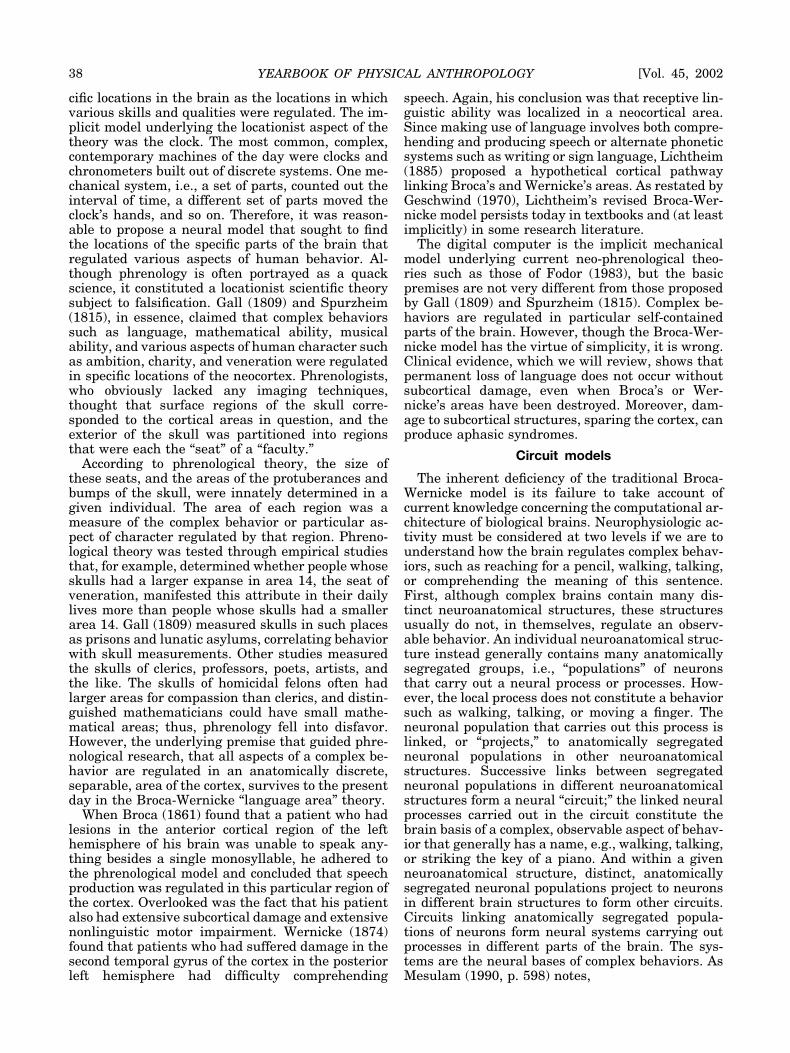
In other words, “local” neural operations occur inparticular regions of the brain. However, these lo-calized operations in themselves do not constitutean observable behavior such as walking or languagefor which we have words. Evidence from hundreds ofindependent studies that span three decades showsthat different regions of the neocortex and differentsubcortical structures are specialized to process par-ticular stimuli (visual or auditory), while other re-gions perform specific operations that regulate as-pects of motor control (such as coding the direction ofa movement or its force), or holding information inshort-term (working) memory (e.g., Marsden andObeso, 1994; Mitchell et al., 1987; Mirenowicz andSchultz, 1996; Monchi et al., 2001; Polit and Bizzi,1978; Sanes et al., 1995). However, these local pro-cesses form part of the neural “computations” that,linked together in complex neural circuits, are man-ifested in behaviors such as walking, pushing a but-ton, speaking, or comprehending the syntax of asentence (Fig. 1).
For example, within the putamen, a subcorticalbasal ganglia structure located within the brain,
anatomically segregated populations of neurons ex-ist that form part of a system that sequences thesubmovements that together constitute an overtmovement of a monkey’s hand, a rat’s groomingsequence, and a person’s walking or speaking (Al-dridge et al., 1993; Cunnington et al., 1995; Lieber-man, 2000; Marsden and Obeso, 1994). But the pu-tamen, in itself, is not the “seat” of the motor act.The putamen, like other neuroanatomical struc-tures, supports anatomically segregated neuronalpopulations that project to different parts of thebrain forming a number of circuits that regulateother aspects of behavior. Distinct, anatomicallysegregated neuronal populations in the putamenproject through other subcortical structures to cor-tical areas implicated in motor control, higher cog-nition, attention, and reward-based learning (e.g.,Aldridge et al., 1993; Alexander et al., 1986; Alex-ander and Crutcher, 1990; Cummings, 1993; Gray-biel, 1995, 1997; Kimura et al., 1993; Lieberman,2000; Marsden and Obeso, 1994; Middleton andStrick, 1994; Parent, 1986). Complex behaviors areregulated by neural circuits that constitute net-works linking activity in many parts of the brain. Inshort, the neural mechanism that carries out theinstruction set manifested in my pecking at my com-
Fig. 1. Anatomically segregated populations of neurons in a particular structure or region of the brain can project to distinct,anatomically segregated populations of neurons in different parts of the brain, forming “circuits” that regulate different aspects ofbehavior. Thus, damage to a particular part of the brain can result in a “syndrome,” an ensemble of seemingly unrelated behavioraldeficits. Here, neuronal populations in different cortical areas project into the putamen, and from there indirectly into different regionsof the cortex, regulating motor control and different aspects of higher cognition, including the comprehension of syntax.
NEURAL BASES OF LANGUAGE 39Lieberman]
puter’s keyboard is a “circuit,” linking neuronal pop-ulations in different neuroanatomical structures inmany parts of the brain.
Some confusion often arises with regard to theprecise meaning of the term “module” in neurophys-iologic and in linguistic studies. The term “module”is often used in neurophysiologic studies (e.g., Gray-biel, 1995, 1997) to refer to complex neural circuitsthat regulate an observable behavior. In contrast,theories of the mind grounded in linguistics, such asthose of Fodor (1983) and Pinker (1998), use theword “module” to refer to localized neuroanatomicalstructures that they claim regulate specific aspectsof language. In these locationist theories, the mod-ule that regulates language, or some aspect of lan-guage such as syntax, has no anatomical or physio-logic relation to other hypothetical neural modulesdevoted to walking, manual motor control, and soon. In principle, these locationist theories claim thatthe functional organization of the human brain issimilar to that of a conventional digital computerthat has a discrete hard disk, a discrete electronicmemory, a display, a modem, and so on.
Aphasia
Studies of aphasia, the permanent loss of lan-guage, which were the basis for the Broca-Wernicketheory, were among the first to note the deficienciesof this traditional model. Doubts had been expressedin the early years of the 20th century (Jackson,1915; Marie, 1926). In the past two decades, com-puter-aided tomography (CT) scans and magneticresonance imaging (MRI) provided noninvasive in-formation on the nature and extent of brain damagethat would result in permanent language loss. Theputative basis of Broca’s syndrome in the model ofLichtheim (1885) is damage to Broca’s neocorticalarea. However, clinical studies have shown that per-manent loss of the linguistic abilities associatedwith the syndrome does not occur unless subcorticaldamage is present (Dronkers et al., 1992; D’Espositoand Alexander, 1995; Stuss and Benson, 1986). Pa-tients with extensive damage to Broca’s area gener-ally recover linguistic ability, unless subcorticaldamage also occurs. Patients suffering brain dam-age that damages subcortical structures but thatleaves Broca’s area intact also can manifest thesigns and symptoms associated with Broca’s syn-drome. As Stuss and Benson (1986, p. 161) note intheir review of studies of aphasia, damage to
. . . the Broca area alone or to its immediate surroundings . . . isinsufficient to produce the full syndrome of Broca’s aphasia.. . . . The full, permanent syndrome (big Broca) invariably indi-cates larger dominant hemisphere destruction . . . deep into theinsula and adjacent white matter and possibly including basalganglia.
Independent studies show that subcortical dam-age that leaves Broca’s area intact can result inBroca-like speech production and language deficits(cf. Alexander et al., 1987; Benson and Geschwind,
1985; Mega and Alexander, 1994; Naeser et al.,1982).
Alexander et al. (1987), for example, reviewed 19cases of aphasia resulting from lesions in these sub-cortical structures. Language impairments occurredthat ranged from fairly mild disorders in the pa-tient’s ability to recall words, to “global aphasia” inwhich the patient produced very limited nonpropo-sitional speech. In general, the severest languagedeficits occurred in patients who had suffered themost extensive subcortical brain damage. Damageto the internal capsule (the nerve fibers that connectthe neocortex to subcortical structures), basal gan-glia structures, the putamen, and the caudate nu-cleus resulted in impaired speech production andagrammatism similar to that of classic aphasias, inaddition to other cognitive deficits. Subsequent stud-ies appear to rule out damage to the internal capsuleas the basis for subcortically induced aphasia. De-liberate surgical lesions of the internal capsuleaimed at mitigating obsessive-compulsive behaviordo not induce aphasia (Greenberg et al., 2000). Thestudies of neurodegenerative diseases, hypoxia, andfocal damage that are discussed below suggest thatdamage to the subcortical basal ganglia and associ-ated subcortical components of cortical-striatal-cor-tical circuits yields Broca’s syndrome.
The situation for Wernicke’s syndrome appears tobe similar. The locus for the brain damage tradition-ally associated with Wernicke’s syndrome includesthe posterior region of the left temporal gyrus (Wer-nicke’s area), but often extends to the supramar-ginal and angular gyrus, again with damage to thesubcortical white matter below (Damasio, 1991). In-deed, recent data indicate that premorbid linguisticcapability can be recovered after complete destruc-tion of Wernicke’s area (Lieberman, 2000). AsD’Esposito and Alexander (1995, p. 141) conclude intheir study of aphasia deriving from subcorticaldamage, it is apparent
. . . that a purely cortical lesion—even a macroscopic one—canproduce Broca’s or Wernicke’s aphasia has never been demon-strated.
CORTICAL-STRIATAL-CORTICAL CIRCUITS
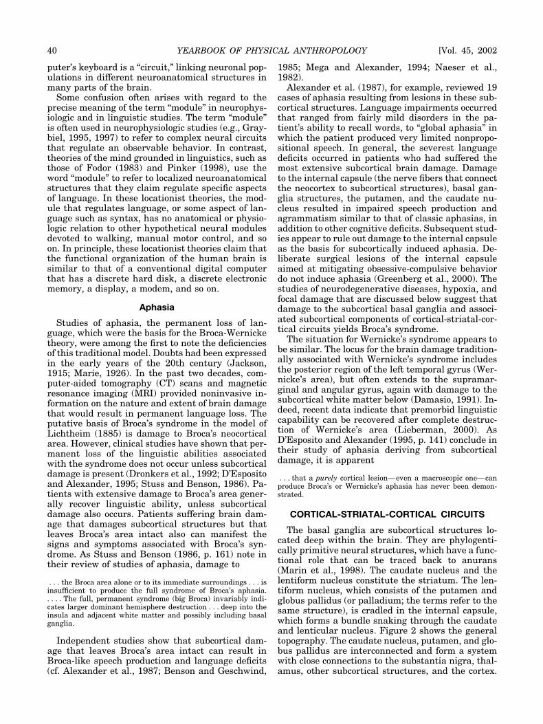
The basal ganglia are subcortical structures lo-cated deep within the brain. They are phylogenti-cally primitive neural structures, which have a func-tional role that can be traced back to anurans(Marin et al., 1998). The caudate nucleus and thelentiform nucleus constitute the striatum. The len-tiform nucleus, which consists of the putamen andglobus pallidus (or palladium; the terms refer to thesame structure), is cradled in the internal capsule,which forms a bundle snaking through the caudateand lenticular nucleus. Figure 2 shows the generaltopography. The caudate nucleus, putamen, and glo-bus pallidus are interconnected and form a systemwith close connections to the substantia nigra, thal-amus, other subcortical structures, and the cortex.
40 YEARBOOK OF PHYSICAL ANTHROPOLOGY [Vol. 45, 2002
The putamen receives sensory inputs from mostparts of the brain. The globus pallidus is an outputstructure receiving inputs from the putamen andcaudate nucleus. Basal ganglia outputs target vari-ous regions of the thalamus, which in turn connectto different cortical areas. Connections with the cor-tex are complex and, as we shall see, not fully un-derstood (Alexander et al., 1986; Alexander andCrutcher, 1990; DeLong, 1993; Marsden and Obeso,1994). However, the probable subcortical locus ofBroca’s aphasia is consistent with one of the majorfindings of contemporary neurophysiological stud-ies.
Disruptions in behavior that are seemingly unre-lated, such as obsessive-compulsive disorder (Green-berg et al., 2000), schizophrenia (Graybiel, 1997),and Parkinson’s disease (Jellinger, 1990), derivefrom the disruption of neural circuits that link cor-tical areas with the basal ganglia structures of thestriatum. Anomalous basal ganglia developmentalso appears to implicated in a genetically transmit-ted deficit affecting speech production and syntax(Lal et al., 2001; Vargha-Khadem et al., 1998;Watkins et al., 2002). Behavioral changes once at-
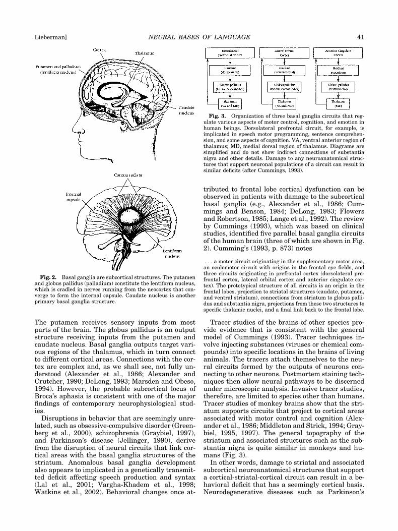
tributed to frontal lobe cortical dysfunction can beobserved in patients with damage to the subcorticalbasal ganglia (e.g., Alexander et al., 1986; Cum-mings and Benson, 1984; DeLong, 1983; Flowersand Robertson, 1985; Lange et al., 1992). The reviewby Cummings (1993), which was based on clinicalstudies, identified five parallel basal ganglia circuitsof the human brain (three of which are shown in Fig.2). Cumming’s (1993, p. 873) notes
. . . a motor circuit originating in the supplementary motor area,an oculomotor circuit with origins in the frontal eye fields, andthree circuits originating in prefrontal cortex (dorsolateral pre-frontal cortex, lateral orbital cortex and anterior cingulate cor-tex). The prototypical structure of all circuits is an origin in thefrontal lobes, projection to striatal structures (caudate, putamen,and ventral striatum), connections from striatum to globus palli-dus and substantia nigra, projections from these two structures tospecific thalamic nuclei, and a final link back to the frontal lobe.
Tracer studies of the brains of other species pro-vide evidence that is consistent with the generalmodel of Cummings (1993). Tracer techniques in-volve injecting substances (viruses or chemical com-pounds) into specific locations in the brains of livinganimals. The tracers attach themselves to the neu-ral circuits formed by the outputs of neurons con-necting to other neurons. Postmortem staining tech-niques then allow neural pathways to be discernedunder microscopic analysis. Invasive tracer studies,therefore, are limited to species other than humans.Tracer studies of monkey brains show that the stri-atum supports circuits that project to cortical areasassociated with motor control and cognition (Alex-ander et al., 1986; Middleton and Strick, 1994; Gray-biel, 1995, 1997). The general topography of thestriatum and associated structures such as the sub-stantia nigra is quite similar in monkeys and hu-mans (Fig. 3).
In other words, damage to striatal and associatedsubcortical neuroanatomical structures that supporta cortical-striatal-cortical circuit can result in a be-havioral deficit that has a seemingly cortical basis.Neurodegenerative diseases such as Parkinson’s
Fig. 2. Basal ganglia are subcortical structures. The putamenand globus pallidus (palladium) constitute the lentiform nucleus,which is cradled in nerves running from the neocortex that con-verge to form the internal capsule. Caudate nucleus is anotherprimary basal ganglia structure.
Fig. 3. Organization of three basal ganglia circuits that reg-ulate various aspects of motor control, cognition, and emotion inhuman beings. Dorsolateral prefrontal circuit, for example, isimplicated in speech motor programming, sentence comprehen-sion, and some aspects of cognition. VA, ventral anterior region ofthalamus; MD, medial dorsal region of thalamus. Diagrams aresimplified and do not show indirect connections of substantianigra and other details. Damage to any neuroanatomical struc-tures that support neuronal populations of a circuit can result insimilar deficits (after Cummings, 1993).
NEURAL BASES OF LANGUAGE 41Lieberman]
disease (PD) and progressive supranuclear palsy(PSP) result in major damage to the subcorticalbasal ganglia, mostly sparing the cortex (Jellinger,1990). Independent studies of these neurodegenera-tive diseases have established the role of the basalganglia in these circuits. The primary deficits ofthese neurodegenerative diseases are motoric: trem-ors, rigidity, and repeated movement patterns occur.However, these subcortical diseases also cause lin-guistic and cognitive deficits. Speech production,syntax, and cognitive deficits similar in nature tothose typical of Broca’s aphasia can occur in evenmild and moderately impaired PD patients (Cools etal., 2001; Gotham et al., 1988; Harrington and Haa-land, 1991; Lange et al., 1992; Lieberman et al.,1992; Morris et al., 1988; Taylor et al., 1990). Inparticular, deficits in the comprehension of and pro-duction of syntax have been noted in independentstudies of PD (e.g., Hochstadt et al., unpublishedfindings; Lieberman, 2000; Lieberman et al., 1990,1992; Grossman et al., 1991, 1993, 2001; Howard etal., 2001; Illes et al., 1998; Natsopoulos et al., 1993;Pickett, 1998). As is the case for Broca’s aphasia(Blumstein, 1995), PD patients have difficulty com-prehending sentences that have moderately complexsyntax, as well as long sentences that tax the brain’scomputational resources (Baum, 1989). In extremeform a dementia occurs, different in kind from Alz-heimer’s dementia (Albert et al., 1974; Cummingsand Benson, 1984; Xuerob et al., 1990). The afflictedpatients retain semantic and real-world knowledge,but are unable to readily form or change cognitivesets (Flowers and Robertson, 1985; Cools et al.,2001). These seemingly unrelated deficits appear toderive from the “local” operations performed by thebasal ganglia in the cortical-striatal-circuits regulat-ing these aspects of behavior.
Probable basal ganglia operationsSequencing. In the era before medication withlevadopa was used to treat Parkinson’s disease,thousands of operations were performed. The effectsof these surgical interventions on motor control inhumans and similar experimental lesions in mon-keys were reviewed in a seminal paper by Marsdenand Obeso (1994). They noted that the basal gangliaappear to have two different motor control functions(Marsden and Obeso, 1994, p. 889).
First, their normal routine activity may promote automatic exe-cution of routine movement by facilitating the desired corticallydriven movements and suppressing unwanted muscular activity.Secondly, they may be called into play to interrupt or alter suchongoing action in novel circumstances. . . . Most of the time theyallow and help cortically determined movements to run smoothly.But on occasions, in special contexts, they respond to unusualcircumstances to reorder the cortical control of movement.
Given the fact that the basal ganglia circuitry reg-ulating motor control does not radically differ fromthat implicated in cognition, Marsden and Obeso(1994, p. 893) concluded that
. . . the role of the basal ganglia in controlling movement mustgive insight into their other functions, particularly if thought ismental movement without motion. Perhaps the basal ganglia arean elaborate machine, within the overall frontal lobe distributedsystem, that allows routine thought and action, but which re-sponds to new circumstances to allow a change in direction ofideas and movement. Loss of basal ganglia contribution, such asin Parkinson’s disease, thus would lead to inflexibility of mentaland motor response.
Advances in brain imaging and behavioral studiesof human subjects support this hypothesis that thebasal ganglia perform cognitive sequencing func-tions. The functional magnetic resonance imaging(fMRI) study of Monchi et al. (2001) monitored brainactivity in neurologically intact subjects as they per-formed a version of the Wisconsin Card Sorting Test(WCST), an instrument that has been used in manystudies to assess cognitive dysfunction. The versionof the WCST used in this experiment assesses sub-jects’ ability to form and shift abstract categories asthey match cards that picture various images toreference cards. The subjects had to match testcards to reference cards based on the color, shape, ornumber of stimuli pictured on each card. Subjectswere informed when they made either correct orincorrect matches, and had to shift the matchingcriterion as the test progressed. As evidence frommany studies of behavioral deficits resulting frombrain damage and neurodegenerative diseases pre-dicted, neural circuits involving the prefrontal cor-tex and basal ganglia were activated during the test.Dorsolateral prefrontal cortical areas (Brodmannareas 9 and 46) were active at points where subjectshad to relate the current match with earlier eventsstored in working memory. In contrast, a cortical tobasal ganglia circuit involving the mid-ventrolateralprefrontal cortex (areas 47/12), caudate nucleus, andthalamus was active when subjects had to shift to adifferent matching criterion. The putamen alsoshowed increased activity during these cognitiveshifts. These findings from neurologically intact hu-man subjects matched data from electrophysiologicstudies of monkey brains (reviewed in Graybiel,1995, 1997).
Reward-based learning. Studies of the brains ofrodents that selectively destroy basal ganglia struc-tures or use direct electrophysiologic recording areconsistent with these human studies. In rodents, thebasal ganglia execute innately determined groomingsequences; electrophysiologic studies of basal gan-glia neurons show firing patterns that release thesequence of submovements that strung together con-stitute a grooming sequence (Aldridge et al., 1993).Animal studies also suggest that basal ganglia areimplicated in reward-based associative learning. Inbirds, a basal ganglia to forebrain circuit belongs toa system that regulates vocal learning and produc-tion. Lesioning this circuit prevented restructuringof adult zebra finches’ songs and young songbirds’acquiring songs (Brainard and Doupe, 2000). Inmonkeys, neuronal populations form in basal gan-
42 YEARBOOK OF PHYSICAL ANTHROPOLOGY [Vol. 45, 2002
glia in the course of adaptive reward-based learning(Kimura et al., 1993; Mirenowicz and Schultz, 1996),Graybiel (1995, 1997), reviewing the data of inde-pendent studies, noted that neurons in the caudatenucleus, putamen, and globus pallidus control motoracts through response patterns that are built upthrough learning and memory. In short, convergingevidence from independent studies suggests that thebasal ganglia and cerebellum (which will be dis-cussed below) are implicated in both learning andexecuting sequences of motor acts or cognitive pro-cesses, forming an internalized repertoire of mean-ingful, goal-directed acts (e.g., Graybiel, 1995, 1997,1998; Kimura et al., 1993; Marsden and Obeso,1994; Mirenowicz and Schultz, 1996).
LINGUISTIC AND COGNITIVE CONSEQUENCESOF SUBCORTICAL BRAIN DAMAGE
As noted earlier, Broca’s aphasia does not occur inthe absence of subcortical brain damage, suggestingthat Broca’s syndrome is the result of impairment ofsubcortical components of cortical-striatal-corticalcircuits. A similar pattern of behavioral deficits wasdocumented in neurodegenerative diseases andbrain damage affecting the basal ganglia and cere-bellum. A brief discussion of the nature of thesedeficits may be useful, insofar as they may be unfa-miliar to nonspecialists.
Speech
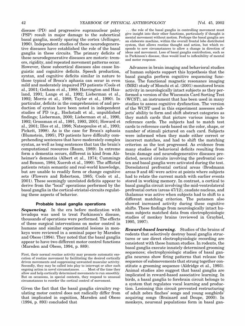
One of the primary speech deficits of Broca’s syn-drome and of compromised basal ganglia function isa breakdown in the sequencing of motor commandsnecessary to produce stop consonants. The primaryacoustic cue that differentiates stop consonants suchas [b] from [p] when they occur before a vowel (as inthe English words bat and pat) is an interval of timethat reflects the sequence of motor commands thatprovide these sounds. These speech sounds are pro-duced by closing the lips, obstructing the flow of airfrom a speaker’s mouth, and then abruptly openingthe lips, which produces a “burst” of turbulent airthat has distinct acoustic properties. At the sametime, the speaker must adjust the muscles of thelarynx to produce phonation subsequent to theburst. In order to produce a [b], phonation mustoccur within 20 msec of lip opening; longer delayswill yield the sound [p]. Similar temporal contrastsinvolving the muscles of the tongue and larynx dif-ferentiate the sounds [d] from [t] (do vs. to), and [g]from [k] (god vs. cod). Lisker and Abramson (1964)coined the term “voice-onset-time” (VOT) to describethis distinction, which appears to hold for all humanlanguages studied to date. In brief, VOT is definedas the time that occurs between the “burst” thatresults from lip or tongue gestures and the onset ofperiodic phonation generated by the larynx. Figure 4shows the waveforms of a [ba] and a [pa] with “cur-sors” superimposed that mark the onsets of thebrusts and phonation.
Speakers must precisely control a sequence of in-dependent motor acts to produce these sounds. Bro-ca’s aphasics are unable to maintain control of thesesequential motor commands: their intended [b] smay be heard as [p] s, [t] s as [d] s, and so on(Blumstein et al., 1980; Baum et al., 1990). Theproblem was not inherently one of maintaining tem-poral control, since Broca’s aphasics maintain theintrinsic durations that differentiate vowels (Baumet al., 1990), for example, the vowel of the word batis three time longer than that of the word bit. Bro-ca’s aphasics, moreover, maintain almost normalcontrol of the magnitude and placement of tongue,lip, and laryngeal gestures; no apparent loss of pe-ripheral motor control occurs. The production of theformant frequency patterns that specify vowels isunimpaired in Broca’s syndrome, though there isincreased variability (Ryalls, 1986; Kent and Rosen-beck, 1983; Baum et al., 1990). Since formant fre-quency patterns are determined by the configura-tion of the supralaryngeal vocal tract (tongue, lips,and larynx height), we can conclude that the controlof these structures is unimpaired in Broca’s aphasicsyndrome. The deficit appears to involve sequenc-ing; Broca’s aphasics also have difficulty executingeither oral, nonspeech, and manual sequential mo-tor sequences (Kimura, 1993). Figure 4 shows thewaveforms of [ba] and [pa] marked for VOT.
Voice-onset-time sequencing deficits, similar innature to those of Broca’s syndrome, can occur in thelater stages of Parkinson’s disease. Computer-im-plemented analysis revealed overlaps between the
Fig. 4. Speech waveforms of syllables [ba] and [pa]. Ampli-tudes of speech signal are plotted at ordinate, and elapse of timeon abscissa. “Cursors” L1 mark beginnings of “bursts” of “stopconsonants” [b] and [p] that occur when the lips open. Cursors. R1mark onset of phonation that occurs when vocal cords (folds) ofspeaker’s larynx start to produce periodic phonation.
NEURAL BASES OF LANGUAGE 43Lieberman]

VOTs of stop consonents such as [t] vs. [d], exceeding19% for some PD subjects; the degree of VOT se-quencing deficits depended on the severity of thedisease state (Lieberman et al., 1992; Hochstadt etal., unpublished findings). The PD subjects, as wasthe case for Broca’s aphasics, maintained controlover vowel duration, other durational speech phe-nomena, and tongue and lip movements. Similarresults occurred for some subjects suffering degen-eration of the cerebellum (Pickett, 1998).
The “phonologic” level, i.e., the knowledge andcoding of sounds that specify the name of a word,appears to be preserved in Broca’s aphasics and inParkinson’s disease for these same sounds. For ex-ample, at the phonologic level, the acoustic cues andarticulatory gestures that specify a particular stopconsonant differ when it occurs in syllable initialposition or after a vowel. The speech-sound [t], forexample, is signalled by a long VOT when it occursin syllable-initial position. In contrast, after a vowel,the acoustic cues for [t] are reduced duration of thevowel that precedes it and increased burst ampli-tude. Broca’s aphasics maintain normal control ofthese cues, although VOT sequencing is disruptedfor syllable-initial [t]s. These distinctions are gen-eral. The duration of a vowel is always longer, tak-ing into account other factors, before a [b], [d], or [g]than for a [p], [t], or [k]. The fact that Broca’s apha-sics preserve these durational cues indicates thatthe phonologic “instruction set” for producing stopconsonants is intact. The preservation of these du-rational cues again indicates that the Broca’s VOTdeficit derives from the disruption of sequencingrather than impaired ability to control duration.Instrumental analyses of the speech of Broca’s apha-sics often reveal waveforms showing irregular pho-nation (Blumstein, 1995). Speech quality is “dysar-thric.” Noisy and irregular phonation occurs,reflecting impaired regulation of the muscles of thelarynx and alveolar air pressure. Similar problemscan also occur in advanced stages of Parkinson’sdisease.
Syntax
“Higher-level” linguistic and cognitive deficits alsooccur in this aphasic syndrome. The utterances pro-duced by Broca’s aphasics often were described as“telegraphic.” In the period when telegrams were ameans of communication, the sender paid by theword, and words were omitted whenever possible.The utterances of English-speaking aphasics, whoomitted prepositions, articles, and tense markers,producing messages such as man sit tree in place ofThe man sat by the tree, had the appearance oftelegrams. These aphasic telegraphic utteranceswere generally thought to be the result of the pa-tient’s compensating for speech production difficul-ties by reducing the utterance’s length, thereby min-imizing difficulties associated with speech production.The presence of language comprehension deficits inBroca’s aphasics that appeared to involve syntax
was established by studies starting in the 1970s.Broca’s aphasics had difficulty comprehending dis-tinctions in meaning conveyed by syntax (Zurif etal., 1972). Although agrammatic aphasics are able tojudge whether sentences are grammatical, albeitwith high error rates (Linebarger et al., 1983;Shankweiler et al., 1989), the comprehension defi-cits of Broca’s aphasics have been replicated inmany independent studies (e.g., Baum, 1989; Blum-stein, 1995). For example, higher error rates occurwhen comprehending distinctions in meaning con-veyed by passive sentences such as, “The boy waskissed by the girl” than for the “cannonical” sen-tence, “The girl kissed the boy.” High error ratesoften occur when comprehending sentences contain-ing embedded relative clauses such as, “The boy whowas wearing a red hat fell down.” Long sentencesgenerally present additional difficulty. Error ratesexceeding 50% can occur using sentences that yieldvirtually error-free performance by neurologicallyintact control subjects; cf. Blumstein (1995) for acomprehensive review.
As is the case for Broca’s syndrome, Parkinson’sdisease (PD) can result in sentence comprehensiondeficits (Grossman et al., 1991, 1993, 2001; Hochs-tadt et al., unpublished findings; Howard et al.,2001; Lieberman et al., 1990, 1992; Natsopoulos etal., 1993; Pickett, 1998). The first study that associ-ated grammatical deficits with PD was reported byIlles et al. (1988); their data showed deficits similarto those noted in Huntington’s disease. The sen-tences produced by PD subjects were often short andhad simplified syntax. However, Illes et al. (1988)attributed these effects to the speakers compensat-ing for their speech motor production difficulties byproducing short sentences. A subsequent study ofcomprehension deficits of PD (Lieberman et al.,1990) showed that syntax comprehension deficitscould occur that could not be attributed to compen-satory motor strategies. The comprehension deficitsnoted clearly were not the result of any compensat-ing strategy, since the motoric component of sub-jects’ responses to both sentences that had complexsyntax and high rates and sentences with simplesyntax and low error rates was identical. The sub-jects simply had to utter the number (one, two, orthree) that identified a line drawing that best rep-resented the meaning of the sentence that theyheard. Deficits in the comprehension of distinctionsof meaning conveyed by syntax occurred for longconjoined simple sentences as well as for sentencesthat had moderately complex syntax. Nine of a sam-ple of 40 nondemented PD subjects had these com-prehension deficits. The test battery used in thisstudy included sentences with syntactic construc-tions that are known to place different processingdemands on normal adult subjects, e.g., center-em-bedded sentences, right-branching sentences, con-junctions, “simple” one-clause declarative sentences,or semantically and constrained and semanticallyunconstrained passives. However, neurologically in-
44 YEARBOOK OF PHYSICAL ANTHROPOLOGY [Vol. 45, 2002
tact subjects made virtually no errors when theytook this test. In contrast, the overall error rate was30% for some PD subjects. The PD subjects’ compre-hension errors typically involved repeated errors onparticular syntactic constructions. Therefore, theobserved syntax comprehension errors could not beattributed to general cognitive decline or attentiondeficits. The highest number of errors (40%) weremade on “left-branching” sentences that departedfrom the canonical pattern of English having theform subject-verb-object (SVO). An example of a left-branching sentence is, “Because it was raining, thegirl played in the house.” Thirty percent errors oc-curred for right-branching sentences with final rel-ative clauses, such as “Mother picked up the babywho is crying.” Twenty percent error rates also oc-curred on long conjoined simple sentences, such as“Mother cooked the food and the girl set the table.”Similar sentence comprehension errors reflecting in-formation conveyed by syntax have been found inindependent studies of nondemented PD subjects(Grossman et al., 1991, 1993, 2001; Howard et al.,2001; Natsopoulos et al., 1993), using proceduresthat monitored either sentence comprehension orjudgments of sentence grammaticality.
The PD subjects studied by Grossman et al. (1991)were asked to interpret information presented insentences in active or passive voices when the ques-tions were posed in passive or active voices. Deficitsin comprehension were noted when PD subjects hadto shift cognitive sets, responding to a questionposed in a passive voice concerning information pre-sented in an active voice or the reverse. Highererrors, for example, occurred when the subjectsheard the sentence The hawk ate the sparrow whenasked Who was the sparrow eaten by? than whenasked Who ate the sparrow? Grossman et al. (1991)also tested PD subjects’ ability to copy unfamiliarsequential manual motor movements (a procedureanalogous to that used by Kimura (1993), who founddeficits in this behavior for Broca’s aphasics). Defi-cits in sequencing manual motor movements andlinguistic sequencing in the sentence comprehensiontask were correlated. The correlation between se-quencing complex manual motor movements andthe cognitive operations implicated in the compre-hension of syntax is consistent with Broca’s areaplaying a role in both verbal working memory andmanual motor control (Rizzolatti and Arbib, 1998) incircuits supported by basal ganglia (Marsden andObeso, 1994).
Grossman et al. (2001) interpreted these deficitsas an attentional rather than a linguistic deficit.However, this is unlikely, since PD subjects andBroca’s aphasics attend to semantic informationwhen they are asked to convey the meaning of asentence. They consistently perform better whenfaced with a sentence such as “The banana waseaten by the boy” than the sentence “The clown waspoked by the cowboy.” They clearly have no atten-tional deficits regarding the fact that an inanimate
banana cannot eat a boy. A study of bilateral dam-age to the putamen and part of the caudate nucleus(Pickett et al., 1998) revealed similar deficits in se-quencing speech motor gestures and the comprehen-sion of distinctions in meaning conveyed by syntax.
Motor and cognitive set-shifting
Motor and cognitive sequencing deficits similar tothose noted and predicted by Marsden and Obeso(1994) generally occur in PD. Speech motor sequenc-ing, verbal working memory, syntactic, and cogni-tive sequencing errors occur in Parkinson’s disease.Manual motor sequencing deficits have been notedin many studies of PD (e.g., Cunnington et al., 1995).As noted above, Grossman et al. (1991) found corre-lated deficits in sequencing manual motor move-ments and linguistic operations in a sentence compre-hension task. Speech production VOT sequencingdeficits have also been found to correlate with syn-tactic comprehension deficits in PD (Lieberman etal., 1992), and with bilateral damage to the putamen(Pickett et al., 1998), as well as for neurologicallyintact human subjects and subjects having cerebelladamage (Pickett, 1998).
Hypoxia (oxygen deficits) commonly occurs inmountain climbers at extreme altitudes, and simi-lar, though generally less extreme, VOT sequencingand syntax comprehension errors again can occur inthese subjects (Lieberman et al., 1994, unpublishedfindings). Histologic studies of the hypoxic brainhave identified regions of “selective vulnerability” inthe hippocampus, cerebellum, basal ganglia, andlayers III, V, and VI of the neocortex (Brierley,1976). Damage to all of these neural structures mayhave motor and cognitive consequences. However,the globus pallidus (the principal basal ganglia out-put structure linking the striatum to the cortexthrough the thalamus and other subcortical struc-tures) is extremely sensitive to hypoxic damage(Laplane et al., 1984, 1989; Strub, 1989); Moreover,the behavioral deficits at extreme altitude that arediscussed below (Lieberman et al., 1994, 1995 sub-mitted; Nelson et al., 1990; Regard et al., 1989) arevirtually identical to those occurring with bilateralsurgical lesions of the globus pallidus (Scott et al.,2002). Long-term memory remains unchanged, as isperformance in a number of psychometric tests thatare generally thought to involve the cortex. Cogni-tive impairment in these subjects appears to be lim-ited to forming and shifting cognitive sets on testssuch as the Odd-Man-Out test (Flowers and Robert-son, 1985), which involves forming a conceptual cat-egory and then shifting to a different category. Forexample, if the subject starts by sorting pictures bytheir shapes, s/he must then switch to sorting themby size. Impaired subjects have difficulty shifting toa different sorting criterion; they tend to persever-ate, holding or shifting back to a previous sortingcriterion. Cognitive perseveration in real-life situa-tions also occurs for hypoxic subjects climbingMount Everest (Lieberman et al., 1994, 1995). It can
NEURAL BASES OF LANGUAGE 45Lieberman]
result in fatal errors of judgment (Lieberman et al.,unpublished findings). Cognitive perseveration inthe domain of language may account for some of thesentence comprehension deficits discussed above.When comprehending the meaning of a sentencethat contains relative clauses, the subject must shiftsyntactic operations at the clause boundary, for ex-ample, as in comprehending the meaning of the sen-tence, “The boy who was fat fell down.”
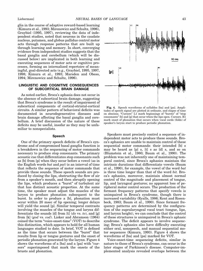
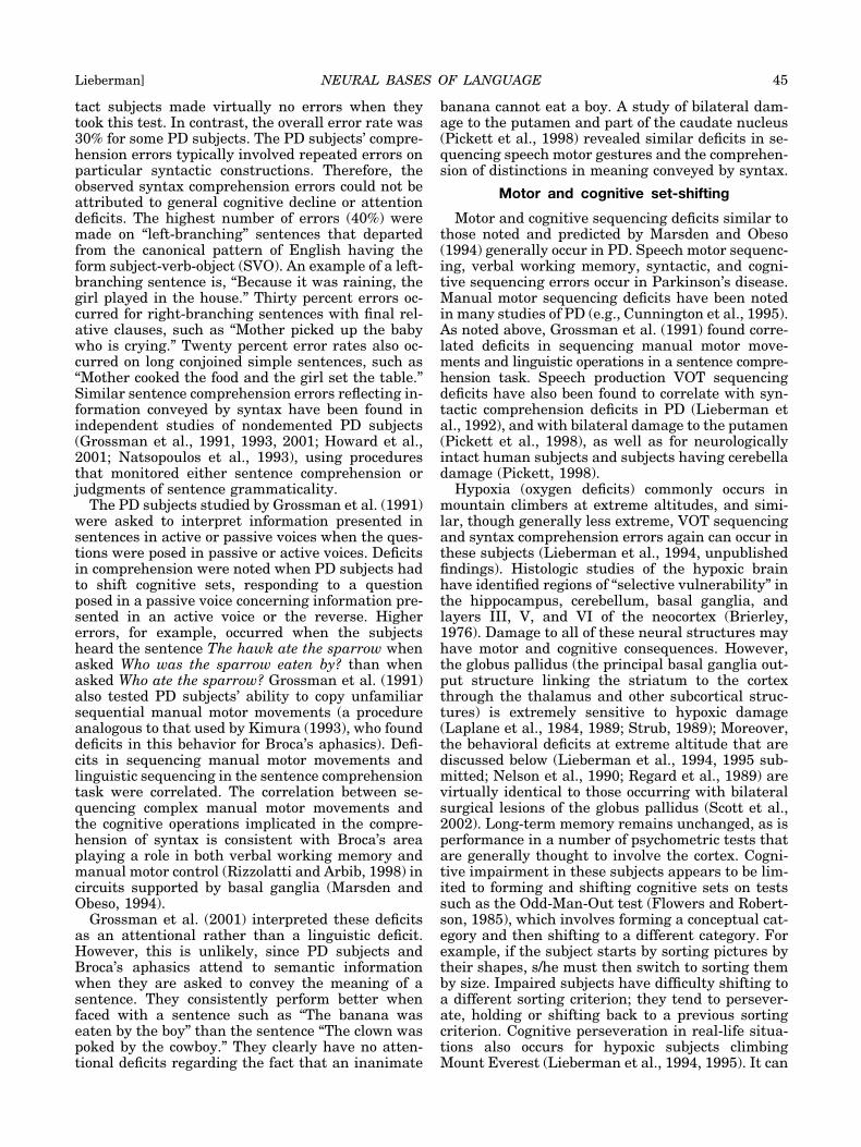
The fMRI study of Monchi et al. (2001) demon-strated the coordinated activity of the cortex andbasal ganglia as human subjects form and shift con-cepts. Brain activity was monitored in neurologi-cally intact subjects in a task similar to the Odd-Man-Out test, a version of the Wisconsin CardSorting Test (WCST), an instrument that has beenused in many studies to assess cognitive dysfunc-tion. The version of the WCST used assesses sub-jects’ ability to form and shift abstract categories asthey match cards that picture various images toreference cards. The subjects had to match testcards to reference cards based on the color, shape, ornumber of stimuli pictured on each card. Subjectswere informed when they made either correct orincorrect matches, and had to shift the matchingcriterion as the test progressed. Figure 5 shows thecortical areas defined by Brodmann (1912). Neuralcircuits involving the prefrontal cortex and basalganglia were activated during the test. Dorsolateralprefrontal cortical areas (Brodmann areas 9 and 46)were active at the points where subjects had to re-late the current match with earlier events stored inworking memory. In contrast, a cortical to basalganglia circuit involving the mid-ventrolateral pre-frontal cortex (areas 47/12), caudate nucleus, andthalamus was active when subjects had to shift to adifferent matching criterion. The putamen alsoshowed increased activity during these cognitive
shifts. These findings from neurologically intact hu-man subjects match data from electrophysiologicstudies of monkey brains (reviewed in Graybiel,1995, 1997).
Cerebellum
Less is known about the cognitive role of the cer-ebellum, a subcortical structure that is linked to theprefrontal and motor cortex as well as to the basalganglia. fMRI and tracer studies show that it isactive in motor learning, apparently acting in con-cert with the prefrontal cortex (Thach, 1996; Dea-con, 1997). More general linguistic and cognitiveroles for the cerebellum, particularly the neocerebel-lum, which is disproportionately large in humans,have been proposed (Leiner et al., 1991). However, itis unclear whether the cerebellum plays a role insentence comprehension and linguistic tasks that donot involve modeling of motor activity (Thach, 1996).A study that tested the ability of persons sufferingcerebellar degeneration to comprehend distinctionsin meaning conveyed by syntax failed to show defi-cits attributable to the neocerebellum (Pickett,1998). The linguistic and cognitive deficits noted byPickett appeared to derive from damage to the neu-ral pathways linking it to the basal ganglia andcortex (Pickett, 1998). The cerebellum has beenlinked to the control of timing motor activity (Ivryand Keele, 1989); although VOT sequencing wasdegraded in some of the subjects studied by Pickett(1998), the intrinsic durations of English vowelswere preserved. This might reflect the highly over-learned nature of the neural pattern generators thatspecify the motor gestures underlying humanspeech.
THE “LANGUAGE GENE” ANDUNIVERSAL GRAMMAR
Some attention has been drawn to the identifica-tion of a putative “language gene” that has beeninterpreted as evidence for the claim by Chomsky(1986) that the syntactic “rules” of all human lan-guages are determined by an innate neural mecha-nism (Gopnik, 1990; Gopnik and Crago, 1991;Pinker, 1994). Over the course of many years, Chom-sky (1966, 1972, 1986) developed the theory of “uni-versal grammar” (UG). No person would disputethat human beings have an innate capacity to ac-quire language. It is clear that neurologically intactinfants and children raised under “normal” circum-stances have the biological capacity to learn anylanguage. However, Chomsky (1966, 1972, 1986)goes further, claiming that the detailed syntax of allhuman languages is an innate attribute of the hu-man brain. For example, English syntax has a “reg-ular plural rule” which predicts the plural form ofmost nouns: dog-dogs, car-cars, etc. Children raisedin an English-speaking environment acquire thisknowledge without explicit tutoring. While manyspecialists would argue that the processes that allow
Fig. 5. Brodmann (1912), by means of microscopic examina-tion, partitioned the surface of the cortex into areas whose cellshad somewhat different anatomical properties. Different localoperations often appear to be performed in these regions. Frontalregions are at left. Areas 44 and 45 are traditional sites of Broca’sarea.
46 YEARBOOK OF PHYSICAL ANTHROPOLOGY [Vol. 45, 2002
children to master other aspects of cognitive behav-ior can account for this and other aspects of theacquisition of language (e.g., Bates et al., 1992; El-man et al., 1997; Greenfield, 1991; Lieberman, 1984,1998, 2000), Chomsky (1966, 1972, 1976, 1986)claims that the UG instantiates innate knowledge ofthis “rule,” in effect “triggering” the genetic programif a child is exposed to regular English plural nounsearly in life. In short, the UG constitutes an innatestore of detailed knowledge of syntax. Other genet-ically transmitted components of the UG wouldspecify the rules governing the formation of yes-noquestions, while other UG genes would confer theability to form passive sentences, and so on. Nowaket al. (2000) make two claims based on a computer-modelling study: 1) that syntax becomes necessaryas a language acquires many words, and 2) that therules of syntax must be innately determined. Thefirst claim is consistent with studies of the develop-ment of lexical ability and syntax in young children.Bates and Goodman (1997) showed that syntax de-velops as vocabulary size increases as children ma-ture. The second claim, that the rules of syntax areinnately specified, reiterates the claim of Chomsky(1966, 1972, 1986) that children could not possiblylearn the rules of syntax. Nowak et al. (2000) disre-gard the body of studies that suggest that childrenacquire words and syntax by means of associativelearning, imitation, and subtle social cues that indi-cate their errors to them (e.g., Bates and Goodman,1997; Elman et al., 1997; Greenfield, 1991; Lieber-man, 1984).
It is clear that the vocal and gestural signals ofmany species are genetically specified and need onlytriggering stimuli. For example, ducklings requirevery limited exposure to duck calls as they hatch todevelop normal duck signaling behavior monthslater (Gottlieb, 1975). In effect, the claim inherent inUG is that human beings possess a vastly moreelaborate set of genetically transmitted linguisticinformation than ducks, allowing children who re-ceive limited exposure to the utterances of a lan-guage to master syntax. The hypothetical UG must,of course, encode the different syntactic schemesthat occur in the world’s languages. Therefore, UGmust contain many detailed syntactic rules. Sincediseases such as diabetes which have a strong ge-netic component result in specific deficits, one sourceof evidence for UG would be a genetic anomaly thatprevented afflicted individuals from mastering aspecific aspect of English syntax, while retainingother aspects of normal linguistic ability. This wasreported to be the case for the afflicted members of alarge extended family (KE) who suffer from a genet-ically transmitted anomaly. Gopnik (1990) andGopnik and Crago (1991) claimed that these individ-uals were unable to master the regular past tense ofEnglish verbs and regular plural nouns. Other as-pects of English syntax, and cognitive and motorbehavior, supposedly were similar to the normalmembers of family KE. However, this is not the case.
Intensive study of family KE reveals the occurrenceof a suite of severe speech and orofacial movementdisorders, cognitive deficits, and linguistic deficitsthat are not limited to specific aspects of the syntaxof English (Lal et al., 2001; Vargha-Khadem et al.,1998; Watkins et al., 2002). Major orofacial sequenc-ing errors (they are not able to stick out theirtongues while closing their lips) occur in these indi-viduals; they have difficulty repeating two words insequence. In a filmed interview of afflicted familyKE children (BBC broadcast, 1994), subtitles wereprovided because their speech was scarcely intelli-gible. On standardized intelligence tests, afflictedmembers of family KE, raised in the same immedi-ate family, have significantly lower scores than theirnonafflicted siblings, which rules out environmentalfactors that might affect intelligence.
Watkins et al. (2002) concluded that these “verbaland non-verbal deficits arise from a common impair-ment in the ability to sequence movement or inprocedural learning.” MRI and PET data on a lim-ited sample of family KE members indicate thatthese cognitive and motor impairments appear toderive from a basal ganglia anomaly. The afflictedmembers of this large extended family have bilater-ally small caudate nuclei. Although other as yetundetermined neural structures may be at risk infamily KE, their conclusion is consistent with thepattern of motor and cognitive sequencing deficitsassociated with the basal ganglia dysfunction notedabove.
Other studies point to damage to components ofcortical-striatal-cortical circuits yielding motor, cog-nitive, and linguistic deficits. Kimura and Watson(1989), in a study of aphasic patients with focalbrain damage, found that their patients, as is thecase for the afflicted members of family KE, hadcoordinate oral sequencing and speech productiondeficits. Developmental verbal apraxia in children,which is defined as an impairment in the program-ming of the sequences of movement, also results inverbal speech production and cognitive deficits de-riving from a breakdown in sequencing (Dewey atal., 1988).
CORTEX
As noted above, subcortical neural structureswork in concert with regions of the cortex in linguis-tic and cognitive tasks as well in motor control.Although the specificity of the traditional Broca-Wernicke theory, localizing language to these corti-cal areas, is incorrect, these and other cortical areasplay critical roles in the neural networks that conferhuman linguistic ability.
Verbal working memory: Broca’sand Wernicke’s areas
Imaging studies that monitor brain activity dur-ing different linguistic tasks consistently show acti-vation of Broca’s and Wernicke’s areas of the cortex,
NEURAL BASES OF LANGUAGE 47Lieberman]

as well as many other cortical areas. In neurologi-cally intact subjects, Broca’s area clearly is impli-cated in sentence comprehension. Stromswold et al.(1996), using PET, studied neurologically intact sub-jects whose task was to decide whether sentenceswere grammatical. The sentences varied in gram-matical complexity; the greatest activation of Bro-ca’s area occurred in the sentences that were mostcomplex, leading to the conclusion that Broca’s areawas implicated in analyzing the syntax of a sen-tence. Indeed, Stromswald et al. (1996) concludedthat Broca’s area is the brain’s “syntax” organ.
But this is not strictly the case; a body of evidencethat extends back 30 years shows that the meaningof a sentence involves recourse to the brain’s neuraldictionary as well as short-term storage and opera-tions in “verbal working memory,” a short-term neu-ral memory buffer (Baddeley, 1986; Gathercole andBaddeley, 1993). Scores of independent studies showthat the words of a sentence are held in verbalworking memory by means of a process of phonetic“rehearsal,” silent speech that makes use of the neu-ral mechanisms that also control overt speech. Thedata of Awh et al. (1996), for example, show thatneurologically intact subjects use neural structuresimplicated in speech production to subvocally “re-hearse” letters of the alphabet, maintaining them inworking memory. Subtractions of PET activityshowed increased metabolic activity (rCBF values)in Broca’s area (Broadmann area 44) as well as thepremotor cortex (area 6), supplementary motor area,cerebellum, and anterior cingulate gyrus when PETdata from a task involving verbal working-memorywere compared with a task that had a substantiallylower working memory load. These brain regions areall implicated in speech motor control. Electrophysi-ologic data from nonhuman primates, for example,show that the anterior cingulate gyrus is implicatedin regulating phonation (Newman and Maclean,1982) as well as in attention (Peterson et al., 1988).Left hemisphere posterior (Wernicke’s area) and su-perior parietal regions also showed greater activityas working memory load increased. These PET dataare consistent with the results of studies of patientshaving lesions in these cortical areas: they showdeficits in verbal working memory that appear toreflect impairment to phonological knowledge, i.e.,the sound pattern of words (Warrington et al., 1971;Vallar et al., 1997).
Imaging studies confirm that Broca’s area andthese cortical areas are involved in overt speech aswell as in silent reading. The PET study of Petersonet al. (1988), in which neurologically intact subjectswere asked to either read or repeat spoken isolatedwords, showed activation of the primary motor cor-tex, premotor cortex, and supplementary motor cor-tex in the subjects’ left hemispheres, and bilateralactivation of areas near Broca’s area and its right-hemisphere homologue. Bilateral activation of areasnear Broca’s region also occurred when subjectswere asked to simply move their mouths and
tongues. This finding is consistent, to a degree, withthe data of many studies of patients having corticallesions, since lesions confined to Broca’s area oftenresult in oral apraxia, i.e., deficits in motor controlinstead of the deficits in motor planning associatedwith aphasia (Stuss and Benson, 1986; Kimura,1993).
Frontal and posterior regions of the cortex alsoactivate when people listen to speech and talk. Aseries of PET studies performed at the MontrealNeurological Institute consistently showed in-creased activity in Brodmann’s areas 47, 46, 45, and8 in the left frontal region, as well as activity in thesubcortical left putamen and posterior secondary“auditory” cortex (Klein et al., 1995; Paus et al.,1996). These studies demonstrate the presence ofpathways from the “motor” to “auditory” cortex. Sig-nals transmitted from neural structures regulatingspeech motor control result in increased activity inregions of the posterior temporal cortex associatedwith speech perception when a person talks.
Broca’s area thus does not constitute a localized“speech production,” “syntax comprehension,” or“sentence-comprehension” organ. The posterior pa-rietal regions, anterior cingulate gyrus, premotorcortex, and supplementary motor area are all impli-cated in these processes. It is also evident that Bro-ca’s area is also implicated in manual motor control(Kimura, 1973). Recent data show that Broca’s areaand its homologue in monkeys support a functionalneural system that generates and monitors graspingand manual gestures (Rizzolatti and Arbib, 1998).
Dynamic neural systems
Moreover, the neural system that carries out sen-tence comprehension is dynamic, recruiting addi-tional resources as task demand increases. ThefMRI study of Just et al. (1996) made use of thesame “subtraction” technique as Stromswold et al.(1996). Neural metabolic activity was monitored assubjects read sentences that expressed the sameconcepts and had the same number of words, butdiffered with respect to syntactic complexity. Thesentences all had two clauses. The sentences withthe simplest syntactic structure were active con-joined sentences (type 1) such as, The reporter at-tacked the senator and admitted the error. The sameinformation was conveyed by the subject relativeclause sentence (type 2), The reporter that attackedthe senator admitted the error, and the object rela-tive clause sentence (type 3), The reporter that thesenator attacked admitted the error. These threesentence types differ with respect to syntactic com-plexity by several generally accepted measures. Pro-gressively longer reading times and higher compre-hension error rates occur in these sentence types.Neurologically intact subjects read sets of exemplarsof each sentence type while activity in their brainswas monitored by means of fMRI. Measures of com-prehension were also obtained, as well as mean pro-cessing time and error rates. Activity in the left
48 YEARBOOK OF PHYSICAL ANTHROPOLOGY [Vol. 45, 2002
temporal cortex, superior temporal gyrus, superiortemporal sulcus, and sometimes the middle tempo-ral gyrus, Wernicke’s area (Brodmann’s areas 22,42, and sometimes 21), increased as subjects readthe sentences with increasing syntactic complexity.Similar increases in activity occurred in the leftinferior frontal gyrus, i.e., Broca’s area (Broad-mann’s areas 44 and 45). The novel finding was thatthe three sentence types resulted in increased activ-ity in areas that were spatially contiguous or prox-imal to the areas activated while reading simplersentences. Furthermore, the right hemisphere ho-mologies of Broca’s and Wernickes’s areas becameactivated, though to a lesser degree, as syntacticcomplexity increased. Moreover, the dorsolateralprefrontal cortex (generally not associated with lan-guage) showed bilateral activation for 3 of the 5subjects who were scanned in an appropriate plane(coronal scans). Activation levels in the dorsolateralprefrontal cortex also increased with sentence com-plexity for these subjects. The dorsolateral prefron-tal cortex is implicated in executive control, visualworking memory, tasks requiring planning, derivingabstract criteria, and changing criteria in cognitivetasks (Grafman, 1989; Paulesu et al., 1993;D’Esposito et al., 1995). In a PET study of bilingualneurologically intact subjects, increased activity inthe left putamen was observed when subjects werespeaking their “second,” less established language(Klein et al., 1994).
It is clear that the neural bases of language arecomplex and appear to involve many different neu-ral circuits (Mesulam, 1990). Moreover, our knowl-edge is imperfect. For example, cortical-striatal-cor-tical circuits linking the prefrontal cortex to otherneural structures do not appear to be implicated inthe linguistic deficits associated with Wernicke’ssyndrome, i.e., fluent, often meaningless speech con-taining neologisms (cf. Blumstein, 1995). While PETstudies show prefrontal hypometabolism in patientswith Broca’s syndrome, this is not the case for Wer-nicke’s syndrome (Metter et al., 1987). The neuralbases of Wernicke’s syndrome are still unclear.
The brain’s dictionary
It is clear that comprehending the meaning of asentence cannot proceed without first identifying itswords, their meanings, and syntactic constraints, forexample, the argument structures of verbs that de-termine, among other things, whether they can referto animate subjects or not (Croft, 1991). And, in fact,a growing body of psycholinguistic research based oninteractive-activation models of linguistic represen-tation and processing indicates that sentence pro-cessing is lexically driven and takes into accountprobabilistic, semantic, and syntactic knowledgecoded in the lexicon (Bates and Goodman, 1997;MacDonald, 1994). Moreover, the neural structuresthat “define” the meaning of a word appear to be theones that are relevant in real life. Neuroimagingstudies show that when we think of a word, the
concepts that are coded by a word result in theactivation of the brain mechanisms that concern thereal-world attributes of the word in question. Forexample, the PET data of Martin et al. (1995b) showthat the primary motor cortex implicated in manualmotor control is activated when we think of thename of a hand tool. Primary visual cortical areasassociated with the perception of shape or color areactivated when we think of the name of an animal.Neurologically intact subjects who were asked toname pictures of tools and animals activated theventral temporal lobes (areas associated with visualperception) and Broca’s area.
A second PET study of neurologically intact sub-jects, who were asked to retrieve information aboutspecific objects and words, reinforces the premisethat the knowledge “coded” in words is stored andaccessed by activating the neuroanatomical struc-tures and circuits that constitute the means bywhich we attain and/or make use of the knowledgecoded by words. Subjects were asked to either namethe color associated with an object or word (e.g.,yellow for a pencil), or state the action associatedwith the word or object (e.g., write for a pencil). AsMartin et al. (1995a) noted.
Generation of color words selectively activated a region in theventral temporal lobe just anterior to the area involved in theperception of color, whereas generation of action words activateda region in the left temporal gyrus just anterior to the areainvolved in the perception of motion.
It is significant that the areas of cortex involved inthese aspects of visual perception are multisensory.Other neural circuits supported in these regions ofthe cortex are implicated in tactile sensation andaudition (Ungerleider, 1995). There may be no cleardistinction between the neural mechanisms in-volved in storing “nonlinguistic” concepts in ourmind-brain and those implicated in perception. Neu-rophysiologic data, for example, show that Brodma-nn’s area 17, an area of the cortex associated withearly stages of visual perception, is activated whensubjects are asked to image simple patterns (Koss-lyn et al., 1999).
As is the case for the dictionaries that we areaccustomed to using, the sound pattern, i.e., theword’s spelling, seems to be its “address” in thebrain’s dictionary. Damasio et al. (1996), by meansof behavioral studies of brain-damaged patients andimaging studies of neurologically intact subjects,again showed that the neural substrate that consti-tutes the brain’s dictionary extends far beyond Wer-nicke’s area. Their data suggest that the brain’slexicon is instantiated in circuits that link concep-tual knowledge to the words’ spellings, i.e., thesounds of speech. Deficits in naming were studied inpatients who had focal brain damage. The subjectswere shown photographs that fell into three generalcategories: 1) the faces of well-known people, 2) an-imals, and 3) tools. Subjects were asked to providethe most specific word for each item and were com-
NEURAL BASES OF LANGUAGE 49Lieberman]
pared with the responses of normal controlsmatched for age and education. Subjects were alsoasked to describe the photograph as best they could.
Twenty-nine subjects were found who could notname the photographs, though they knew what theyrepresented. Seven patients were impaired solely onpersons, 2 on persons and animals, 5 only on ani-mals, 5 on animals and tools, 7 only on tools, and 4on persons, animals, and tools. All of these subjectshad cortical and underlying subcortical lesions local-ized along the temporal pole and inferotemporal re-gions inferior to Wernicke’s area (all but one had lefthemisphere damage). The naming deficits roughlycorrelated with damage to three adjoining corticalareas and the underlying subcortical structures inthis region. fMRI activation of these same regionsoccurred when these photographs were shown tonine neurologically intact subjects. Variations oc-curred from subject to subject, which Damasio et al.(1996) considered to have resulted from different lifehistories; the detailed circuitry in their view wasacquired rather than genetically specified.
Cortical plasticity
One of the surprising findings of current researchon the brain is the presence of cortical plasticity. Thesupposition of Damasio et al. (1996) regarding thephenotypic acquisition of the circuits that neurallyinstantiate words is well-founded. Neurophysiologi-cal studies indicate, beyond reasonable doubt, thatthe particular neural circuits that regulate complexaspects of human and animal behavior are shapedby exposure to an individual’s environment. For ex-ample, they show that inputs to the visual cortexdevelop in early life in accordance with visual input;different visual inputs yield different input connec-tions (Edelman, 1987; Hata and Stryker, 1994). Cor-tical regions that normally respond to visual stimuliin cats respond to auditory and tactile stimuli invisually deprived cats. Rauschecker and Korte(1993) monitored single-neuron activity in the ante-rior ectosylvian visual cortical area of normal catsand cats that had been vision-deprived. Neurons inthis area in normal cats had purely visual responses.In young cats who had been deprived of vision frombirth, only a minority of cells in this area respondedto visual stimuli: most responded vigorously to au-ditory and to some extent somasensory stimuli. Im-aging and behavioral data indicate that similar pro-cesses account for the formation of such basicaspects of vision as depth perception in children.
PET data show that both the primary and second-ary visual cortex in persons blinded early in life areactivated by tactile sensations when they readBraille text (Sadato et al., 1996). Children who havesuffered large lesions to the classic “language areas”of the cortex usually recover language abilities thatcannot be differentiated from those of other normalchildren (Bates et al., 1992). Sign language is“heard” in the auditory cortex of deaf people(Nisimura et al., 1999). The cortical representation
of the tactile finger receptors involved in playingstringed instruments is a function of the age atwhich musicians started musical lessons (Pantev etal., 1998).
Acquisition of plans for motor control,cognition, and language
The details of the motor programs instantiated inthe motor cortex likewise are phenotypically ac-quired (Edelman, 1987; Nudo et al., 1996; Sanes andDonoghue, 1994, 1997). Given the architectural sim-ilarity of the cortical-striatal-cortical circuits impli-cated in motor control and cognition noted earlier(Cummings, 1993; Graybiel et al., 1994; Kimura etal., 1993; Marsden and Obeso, 1994; Middleton andStrick, 1994; Mirenowicz and Schultz, 1996), it ismost unlikely that the cognitive “pattern genera-tors” (Graybiel, 1997, 1998) that specify syntacticoperations (Lieberman, 2000) are innately specified.The neural mechanisms implicated in motor controlare massively parallel (Alexander et al., 1986, 1992;Alexander and Crutcher, 1990; Marsden and Obeso,1994; Sanes and Donoghue, 1996, 1997), and cannotusefully be described by means of sequential algo-rithms similar to those commonly used by linguiststo describe the “rules” of syntax (Alexander et al.,1992). As Croft (1991) noted, the inability of formallinguistics to describe the sentences of English, ar-guably the most intensively studied language onearth, may derive from an overreliance on algorith-mic procedures that do not take account of gradedsemantic and “real-world” knowledge (MacDonald,1994). Even formal linguists committed to theChomskian school, such as Jackendoff (1994), notethat it has not been possible to describe the syntax ofEnglish by these procedures. The problem mostlikely rests in the fact that language is the product ofa biological brain that does not resemble the digitalcomputers programmed by means of sequential al-gorithms that linguists implicitly use as a model ofthe mind (e.g., Pinker, 1994).
Brain lateralization and language
The human brain is lateralized, and the left hemi-sphere in about 90% of the present human popula-tion has a dominant role in regulating both motorcontrol and language. Lenneberg (1967), in one ofthe first modern studies of the biology of language,believed that brain lateraliziation was the key to thepresence of language; many subsequent studieshave sought to establish brain lateralization in ex-tinct hominids. However, lateralization is a primi-tive feature (Bradshaw and Nettleton, 1981). Thebrains of some species of frogs are lateralized; theleft hemisphere of their brains regulates their vocal-izations (Bauer, 1993). Moreover, studies of manymammalian species show that paw movements areunder lateralized neural control (Denneberg, 1981;MacNeilage, 1991). And as noted earlier, currentneural imaging studies show that although one
50 YEARBOOK OF PHYSICAL ANTHROPOLOGY [Vol. 45, 2002
hemisphere generally is more active during linguis-tic tasks, both hemispheres of the brain are acti-vated (Just et al., 1996). Theories that identifiedasymmetric development of the traditional “lan-guage areas” of the neocortex with linguistic abilityhave not stood the test of time. The planum tempo-rale of Wernicke’s area was thought to be symmetricin apes, in contrast to the asymmetrically largerplanum temporale in the human dominant hemi-sphere. However, further study shows that apes andhumans both have a similar asymmetric planumtemporale (Gannon et al., 1998). Broca’s area like-wise has been found to be asymmetric in apes (Can-talupo and Hopkins, 2001). Since apes who haveasymmetric neocortical “language areas” lack hu-man language, it is apparent that the asymmetriccharacteristics of the human brain by themselves donot confer linguistic ability.
SUMMARY: NEURAL BASES OF LANGUAGE
Summarizing the discussion above, the neuralbases of human language are not localized in Broca’sand Wernicke’s areas of the cortex. The brain’s dic-tionary appears to be instantiated by means of adistributed network in which neuroanatomicalstructures that play a part in the immediate percep-tion of objects and animals as we view them, or thegestures associated with tools as we use them, areactivated. The lexicon appears to connect real-worldknowledge with the sound-patterns by which wecommunicate the concepts coded by words. It, likeother neural structures implicated in language, isplastic and is shaped by life’s experiences.
Human beings possess a verbal working memorysystem that allows us to comprehend the meaning ofa sentence, taking into account the syntactic, se-mantic information coded in words as well as prag-matic factors. Verbal working memory appears to beinstantiated in the human brain by a dynamic dis-tributed network that recruits neural “computational”resources in response to task demands such as syn-tactic complexity and sentence length. The neuralnetwork that is the basis of verbal working memorylinks activity in posterior, temporal regions of theneocortex, including Wernicke’s area, with frontalregions such as Broca’s area (Brodmann areas 44and 45), frontal regions adjacent to Broca’s area, thepremotor cortex (area 6), motor cortex, supplemen-tary motor area, right hemisphere homologies ofWernicke’s and Broca’s areas, and prefrontal cortex.Frontal regions of the cortex generally associatedwith “nonlinguistic” cognition are activated as taskdifficulty increases. The anterior cingulate cortex,basal ganglia, and other subcortical structures suchas the thalamus and cerebellum are also implicated.
Cortical-striatal-cortical neural circuits are impli-cated in sentence comprehension, cognitive sequenc-ing, and speech and other aspects of motor control.The subcortical neuroanatomical structures thatsupport the neuronal populations that constitutethese circuits also play a part in regulating emotion.
The basal ganglia play a critical role in these circuitsand carry out at least three motor and cognitivecontrol functions:
1. They are involved in learning activities that yielda reward.
2. They play a part in sequencing the individualelements that constitute a motor or cognitive“pattern generator.”
3. They interrupt an ongoing sequence, contingenton external events and prior knowledge.
The motor patterns that generate the articulatorygestures that produce human speech appear to belearned (Lieberman, 1984), as is the case for otheracquired motor patterns. Cognitive, proceduralknowledge, including the syntactic operations spe-cific to a particular language, also appear to belearned by children (Bates et al., 1992; Bates andGoodman, 1997; Deacon, 1997). The cerebellum isimplicated in motor learning and may play a part incognitive and linguistic tasks involving motor imag-ery. Our knowledge of the brain is imperfect. How-ever, localized areas of the brain do not appear toconstitute “language organs” devoted to languageand language alone.
ON THE EVOLUTION OF THE BRAINBASES OF LANGUAGE
Here, given the imperfection of human knowledge,we go with some trepidation. However, some hy-potheses concerning the evolution of language canbe rejected, while other hypotheses may lead to use-ful insights on both the nature and evolution of thehuman brain and human nature. It is most improb-able that one single factor could account for theevolution of language, given the complexity of theneural bases of human linguistic ability. Indeed,linguistic theories since the time of the Sanskritgrammarians generally propose that the ability tocommunicate and think by means of language in-volves different elements. Syntax, the store of wordsin the brain’s dictionary, and the ability to talk (oralternate manual systems) all make human lan-guage possible, and different evolutionary processesand timetables may account for their evolution.
The principles and techniques of evolutionary bi-ology noted at the start of this discussion can clarifythe issues that must be addressed. Biologists differ-entiate a species from its ancestral species and“cousins” (related species that can be traced to acommon ancestor) by means of its “derived” charac-teristics that differentiate it from its ancestors andcousins. But in order to track derived features, wefirst must identify the “primitive” characteristics(aspects of morphology, physiology, and behavior)shared with the ancestral species and the particularspecies’ “cousins.” Comparative studies clearly showthat human language shares many primitive fea-tures with the communication systems of other spe-cies.
NEURAL BASES OF LANGUAGE 51Lieberman]
Lexical ability
Lexical ability, the ability to name objects, ac-tions, and states of being, clearly is a primitive fea-ture of human language. Although Chomsky (1976)claimed that no other living species can understandor communicate by means of words, dogs can under-stand a limited number of spoken words (Wardenand Warner, 1928). Present-day chimpanzees ex-posed to nonverbal forms of human language canacquire about 150 words and devise new words andcan modify the meaning of words that they alreadyhave (Gardner and Gardner, 1969, 1984; Savage-Rumbaugh et al., 1985; Savage-Rumbaugh andRumbaugh, 1993). Moreover, monkeys develop pred-ator-specific calls (Zuberbuhler, 2001). Baboonslearn to respond to conspecific barks that have dif-ferent referents, but have subtle acoustic distinc-tions (Fischer et al., 2000). It is quite probable thatsome aspects of lexical ability were present at thedawn of hominid evolution.
Syntax
Virtually all theoretical linguists have focused onsyntax as the defining aspect of human language.Many linguists have claimed that syntax is totallyabsent in the communication of other species andpropose that “protolanguage,” that lacked any as-pect of syntax, constituted the early stages of hom-inid language (e.g., Bickerton, 1990; Calvin andBickerton, 2000; Pinker, 1994). This position is per-haps based on the erroneous claim of Terrace et al.(1979), who stated that chimpanzees using Ameri-can Sign Language (ASL) were simply imitating thesigns used by their human attendants. This clearlywas not the case, since the chimpanzees in the studyby Gardner and Gardner (1969) were observed sign-ing the names of objects pictured in magazines thatthey were “reading” by themselves in much the samemanner as young children. Moreover, those chim-panzees correctly answered questions such as,“What are you holding?” No chimpanzee has demon-strated syntactic ability equivalent to a neurologi-cally intact human, raised in a “normal” environ-ment. However, rudimentary syntactic abilitylimited to short sentences lacking embedded clausesis present in chimpanzees exposed to human lan-guage for prolonged periods starting in infancy.Chimpanzees can also understand simple spokensentences that lack embedded clauses, derivingmeaning from the sentence’s syntax (Savage-Rum-baugh and Rumbaugh, 1993). Chimpanzees exposedto ASL use simple phrases (Gardner and Gardner,1994). Moreover, a simple syntactic “rule” has beenobserved in the responses of monkeys to alarm calls:Diana monkeys respond differentially to the alarmcalls of Cambell’s monkeys (a different species), de-pending on the particular sequence of calls emittedby the Cambell’s monkeys (Zuberbuhler, 2002).Therefore, it is most unlikely that the earliest homi-nids’ command of syntax was less than that of lan-
guage-trained chimpanzees. We can conclude that“protolanguage” most likely never existed in anyhominid. The ability to generate and comprehendcomplex syntax, which involves switching sequencesat clause boundaries, may be tied to the evolution ofthe cortical-striatal-circuits that regulate the pro-duction of human speech.
Brain size
In short, lexical ability and simple syntax areprimitive characteristics. Though reduced in extentin living apes, these aspects of language are presentin them. Evolution by means of natural selection,gradually increasing the neural memory base thatconstitutes the brain’s dictionary, could in part ac-count for our enhanced lexical ability. The gradualincrease in brain size noted by Jerison (1973) andDeacon (1997) could, in part, account for increases inthe size of the human neural dictionary. The focusby Deacon (1997) on the role of the prefrontal cortexand cerebellum in acquiring conceptual knowledgeis consistent with research showing their increasedactivation during motor learning (Thach, 1996). Al-though neither the prefrontal cortex nor cerebellumappears to be disproportionately larger in the hu-man brain compared to apes and other primates(Semendeferi et al., 1997; Stephan et al., 1981), thealmost threefold increase in the volume of thesestructures and the basal ganglia, compared to chim-panzees, could have yielded the computational baseand memory size necessary to rapidly learn andstore the meanings of new words. Recent findings(Semendeferi et al., 2002) suggest that the posteriorhuman brain, which current studies suggest is crit-ical for accessing words from the lexicon (Damasio etal., 1996), is disproportionately large in humanscompared to apes. However, memory size, in itself, isuseless without the capacity to learn words. Asnoted above (Graybiel, 1995, 1997, 1998; Kimura etal., 1993; Lieberman, 2000; Marsden and Obeso,1994; Mirenowicz and Schultz, 1996), the basal gan-glia and prefrontal cortex play a critical role in cor-tical-striatal-cortical circuits implicated in learningmotor control programs, cognitive sets, and mostlikely new words. The cerebellum clearly is impli-cated in motor learning, and may also take part inlearning other aspects of behavior (Thach, 1996).
Fossil evidence for lateralization: Broca’sand Wernicke’s areas
A number of claims (e.g., Wilkins and Wakefield,1995) concerning the presence of fully developedlanguage in extinct hominids have been made thatare based on being able to discern brain lateraliza-tion in the form of asymmetric Broca’s and Wer-nicke’s areas in the endocasts of fossil skulls.Wilkins and Wakefield (1995) claimed to discernevidence in fossil endocasts for a Chomskian univer-sal grammar. Apart from the difficulties of discern-ing the presence of these cortical areas from endo-
52 YEARBOOK OF PHYSICAL ANTHROPOLOGY [Vol. 45, 2002
casts, a process likened by Holloway (1995) toreading tea leaves, the presence of Broca’s area or itshomologue in an archaic hominid would not be anindisputable “proof” of fully developed human lin-guistic ability, since Broca’s area is also implicatedin manual motor control. Studies of aphasia havelinked lesions in or near Broca’s area to deficits inprogramming manual motor control (Kimura, 1993).Moreover, the homologue of Broca’s area in monkeyscontains mirror neurons implicated in manual mo-tor control (Rizzolati and Arbib, 1998). Therefore,even if an extinct hominid’s brain had a cortical arearesembling Broca’s area, that fact might reflect theresult of adaptation for manual motor control.
Gestural language
Rizzolati and Arbib (1998) proposed that naturalselection that initially enhanced manual motor con-trol played a part in the evolution of Broca’s areaand human language, a view previously presentedby Greenfield (1991), Hewes (1973), Kimura (1993),Lashley (1951), Lieberman (1975, 1984), and others.A stage at which manual gestures rather thanspeech was the phonetic medium for language hasbeen proposed many times (e.g., Burling, 1993). Thebasis for this claim is the apparent discontinuitybetween the stereotyped vocalizations of nonhumanprimates and human speech. However, studies thatclaim a total discontinuity between human speechand primate vocalizations fail to take account of therepertoire of phonetic contrasts shared by humansand other primates, which is not evident in theabsence of quantitative acoustic analysis (Hauser,1996; Lieberman, 1968, 1975; Fitch, 1997; Fischer etal., 2002).
Manual gestures continue to play a part in lin-guistic communication, even in hearing individuals(MacNeill, 1985), and adaptations that enhancedmanual gestures may have played a part in theevolution of language. These adaptations may alsohave played a role in the evolution of the neuralbases of vocal control, but speech is the default me-dium for human language, and the human brain issingularly adapted to regulate speech. Any theoryfor the evolution of human language must accountfor that fact. Hewes (1973), to whom contemporarygestural language theories are indebted, traced thedevelopment of gestural theories for the evolution ofhuman language back to the 19th century.
Social factors
Many social factors have been cited as stimulidriving the natural selection that increased neocor-tical size, thereby enhancing lexical ability. In thiscontext, Dunbar (1993) noted group size, cohesion,and grooming (though the brain size of solitary or-angutans does not fit his model). Lieberman (1984)cited “collective insight,” i.e., sharing of informationamong the members of a group so as to enhanceproblem-solving. However, it is the case that lan-
guage enhances the conduct of virtually every aspectof human behavior that could enhance biologicalfitness. Paraphrasing Darwin (1859), in the infi-nitely complex relations of hominids to each otherand external nature, language would have been anasset.
SPEECH AND COMPLEX SYNTAX
The derived features of human language clearlyare the ability to talk and to regulate complex syn-tax. Although apes can, to a degree, comprehendhuman speech, they cannot vocally reply. The abilityto produce human speech is a derived feature ofhuman language, which as we shall see, enhancesbiological fitness—the edge in the Darwinian strug-gle for existence. The mechanism first proposed byCharles Darwin in On the Origin of Species (1859, p.190), that “an organ might be modified for someother and quite distinct purpose,” may provide in-sights into the evolution of this derived feature ofhuman language and the nature of the “rules” ofsyntax. The role of the striatal sequencing engine inboth speech and syntax (as well as in other aspectsof cognition) suggests that the neural substrate thatregulated motor control in the common ancestor ofapes and humans apparently was modified to en-hance cognitive and linguistic ability. Similar neuralcomputational processes appear to govern motor ac-tivity and syntax; adaptations for speech communi-cation may have played a central role in this process.
Speech
Despite many attempts, spanning several centu-ries, to teach apes to talk, they cannot speak. Hu-man speech achieves its productivity by altering thesequence in which a limited number of speechsounds occur. The words “see” and “me” contain thesame vowel; the initial consonants signify differentconcepts. Changing vowels also signifies differentwords: “sue, ma, sit, mat.” Human speakers are ableto alter the sequence of articulatory gestures thatgenerate the meaningful speech sounds, the “pho-nemes,” that convey the words of their language.Different languages have particular constraints, butany neurologically intact child raised in a “normal”environment learns to speak his or her native lan-guage or languages. But speech production arguablyis the most complex motor activity “normally” at-tained in the course of human development. Normalchildren do not even attain the ability to talk withthe articulatory precision or speed of adults untilage 10 years (Smith, 1978).
In contrast, apes appear to lack the neural capac-ity to even freely alter the sequence of muscle com-mands that generate phonemes. Anatomical limita-tions, which will be discussed below, limit the rangeof phonetic forms that apes could produce. However,acoustic analysis of the vocal signals that they pro-duce in a state of nature reveals many of the seg-mental phonetic elements that could be used to form
NEURAL BASES OF LANGUAGE 53Lieberman]

spoken words. Chimpanzee vocalizations, for exam-ple, include segments that could convey the conso-nants [m], [b], and [p] and the vowel of the word“but” (Lieberman, 1968; Hauser, 1996). Computermodelling studies (Lieberman et al., 1972) show thatchimpanzee speech apparatus is capable of produc-ing the sounds [n], [d], and [t] and most vowels otherthan [i], [u], and [a] (the vowels of the words “tea,”“too,” and “ma”). However, chimpanzees are unableto voluntarily speak any of the words of English thatcould be formed from the range of speech soundsthat their vocal apparatus could generate. Exhaus-tive field observations of chimpanzee communica-tion reveal that their vocal signals are bound toparticular emotions or situations. They cannot alterthese stereotyped vocal calls, and even have greatdifficulty suppressing their calls in situations wherethat would appear to be warranted (Goodall, 1986).Similar constraints limit the vocal communicationsof other primate species. Male baboon cries signal-ing different “semantic” concepts (predator alarmcalls vs. contact with other baboons) are limited to anarrow acoustic range (Fischer et al., 2002). Thus, ifwe follow the logic of evolutionary biology, we mustconclude that speech production is perhaps the pri-mary derived characteristic of human language.And, although virtually all theoretical linguistshave focused on syntax for the past 40 years, wemust account for the evolution of the anatomy andneural apparatus that makes human speech possi-ble.
The selective advantages of human speech
In most discussions of human language, little at-tention is paid to the rate of information transfer. Atnormal speaking rates, 20–30 phonemes per secondare transmitted from a speaker to listeners. Thisrate exceeds the temporal resolving power of thehuman auditory system; individual nonspeechsounds merge into a buzz at rates in excess of 15 persecond (Liberman et al., 1967). Indeed, it is difficultto even count more than 7 sounds in one second.Studies of the evolution of the physiology of speechproduction indicate that the anatomical structuresinvolved have been modified from the common an-cestor of apes and humans to facilitate this process(Bosma, 1975; Lieberman and Crelin, 1971; Lieber-man et al., 1972; Lieberman, 1984; Negus, 1949).The relevant point is that the pongid airway inher-ently lacks the capacity to produce speech soundsthat facilitate the process of speech perception.
A short discussion of the anatomy and physiologyof speech production may be useful in understand-ing the contribution of human speech to human lin-guistic ability. The physiology of speech productionhas been studied since the time of Muller (1826).The lungs power speech production. The outwardflow of air from the lungs is converted to audiblesound by the action of the larynx, which during“phonation” generates a series of quasiperiodic puffsof air as the vocal cords rapidly open and close. The
larynx operates in much the same manner as thereed of a woodwind instrument, producing acousticenergy which is then shaped into the notes of amusical composition by the air passages above it.Acoustic energy (“noise”) can also be generated byair turbulence at a constriction, in much the samemanner as the source of acoustic energy in a flute ororgan. The length of the air passage above the reedor turbulent energy source acts as a “filter,” allowingmaximum acoustic energy through at a particularfrequency or “note.” The differing tube lengths of apipe organ act in a similar manner: long pipes pro-duce “low” notes, short pipes “high” notes. The air-way above the human larynx filters the acousticenergy produced by the larynx or the noise sourcesof consonants, except that the perceptual bases ofthe sounds of speech are the particular frequenciesat which maximum acoustic energy occurs.
The supralaryngeal vocal tract
The shape of the airway above the larynx, termedthe “supralaryngeal vocal tract” (SVT), is continu-ally modified as a person talks, in effect yielding aplastic air passage that can dramatically change itsshape. Major changes in SVT shape are produced bythe tongue. The human tongue, which extends downinto the pharynx, can be depressed or elevated back-wards or forward to dramatically change the shapeof the SVT (Nearey, 1979). The position and degreeof constriction of the lips and vertical position of thelarynx, which can move up or down about 25 mm,also can change the SVT configuration. The filteringproperties of the SVT are determined by its shapeand overall length (Chiba and Kajiyama, 1941; Fant,1960; Perkell, 1969; Stevens, 1972). Peak energy canpotentially be transmitted through the SVT at par-ticular “formant frequencies.” Systematic researchsince the end of the 18th century (Hellwag, 1781)shows that the phonetic properties of many speechsounds are determined by these formant frequen-cies. The vowel [i] of the word “see,” for example,differs from the vowel of [a] of the word “ma” solelybecause of its different formant frequencies. Thepitch of a person’s voice, which is determined by therate at which the vocal cords of the larynx open andclose, has no effect on formant frequencies.
The rapid transmission rate of human speech fol-lows from the “encoding” of these formant frequen-cies. As a person talks, the SVT continually changesits shape, thereby changing the formant frequenciesthat specify the “phonemes” that make up words.However, the shape of the SVT changes gradually;for example, it is impossible to move the lips openinstantly from the position that is necessary to pro-duce the consonant [b] of the word “bat” into theposition necessary to produce the word’s vowel. Norcan a person’s tongue instantly move from the posi-tion that produces the formant frequencies that con-vey the vowel of “bat” to produce the [t] sound.Therefore, the articulatory gestures that make upthe initial and final consonants and vowel of “bat”
54 YEARBOOK OF PHYSICAL ANTHROPOLOGY [Vol. 45, 2002
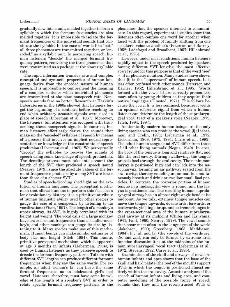
gradually flow into a unit, melded together to form asyllable in which the formant frequencies are alsomelded together. It is impossible to isolate the for-mant frequencies of the individual sounds that con-stitute the syllable. In the case of words like “bat,”all three phonemes are transmitted together, or “en-coded,” as a syllabic unit. In perceiving speech, hu-man listeners “decode” the merged formant fre-quency pattern, recovering the three phonemes thatwere transmitted as a package at the slower syllabicrate.
The rapid information transfer rate and complexconceptual and syntactic properties of human lan-guage derive from the encoded nature of humanspeech. It is impossible to comprehend the meaningof a complex sentence when individual phonemesare transmitted at the slow nonspeech rate. Non-speech sounds fare no better. Research at Haskin’sLaboratories in the 1960s showed that listeners for-got the beginning of a sentence before reaching itsend when arbitrary acoustic signals were used inplace of speech (Liberman et al., 1967). Moreover,the listeners’ full attention was occupied with tran-scribing these nonspeech signals. In contrast, hu-man listeners effortlessly derive the sounds thatmake up the “encoded” syllables of speech by meansof a process that involves an implicit neural repre-sentation or knowledge of the constraints of speechproduction (Liberman et al., 1967). We perceptually“decode” the syllables to recover the sounds ofspeech using some knowledge of speech production.The decoding process must take into account thelength of the SVT that produced a speech sound(Nearey, 1979), since the absolute values of the for-mant frequencies produced by a long SVT are lowerthan those of a shorter SVT.
Studies of speech-decoding shed light on the evo-lution of human language. The perceptual mecha-nism that allows humans to perform this feat has along evolutionary history; it is a primitive attributeof human linguistic ability used by other species togauge the size of a conspecific by listening to itsvocalizations (Fitch, 1997). The length of a monkey’supper airway, its SVT, is highly correlated with itsheight and weight. The vocal calls of a large monkeyhave lower formant frequencies than a smaller mon-key’s, and other monkeys can gauge its size by lis-tening to it. Many species make use of this mecha-nism. Human beings can make similar estimates ofbody size and height (Fitch, 1994). This innate,primitive perceptual mechanism, which is apparentat age 3 months in infants (Lieberman, 1984), isused by human listeners as they perceive speech todecode the formant frequency patterns. Talkers withdifferent SVT lengths can produce different formantfrequencies when they say the same words. For ex-ample, an adult woman’s [a] can have the sameformant frequencies as an adolescent girl’s [ae]vowel. Listeners, therefore, must have some knowl-edge of the length of a speaker’s SVT in order torelate specific formant frequency patterns to the
phonemes that the speaker intended to communi-cate. In this regard, experimental studies show thatlisteners often confuse one word for another whenfaced with the problem of rapidly shifting from onespeaker’s voice to another’s (Peterson and Barney,1952; Ladefoged and Broadbent, 1957; Hillenbrandet al., 1995).
However, under most conditions, human listenersrapidly adjust to the speech produced by speakershaving different SVT lengths; the most effectivevowel sound for this purpose is that of the word “see”-- [i] in phonetic notation. Many studies have shownthat [i] is the “supervowel” of human speech. It isless often confused with other sounds (Peterson andBarney, 1952; Hillenbrand et al., 1995). Wordsformed with the vowel [i] are correctly pronouncedmore often by young children as they acquire theirnative languages (Olmsted, 1971). This follows be-cause the vowel [i] is less confused, because it yieldsan optimal reference signal from which a humanlistener can determine the length of the supralaryn-geal vocal tract of a speaker’s voice (Nearey, 1979;Fitch, 1994, 1997).
Anatomically modern human beings are the onlyliving species who can produce the vowel [i] (Lieber-man and Crelin, 1971; Lieberman et al., 1972;Lieberman, 1968, 1975, 1984; Carre et al., 1995).The adult human tongue and SVT differ from thoseof all other living animals (Negus, 1949). In apes,the body of the tongue is long and relatively flat, andfills the oral cavity. During swallowing, the tonguepropels food through the oral cavity. The nonhumanlarynx is positioned high and can lock into the na-sopharynx, forming an air pathway sealed from theoral cavity, thereby enabling an animal to simulta-neously breath and drink or swallow small food par-ticles. In contrast, the posterior portion of humantongue in a midsaggital view is round, and the lar-ynx is positioned low. The resulting human suprala-ryngeal airway has an almost right-angle bend at itsmidpoint. As we talk, extrinsic tongue muscles canmove the tongue upwards, downwards, forwards, orbackwards, yielding abrupt and extreme changes inthe cross-sectional area of the human supralaryn-geal airway at its midpoint (Chiba and Kajiyama,1941; Fant, 1960; Nearey, 1979). The vowel soundsthat occur most often in the languages of the world(Jakobson, 1990; Greenberg, 1963; Maddieson,1984), [i], [u], and [a] (the vowels of the words see,do, and ma), can only be formed by extreme areafunction discontinuities at the midpoint of the hu-man supralaryngeal vocal tract (Lieberman et al.,1972; Stevens, 1972; Carre et al., 1995).
Examination of the skull and airways of newbornhuman infants and apes shows that the base of theskull and hard palate (the roof of the mouth) supportSVTs in which the tongue is positioned almost en-tirely within the oral cavity. Acoustic analyses of thespeech of human infants and living apes, and com-puter modelling of the possible range of speechsounds that they and the reconstructed SVTs of
NEURAL BASES OF LANGUAGE 55Lieberman]
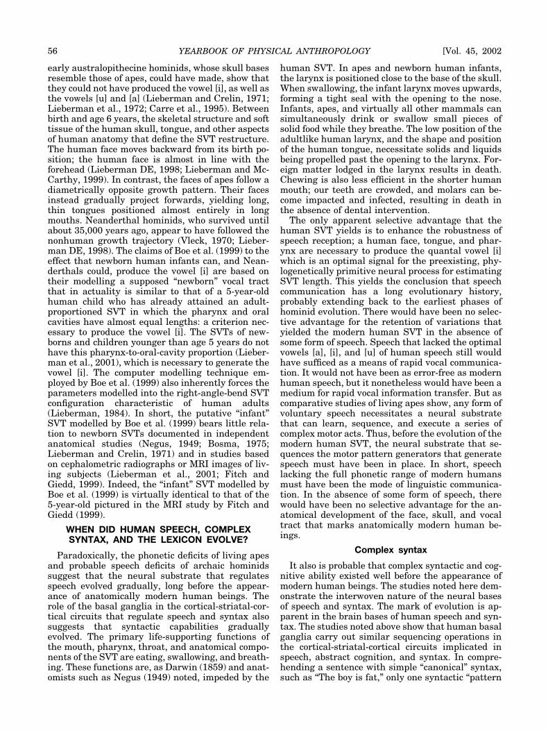
early australopithecine hominids, whose skull basesresemble those of apes, could have made, show thatthey could not have produced the vowel [i], as well asthe vowels [u] and [a] (Lieberman and Crelin, 1971;Lieberman et al., 1972; Carre et al., 1995). Betweenbirth and age 6 years, the skeletal structure and softtissue of the human skull, tongue, and other aspectsof human anatomy that define the SVT restructure.The human face moves backward from its birth po-sition; the human face is almost in line with theforehead (Lieberman DE, 1998; Lieberman and Mc-Carthy, 1999). In contrast, the faces of apes follow adiametrically opposite growth pattern. Their facesinstead gradually project forwards, yielding long,thin tongues positioned almost entirely in longmouths. Neanderthal hominids, who survived untilabout 35,000 years ago, appear to have followed thenonhuman growth trajectory (Vleck, 1970; Lieber-man DE, 1998). The claims of Boe et al. (1999) to theeffect that newborn human infants can, and Nean-derthals could, produce the vowel [i] are based ontheir modelling a supposed “newborn” vocal tractthat in actuality is similar to that of a 5-year-oldhuman child who has already attained an adult-proportioned SVT in which the pharynx and oralcavities have almost equal lengths: a criterion nec-essary to produce the vowel [i]. The SVTs of new-borns and children younger than age 5 years do nothave this pharynx-to-oral-cavity proportion (Lieber-man et al., 2001), which is necessary to generate thevowel [i]. The computer modelling technique em-ployed by Boe et al. (1999) also inherently forces theparameters modelled into the right-angle-bend SVTconfiguration characteristic of human adults(Lieberman, 1984). In short, the putative “infant”SVT modelled by Boe et al. (1999) bears little rela-tion to newborn SVTs documented in independentanatomical studies (Negus, 1949; Bosma, 1975;Lieberman and Crelin, 1971) and in studies basedon cephalometric radiographs or MRI images of liv-ing subjects (Lieberman et al., 2001; Fitch andGiedd, 1999). Indeed, the “infant” SVT modelled byBoe et al. (1999) is virtually identical to that of the5-year-old pictured in the MRI study by Fitch andGiedd (1999).
WHEN DID HUMAN SPEECH, COMPLEXSYNTAX, AND THE LEXICON EVOLVE?
Paradoxically, the phonetic deficits of living apesand probable speech deficits of archaic hominidssuggest that the neural substrate that regulatesspeech evolved gradually, long before the appear-ance of anatomically modern human beings. Therole of the basal ganglia in the cortical-striatal-cor-tical circuits that regulate speech and syntax alsosuggests that syntactic capabilities graduallyevolved. The primary life-supporting functions ofthe mouth, pharynx, throat, and anatomical compo-nents of the SVT are eating, swallowing, and breath-ing. These functions are, as Darwin (1859) and anat-omists such as Negus (1949) noted, impeded by the
human SVT. In apes and newborn human infants,the larynx is positioned close to the base of the skull.When swallowing, the infant larynx moves upwards,forming a tight seal with the opening to the nose.Infants, apes, and virtually all other mammals cansimultaneously drink or swallow small pieces ofsolid food while they breathe. The low position of theadultlike human larynx, and the shape and positionof the human tongue, necessitate solids and liquidsbeing propelled past the opening to the larynx. For-eign matter lodged in the larynx results in death.Chewing is also less efficient in the shorter humanmouth; our teeth are crowded, and molars can be-come impacted and infected, resulting in death inthe absence of dental intervention.
The only apparent selective advantage that thehuman SVT yields is to enhance the robustness ofspeech reception; a human face, tongue, and phar-ynx are necessary to produce the quantal vowel [i]which is an optimal signal for the preexisting, phy-logenetically primitive neural process for estimatingSVT length. This yields the conclusion that speechcommunication has a long evolutionary history,probably extending back to the earliest phases ofhominid evolution. There would have been no selec-tive advantage for the retention of variations thatyielded the modern human SVT in the absence ofsome form of speech. Speech that lacked the optimalvowels [a], [i], and [u] of human speech still wouldhave sufficed as a means of rapid vocal communica-tion. It would not have been as error-free as modernhuman speech, but it nonetheless would have been amedium for rapid vocal information transfer. But ascomparative studies of living apes show, any form ofvoluntary speech necessitates a neural substratethat can learn, sequence, and execute a series ofcomplex motor acts. Thus, before the evolution of themodern human SVT, the neural substrate that se-quences the motor pattern generators that generatespeech must have been in place. In short, speechlacking the full phonetic range of modern humansmust have been the mode of linguistic communica-tion. In the absence of some form of speech, therewould have been no selective advantage for the an-atomical development of the face, skull, and vocaltract that marks anatomically modern human be-ings.
Complex syntax
It also is probable that complex syntactic and cog-nitive ability existed well before the appearance ofmodern human beings. The studies noted here dem-onstrate the interwoven nature of the neural basesof speech and syntax. The mark of evolution is ap-parent in the brain bases of human speech and syn-tax. The studies noted above show that human basalganglia carry out similar sequencing operations inthe cortical-striatal-cortical circuits implicated inspeech, abstract cognition, and syntax. In compre-hending a sentence with simple “canonical” syntax,such as “The boy is fat,” only one syntactic “pattern
56 YEARBOOK OF PHYSICAL ANTHROPOLOGY [Vol. 45, 2002

generator” (syntactic scheme or set of “rules”) needbe invoked. The words “the boy” constitute a noun-phrase (NP) that is the sentence’s subject. Thewords “is fat” constitute a verb phrase (VP) that isthe sentence’s predicate. The syntactic “formula”Sentence � NP � VP describes the basic syntax ofthe sentence. In contrast, comprehending a complexsentence, such as “The boy who is fat fell down,”entails being able to interrupt the sequence of cog-nitive acts involved in interpreting syntax at therelative clause’s boundary (the word “who”), wherean “embedded” sentence has been inserted into thecanonical form. As syntactic complexity increases,different “rules” of syntax must be continually sup-pressed and invoked. As Marsden and Obeso (1994)and Graybiel (1997, 1998) noted, the basal gangliaappear to perform similar cognitive and motor pro-cessing sequencing functions, i.e., interrupting anongoing activity and starting a different act whenappropriate. The basal ganglia most likely are activein acquiring the cognitive pattern generators thatare relevant to the syntax of language: the “rules ofgrammar,” and perhaps the acquisition of the in-creased store of words that marks human lexicalability. Further study obviously is necessary.
Walking
As noted above, a half century ago, Lashley (1951)explicitly pointed out the similarities between thesyntax of motor control and language. Data-linkingdeficits in serial manual motor control, speech, andsyntax in aphasia suggest that neural mechanismsinitially adapted for motor control were modified inthe course of human evolution to yield syntacticability (Lieberman, 1975, 1984, 1985), a view inde-pendently shared by Kimura (1993). Studies of theacquisition of gesture and speech by children sup-port this view (Greenfield, 1991); a seamless transi-tion between gesture and speech occurs in the courseof maturation. Neurophysiologic studies of motorcontrol and cognition (Graybiel, 1997, 1998), and thestudies of neurodegenerative diseases noted herein,show that the basal ganglia support neuronal popu-lations that carry out similar operations in the do-mains of motor control, cognition, and syntax. Aninsight regarding the start of the evolutionary pro-cess that yielded the neural bases that allow humanlanguages to sequence the sounds of speech (syntac-tic rules), and that confer flexability to humanthought, may follow if we take account of the role ofbasal ganglia in walking (Hochstadt, personal com-munication). Although the striatal components ofhuman cortical-striatal-cortical neural circuits playa role in language and cognition, they continue toregulate motor control. In Parkinson’s disease, oneof the most apparent behavioral deficits of degradedbasal ganglia function is the marked deterioration ofwalking and upright balance. The PD rating scale ofHoehn and Yahr (1967) is essentially a measure of apatient’s ability to recover a stable upright positionafter being gently perturbed. Moderate to severe PD
generally results in deficits in a person’s ability tosequence internally generated motor sequences(Cunnington et al., 1995; Harrington and Haaland,1991; Jellinger, 1990), and one of the most symptom-atic signs is impaired ability to execute the complexsequence of motor acts involved in upright bipedallocomotion. Moreover, upright human bipedal loco-motion appears to be learned rather than innate(Thelen and Cooke, 1987), and the basal ganglia(Mirenowicz and Schultz, 1996; Graybiel, 1995) aswell as cerebellum and prefrontal cortex (Thach,1996) are all implicated in motor learning. Given therole of the basal ganglia in the motor control neces-sary for upright locomotion, natural selection di-rected at enhancing upright bipedal locomotion andsubsequent Darwinian “preadatation” (the directionof the basal ganglia “sequencing engine” to speechproduction, syntax, and thinking) may have beentriggered in the earliest phases of hominid evolu-tion.
The archaelogical record
The archaeological record has often been used todate the appearance of the modern human mind(Klein, 1999). Until recently, the artifacts associatedwith modern human beings appeared to occur after40,000 years ago. However, it is apparent that arti-facts associated with anatomically modern humansappeared in Africa well before that time (McBreartyand Brooks, 2000). Moreover, the modern humanmind and full linguistic capabilities must haveevolved before the probable dispersal of modern hu-mans from Africa, since any neurologically intactchild from any location on earth can effortlesslyacquire any language when exposed to it in a normalenvironment before age 7 years. Since modern hu-mans reached Australia about 60,000 years ago, thehuman brain must have reached its present state ofdevelopment long before that date.
CONCLUDING COMMENTS
The neural bases of human linguistic ability arecomplex, involving structures other than Broca’sand Wernicke’s areas. Although our knowledge is atbest incomplete, it is clear that many other corticalareas and subcortical structures form part of theneural circuits implicated in the lexicon, speech pro-duction and perception, and syntax. The subcorticalbasal ganglia support the cortical-striatal-corticalcircuits that regulate speech production, complexsyntax, and the acquisition of the motor and cogni-tive pattern generators that underlie speech produc-tion and syntax. They most likely are involved inlearning the semantic referents and sound patternsthat are instantiated as words in the brain’s dictio-nary. The cerebellum and prefrontal cortex are alsoinvolved in learning motor acts. Frontal regions ofthe cortex are implicated in virtually all cognitiveacts and the acquisition of cognitive criteria; poste-rior cortical regions are clearly active elements of
NEURAL BASES OF LANGUAGE 57Lieberman]
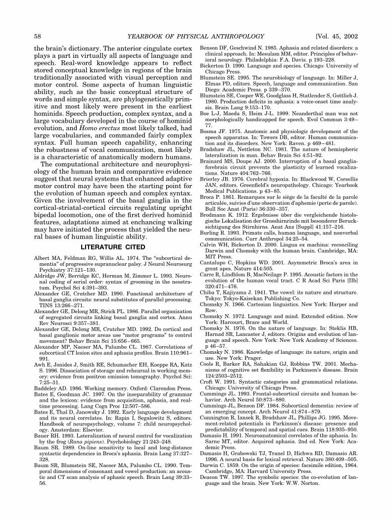
the brain’s dictionary. The anterior cingulate cortexplays a part in virtually all aspects of language andspeech. Real-word knowledge appears to reflectstored conceptual knowledge in regions of the braintraditionally associated with visual perception andmotor control. Some aspects of human linguisticability, such as the basic conceptual structure ofwords and simple syntax, are phylogenetically prim-itive and most likely were present in the earliesthominids. Speech production, complex syntax, and alarge vocabulary developed in the course of hominidevolution, and Homo erectus most likely talked, hadlarge vocabularies, and commanded fairly complexsyntax. Full human speech capability, enhancingthe robustness of vocal communication, most likelyis a characteristic of anatomically modern humans.
The computational architecture and neurophysi-ology of the human brain and comparative evidencesuggest that neural systems that enhanced adaptivemotor control may have been the starting point forthe evolution of human speech and complex syntax.Given the involvement of the basal ganglia in thecortical-striatal-cortical circuits regulating uprightbipedal locomotion, one of the first derived hominidfeatures, adaptations aimed at enchancing walkingmay have initiated the process that yielded the neu-ral bases of human linguistic ability.
LITERATURE CITED
Albert MA, Feldman RG, Willis AL. 1974. The “subcortical de-mentia” of progressive supranuclear palsy. J Neurol NeurosurgPsychiatry 37:121–130.
Aldridge JW, Berridge KC, Herman M, Zimmer L. 1993. Neuro-nal coding of serial order: syntax of grooming in the neostra-tum. Psychol Sci 4:391–393.
Alexander GE, Crutcher MD. 1990. Functional architecture ofbasal ganglia circuits: neural substitutes of parallel processing.TINS 13:266–271.
Alexander GE, Delong MR, Strick PL. 1986. Parallel organizationof segregated circuits linking basal ganglia and cortex. AnnuRev Neurosci 9:357–381.
Alexander GE, Delong MR, Crutcher MD. 1992. Do cortical andbasal ganglionic motor areas use “motor programs” to controlmovement? Behav Brain Sci 15:656–665.
Alexander MP, Naeser MA, Palumbo CL. 1987. Correlations ofsubcortical CT lesion sites and aphasia profiles. Brain 110:961–991.
Awh E, Jonides J, Smith RE, Schumacher EH, Koeppe RA, KatzS. 1996. Dissociation of storage and rehearsal in working mem-ory: evidence from positron emission tomography. Psychol Sci:7:25–31.
Baddeley AD. 1986. Working memory. Oxford: Clarendon Press.Bates E, Goodman JC. 1997. On the inseparability of grammar
and the lexicon: evidence from acquisition, aphasia, and real-time processing. Lang Cogn Proc 12:507–586.
Bates E, Thal D, Janowsky J. 1992. Early language developmentand its neural correlates. In: Rapin I, Segalowitz S, editors.Handbook of neuropsychology, volume 7: child neuropsychol-ogy. Amsterdam: Elsevier.
Bauer RH. 1993. Lateralization of neural control for vocalizationby the frog (Rana pipiens). Psychobiology 21:243–248.
Baum SR. 1989. On-line sensitivity to local and long-distancesyntactic dependencies in Broca’s aphasia. Brain Lang 37:327–328.
Baum SR, Blumstein SE, Naeser MA, Palumbo CL. 1990. Tem-poral dimensions of consonant and vowel production: an acous-tic and CT scan analysis of aphasic speech. Brain Lang 39:33–56.
Benson DF, Geschwind N. 1985. Aphasia and related disorders: aclinical approach. In: Mesulam MM, editor. Principles of behav-ioral neurology. Philadelphia: F.A. Davis. p 193–228.
Bickerton D. 1990. Language and species. Chicago: University ofChicago Press.
Blumstein SE. 1995. The neurobiology of language. In: Miller J,Elmas PD, editors. Speech, language and communication. SanDiego: Academic Press. p 339–370.
Blumstein SE, Cooper WE, Goodglass H, Statlender S, Gottlieb J.1980. Production deficits in aphasia: a voice-onset time analy-sis. Brain Lang 9:153–170.
Boe L-J, Maeda S, Heim J-L. 1999. Neanderthal man was notmorphologically handicapped for speech. Evol Commun 3:49–77.
Bosma JF. 1975. Anatomic and physiologic development of thespeech apparatus. In: Towers DB, editor. Human communica-tion and its disorders. New York: Raven. p 469–481.
Bradshaw JL, Nettleton NC. 1981. The nature of hemisphericlateralization in man. Behav Brain Sci 4:51–92.
Brainard MS, Doupe AJ. 2000. Interruption of a basal ganglia-forebrain circuit prevents the plasticity of learned vocaliza-tions. Nature 404:762–766.
Brierley JB. 1976. Cerebral hypoxia. In: Blackwood W, CorsellisJAN, editors. Greenfield’s neuropathology. Chicago: YearbookMedical Publications. p 43–85.
Broca P. 1861. Remarques sur le siege de la faculte de la parolearticulee, suivies d’une observation d’aphemie (perte de parole).Bull Soc Anat (Paris) 36:330–357.
Brodmann K. 1912. Ergebnisse uber die vergleichende histolo-gische Lokalisation der Grosshirnrinde mit besonderer Beruck-sichtigung des Stirnhirns. Anat Anz [Suppl] 41:157–216.
Burling R. 1993. Primate calls, human language, and nonverbalcommunication. Curr Anthropol 34:25–54.
Calvin WH, Bickerton D. 2000. Lingua ex machina: reconcilingDarwin and Chomsky with the human brain. Cambridge, MA:MIT Press.
Cantalupo C, Hopkins WD. 2001. Asymmetric Broca’s area ingreat apes. Nature 414:505.
Carre R, Lindblom B, MacNeilage P. 1995. Acoustic factors in theevolution of the human vocal tract. C R Acad Sci Paris [IIb]320:471–476.
Chiba T, Kajiyama J. 1941. The vowel: its nature and structure.Tokyo: Tokyo-Kaisekan Publishing Co.
Chomsky N. 1966. Cartesian linguistics. New York: Harper andRow.
Chomsky N. 1972. Language and mind. Extended edition. NewYork: Harcourt, Brace and World.
Chomsky N. 1976. On the nature of language. In: Steklis HB,Harnad SR, Lancaster J, editors. Origins and evolution of lan-guage and speech. New York: New York Academy of Sciences.p 46–57.
Chomsky N. 1986. Knowledge of language: its nature, origin anduse. New York: Prager.
Cools R, Barker RA, Sahakian GJ, Robbins TW. 2001. Mecha-nisms of cognitive set flexibility in Parkinson’s disease. Brain124:2503–2512.
Croft W. 1991. Syntactic categories and grammatical relations.Chicago: University of Chicago Press.
Cummings JL. 1993. Frontal-subcortical circuits and human be-havior. Arch Neurol 50:873–880.
Cummings JL, Benson DF. 1984. Subcortical dementia: review ofan emerging concept. Arch Neurol 41:874–879.
Cunnington R, Iansek R, Bradshaw JL, Phillips JG. 1995. Move-ment-related potentials in Parkinson’s disease: presence andpredictability of temporal and spatial cues. Brain 118:935–950.
Damasio H. 1991. Neuroanatomical correlates of the aphasia. In:Sarno MT, editor. Acquired aphasia. 2nd ed. New York: Aca-demic Press.
Damasio H, Grabowski TJ, Tranel D, Hichwa RD, Damasio AR.1996. A neural basis for lexical retrieval. Nature 380:409–505.
Darwin C. 1859. On the origin of species: facsimile edition, 1964.Cambridge, MA: Harvard University Press.
Deacon TW. 1997. The symbolic species: the co-evolution of lan-guage and the brain. New York: W.W. Norton.
58 YEARBOOK OF PHYSICAL ANTHROPOLOGY [Vol. 45, 2002

DeLong MR. 1993. Overview of basal ganglia function. In: ManoN, Hamada I, deLong MR, editors. Role of the cerebellum andbasal ganglia in voluntary movement. Amsterdam: Elsevier.
Denneberg VH. 1981. Hemispheric laterality in animals and theeffects of early experience. Behav Brain Sci 4:1–50.
D’Esposito M, Alexander MP. 1995. Subcortical aphasia: distinctprofiles following left putaminal hemorrhage. Neuorology 45:38–41.
D’Esposito M, Detre JA, Alsop DC, Shin RK, Atlas S, GrossmanM. 1995. The neural basis of the central executive system ofworking memory. Nature 378:279–281.
Dewey D, Roy EA, Square-Storer PA, Hayden DC. 1988. Limband oral praxic abilities of children with verbal sequencingdeficits. Dev Med Child Neurol 30:743–751.
Dronkers NF, Shapiro JK, Redfern B, Knight RT. 1992. The roleof Broca’s area in Broca’s aphasia. J Clin Exp Neuropsychol14:198.
Dunbar RIM. 1993. Coevolution of neocortical size and languagein humans. Behav Brain Sci 16:681–735.
Edelman GM. 1987. Neural Darwinism. New York: Basic Books.Elman J, Bates E, Johnson M, Karmiloff-Smith A, Parisi D,
Plunkett K. 1997. Rethinking innateness: a connectionist per-spective on development. Cambridge, MA: MIT Press/BradfordBooks.
Fant G. 1960. Acoustic theory of speech production. The Hague:Mouton.
Fischer J, Cheney DL, Seyfarth RM. 2000. Development of infantbaboons’ responses to graded bark variants. Proc R Soc Lond[Biol] 267:2317–2321.
Fischer J, Hammerschmidt K, Cheney DL, Seyfarth RM. 2002.Acoustic features of male baboon loud calls: influences of con-text, age and individuality. J Acoust Soc Am 111:1485–1474.
Fitch WT III. 1994. Vocal tract length and the evolution of lan-guage. Ph.D. dissertation, Brown University.
Fitch WT III. 1997. Vocal tract length and formant frequencydispersion correlate with body size in macaque monkeys. JAcoust Soc Am 102:1213–1222.
Fitch WT, Giedd J. 1999. Morphology and development of thehuman vocal tract: a study using magnetic resonance imaging.J Acoust Soc Am 106:1511–1522.
Flowers KA, Robertson C. 1985. The effects of Parkinson’s diseaseon the ability to maintain a mental set. J Neurol NeurosurgPsychiatry 48:517–529.
Fodor J. 1983. Modularity of mind. Cambridge, MA: MIT Press.Gall FJ. 1809. Recherches sur le systeme nerveux. Paris: B.
Bailliere.Gannon PJ, Holloway RL, Broadfield DC, Braun AR. 1998. Asym-
metry of chimpanzee planum temporale: humanlike pattern ofWernicke’s brain language area homolog. Science 279:220–222.
Gardner RA, Gardner BT. 1969. Teaching sign language to achimpanzee. Science 165:664–672.
Gardner RA, Gardner BT. 1984. A vocabulary test for chimpan-zees (Pan troglodytes). J Comp Psychol 4:381–404.
Gardner BT, Gardner RA. 1994. Developmant of phrases in theutterances of children and cross-fostered chimpanzees. In:Gardner RA, Gardner BT, Chiarelli B, Plooj R, editors. Theethological roots of culture. Dordrecht: Kluwer Academic Pub-lishers. p 223–255.
Gathercole SE, Baddeley AD. 1993. Working memory and lan-guage. Hillside, NJ: Lawrence Erlbaum Associates.
Geschwind N. 1970. The organization of language and the brain.Science 170:940–944.
Goodall J. 1986. The chimpanzees of Gombe: patterns of behavior.Cambridge, MA: Harvard, University Press.
Gopnik M. 1990. Feature-blind grammar and dysphasia in anextended family. Nature 344:715.
Gopnik M, Crago M. 1991. Familial segregation of a developmen-tal language disorder. Cognition 39:1–50.
Gotham AM, Brown RG, Marsden CD. 1988. “Frontal” cognitivefunction in patients with Parkinsons’s disease “on” and “off”levadopa. Brain 111:199–321.
Gottlieb G. 1975. Development of species identification in duck-lings: I. Nature of perceptual deficits caused by embryonicauditory deprivation. J Comp Physiol Psychol 89:387–389.
Grafman J. 1989. Plans, actions and mental sets: the role of thefrontal lobes. In: Perecman E, editor. Integrating theory andpractice in clinical neuropsychology. Hillsdale, NJ: Erlbaum.
Graybiel AM. 1995. Building action repertoires: memory andlearning functions of the basal ganglia. Curr Opin Neurobiol5:733–741.
Graybiel AM. 1997. The basal ganglia and cognitive pattern gen-erators. Schizophr Bull 23:459–469.
Graybiel A. 1998. The basal ganglia and chunking of action rep-ertoires. Neurobiol Mem Learn 70:119–136.
Graybiel AM, Aosaki T, Flaherty AW, Kimura M. 1994. The basalganglia and adaptive motor control. Science 265:1826–1831.
Greenberg BD, Murphy DL, Rasmussen SA. 2000. Neuroana-tomically based approaches to obsessive-compulsive disorder:neurosurgery and transcranial magnetic stimulation. Psychi-atr Clin North Am 23:671–685.
Greenberg J. 1963. Universals of language. Cambridge, MA: MITPress.
Greenfield PM. 1991. Language, tools and brain: the ontogenyand phylogeny of hierarchically organized sequential behavior.Behav Brain Sci 14:531–577.
Grossman MG, Carvell S, Gollomp S, Stern MB, Vernon G, Hur-tig HI. 1991. Sentence comprehension and praxis deficits inParkinson’s disease. Neurology 41:1620–1628.
Grossman MG, Carvell S, Gollomp S, Stern MB, Reivich M,Morrison D, Alavi A, Hurtig HI. 1993. Cognitive and physio-logical substrates of impaired sentence processing in Parkin-son’s disease. J Cogn Neurosci 5:480–498.
Grossman MG, Glosser J, Kalmanson J, Morris MB, Stren, Hur-tig HI. 2001. Dopamine supports sentence comprehension inParkinson’s disease. J Neurol Sci 184:123–130.
Harrington DL, Haaland L. 1991. Sequencing in Parkinson’sdisease: abnormalities in programming and controlling move-ment. Brain 114:99–115.
Hata Y, Stryker MP. 1994. Control of thalamocortical afferentrearrangement by postsynaptic activity in developing visualcortex. Science 263:1732–1735.
Hauser MD. 1996. The evolution of communication. Cambridge,MA: MIT Press.
Hellwag C. 1781. De formatione loquelae. Dissertation, Tubingen.Hewes GW. 1973. Primate communication and the gestural origin
of language. Curr Anthropol 14:5–24.Hillenbrand JL, Getty A, Clark MJ, Wheeler K. 1995. Acoustic
characteristics of American English vowels. J Acoust Soc Am97:3099–3111.
Hochstadt J, Lieberman P. 2000. Eye tracking of sentence picturematching: comparisons between normal and Parkinsonian sub-jects. CUNY 2000 conference. San Diego, CA.
Hoehn MM, Yahr MD. 1967. Parkinsonism: onset, progressionand mortality. Neurology 17:427–442.
Holloway RL. 1995. Evidence for POT expansion in early Homo:a pretty theory with ugly (or no) paleoneurological facts. BehavBrain Sci 18:191–193.
Howard LA, Binks MG, Moore AP, Playfer JR. 2001. The contri-bution of apraxic speech to working memory deficits in Parkin-son’s disease. Brain Lang 74:269–288.
Illes J, Metter EJ, Hanson WR, Iritani S. 1988. Language pro-duction in Parkinson’s disease: acoustic and linguistic consid-erations. Brain Lang 33:146–160.
Ivry RB, Keele SW. 1989. Timing functions of the cerebellum. JCogn Neurosci 1:134–150.
Jackendoff R. 1994. Patterns in the mind: language and humannature. New York: Basic Books.
Jackson JH. 1915. On affectations of speech from diseases of thebrain. Brain 38:106–174.
Jakobson R. 1990. On language. In: Waugh LR, Monville-BurstonM, editors. Cambridge, MA: Harvard University Press.
Jellinger K. 1990. New developments in the pathology of Parkin-son’s disease. Adv Neurol 53:1–15.
Jerison HJ. 1973. Evolution of the brain and intelligence. NewYork: Academic Press.
Just MA, Carpenter PA, Keller TA, Eddy WF, Thulborn B. 1996.Brain activation modulated by sentence comprehension. Sci-ence 274:114–116.
NEURAL BASES OF LANGUAGE 59Lieberman]
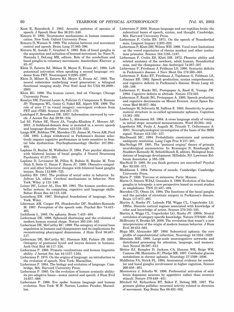
Kent R, Rosenbeck J. 1983. Acoustic patterns of apraxia ofspeech. J Speech Hear Res 26:231–248.
Kimura D. 1993. Neuromotor mechanisms in human communi-cation. New York: Oxford University Press.
Kimura D, Watson N. 1989. The relation between oral movementcontrol and speech. Brain Lang 37:565–590.
Kimura M, Aosaki T, Graybiel A. 1993. Role of basal ganglia inthe acquisition and initiation of learned movement. In: Nano N,Hamada I, DeLong MR, editors. Role of the cerebellum andbasal ganglia in voluntary movements. Amsterdam: Elsevier. p83–87.
Klein D, Zatorre RJ, Milner B, Meyer E, Evans AC. 1994. Leftputaminal activation when speaking a second language: evi-dence from PET. Neuroreport 5:2295–2297.
Klein D, Milner B, Zatorre RJ, Meyer E, Evans AC. 1995. Theneural substrates underlying word generation: a bilingualfunctional imaging study. Proc Natl Acad Sci USA 92:2899–2903.
Klein RG. 1999. The human career, 2nd ed. Chicago: ChicagoUniversity Press.
Kosslyn SM, Pascual-Leone A, Felician O, Camposano S, KeenanJP, Thompson WL, Ganis G, Sukel KE, Alpert NM. 1999. Therole of area 17 in visual imagery: convergent evidence fromPET and rTMS. Science 284:167–170.
Ladefoged P, Broadbent DE. 1957. Information conveyed by vow-els. J Acoust Soc Am 29:98–104.
Lal SJ, Fisher SE, Hurst JA, Vargha-Khadem F, Monaco AP.2001. A forkhead-domain dene is mutated in a severe speechand language disorder. Nature 413:519–523.
Lange KW, Robbins TW, Marsden CD, James M, Owen AM, PaulGM. 1992. L-dopa withdrawal in Parkinson’s disease selec-tively impairs cognitive performance in tests sensitive to fron-tal lobe dysfunction. Psychopharmacology (Berlin) 107:394–404.
Laplane D, Baulac M, Widlocher D. 1984. Pure psychic akinesiawith bilateral lesions of basal ganglia. J Neurol NeurosurgPsychiatry 47:377–385.
Laplane D, Levasseur M, Pillon B, Bubois R, Baulac M, TranDinh S, Sette G, Danze F, Baron JC. 1989. Obsessive-compul-sive and other behavioral changes with bilateral basal ganglialesions. Brain 112:699–725.
Lashley KS. 1951. The problem of serial order in behavior. In:Jefress LA, editor. Cerebral mechanisms in behavior. NewYork: Wiley. p 112–146.
Leiner HC, Leiner AL, Dow RS. 1991. The human cerebro-cere-bellar system: its computing, cognitive and language skills.Behav Brain Res 44:113–128.
Lenneberg EH. 1967. Biological foundations of language. NewYork: Wiley.
Liberman AM, Cooper FS, Shankweiler DP, Studdert-KennedyM. 1967. Perception of the speech code. Psychol Rev 74:431–461.
Lichtheim L. 1885. On aphasia. Brain 7:433–484.Lieberman DE. 1998. Sphenoid shortening and the evolution of
modern human cranial shape. Nature 393:158–162.Lieberman DE, McCarthy RC. 1999. The ontogeny of cranial base
angulation in humans and chimpanzees and its implications forreconstructing pharyngeal dimensions. J Hum Evol 36:487–517.
Lieberman DE, McCarthy RC, Hieemae KM, Palmer JB. 2001.Ontogeny of postnatal hyoid and larynx descent in humans.Arch Oral Biol 46:117–128.
Lieberman P. 1968. Primate vocalizations and human linguisticability. J Acoust Soc Am 44:1157–1164.
Lieberman P. 1975. On the origins of language: an introduction tothe evolution of speech. New York: Macmillan.
Lieberman P. 1984. The biology and evolution of language. Cam-bridge, MA: Harvard University Press.
Lieberman P. 1985. On the evolution of human syntactic ability:its pre-adaptive bases—motor control and speech. J Hum Evol14:657–668.
Lieberman P. 1998. Eve spoke: human language and humanevolution. New York: W.W. Norton; London: Picador, Macmil-lan.
Lieberman P. 2000. Human language and our reptilian brain: thesubcortical bases of speech, syntax, and thought. Cambridge,MA: Harvard University Press.
Lieberman P, Crelin ES. 1971. On the speech of Neanderthalman. Linguist Inquiry 2:203–222.
Lieberman P, Klatt DH, Wilson WH. 1969. Vocal tract limitationson the vowel repertoires of rhesus monkey and other nonhu-man primates. Science 164:1185–1187.
Lieberman P, Crelin ES, Klatt DH. 1972. Phonetic ability andrelated anatomy of the newborn, adult human, Neanderthalman, and the chimpanzee. Am Anthropol 74:287–307.
Lieberman P, Friedman J, Feldman LS. 1990. Syntactic deficitsin Parkinson’s disease. J Nerv Ment Dis 178:360–365.
Lieberman P, Kako ET, Friedman J, Tajchman G, Feldman LS,Jiminez EB. 1992. Speech production, syntax comprehension,and cognitive deficits in Parkinson’s disease. Brain Lang 43:169–189.
Lieberman P, Kanki BG, Protopapas A, Reed E, Youngs JW.1994. Cognitive defects at altitude. Nature 372:325.
Lieberman P, Kanki BG, Protopapas A. 1995. Speech productionand cognitive decrements on Mount Everest. Aviat Space En-viron Med 66:857–864.
Linebarger M, Schwartz M, Saffran E. 1983. Sensitivity to gram-matical structure in so-called agrammatic aphasics. Cognition13:361–392.
Lisker L, Abramson AS. 1964. A cross language study of voicingin initial stops: acoustical measurements. Word 20:384–442.
Logothetis NK, Pauls J, Augath M, Trinath T, Oeltermann A.2001. Neurophysiological investigation of the basis of the fMRIsignal. Nature 412:150–157.
MacDonald MC. 1994. Probabilistic constraints and syntacticambiguity resolution. Lang Cogn Proc 9:157–201.
MacNeilage PF. 1991. The “postural origins” theory of primateneurobiolgical asymmetries. In: Krasnegor N, Rumbaugh D,Studdert-Kennedy M, Schiefelbusch R, editors. Biological foun-dations of language development. Hillsdale, NJ: Lawrence Erl-baum Associates. p 165–188.
MacNeill D. 1985. So you think gestures are nonverbal? PsycholRev 92:350–371.
Maddieson I. 1984. Patterns of sounds. Cambridge: CambridgeUniversity Press.
Marie P. 1926. Travaux et memoires. Paris: Masson.Marin O, Smeets WJAJ, Gonzalez A. 1998. Evolution of the basal
ganglia in tetrapods: a new perspective based on recent studiesin amphibians. TNN 21:487–494.
Marsden CD, Obeso JA. 1994. The functions of the basal gangliaand the paradox of sterotaxic surgery in Parkinson’s disease.Brain 117:877–897.
Martin A, Haxby JV, Lalonde FM, Wiggs CL, Ungerleider LG.1995a. Discrete cortical regions associated with knowledge ofcolor and knowledge of action. Science 270:102–105.
Martin A, Wiggs CL, Ungerleider LG, Haxby JV. 1995b. Neuralcorrelates of category-specific knowledge. Nature 379:649–652.
McBrearty S, Brooks AS. 2000. The revolution that wasn’t: a newinterpretation of the origin of modern human behavior. J HumEvol 39:453–563.
Mega MS, Alexander MF. 1994. Subcortical aphasia: the coreprofile of capsulostriatal infarction. Neurology 44:1824–1829.
Mesulam MM. 1990. Large-scale neurocognitive networks anddistributed processing for attention, language, and memory.Ann Neurol 28:597–613.
Metter EJ, Kempler D, Jackson CA, Hanson WR, Reige WH,Camras JM, Mazziotta JC, Phelps ME. 1987. Cerebular glucosemetabolism in chronic aphasia. Neurology 37:1599–1606.
Middleton FA, Strick PL. 1994. Anatomical evidence for cerebel-lar and basal ganglia involvement in higher cognition. Science266:458–461.
Mirenowicz J, Schultz W. 1996. Preferential activation of mid-brain dopamine neurons by appetitive rather than aversivestimuli. Nature 379:449–451.
Mitchell SJ, Richardson RT, Baker F, Delong MR. 1987. Theprimate globus pallidus: neuronal activity related to directionof movement. Exp Brain Res 68:491–505.
60 YEARBOOK OF PHYSICAL ANTHROPOLOGY [Vol. 45, 2002
Monchi O, Petrides P, Petre V, Worsley K, Dagher A. 2001.Wisconsin Card Sorting revisited: distinct neural circuits par-ticipating in different stages of the task identified by event-related functional magnetic resonance imaging. J Neurosci 21:7733–7741.
Morris RG, Downes JJ, Sahakian BJ, Evenden JL, Heald A,Robbins TW. 1988. Planning and spatial working memory inParkinson’s disease. J Neurol Neurosurg Psychiatry 51:757–766.
Muller J. 1848. The physiology of the senses, voice and muscularmotion with the mental faculties. Baly W, translator. London:Walton and Maberly.
Naeser MA, Alexander MP, Helms-Estabrooks N, Levine HL,Laughlin SA, Geschwind N. 1982. Aphasia with predomini-nantly subcortical lesion sites; description of three capsular/putaminal aphasia syndromes. Arch Neurol 39:2–14.
Natsopoulos D, Grouios G, Bostantzopoulou S, Mentenopoulos G,Katsarou Z, Logothetis J. 1993. Algorithmic and heuristicstrategies in comprehension of complement clauses by patientswith Parkinson’s disease. Neuropsychologia 31:951–964.
Nearey T. 1979. Phonetic features for vowels. Bloomington: Indi-ana University Linguistics Club.
Negus VE. 1949. The comparative anatomy and physiology of thelarynx. New York: Hafner.
Nelson TO, Dunlosky J, White DM, Steinberg J, Townes BD,Anderson D. 1990. Cognition and metacognition at extremealtitudes on Mount Everest. J Exp Psychol [Gen] 119:367–374.
Newman JD, Maclean PD. 1982. Effects of tegmental lesions onthe isolation call of squirrel monkeys. Brain Res 232:317–329.
Nisimura H, Hashikawa K, Doi K, Iwaki T, Watanabe Y,Kusuoka H, Nishimura T, Kubo T. 1999. Sign language “heard”in the auditory cortex. Nature 397:116.
Nowak MA, Plotkin JB, Jansen VAA. 2000. The evolution ofsyntactic communication. Science 404:495–498.
Nudo RJ, Milliken GW, Jenkins WM, Merzenich MM. 1996. Use-dependent alterations of movement representations in primarymotor cortex of adult squirrel monkeys. J Neurosci 16:785–807.
Olmsted DL. 1971. Out of the mouth of babes. The Hague: Mou-ton.
Pantev C, Oostenveld A, Engelien A, Ross B, Roberts LE, Hoke M.1998. Increased cortical representation in musicians. Nature392:811–813.
Parent A. 1986. Comparative neurobiology of the basal ganglia.New York: John Wiley.
Paulesu E, Firth C, Frackowiak R. 1993. The neural correlates ofthe verbal component of working memory. Nature 362:342–345.
Paus T, Perry DW, Zatorre RA, Worsley KJ, Evans AC. 1996.Modulation of cerebral blood flow in the human auditory cortexduring speech: role of motor-to-sensory discharges. Eur J Neu-rosci 8:2236–2246.
Perkell JS. 1969. Physiology of speech production: results andimplications of a quantitative cineradiographic study. Cam-bridge, MA: MIT Press.
Peterson GE, Barney HL. 1952. Control methods used in a studyof the vowels. J Acoust Soc Am 24:175–184.
Peterson SE, Fox PT, Posner MI, Mintun M, Raichle ME. 1988.Positron emission tomographic studies of the cortical anatomyof single-word processing. Nature 331:585–589.
Pickett ER. 1998. Language and the cerebellum. Ph.D. disserta-tion; Brown University.
Pickett ER, Kuniholm E, Protopapas A, Friedman J, LiebermanP. 1998. Selective speech motor, syntax and cognitive deficitsassociated with bilateral damage to the head of the caudatenucleus and the putamen. A single case study. Neuropsycholo-gia 36:173–188.
Pinker S. 1994. The language instinct; how the mind createslanguage. New York: William Morrow.
Pinker S. 1998. How the mind works. New York: W.W. Norton.Polit A, Bizzi E. 1978. Processes controling arm movements in
monkeys. Science 201:1235–1237.Rauschecker JP, Korte M. 1993. Auditory compensation for early
blindness in cat cerebral cortex. J Neurosci 18:4538–4548.
Regard M, Oelz O, Brugger P, Landis T. 1989. Persisitent cogni-tive impairment in climbers after repeated exposure to alti-tude. Neurology 39:210–213.
Rizzolatti G, Arbib MA. 1998. Language within our grasp. TrendsNeurosci 21:188–194.
Ryalls J. 1986. An acoustic study of vowel production in aphasia.Brain Lang 29:48–67.
Sadato NA, Pasual-Leone J, Grafman V, Ibanez M-P, Delber G,Dold D, Hallet M. 1996. Activation of the primary visual cortexby Braille reading in blind subjects. Nature 380:525–528.
Sanes JN, Donoghue JP. 1996. Static and dynamic organizationof motor cortex. Adv Neurol. Brain Plastic 73:277–296.
Sanes JN, Donoghue JP. 1997. Dynamic motor cortical organiza-tion. Neuroscientist 3:158–165.
Sanes JN, Donoghue JP, Thangaraj V, Edelman RR, Warach S.1995. Shared neural substrates controlling hand movements inhuman motor cortex. Science 268:1775–1777.
Savage-Rumbaugh ES, Rumbaugh D. 1993. The emergence oflanguage. In: Gibson KR, Ingold T, editors. Tools, language andcognition in human evolution. Cambridge: Cambridge Univer-sity Press. p 86–100.
Savage-Rumbaugh ES, Rumbaugh D, McDonald K. 1985. Lan-guage learning in two species of apes. Neurosci Biobehav Rev9:653–665.
Scott RB, Harrison J, Boulton C, Wilson J, Gregory R, Parkin S,Bain PG, Joint C, Stein J, Aziz TZ. 2002. Global attentional-executive sequelae following surgical lesions to globus pallidusinterna. Brain 125:562–574.
Semendeferi K, Damasio H, Frank R, Van Hoesen GW. 1997. Theevolution of the frontal lobes: a volumetric analysis based onthree-dimensional reconstructions of magnetic resonance scansof human and ape brains. J Hum Evol 32:375–378.
Semendeferi K, Lu A, Schenker N, Damasio H. 2002. Humansand apes share a large frontal cortex. Nat Neurosci 5:272–276.
Shankweiler D, Crain S, Gorell P, Tuller B. 1989. Reception oflanguage in Broca’s aphasia. Lang Cogn Proc 4:1–33.
Smith BL. 1978. Temporal aspects of English speech production:a developmental perspective. J Phonetics 6:37–68.
Spurzheim JK. 1815. The physiognomical system of Gall andSpurzheim. London.
Stephan HH, Frahm B, Baron G. 1981. New and revised data onvolumes of brain structures in insectivores and primates. FoliaPrimatol (Basel) 35:1–29.
Stevens KN. 1972. Quantal nature of speech. In: David EE Jr,Denes PB, editors. Human communication: a unified view. NewYork: McGraw Hill. p 51–66.
Stringer CB. 1992. Evolution of early humans. In: Jones S, Mar-tin R, Pilbeam D, editors. The Cambridge encylopedia of hu-man evolution. Cambridge: Cambridge University Press. p241–251.
Stromswold K, Caplan D, Alpert N, Rausch S. 1996. Localizationof syntactic processing by positron emission tomography. BrainLang 51:452–473.
Strub RL. 1989. Frontal lobe syndrome in a patient with bilateralglobus pallidus lesions. Arch Neurol 46:1024–1027.
Stuss DT, Benson DF. 1986. The frontal lobes. New York: Raven.Taylor AE, Saint-Cyr JA, Lang AE. 1990. Memory and learning in
early Parkinson’s disease: evidence for a “frontal lobe syn-drome.” Brain Cogn 13:211–232.
Terrace HS, Petitto LA, Sanders RJ, Bever TG. 1979. Can an apecreate a sentence? Science 206:821–901.
Thach WT. 1996. On the specific role of the cerebellum in motorlearning and cognition: clues from PET activation and lesionstudies in man. Behav Brain Sci 19:411–431.
Thelen E, Cooke DW. 1987. Relationship between newborn step-ping and later walking: a new interpretation. Dev Med ChildNeurol 29:380–393.
Ungerleider LG. 1995. Functional brain imaging studies of corti-cal mechanisms for memory. Science 270:769–775.
Vallar G, Betta AMD, Silveri MC. 1997. The phonological short-term store-rehearsal system. Neuropsychologia 35:795–812.
Vargha-Khadem F, Watkins KE, Price CJ, Ashbruner J, AlcockKJ, Connelly A, Frackowiak RSJ, Friston KJ, Pembrey ME,
NEURAL BASES OF LANGUAGE 61Lieberman]
Mishkin M, Gadian DG, Passingham RE. 1998. Neural basis ofan inherited speech and language disorder. Proc Natl Acad SciUSA 95:2695–12700.
Vleck E. 1970. Etude comparative onto-phylogenetique del’enfant du Pech-de-L’Aze par rapport a d’autres enfants nean-derthaliens. In: Feremback D, editor. L’enfant Pech-de-L’Aze.Paris: Masson. p 149–186.
Warden CJ, Warner LH. 1928. The sensory capacities and intel-ligence of dogs, with a report on the ability of the noted dog“Fellow” to respond to verbal stimuli. Q Rev Biol 3:1–28.
Warrington E, Logue V, Pratt R. 1971. The anatomical localiza-tion of the selective impairment of auditory verbal short-termmemory. Neuropsychologia 9:377–387.
Watkins KE, Vargha-Khadem F, Ashburner J, PassinghamRE, Connelly A, Friston KJ, Frackiwiak RSJ, Miskin M,Gadian DG. 2002. MRI analysis of an inherited speech and
language disorder: structural brain abnormalities. Brain125:465– 478.
Wernicke C. 1874. The aphasic symptom complex: a psychologicalstudy on a neurological basis. Breslau: Kohn and Weigert.Reprinted in: Cohen RS, Wartofsky MW, editors. Boston stud-ies in the philosophy of science, volume 4. Boston: Reidel.
Wilkins WK, Wakefield J. 1995. Brain evolution and neurolin-guistic preconditions. Behav Brain Sci 18:161–226.
Xuerob JH, Tomlinson BE, Irving D, Perry RH, Blessed G, PerryEK. 1990. Cortical and subcortical pathology in Parkinson’sdisease: relationship to Parkinsonian dementia. Adv Neurol53:35–39.
Zuberbuhler K. 2002. A syntactic rule in forest monkey commu-nication. Anim Behav 63:293–299.
Zurif EB, Caramazza A, Meyerson R. 1972. Grammatical judge-ments of agrammatic aphasics. Neuropsychologia 10:405–418.
62 YEARBOOK OF PHYSICAL ANTHROPOLOGY [Vol. 45, 2002