Embed Size (px)
Citation preview
Neural bases of emotion regulation associated with clinical improvement
Changes in the neural bases of emotion regulation associated with
clinical improvement in children with behavior problems
Marc D. Lewisa, Isabela Granicb, Connie Lamma, Jim Stiebenc, Rebecca M. Todda, Ida Moadabd,
& Debra Peplere
aDepartment of Human Development and Applied Psychology, University of Toronto
bCommunity Health Systems Resource Group, Hospital for Sick Children
cMilton and Ethel Harris Research Initiative, York University
dPsychology Department, University of Oregon
ePsychology Department, York University
Corresponding author:
Marc D. Lewis
University of Toronto
252 Boor St. West
Toronto, ON M5S 1V6
Canada
To appear in Development and Psychopathology.
Neural bases of emotion regulation associated with clinical improvement
2
Changes in the neural bases of emotion regulation associated with
clinical improvement in children with behavior problems
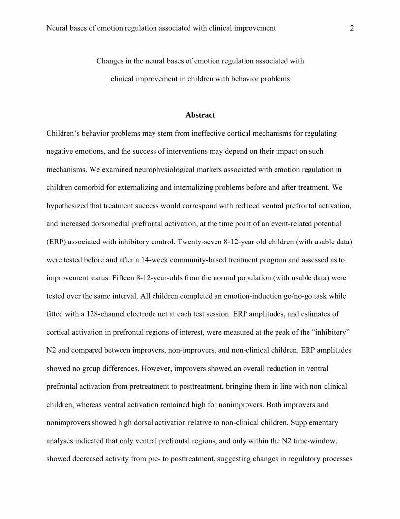
Abstract
Children’s behavior problems may stem from ineffective cortical mechanisms for regulating
negative emotions, and the success of interventions may depend on their impact on such
mechanisms. We examined neurophysiological markers associated with emotion regulation in
children comorbid for externalizing and internalizing problems before and after treatment. We
hypothesized that treatment success would correspond with reduced ventral prefrontal activation,
and increased dorsomedial prefrontal activation, at the time point of an event-related potential
(ERP) associated with inhibitory control. Twenty-seven 8-12-year old children (with usable data)
were tested before and after a 14-week community-based treatment program and assessed as to
improvement status. Fifteen 8-12-year-olds from the normal population (with usable data) were
tested over the same interval. All children completed an emotion-induction go/no-go task while
fitted with a 128-channel electrode net at each test session. ERP amplitudes, and estimates of
cortical activation in prefrontal regions of interest, were measured at the peak of the “inhibitory”
N2 and compared between improvers, non-improvers, and non-clinical children. ERP amplitudes
showed no group differences. However, improvers showed an overall reduction in ventral
prefrontal activation from pretreatment to posttreatment, bringing them in line with non-clinical
children, whereas ventral activation remained high for nonimprovers. Both improvers and
nonimprovers showed high dorsal activation relative to non-clinical children. Supplementary
analyses indicated that only ventral prefrontal regions, and only within the N2 time-window,
showed decreased activity from pre- to posttreatment, suggesting changes in regulatory processes
Neural bases of emotion regulation associated with clinical improvement
3
rather than in overall emotional arousal. These cortically-mediated changes may permit a
reduction in the over-engaged, rigid style of emotion regulation characteristic of children with
behavior problems.
Keywords: emotion regulation, prefrontal cortex, comorbid internalizing and externalizing
problems, EEG/ERP, clinical improvement
Neural bases of emotion regulation associated with clinical improvement
4
Introduction
Given the prevalence, stability, and negative outcomes associated with children’s aggressive
behavior, finding effective interventions has been a top priority. Much progress has been made in
identifying evidence-based treatments that decrease children’s aggression (Dishion & Andrews,
1995; Dishion, Bullock, & Granic, 2002; Henggeler, 1999; Henggeler, et al., 1998; Kazdin, 2002;
Snyder & Ingram, 2000) and several randomized clinical trials have shown the efficacy of
various treatment modalities (for reviews, see Brestan & Eyberg, 1998; Kazdin, 2001).
Specifically, two of the most well-recognized, evidence-based interventions are Parent
Management Training (PMT) and cognitive-behavioral therapy (CBT; Dumas, 1989; Kazdin,
1997; Brestan & Eyeberg, 1998). Despite these promising efforts, the outcomes of these
interventions still show enormous variability, and this variability is difficult to explain because
we have little understanding of the psychosocial and biological mechanisms underlying clinically
significant change (Kazdin, 2001). To improve our knowledge of these mechanisms, several steps
are needed. First, although many investigators link aggressive behavior problems with inadequate
or inappropriate emotion regulation (e.g., Eisenberg et al., 2001; Eisenberg et al., 2007; Zhou et
al., 2007), treatment effectiveness is still measured as change in overt behavior. To understand
why some children learn to control their aggression following treatment, it will be important to
study the impact of treatment on emotion regulation directly. Second, aggressive children
referred for treatment often display internalizing (e.g., anxiety, depression) as well as
externalizing problems. Epidemiological evidence shows that a significant proportion of
aggressive youth exhibit clinically elevated levels of anxiety and/or depression (see Zoccolillo,
1992, for review), and the majority of aggressive children present with serious co-occurring
internalizing symptoms in community settings (Hinshaw, 2002; Kazdin, 2002). Treatment
Neural bases of emotion regulation associated with clinical improvement
5
effectiveness may be better understood when these internalizing problems are acknowledged and
the regulatory difficulties underlying them are systematically studied. Third, there is increasing
interest in the neurobiological mechanisms of emotion regulation in children with behavior
problems. However, little or no research has examined neural measures of emotion regulation as
a means for explaining the effectiveness of treatment for children’s aggressive behavior
problems. If treatment is to have a lasting impact, it may need to alter not only the psychology
but also the biology of emotion regulation in children with behavior problems.
The research reported here begins by acknowledging that children referred for aggressive
behavior problems often suffer with internalizing as well as externalizing symptoms. For this
population of comorbid children, we sought to understand the changes in emotion regulation
habits (underlying both externalizing and internalizing problems) that appeared to correspond
with successful treatment. To examine these changes at a biological level, we tested children’s
neurocognitive responses to a negative emotion induction before and after treatment, using dense-
array electroencephalography (EEG), event-related potentials (ERPs), and cortical source
analysis. Specifically, we looked for changes in these measures from pre- to posttreatment for
children who improved clinically, as compared with those who continued to behave aggressively
and age-matched, non-clinical participants. Our intention was not to evaluate a particular
treatment approach, but to utilize an evidence-based treatment already common at community
mental health agencies. Nor was it our goal to achieve clinical improvement in all or most of our
participants—even if that were possible. Rather, our objective was to determine which children
improved with treatment, based on commonly-used clinical and behavioral markers, and then to
see what characterized these children’s cortical response to a challenging, emotion-inducing task
in comparison with those who did not improve as well as non-clinical age-mates.
Neural bases of emotion regulation associated with clinical improvement
6
Because our sample, like most community samples, generally showed internalizing as well as
externalizing problems, we were particularly interested in the neurobiology of cognitive
overcontrol in response to negative cues (Eisenberg Hofer & Vaughn, 2007). Overcontrol or
overengagement describes the ruminative, stimulus-bound style of emotion regulation
characteristic of anxious children, thought to be mediated by ventral prefrontal and/or amygdala
activation in response to threatening cues (McClure et al., 2007; Monk et al., 2006; Perez-Edgar
et al., 2007; Thomas et al., 2001). We therefore hypothesized that excessive ventral prefrontal
activation would be replaced by more normal patterns of self-regulation for comorbid
(internalizing/externalizing) children who improved with treatment. Specifically, we predicted
that, for children who improved with treatment, cortical activation underlying inhibitory ERPs
would diminish in ventral regions of PFC and correspondingly increase in dorsomedial regions in
the vicinity of the anterior cingulate.
Emotion regulation and childhood psychopathology
Clinically significant externalizing and internalizing problems can be understood as disorders
of emotion regulation (e.g., Bradley, 2000; Calkins, 1994; Calkins, Howse, & Philippot, 2004).
Children with these problems have failed to develop the capacity to appropriately modulate their
feelings of anger and anxiety and the behaviors that flow from them. There has been a good deal
of research with young children supporting the association between poor emotion regulation and
aggressive outcomes. Young children who are less able to voluntarily shift their attention and
inhibit their emotional impulses have higher levels of aggression (Rothbart, Ahadi, & Hershey,
1994). In contrast, children with good emotional control are able to shift attention away from
anger-inducing cues and use nonhostile verbal methods (Eisenberg et al., 1994). Inhibitory
Neural bases of emotion regulation associated with clinical improvement
7
control contributes to the development of conscience in young school-aged children (Kochanska,
Murray, & Coy, 1997), and children’s emotion regulation fosters awareness of responsibility for
their own actions and negative consequences for other people (Derryberry & Reed, 1996).
Eisenberg et al. (1997) found associations between good self-regulation and high-quality social
functioning in conflict situations to be as strong in middle childhood as in the preschool period.
All these findings indicate that the capacity for self-control is prerequisite for inhibiting angry
impulses and engaging in prosocial behavior.
Pure externalizing behavior patterns are associated with poor inhibitory control, sometimes
due to low physiological reactivity and/or reduced fear of consequences (see van Goozen et al.,
2007, for a review). However, children comorbid for externalizing and internalizing problems
have more complex difficulties in emotion regulation. In particular, in addition to anger and
aggression, these children often experience anxiety and depression, which have been linked to
excessive or inappropriate cognitive activity in attempts to control negative emotions and their
outcomes. Anxious or depressed children become overly focused on negative cues, find it
difficult to stop thinking about them, or attempt to regulate them using rigid, overlearned
strategies. Specifically, anxious children can amplify their fears by focusing on stress-inducing
stimuli rather than recruiting a repertoire of coping strategies (Bradley, 2000; Pérez-Edgar & Fox,
2003). Vigilance in relation to threatening cues prevents these children from flexibly allocating
attention elsewhere (Kagan et al., 1984; Kagan & Fox, 2006). According to Eisenberg and
colleagues (Eisenberg, Hofer, & Vaughn, 2007; Eisenberg & Morris, 2002; Murphy et al., 1999),
overcontrol and undercontrol are both maladaptive strategies of emotion regulation, and
overcontrol is associated with the inability to disengage from the negative emotional content of
situations. Thus, for children with both aggression and anxiety problems, angry impulses may be
Neural bases of emotion regulation associated with clinical improvement
8
difficult to regulate because it is difficult to disengage from the threatening or shaming aspects of
challenging situations. For these children, perhaps because they are less skilled at inhibitory
control in general, social isolation, shame, and low self-esteem may lead to recurring aggressive
behavior (Granic & Patterson, 2006).
Neurocognitive mechanisms of emotion regulation
We use the term emotion regulation to refer to cognitive processes involved in response control—
that is, the control of attention, thought, and action impulses—in the presence of emotional states. We
assume a suite of executive functions (e.g., reappraisal, response inhibition, action-monitoring, and
effortful attention) that work together to provide cognitive control (Eisenberg, Hofer, & Vaughn, 2007;
Ochsner & Gross, 2007). However, styles of emotion regulation vary enormously among same-aged
children. Moreover, individual differences in emotion regulation become deeply entrenched, they
reliably predict psychopathological outcomes, and they become increasingly resistant to
intervention as children mature. For these reasons, most investigators assume that styles of
emotion regulation are embedded in neurobiological differences. Behavioral research can only go
so far in measuring these mechanisms, and even the definition of emotion regulation seems to
require biological grounding (Cole, Martin, & Dennis, 2004; Thompson, Lewis, & Calkins, in
press). That may be why developmental psychopathologists are becoming increasingly interested
in the neurobiological substrates of these mechanisms (Pollak, 2005). Neural approaches use
imaging techniques, lesion studies, and electrophysiological methods to specify cortical regions
and activation profiles. Research with adults has made progress linking these control mechanisms
with normal and abnormal emotional processes. However, developmental neuroscience is only
beginning to tackle emotion and its regulation, despite wide agreement on the importance of this
Neural bases of emotion regulation associated with clinical improvement
9
agenda (Dahl, 2001; Goldsmith & Davidson, 2004; Lewis & Stieben, 2004; Pollak, 2005; Posner
& Rothbart, 2000).
Neuroimaging and lesion studies have focused on prefrontal systems that mediate appraisal,
inhibitory control, and self-monitoring. These systems are implicated in normal emotion
regulation. The dorsal anterior cingulate cortex (ACC), on the medial wall of each frontal lobe, is
a key structure for selecting among competing choices, making judgements, monitoring one’s
performance, and learning (Frith et al. 1991; van Veen & Carter, 2002, see Paus et al., 2001 for a
review). The ACC can also be involved in processing emotion, and it is specifically implicated
when individuals are in control of their emotional responses or judgements (Lane et al., 1998;
Taylor et al., 2003). The orbitofrontal cortex (OFC), on the ventral surface of the prefrontal
cortex (PFC), is responsible for assigning emotional significance, especially in social situations,
and for maintaining a response set such as avoidance or inhibition in anticipation of emotional
consequences (Blair et al., 1999; Rolls, 1999). Importantly, both children and adults show
increased fMRI activation in both the ACC and OFC during response inhibition (e.g., Casey et
al., 1997). Hence, both structures may play a role in emotion regulation in children as well as adults
(Lewis et al., 2006).
In adults, externalizing and internalizing psychopathologies are linked with emotion
dysregulation corresponding to anomalies in both these frontal systems. Aggressive individuals
typically show deficits in both ACC and OFC activation (Davidson, Putnam, & Larson, 2000),
implying underregulation of behavior. Blair (2001) suggests that the OFC is especially important
for the regulation of reactive aggression, and Hoptman (2003) found aggression to be associated
with decreased metabolism in anterior, inferior, and medial frontal systems. Conversely, anxious
and depressed individuals show greater-than-normal activation in ventral systems including the
Neural bases of emotion regulation associated with clinical improvement
10
OFC and ventral ACC (Drevets & Raichle, 1998; Mayberg et al., 1999). In fact, dorsal and
ventral prefrontal systems appear to compete for activation, with emotional demands deactivating
dorsal systems and activating ventral systems in their place (Bush, Luu, & Posner, 2000). This
ventral dominance may be chronic in the case of anxiety disorders (Drevets & Raichle, 1998),
and ventral activation has been found to normalize (i.e., activation shifts dorsally) when treatment
for depression is successful (Drevets, 2000; Mayberg et al., 1999). The roles of these
frontocortical systems in emotion regulation have not been as thoroughly investigated in children,
especially with regard to aggressive disorders. However, two studies have now shown reduced
dorsal ACC activation in aggressive children and adolescents (9-15 years), in response to
negative stimuli, when compared with controls (Stadler et al., 2007; Sterzer et al., 2005). Both
dorsal and ventral prefrontal systems showed reduced activation when adolescents made risky
decisions (Eshel et al., 2007). With respect to anxiety, two recent studies showed greater right
ventral PFC activation in children with anxiety disorders than controls when viewing negative
facial expressions (McClure et al., 2007; Monk et al., 2006). In addition, several studies have
found anxious or inhibited children 8 years old and older to show exaggerated amygdala
responses to fear-eliciting situations (Perez-Edgar et al., 2007; Thomas et al., 2001).
We have reviewed these findings in some detail to establish three key points. (1) Activation
of dorsal vs. ventral prefrontal systems is associated with unique cognitive styles: dorsal systems
(e.g., dorsal ACC) appear to mediate the smooth, deliberate control of behavior, including
emotional behavior, in a supervisory or top-down fashion, whereas ventral systems (e.g., ventral
ACC and OFC) control impulses rigidly, in anticipation of negative consequences. In essence,
ventral structures appear to monitor the expectation of further negative events, thus managing
self-control defensively rather than opportunistically. (2) Over- or underactivation of these
Neural bases of emotion regulation associated with clinical improvement
11
systems has been systematically linked with psychopathology both in adults and children.
Underactivation of both dorsal and ventral prefrontal systems characterizes (pure) aggressive
problems and overactivation of ventral systems characterizes anxiety problems. (3) Whereas
behavioral neuroscience generally assumes that control functions are hard-wired in the brain,
there is some evidence that the relative activation levels of prefrontal control systems can be
altered with successful treatment. We aimed to examine children comorbid for internalizing and
externalizing problems pre- and post-treatment, to the extent possible using EEG techniques and
source modeling, in order to assess neural changes associated with successful treatment.
EEG, medial-frontal negativities, and source modeling
Of the various techniques available to neuroscientists, electroencephalographic (EEG)
methods are particularly appealing for clinical research because they are noninvasive, versatile,
and relatively inexpensive. EEG or electrical brain wave activity is recorded at the scalp from an
array of electrodes. Event-related potentials (ERPs) are computed by averaging EEG data over
many trials on a given task. Several ERP components recorded over prefrontal (or frontocentral)
cortex are thought to index aspects of cognitive control, and these have been linked with the
inhibition or regulation of emotional responses in several studies.
The frontal N2 is seen 200-400 ms post-stimulus on trials requiring participants to withhold a
prepotent response, and it is often assumed to tap inhibitory control mechanisms (it is sometimes
dubbed the “inhibitory N2”). That the N2 is also a marker of emotion regulation is implied by its
association with negative emotion in several studies with adults. For example, negatively-
valenced emotional evaluations of self and other predicted higher-amplitude N2s (Tucker et al.,
2003a), and an N2-like “medial-frontal negativity” was found to be enhanced by negative
Neural bases of emotion regulation associated with clinical improvement
12
feedback concerning one’s performance (Luu et al., 2003). In terms of psychopathology, Tucker
et al. (2003b) found medial-frontal amplitudes (specifically, the feedback-related negativity) to
correlate with the intensity of depressive symptoms. Thus, greater-amplitude medial-frontal
negativities probably reflect the augmentation of inhibitory controls when negative emotions
arise, and these controls may be recruited more when people are depressed. Other medial-frontal
negativities, such as the error-related negativity (ERN), are also thought to tap action monitoring
or response control (Falkenstein, Hoorman, & Hohnsbein, 1999; Gehring et al., 1993), and
higher-amplitude ERNs have also been linked to anxiety and negative affect (Gehring, Himle, &
Nisenson, 2000; Hajcak, McDonald, & Simons, 2004; Luu, Collins, & Tucker, 2000; Pailing et
al., 2002).
Researchers have also begun to examine the N2 and related components in children (Davies,
Segalowitz, & Gavin, 2004; Davis et al., 2003; Johnstone et al., 2005; Jonkman, Lansbergen, &
Stauder, 2003; Santesso, Segalowitz, & Schmidt, 2005). These studies compare amplitudes and
latencies between children and adults or test differences across different trial types (e.g., go vs.
no-go trials). However, very few studies to date have utilized medial-frontal ERPs to examine
children’s emotional processes. Exceptions are Santesso et al. (2005), who report lower ERN
amplitudes for undersocialized children, Ladouceur, Dahl, Birmaher, Axelson & Ryan ( 2006),
who report higher ERN amplitudes for anxious children, and Nelson and Nugent (1990), who
found greater-amplitude N2-like components to angry than happy faces in normal children. Four
of our own studies have contributed to this line of investigation. In two of these, N2 amplitudes
were greater to angry than happy faces in 4-6 year olds (Lewis, Todd, & Honsberger, 2007;
Todd, Lewis, Meusel, & Zelazo, 2008). In the first of these, N2 latencies also correlated with
fearful temperament (Lewis et al., 2007). In the second, an N2-like component was greatest to
Neural bases of emotion regulation associated with clinical improvement
13
mothers’ angry faces (Todd et al., 2008). Two studies of older children employed the same task
as the present research. In one of these, a negative mood induction (based on the loss of earned
points) increased N2 amplitudes in normal children aged 13 to 16 years (Lewis et al., 2006). In the
other study, which investigated subtypes of aggressive children, comorbid
(internalizing/externalizing) children showed greater N2s than pure externalizers in response to
the same mood induction (Stieben et al., 2007). In sum, N2s (and related components) tapping
cognitive control are larger in the presence of negative emotion, and more rapid for children with
trait-like anxiety, suggesting a cortical locus of emotion regulation that varies in activation
strength.
Dense-array EEG techniques (e.g., recording from 128 channels rather than just a few) allow
researchers to model the cortical activity underlying ERPs using source analysis methods. We
have made use of these techniques in order to test hypotheses about the approximate location of
cortical activities that may underpin unique mechanisms of emotion regulation. Source analyses
of medial-frontal ERPs (including the N2 and ERN) indicate a key generator in the region of the
ACC for adults (e.g., Bekker, Kenemans, & Verbaten, 2005; Bokura, Yamaguchi, & Kobayashi,
2001; Dehaene, Posner, & Tucker, 1994; Fallgatter, Mueller, & Strik, 1999; Nieuwenhuis et al.,
2003; van Veen & Carter, 2002). Similarly, the region of the OFC has been identified as a
probable source of the N2 in studies of adults and children (Bokura et al., 2001; Lavric, Pizzagalli,
& Forstmeier, 2004; Pliszka, Liotti, & Waldorff, 2000). Recall that these are the very same
prefrontal regions that have been implicated in supervisory vs. stimulus-bound styles of emotion
regulation. Source analysis of scalp EEG cannot provide definitive anatomical information, because
more than one source solution can produce similar scalp topographies. Moreover, reliable anatomical
hypotheses concerning children’s neural activation patterns have only recently emerged in the literature.
Neural bases of emotion regulation associated with clinical improvement
14
Nevertheless, findings from cortical source modeling, and their correspondence with fMRI data,
suggest that ERPs tapping inhibitory control or action-monitoring reflect activation of frontal
regions targeted by imaging studies of emotion regulation. We therefore wished to utilize source
modeling to examine the relative contributions of dorsal and ventral prefrontal systems to the cortical
underpinnings of emotion regulation in children who either improve or do not improve with treatment.
Evidence-based treatments
Among the most effective treatments for aggressive children are family-based Parent
Management Training (PMT) with or without child-focused cognitive behavioral therapy (CBT)
(Dumas, 1989; Kazdin, 1997; Brestan & Eyeberg, 1998). Both PMT and CBT focus on
increasing children’s capacity to regulate their distressing emotions and destructive behaviors.
PMT for aggressive children grew, in part, from Patterson and colleagues’ applied observational
research (Patterson, 1982; Patterson, Reid, & Dishion, 1992) and Forgatch’s research on family
problem-solving interactions (Forgatch, 1984). PMT directly targets coercive family interactions
and attempts to replace lax and aversive parenting practices with mild sanctions (e.g., time-out)
that contingently target misbehavior (Forehand, 1986, 1988). PMT also promotes positive
parenting practices such as skill encouragement, problem-solving, and monitoring (Forgatch &
Degarmo, 1999; Martinez & Forgatch, 2001). Several randomized control studies have examined
the impact of PMT on children’s aggressive behavior (Forgatch & Degarmo, 1999; Martinez &
Forgatch, 2001; Patterson, Chamberlain, & Reid, 1982). Results confirmed that (a) on average,
PMT decreases children’s level of aggressive behavior and (b) reduced coercive parenting is one
of the means by which children’s behavior improves.
Neural bases of emotion regulation associated with clinical improvement
15
Combining PMT with child-focused CBT is another evidence-based strategy for improving
children’s problem behavior. Aggressive children often misunderstand social cues and they have
difficulty regulating their resulting negative emotions (Dodge, 1991; Larson & Lochman, 2002).
CBT targets aggressive behaviors and cognitions through techniques such as behavior
management, role-playing, modeling, problem-solving, cognitive restructuring, social and token
reinforcements, contingent consequences and generalization activities (Barkley, 2000;
Bloomquist & Schnell, 2002). Several studies have documented the effectiveness of combined
PMT and CBT interventions for aggressive children (Brestan & Eyberg, 1998; Tremblay et al.,
1995; Webster-Stratton & Hammond, 1997). In studies that have compared treatment effects of
CBT, PMT, and combined programs, combined CBT/PMT has been found to be most effective,
at least with children from 5 to 12 years of age (Kazdin, Siegel, & Bass, 1992; Lochman &
Wells, 2004; Webster-Stratton & Hammond, 1997). In the current study, we partnered with
community-based agencies who deliver CBT/PMT in order to examine neural correlates of
clinical change.
Design and Hypotheses
The current study was designed to identify a frontal ERP—the inhibitory N2—and to assess
differences in the activation strengths of two broad regions of PFC that generate it: the ventral
PFC (including ventral ACC) region and the dorsomedial region suggestive of dorsal ACC,
before and after a community treatment program for children with behavior problems. We wished
to determine any scalp differences, and especially differences in cortical activation,
corresponding with successful vs. nonsuccessful treatment, under the assumption that these
differences tapped neurocognitive mechanisms of emotion regulation important for behavioral
Neural bases of emotion regulation associated with clinical improvement
16
improvement. All participants completed 14 weeks of a combined PMT/CBT program. Before
the start of the program and immediately afterwards, children were brought to the EEG lab with
their mothers where they took part in a go/no-go task integrated with an emotion induction
procedure. However, because we were interested in measuring change through the administration
of the same task twice, we needed to determine whether and to what extent practice effects
produced a change in the neural response to the task. Therefore, a group of normal same-aged
children went through the same two assessments, also 14 weeks apart. As opposed to a
conventional "control group," this "non-clinical group" was included so as to avoid over-
interpreting practice effects as indicators of real behavioral change. Children earned points for
successful task performance in the first block of trials (block A), then lost all their points due to
more rapid stimulus presentation during block B, and then had a chance to regain their points
when stimulus presentation slowed down again in the third block of trials (block C). ERP and
source data were analyzed for block A (pre-emotion-induction) and block C (post-emotion-
induction), to see whether brain activation differences thought to tap emotion regulation varied
with the added challenge of induced negative emotion. For the clinical children, treatment
effectiveness was assessed using parent- and clinician-reported standard instruments. Based on
these measures, children were grouped into “improvers” (IMPs) and “non-improvers” (NIMPs),
and these groups were compared on each of the neural variables. Finally, standardized parent-,
teacher-, and clinician-rated measures were used to determine the degree of internalizing and
externalizing behavior problems characteristic of our sample, in order to help us interpret the
neurocognitive differences we observed.
Given that our clinical participants were mostly comorbid for internalizing and externalizing
behavior problems, the following three hypotheses were tested:
Neural bases of emotion regulation associated with clinical improvement
17
1. Within sessions, the N2 amplitudes of both clinical and non-clinical children would
increase following the emotion-induction (block B). However, we had no specific predictions as
to changes in N2 amplitudes corresponding with treatment outcomes.
2. Clinical children would show greater activation of ventral regions of PFC than their non-
clinical counterparts. Children who improved with treatment would show decreased activation of
ventral PFC compared with nonimprovers, underlying decreased reliance on a rigid, stimulus-
bound style of emotion regulation.
3. Clinical children would show less activation of dorsal midline frontal regions than non-
clinical children. However, children who improved with treatment would show increased
activation of dorsal regions compared to nonimprovers, underlying increased utilization of a
voluntary, supervisory style of emotion regulation.
Method
Participants
Forty-five children, 8 to 12 years of age (40 boys), were recruited from two outpatient
treatment programs for aggressive children. Participants were referred to the program by mental
health professionals, teachers, and/or parents. To be included in the study, referred children had
to score within the clinical or borderline-clinical range on the externalizing subscale of either the
parent- or teacher-report form of the Child Behavior Checklist (CBCL; Achenbach, 1991a,b).
Additionally, 19 non-clinical children between the ages of 8 and 12 years were included as part of
a larger community sample. These children were recruited through advertising in a city-wide
newspaper. Exclusion criteria for clinically-referred children and their non-clinical age mates
included significant developmental delay and residence outside the large urban center where the
Neural bases of emotion regulation associated with clinical improvement
18
study was conducted. Eighteen clinically-referred children and four non-clinical children were
eliminated from all analyses due to insufficient ERP trial counts. The 18 excluded clinical
children were compared to the sample of 27 included clinical children on demographic variables
and CBCL scores. T-tests (on child’s age and CBCL internalizing and externalizing scores) and
chi-square analyses (of ethnicity, family structure, mother’s highest level of education, father’s
highest level of education, and family income) revealed no significant differences with one
exception: children included in the study scored significantly higher on pre-treatment levels of
externalizing scores than children excluded from the study, t (26) = 2.06, p = .05. Table 1 shows
the demographics of our final sample of 27 clinically-referred children.
Intervention
The treatment program was an evidence-based intervention for children between 8 and 12
years of age and their parents. The program is called SNAPTM (Stop Now and Plan; Earlscourt
Child and Family Centre, 2001a, 2001b; Goldberg & Leggett, 1990) and it combines PMT and
CBT. The clinical directors of the program have been consulting with the original developer of
PMT (Marion Forgatch at the Oregon Social Learning Center) for over 10 years in order to
ensure fidelity to the original PMT model. Therapists were either social workers, child-care
workers or M.A.- or Ph.D.-level clinical psychology students. Like most social welfare programs
in Canada, families were not charged for treatment services. The program was delivered to both
parents (PMT) and children (CBT) once a week for 14 weeks in a group format. The groups met
for 3 hours during the evening at the community agencies. In the PMT groups, parents were
taught to replace coercive or lax discipline strategies with mild sanctions (e.g., time-out) that
contingently target misbehavior (Forehand, 1986). The groups also promoted positive parenting
Neural bases of emotion regulation associated with clinical improvement
19
practices such as skill encouragement (e.g., providing contingent praise for success, prompting
for appropriate behavior), problem-solving, and monitoring (Forgatch & Degarmo, 1999;
Martinez & Forgatch, 2001). In the CBT groups, aggressive behaviors and negatively-biased
cognitions were targeted for change through well-documented strategies such as behavior
management, role-playing, problem-solving, cognitive restructuring, social and token
reinforcements, and generalization activities (Barkley, 2000; Bloomquist & Schnell, 2002).
As previously reviewed, there are numerous randomized control trials that have established
the efficacy of PMT and CBT. In addition, the SNAPTM program itself has undergone two
evaluations to assess its effectiveness. A within-group design comparing baseline, discharge (3
months later), and 6- and 12-month follow-up data for 104 children admitted between 1985 and
1988 showed significant decreases in children’s externalizing behavior (as measured by the
CBCL; Achenbach, 1991a). These treatment gains were maintained over the 6- and 12-month
follow-up period (Hrynkiw-Augimeri, Pepler, & Goldberg, 1993). A recently completed
randomized control trial (Augimeri et al., 2007) indicated that children randomly assigned to the
treatment group, compared to an “attention” control group, showed decreases in externalizing
scores; treatment gains were maintained over 6- and 12-month follow-up periods. For child- and
parent-reported delinquency, effect sizes (d) exceeded 1.2.
Control for Practice Effects
As noted above, a group of non-clinical children between the ages of 8 and 12 years old was
also assessed on two sessions, 14 weeks apart, a lag that was approximately the same as the lag
between the pre- and posttreatment assessments carried out with clinically-referred children. We
do not refer to this group as a control group because they were not matched with the other
Neural bases of emotion regulation associated with clinical improvement
20
children (except on a few global parameters such as approximate age and absence of mental
illness) and they were not intended to ascertain the effect of treatment in any direct way. Rather,
they were intended to ascertain the effect of the repeated administration of our task. If performing
the task on repeated occasions increased the ease of performance, or in some other way decreased
anxiety, discomfort, or other negative emotional states, then any change in cortical response may
have reflected that difference rather than an effect of treatment. The inclusion of a non-clinical
group allowed us to isolate such an effect, if it existed. Thus, the non-clinical children were
included so as to avoid over-interpreting practice effects as an indication of meaningful
behavioral change.
Procedure
Just prior to the beginning of treatment and then again after treatment, clinically-referred
children were accompanied to the laboratory by a parent. Non-clinical children were also tested
twice with roughly 14 weeks between testing sessions. Following a brief introduction to the
testing environment, electrode sensor nets, and recording system, parental consent and child
assent were obtained. Parents were seated in an adjacent room and asked to complete the CBCL.
Children were then informed that they could win a prize for playing the EEG computer game and
were shown two toy bins. One of the bins contained small, undesirable toys such as small plastic
cars, whereas the second bin contained a wide selection of more desirable, age-appropriate toys
such as large action figures, stuffed animals, games, and $10.00 gift certificates from a local
music store. The children were informed that, with successful performance (accumulation of
points) in the game, they would be able to choose their desired prize, but that less successful
performance would limit their choice to the less desirable toy bin. Children were then seated in
Neural bases of emotion regulation associated with clinical improvement
21
front of a computer monitor with the distance and alignment to the monitor controlled by the use
of a chin rest. The electrode sensor net was applied. Children were instructed to make responses
during the game by clicking a button on the response pad with the index finger of their dominant
hand (writing hand). They were given a practice block of 30 trials to ensure proficiency with the
task.
Measures and tasks
Child Behavior Checklist (CBCL; Achenbach, 1991a). The CBCL is a standardized, highly
reliable, and valid measure of children’s emotional and behavioral problems. At pre- and post-
treatment, parents were asked to indicate whether, and to what degree, their child exhibited a list
of symptoms. The instrument yields standardized T-scores for numerous subscales. For this
study, only the internalizing and externalizing problems subscales were used.
Teacher Report Form (TRF; Achenbach, 1991b). The TRF is a parallel measure to the CBCL
but is completed by the child’s teacher at pre- and post-treatment. It is also a standardized, highly
reliable, and valid measure. It generates the same standardized T-scores as the CBCL and again,
only the internalizing and externalizing subscales were used for this study.
Child and Adolescent Functional Assessment Scale (CAFAS; Hodges & Wong, 1996). The
CAFAS is completed by the clinician at pre- and posttreatment. Before clinicians can complete
the CAFAS, they undergo a training period conducted by a CAFAS-certified trainer and are
subsequently tested on a number of vignettes; they must achieve a pre-specified level of
reliability before they are CAFAS-certified. The CAFAS measures the degree of disruption in the
child’s current functioning in eight psychosocial areas. To rate the child, the clinician collects
information from multiple informants in different settings including the child’s parents, teachers,
Neural bases of emotion regulation associated with clinical improvement
22
and any other significant adults that know the child (e.g., grandparent, school counselor). Each of
the eight subscales is rated and scored for level of severity: severe (30), moderate (20), mild (10),
and minimal or none (0). For our purposes, we focused on four scales: “school”, “home”,
“community”, and “behavior toward others.” The reliability and validity of the instrument have
been well established (e.g., Hodges & Wong, 1996; Hodges & Gust, 1995). Critically, the
CAFAS has been shown to be sensitive to clinical change over time (Hodges & Wong, 1996;
Hodges, Wong, & Latessa, 1998; Hodges, 1999). A decrease of 20 points or more from pre- to
post-treatment is considered clinically significant improvement (Hodges & Wong, 1996; Hodges
et al., 1998).
ERP task. The emotion induction go/no-go task that was used in the present study was partly
adapted from a task developed by Garavan, Ross, and Stein (1999), and was presented using E-
Prime software (Psychological Software Tools, Pittsburgh, PA). In standard go/no-go paradigms,
participants are required to press a button as fast as possible given a particular category of stimuli
(the go condition) and withhold responding given another category of stimuli (the no-go
condition). Participants in this study were instructed to click the button for each letter presented
but to avoid clicking when a letter was repeated a second time in succession. Different pairs of
similarly shaped letters were used for each block (block A: x, y; block B: o, p; block C: u, d) to
enhance novelty without modifying the level of difficulty and to facilitate guided recall during a
self-report scale administered at the end. The no-go error rate for the task was maintained at 50%
± 10% by dynamically adjusting the stimulus duration and thus the inter-trial interval. Stimulus
duration was increased with each erroneous response made on no-go trials. Stimulus duration was
decreased following correct no-go trials, but only when the no-go trial followed a correct go trial.
This constraint was incorporated to prevent stimulus time adjustments due to chronic non-
Neural bases of emotion regulation associated with clinical improvement
23
responding. The dynamic adjustment of the stimulus time was intended to provide the same level
of challenge for all participants at all ages, and to obtain a sufficient number of correct no-go
trials for ERP averaging. Error feedback was provided by a red bar in the middle of the screen
following incorrect responses, omitted responses, and late responses.
Children were presented with a practice block and three blocks of trials (blocks A, B, and C).
In blocks A and C children gained points quite steadily. These blocks were structurally identical,
each consisting of 200 trials, including 66 no-go trials, in pseudorandom sequence. In block B,
children immediately began to lose their points, due to a change in the point-adjustment
algorithm. By the end of block B, children had lost all, or almost all, their points. The loss of
points was intended to induce negative emotions, such as anxiety, sadness, and anger. To limit
the duration of children’s distress, block B consisted of only 150 trials, including 40 no-go trials.
With a return to the more generous algorithm in block C, children regained their points to win the
desired prize. For each block, their accumulated points were displayed approximately every 20
trials in the center of the computer screen. Points were added for correct no-go responses and
deducted for response errors on both go and no-go trials. Children were reminded at the
beginning of the task, and the onset of each block, that a high number of points were required to
win the “big prize.”
Self-report emotion-induction check. The emotion induction scheme was assessed with a
subjective rating scale administered directly after the go/no-go task. An 8.5 by 11 inch card with
animated faces of five different emotions was presented to the children. The five emotions were
upset, mad, nervous, satisfied, and excited. Children were asked to rate the intensity of each of
these emotions on a 10-point Likert scale for each of the three blocks. Cards showing animated
emotion faces of different intensities were used to help children identify the intensity of their
Neural bases of emotion regulation associated with clinical improvement
24
emotions. Furthermore, to help children recall how they felt in each of the blocks, researchers
indicated which letter combination was used for each block (e.g., x/y in block A, o/p in block B).
Analyses
EEG data collection and analysis. EEG was recorded using a 128-channel Geodesic Sensor
Net and sampled at 250 Hz, using EGI software (Electrical Geodesic, Inc., Eugene, OR). Data
acquisition was started after all impedances for all EEG channels were reduced to below 50 kΩ.
All channels were referenced to Cz (channel 129) during recording and later rereferenced against
an average reference (Tucker et al., 1993; Bertrand, Perrin, & Pernier, 1985). Eye blink and eye
movement artifacts (70 µV threshold), signals exceeding 200 µV, and fast transits exceeding 100
µV were removed during the averaging process. Data was filtered using a FIR bandpass filter
with a lowpass frequency of 30 Hz and a highpass frequency of 1 Hz. Correct no-go data were
segmented into epochs from 400 ms before to 1000 ms after stimulus onset and baseline
corrected for the 400 ms preceding the stimulus. Correct no-go trials that did not have a correct
go trial preceding and following them (or preceding or following them, in the case of consecutive
no-go trials) were removed, because they most likely reflected attentional lapses or chronic
nonresponding. The mean number of trials comprising correct no-go ERPs for the pretreatment
session was 23.69 (ranging from 11 to 37 trials) and for the posttreatment session was 25.28
(ranging from 8 to 50 trials). To avoid the confounding effect of trial count on amplitude values,
amplitude analyses were conducted with trial count as a covariate. The no-go N2 was scored as
the largest negative deflection with a medial-frontocentral topography between 200 and 500 ms
post-stimulus. Scoring was performed by two independent coders and intercoder agreement was
Neural bases of emotion regulation associated with clinical improvement
25
90%. N2 amplitudes for blocks A and C were analyzed. Block B ERPs were not analyzed due to
insufficient trial counts.
Source-space analysis. Source modeling programs often fit hypothetical generators or
“dipoles” in a model of the cortex and test for goodness-of-fit against the scalp data (e.g., BESA,
MEGIS Software, GmbH). Other programs compute activation voxel by voxel, producing images
that somewhat resemble fMRI images but again on the basis of scalp voltage patterns. In the
current study we utilized the second of these approaches, partly in response to criticisms of
dipole-based source modeling techniques. Specifically, we utilized an algorithm called LAURA
(local autoregressive average), a constraint applied to the minimum-norm method which
minimizes the discrepancy between values of adjacent voxels (to achieve the most realistic
model). Although LAURA source-modeling does not permit precise anatomical localization, its
estimates of regions of activation are more reliable than those of dipole-fitting methods. This
makes it ideal for testing global hypotheses, in this case concerning the relative weight of dorsal
versus ventral sources of frontomedial activation.
In order to estimate the cortical generators for the N2, LAURA constraints were applied to
calculate the inverse solution within the GeoSource (EGI) interface (for a review of these
constraints and other minimum norm solutions, see Michel et al., 2004). Before any group or
block differences were assessed, the “fit” between the inverse solution and the scalp topography
was evaluated. Morphology-based regions of interest (ROIs) were generated using the Montreal
Neurological Institute (MNI) average adult MRI. The ventral ROI, shown in Figure 1,
approximates activation in the ventromedial PFC, orbitofrontal cortex, and subgenual ACC. The
dorsal ROI, also shown in Figure 1, approximates activation in the dorsal ACC. Each ROI was
comprised of a subset of dipoles (or voxels). Source waveform amplitudes (nA) for all dipoles
Neural bases of emotion regulation associated with clinical improvement
26
within a ROI were baseline corrected (400 ms before stimulus onset). Because source waveforms
do not have distinct components as found in scalp waveforms (e.g., N1, P2, N2, and P3), they
were not individually visualized and hand coded. However, we still wanted to extract values that
were most representative of the N2 and that did not include activation subserving other frontal
components, such as a later negativity often found after the parietal P3. The latency range for the
N2 was subdivided into 50 ms bins, and the three bins closest in time to the peak grand-averaged
N2 were sufficient to exclude activation from other ERP components for nearly all children.
Thus, we ended up with a 150-ms window (250-400 ms) from which the maximal activation
value for each voxel was exported. Lastly, for each participant, and for each ROI, we selected the
maximal value across all these voxels. In other words, each participant’s data was reduced further
to capture the maximal activation value within each ROI (for the 150-ms time window
corresponding with the N2).
Results
Preliminary analyses
Outcome group classification of clinically-aggressive children. Children were classified as
“Improvers” (IMPs) or “Non-improvers” (NIMPs) based on a combination of information from
the CBCL and CAFAS. Clinically significant improvement was operationalized as a drop in
score of at least a half a standard deviation (a T-score of 5 or more) on the CBCL and a drop of
20 or more points on the CAFAS (Hodges & Wong, 1996; Hodges et al., 1998). If the two
measures were inconsistent (i.e., if one measure indicated clinical improvement and the other did
not), then priority was given to the CAFAS, because it combined information from multiple
informants, not just the parent. Based on these criteria, 15 children were classified as IMPs and
Neural bases of emotion regulation associated with clinical improvement
27
12 were classified as NIMPs. Means and standard deviations on the CAFAS and CBCL subscales
are presented in Table 2.
It is important to note that, consistent with epidemiological and clinical studies, our sample
was clinically impaired in terms of internalizing problems as well as externalizing problems. The
sample mean was above the borderline clinical cut-off (T = 67) on internalizing, M = 68.13, SD =
7.22. Furthermore, 20 participants (74%) had internalizing scores in the clinical or borderline-
clinical range and all but 1 child scored above T= 60. Because there was no scree or inflection
point in the distribution of internalizing scores, it did not make sense to treat these children as a
separate group — a group that would have been too small for statistical analysis in any case. We
therefore viewed the entire sample as a single group comorbid for internalizing and externalizing
symptoms.
Go/no-go behavioral data analyses
A 2 (session) x 3 (group) x 3 (block) repeated-measures ANOVA was conducted for go
response times and go/no-go performance accuracy. Furthermore, a 2 (session) x 3 (group)
repeated-measures ANOVA was conducted on post-feedback slowing for the mood induction
block only (the only block in which feedback was consistently negative). Age, sex, and
medication were entered as covariates in all behavioral analyses. The response time measure was
straightforward. However, because perseverative responding leads to high accuracy on go trials
and low accuracy on no-go trials, whereas chronic nonresponding leads to high accuracy on no-
go trials and low accuracy on go trials, each of these measures is misleading on its own.
Therefore, to better reflect the overall quality of performance, we also report accuracy scores
averaged across both trial types (see Table 3). Next, the post-feedback response slowing score
Neural bases of emotion regulation associated with clinical improvement
28
consisted of the difference in response time between the average of three consecutive go trials
before the appearance of a points feedback window and the average of three trials after the
window. Negative values indicated response slowing, which is generally taken to mean that the
individual is engaging in performance monitoring triggered by negative feedback. Means and
standard deviations for response time, performance accuracy, and post-feedback slowing are
displayed in Table 3.
Following adjustments for covariates, performance accuracy results revealed a main effect for
session, F(1, 35) = 11.36, p = .002, with greater accuracy observed in the posttreatment session (p
< .001). This pattern of results may be due to increased familiarity with the task at posttreatment.
Furthermore, a group-by-session interaction was found, F(2, 35) = 3.74, p = .03. Planned
contrasts revealed greater accuracy for normal children than improvers in posttreatment only (p =
.05). No other significant or trend-level effects were found (p values ranging from .14 to .83).
Thus, as shown in Figure 2, improvers and non-improvers did not differ in performance accuracy.
Furthermore, both the go response time and post-feedback slowing results showed no differences
for group or session.
Emotion-scale analyses
Emotion-scale data were only collected for the clinically-referred children. A 2 (session) x 2
(group) x 3 (block) x 3 (negative emotion) repeated-measures ANOVA revealed no differences
among the three negative emotions (i.e., no main or interaction effects). Thus, we averaged the
three negative-emotion scales to form a global measure of experienced negative affect. We then
conducted a 2 (session) x 2 (group) x 3 (block) repeated-measures ANOVA. Because no
significant differences were observed for this analysis, we re-ran the ANOVA without group as a
Neural bases of emotion regulation associated with clinical improvement
29
factor, to increase power. Additionally, because we were only concerned with block and session
differences, age, sex, and medication were removed from the analysis as covariates, further
increasing power. This time, results revealed a substantial quadratic main effect of block, F(1,
24) = 45.24, p < .001, and a block-by-session interaction, F(1, 24) = 12.28, p = .002. Planned
contrasts, for both sessions, revealed that the emotion induction block (block B), was perceived
as significantly more negative than both block A and block C, p < .001. Contrasts also indicated
that block A was perceived as more negative in pretreatment than in posttreatment, p = .008.
Thus, a negative mood induction in block B was confirmed by children’s self-report. However,
we have no evidence for a continuation of this negative mood into block C. We infer that, after
the task was completed and the game had been “won”—which is when the self-report scale was
administered—participants appraised block C as more positive. Nevertheless, children’s
emotional state during block C was probably more negative (compared to block A), partly
because this period immediately followed the loss of all points in block B, and partly because of
anecdotal descriptions by the examiners of tension, anxiety, moodiness, and vigilance indicated
by children’s behavior (e.g., posture, utterances) during block C.
ERP analyses
All analyses of N2 amplitudes were conducted on correct no-go stimulus-locked waveforms.
These waveforms, shown in Figure 3, reveal N1, P2, and N2 components that are distinct and
well-formed at both pre- and posttreatment for all groups. However, grand-average waveforms
can be visually deceptive and give the appearance of group differences that are not actually
present. Statistical analyses were performed on values averaged across electrodes Fz and FCz,
where grand averaged ERPs revealed maximal scalp activation. Age, sex, trial count, and
Neural bases of emotion regulation associated with clinical improvement
30
medication were included as covariates in all analyses, because each of these variables can affect
ERP amplitudes. A 2 (session) x 2 (block) x 3 (group) repeated-measures ANOVA revealed no
significant or trend-level main effects or interactions (F values ranged from .009 to 1.38, ns).
Thus, the N2 amplitudes of clinically referred children did not differ from each other as a
function of treatment success, nor did they differ from those of non-referred children.
No hypotheses had been specified concerning changes in N2 amplitudes, partly because ERP
amplitudes can be influenced by contrasting factors such as efficiency and vigilance, and partly
because ERP amplitudes are compilations of the activities of several cortical generators. Our
hypotheses were specific to generators that may well have had counteracting effects on N2
amplitudes. Thus, differences in cortical source waves (e.g., those emanating from ventral vs.
dorsal prefrontal regions) could cancel each other out by the time they got to the scalp!
Nevertheless, the N2 is thought to index high-level executive processes such as those involved in
self-regulation or inhibitory self-control. Therefore, it made sense to use the N2 as a temporal
marker for a presumed cognitive event and to compare ROI activation values at that time-point,
despite the absence of group or session differences in N2 amplitudes.
Source-space analyses
Similar to ERP analyses, all analyses of source-space activity were conducted on correct no-
go stimulus-locked source waveforms. However, as outlined in the Method section, source
waveforms do not have distinct components (e.g., N1, P2, N2, and P3) to guide individualized
coding. Therefore, we shrunk the latency range for the source waveforms to the 150 ms window
surrounding the average N2 latency for the sample as a whole, thus minimizing the possibility of

Neural bases of emotion regulation associated with clinical improvement
31
the (automatic) extraction of a value that was unrelated to the N2. As described earlier, activation
levels were estimated for two regions of interest (ROIs): a dorsomedial and ventromedial region.
We then ran a 3 (group: IMPs, NIMPs, and normals) x 2 (session: pre vs. post) x 2 (region:
ventral vs. dorsal) x 2 (block: A vs. C) repeated-measures ANOVA as an omnibus test of
differences. As with ERP analyses, all source-space analyses had sex, age, medication, and trial
counts covaried out. Results revealed a main effect of region, F (1, 31) = 5.86, p = .02, with the
ventral region showing higher activation than the dorsal region, and a main effect of block, F (1,
31) = 4.93, p = .03, with the postinduction block (C) showing greater activation than the
preinduction block (A). Moreover, as shown in Figure 4, a session-by-region-by-group
interaction was found, F (2, 31) = 4.58, p = .02. Planned contrasts, collapsing across blocks,
revealed a substantial decrease in ventral activation from pretreatment to posttreatment for the
IMPs (p = .008), but no change for NIMPs (p = .93) or non-clinical children (p = .45), as
hypothesized (see panel A). There was also less ventral activation for IMPs than NIMPs at
posttreatment only (p = .04). Thus, as predicted, children whose behavior improved with
treatment showed less ventral activation, compared with their own previous levels and compared
with children who did not improve. Additionally, as shown in panel B of Figure 4, the improvers
and non-improvers had very similar levels of activation in the dorsal ROI, and no change from
pretreatment to posttreatment (ps ranging from .15 to .85 in planned contrasts). Moreover, as
highlighted in the figure and confirmed by contrasts, dorsal activation was lower for the non-
clinical children than for improvers at a borderline-significance level, p = .06. Thus, contrary to
our hypotheses, improvers showed no increase their dorsomedial activation compared to
nonimprovers, nor did they come to resemble their non-clinical age-mates. In fact, again contrary
Neural bases of emotion regulation associated with clinical improvement
32
to our predictions, dorsomedial activation was consistently lower—not higher—for non-clinical
children.
Supplementary source-space analyses
Regulation vs. emotion: testing for general cortical arousal. The decrease in ventral
activation with treatment for the improvers, outlined above, could be associated with a number of
processes. It may be that, at posttreatment compared to pretreatment, improvers simply had less
cortical arousal, reflecting reduced emotional arousal. Or children who improved with treatment
may have relied less on ventrally-mediated regulatory activities, as hypothesized. It was
important to try to resolve this ambiguity. The fact that improvers did not show a drop in dorsal
activation at posttreatment argued against a general decrease in cortical arousal. However, to
further test this possibility, we exported the average cortical activation value for the entire brain
and then computed each child’s maximal activation for the same time range used in the source-
space analysis (250 to 400 ms post-stimulus). We conducted a 3 (group) x 2 (session) x 2 (block)
repeated-measures ANOVA that revealed a session-by-group interaction, at the level of a trend, F
(2, 31) = 3.19, p = .06. Contrasts, corrected for multiple comparisons, revealed an increase in
cortical activation from pretreatment to posttreatment at a borderline-level of significance (p =
.05). The values for each group are shown in Figure 5. Thus, our principal finding of a pre-to-
post decrease in ventral activation (for IMPs) cannot be explained by a general decrease in
cortical arousal and a corresponding drop in emotional reactivity.
Regulation vs. emotion: testing for the temporal specificity of activation change. The N2,
occurring at roughly 200-400 ms, has been linked with various regulatory functions, such as
inhibitory control. The treatment-related decrease in ventral activation for the improvers may
Neural bases of emotion regulation associated with clinical improvement
33
have been specific to this time window (and its corresponding functions) or it could have
extended more broadly across the ERP waveform. The former, more specific timing of the
decrease would support the argument for a change in regulatory activities, whereas the latter,
more extended period of decreased activation would support the argument for a more general
change in cortical activation, possibly reflecting emotional arousal. To select between these
possibilities, we tested a second 150-ms time window, separated from the N2 window by 100 ms
lag in order to minimize potential overlap with activation subserving the N2. A window from 500
to 650 ms was tested. We conducted a 3 (group) x 2 (session) x 2 (region) x 2 (block) repeated-
measures ANOVA that revealed no significant or trend-level results. Additionally, even contrasts
uncorrected for multiple comparisons did not reveal a treatment-related decrease in ventral
activation for the improvers during this second time window (p = .70). As shown in Figure 6
(panel A), the slope of the line plotting improvers’ values from pretreatment to posttreatment is
almost flat (as is the case for the other groups as well). These results indicate that the pre-to-post
decrease in ventral activation for the improvers was unique to the time period of the N2. Thus,
the change in ventral activation corresponding with treatment success appears specific to a time
period characterized by inhibitory control processes.
Discussion
This study set out to determine whether cortical activities thought to underlie emotion
regulation changed as a function of successful treatment, for children with serious behavior
problems. Because our sample of clinically-referred children was characterized by comorbidity,
with high scores on both externalizing and internalizing scales, their difficulties in emotion
regulation were thought to involve both overcontrol and undercontrol. Overcontrol would include
Neural bases of emotion regulation associated with clinical improvement
34
excessive attention to threat, overengagement, and/or rumination, a regulatory style thought to be
mediated by activation in ventral prefrontal systems. However, undercontrol would include a
failure to inhibit aggressive impulses, a regulatory style associated with underactivation of both
dorsal and ventral prefrontal regions but especially linked with dorsal ACC. Thus, we expected
the clinically-referred children in our sample to show overactivation of ventral prefrontal
systems, consistent with their internalizing dynamics, and underactivation of dorsal systems,
consistent with their externalizing problems. Based on this reasoning, we hypothesized that
successful treatment outcomes would correspond with a regularization of both cortical systems: a
decrease in the activation of ventral PFC and an increase in the activation of dorsomedial PFC.
These brain changes should allow children to disengage more easily from fixation on negative
events while asserting more voluntary control over their behavioral choices.
The results supported our hypotheses concerning ventral but not dorsal cortical regions.
Children who improved showed a decrease in ventral PFC activation with treatment,
demonstrating less ventral activation at posttreatment than children who did not improve. While
these results are preliminary and require replication, they constitute the first record of brain
changes corresponding with the successful treatment of children’s behavior problems.
We have argued that ventral prefrontal activation underlies a particular mechanism of
emotion regulation for children with behavior problems, and we suggest that the drop in ventral
activation shown by our improvers mediated a shift in their emotion regulatory habits. There are
several lines of evidence to support this contention. First, using an admittedly coarse
measurement protocol, only ventral PFC appeared to show reduced activation at posttreatment.
Other regions of PFC, and a global measure of cortical activation, showed either no change or
else increased activation from pre- to posttreatment. The ventral PFC is indeed one of the cortical
Neural bases of emotion regulation associated with clinical improvement
35
regions most consistently associated with emotion. Ventral PFC is thought to be a center for
emotion processing, recruited to appraise the impact of a stimulus on one’s wellbeing (e.g.,
Barbas, 2000; Ressler & Mayberg, 2007; Rolls, 1999; Schmitz & Johnson, 2006). More
specifically, anxiety, depression, and inhibited temperament have been associated with greater
levels of activity in ventral PFC, both in adolescents and in adults (Bush et al., 2000; Drevets et
al., 1992, 1997; Hasler et al., 2007; Monk et al., 2006; Steele et al., 2007). Second, studies of
depressed or anxious adults have shown decreased or normalized ventral activation resulting
from successful treatment (Drevets & Raichle, 1998; Fu et al., 2004; Mayberg et al., 1999; for
review see Ressler & Mayberg, 2007). According to Drevets (2000), this normalization may
indicate that ventral PFC is able to “relax” following successful intervention. Moreover, this
change probably corresponds with reduced activity in the amygdala, to which the ventral PFC is
positively coupled in internalizing individuals (Heinz et al., 2005, McClure et al., 2007; Schmitz
& Johnson, 2006; for review see Phillips et al., 2003).
Third, our improvers showed reduced ventral activation only in the time window of the N2, a
component that is understood to tap self-regulatory functions such as response inhibition or
inhibitory control. A link between the N2 and ventral PFC has turned up in a number of studies.
For example, the orbitofrontal region has been identified as a likely generator of the N2 in studies
of adults and children (Bokura et al., 2001; Lavric et al., 2004; Pliszka et al., 2000), even though
the dorsal ACC is associated with the N2 as frequently or more frequently. In our own previous
work, we have used a version of the present paradigm and another paradigm utilizing negative
emotion faces to induce negative emotions in children. In these studies, children of different ages
showed enhanced N2 amplitudes corresponding with negative emotion blocks or trials (e.g.,
Lewis et al., 2006; Lewis, Todd, & Honsberger, 2007; Stieben et al., 2007). A robust dipole in
Neural bases of emotion regulation associated with clinical improvement
36
the right OFC area was the primary generator of the N2 in some of this research (e.g., Lewis et
al., 2006; Lewis, Granic, & Lamm, 2006). Thus, a body of interrelated findings points to a
convergence among the N2, ventral prefrontal activity, and the regulation of negative emotion.
A recent meta-analysis found that normal individuals only recruit ventral prefrontal regions in
“emotional” tasks, but depressed subjects recruit ventral regions in tasks of all kinds—
“cognitive” tasks as well as tasks involving negative emotions (Steele et al., 2007). In other
words, they are less discriminating and they overgeneralize expectations and interpretations
focused on threat, rejection, or unpleasantness. A similar interpretation has been used to explain
the threat-focused attentional bias of anxious and inhibited adolescents (Perez-Edgar at al., 2007).
Like these populations, our clinical participants may have interpreted the task, the loss of points,
and the social situation in which they were embedded as threatening, despite cues to the contrary.
This may be the bias they bring to social situations in general, initiating defensive reactions,
counter-threats, withdrawal and/or aggression. Emotion-regulation mechanisms based on ventral
prefrontal activities would be expected to maintain these threat-focused expectations, resulting in
a defensive, rigid style of thinking about and responding to all kinds of social circumstances. The
children in our sample whose behavior improved with treatment may have been able to "relax"
this ventral predisposition, to paraphrase Drevets (2000), thereby reducing ventral activation in
the time range of the N2 and showing greater flexibility and openness in social situations. Those
who did not improve, for whatever reason, may have remained stuck in their ventral style of
emotion regulation, making it difficult to process social cues without defensiveness.
Indeed, the purpose of therapeutic intervention for these children was to foster greater
flexibility in their appraisals of the emotional meaning of events, greater flexibility in their
response to those events, and greater capacity to view events as neutral or potentially positive
Neural bases of emotion regulation associated with clinical improvement
37
rather than challenging or threatening. The CBT portion of treatment included specific strategies
for reappraising social situations and for delaying responses until those reappraisals took hold.
The PMT portion of treatment was intended to reduce hostile interactions between children and
their parents, so that everyday situations would not be as emotionally loaded, threatening, and
doomed to failure. The reduction in ventral activation we observed in our improvers may
constitute the beginnings of a shift in cortical habits of emotion regulation in response to the
thrust of these treatment goals. If this interpretation is borne out by future studies, it would imply
that prefrontal regulatory mechanisms can be rapidly retrained when the social world is shown to
be less threatening and more supportive.
However, a second prefrontal mechanism of emotion regulation, centered in dorsomedial systems for
voluntary control, was also expected to change with successful treatment. Increases in dorsal ACC
activity were expected to allow children to flexibly monitor their behavior and inhibit aggressive acts.
This prediction was not borne out. In fact, non-clinical children showed marginally lower levels of
dorsomedial activation than clinical children, and clinical children maintained relatively high dorsal
activation whether or not they improved. Because our non-clinical group did not constitute a matched
control group, we do not want to make too much of these findings, but Figure 4 reveals remarkably little
differentiation between the profiles of improvers and nonimprovers. Perhaps clinical children, whether
showing behavioral improvement or not, still needed to recruit dorsal ACC regulatory systems as much as
possible, in order to maximize self-regulation in the face of a challenging task. Another interesting speculation
is that anxiety-related mechanisms may be primary, not secondary, for children with comorbid
symptomatology. There has been much debate as to whether anxiety stems from the consequences of
aggressive behavior or whether aggressive behavior counters the isolation and anguish that accompany anxiety
in comorbid children. Our findings may suggest that behavior improves as soon as attention shifts away from
Neural bases of emotion regulation associated with clinical improvement
38
threat. Once their ventrally-mediated threat bias starts to change, children may find it easier to stay out of
trouble by choosing to avoid particular situations.
Limitations of this research include a relatively small sample size; to increase confidence in our
interpretations of the data, replication with a larger sample will be needed. A second limitation is that changes in
neural activation were assessed at about the same time as measures of behavioral improvement, making it
impossible to determine whether neural changes were causal antecedents of behavior change. To overcome this
limitation, it will be necessary to carry out research with multiple time points, so that neural changes assessed at
one time can be associated with later behavioral changes. We are gathering one-year follow-up data on the
children who participated in this research in hopes of addressing this issue. Next, the use of children comorbid
for externalizing and internalizing symptoms prevented us from isolating neural mechanisms specific to each.
However, this choice constituted a strength as well as a weakness. We studied a community sample
representing real-world behavior problems. Therefore, the results of our research can be generalized more easily
to a clinically relevant population. Another limitation was the absence of a proper wait-list control group. We
decided that it was most important to control for the practice effects of our task, so we utilized a group of non-
clinical children to confirm that practice effects were not responsible for the observed changes in ventral
prefrontal activation. Contrasting improvers and nonimprovers provided a within-group control, but we still
cannot rule out some fundamental difference between these children that influenced their response to the task.
Finally, we did not use a randomized control design because we were primarily interested in individual
differences in treatment outcomes rather than treatment efficacy more broadly. Nevertheless, without the benefit
of random assignment, we can't be absolutely sure that the changes we observed were a result of treatment. It
would be possible to integrate random assignment with neural and behavioral measures across several time
points in a larger study.
Neural bases of emotion regulation associated with clinical improvement
39
Despite these limitations, we managed to bring children with serious behavior problems into a neural
assessment environment for multiple testing sessions, expose them to a task designed to elicit anxiety and
frustration, and derive sufficient artifact-free data to conduct statistically meaningful comparisons. These
comparisons suggest partial support for a model of prefrontally-mediated emotion regulation processes,
whereby different cortical systems play unique roles in the regulation of negative emotions underlying
children’s behavior problems. In conclusion, we have compiled a preliminary data set linking treatment success
with a reduction in ventral prefrontal activation at the time of an inhibitory control function indexed by the
frontal N2. We see this neural change as a shift in the cortical underpinnings of emotion regulation habits that
have maintained patterns of social interaction that are unrewarding and ultimately destructive. Our
interpretation of these findings is provisional at present, but we see this as the first of many steps toward
understanding the biological correlates of effective treatment for children with behavioral difficulties.
Neural bases of emotion regulation associated with clinical improvement
40
References
Achenbach, T.M. (1991a). Manual for the Child Behavior Checklist/4-18 and 1991 Profile.
Burlington, VT: University of Vermont Department of Psychiatry.
Achenbach, T.M. (1991b). Manual for the Child Behavior Checklist/4-18 and 1991 Profile –
Teacher Version. Burlington: University of Vermont, Department of Psychiatry.
Augimeri, L. K., Farrington, D. P., Koegl, C. J., & Day, D. M. (2007). A community based
program for children with conduct problems: Immediate and long-term effects.
Barbas, H. (2000). Connections underlying the synthesis of cognition, memory, and emotion in
primate prefrontal cortices. Brain Research Bulletin, 52, 319-330.
Barkley, R. A. (2000). Commentary on the multimodal treatment study of children with ADHD.
Journal of Abnormal Child Psychology, 28, 595-599.
Bloomquist, M. L. & Schnell, S. V. (2002). Helping children with aggression and conduct problems:
Best practices for intervention. New York, NY, US: Guildford Press. Xiv, 418 pp.
Bekker, E. M., Kenemans, J. L., & Verbaten, M. N. (2005). Source analysis of the N2 in a cued
Go/No-go task. Cognitive Brain Research, 22, 221-231.
Bertrand, O., Perrin, F., & Pernier, J. (1985). A theoretical justification of the average-reference
in topographic evoked potential studies. Electroencephalography and Clinical
Neurophysiology, 62, 462-464.
Blair, R. J. R. (2001). Neurocognitive models of aggression, the antisocial personality disorders, and
psychopathy. Journal of Neurology, Neurosurgery & Psychiatry, 71, 727-731.
Blair, R. J. R., Morris, J. S., Frith, C. C., Perrett, D. I., & Dolan, R. J. (1999). Dissociable neural
responses to facial expressions of sadness and anger. Brain, 122, 883-893.
Neural bases of emotion regulation associated with clinical improvement
41
Bloomquist, M. L. & Schnell, S. V. (2002). Helping children with aggression and conduct
problems: Best practices for intervention. New York, NY, US: Guildford.
Bokura, H., Yamaguchi, S., & Kobayashi, S. (2001). Electrophysiological correlates for response
inhibition in a Go/No-go task. Clinical Neurophysiology, 112, 2224-2232.
Bradley, S. (2000). Affect regulation and the development of psychopathology. New York: Guilford.
Brestan, E. V. & Eyberg, S. M. (1998). Effective psychosocial treatments of conduct disordered
children and adolescents: 29 Years, 82 Studies, and 5,272 Kids. Journal of Clinical Child
Psychology, 27, 180-189.
Bush, G., Luu, P., & Posner, M. I. (2000). Cognitive and emotional influences in anterior cingulate
cortex. Trends in Cognitive Sciences, 4, 215-222.
Calkins, S. D. (1994). Origins and outcomes of individual differences in emotion regulation.
Monographs of the Society for Research in Child Development, 59, 53-72.
Calkins, S. D., Howse, R. B., Philippot, P. (2004). Individual differences in self-regulation:
Implications for childhood adjustment. In R. S. Feldman (Ed.), The regulation of emotion
(pp. 307-332). Mahwah, NJ, US: Erlbaum.
Casey, B. J., Trainor, R. J., Orendi, J. L., Schubert, A. B., Nystrom, L. E., Giedd, J. N., et al.
(1997). A developmental functional MRI study of prefrontal activation during
performance of a Go-No-Go task. Journal of Cognitive Neuroscience, 9, 835-847.
Cole, P. M., Martin, S. E., & Dennis, T. A. (2004). Emotion regulation as a scientific construct:
Methodological challenges and directions for child development research. Child
Development, 75, 317-333.
Dahl, R. E. (2001). Affect regulation, brain development, and behavioral/emotional health in
adolescence. CNS Spectrums, 6, 60-72.
Neural bases of emotion regulation associated with clinical improvement
42
Davidson, R. J., Putnam, K. M., & Larson, C. L. (2000). Dysfunction in the neural circuitry of
emotion regulation—a possible prelude to violence. Science, 289, 591-594.
Davies, P. L., Segalowitz, S. J., & Gavin, W. J. (2004). Development of response-monitoring ERPs
in 7- to 25-year-olds. Developmental Neuropsychology, 25, 355-376.
Davis, E. P., Bruce, J., Snyder, K., & Nelson, C. (2003). The X-trials: Neural correlates of an
inhibitory control task in children and adults. Journal of Cognitive Neuroscience, 13, 432-
443.
Dehaene, S., Posner, M. I., & Tucker, D. M. (1994). Localization of a neural system for error
detection and compensation. Psychological Science, 5, 303-305.
Derryberry, D. & Reed, M. A. (1996). Regulatory processes and the development of cognitive
representations. Development and Psychopathology, 8, 215-234.
Dishion, T. J. & Andrews, D. W. (1995). Preventing escalation in problem behaviors with high-
risk young adolescents: Immediate and 1-year outcomes. Journal of Consulting and
Clinical Psychology, 63, 538-548.
Dishion, T. J., Bullock, B. M., & Granic, I. (2002). Pragmatism in modeling peer influence:
Dynamics, outcomes, and change processes. How prevention intervention studies in the
field of developmental psychopathology can inform developmental theories and models.
[Special Issue]. Development and Psychopathology, 14, 969-981.
Dishion, T. J. & Granic, I. (2004). Naturalistic observation of relationship processes. Hoboken,
NJ, US: John Wiley & Sons.
Dodge, K. A. (1991). The structure and function of reactive and proactive aggression. In D. J.
Pepler & K. H. Rubin (Eds.), The development and treatment of childhood aggression
(pp. 201-218). Hillsdale, NJ: Erlbaum.
Neural bases of emotion regulation associated with clinical improvement
43
Drevets, W. C. (2000). Neuroimaging studies of mood disorders. Biological Psychiatry, 48, 813-829.
Drevets, W. C., Price, J. L., Simpson Jr., J. R., Todd, R. D., Reich, T., Vannier, M., & Raichle, M. E.
(1997). Subgenual prefrontal cortex abnormalities in mood disorders. Nature, 386, 824-827.
Drevets, W. C. & Raichle, M. E. (1998). Reciprocal suppression of regional cerebral blood flow
during emotional versus higher cognitive processes: Implications for interactions between
emotion and cognition. Cognition and Emotion, 12, 353-385.
Drevets, W. C., Videen, T. O., Price, J. L., Preskorn, S. H., Carmichael, S. T., & Raichle, M. E.
(1992). A functional anatomical study of unipolar depression. Journal of Neuroscience, 12,
3628-3641.
Dumas, J. E. (1989). Treating antisocial behavior in children: Child and family approaches.
Clinical Psychology Review, 9, 197-222.
Earlscourt Child and Family Centre (2001b). SNAP™ parent group manual. Toronto, ON:
Earlscourt Child and Family Centre.
Eisenberg, N., Cumberland, A., Spinrad, T. L., Fabes, R. A., Shepard, S. A., & Reiser, M. et al.
(2001). The relations of regulation and emotionality to children's externalizing and
internalizing problem behavior. Child Development, 72, 1112-1134.
Eisenberg, N., Fabes, R. A., Nyman, M., Bernzweig, J., & Pinuelas, A. (1994). The relations of
emotionality and regulation to children’s anger-related reactions. Child Development, 65,
109-128.
Eisenberg, N., Fabes, R. A., Shepard, S. A., Murphy, B. C., Guthrie, I. K., Jones, S., Friedman, J.,
Poulin, R., & Maszk, P. (1997). Contemporaneous and longitudinal prediction of children's
social functioning from regulation and emotionality. Child Development, 68, 642-664.
Neural bases of emotion regulation associated with clinical improvement
44
Eisenberg, N., Hofer, C., & Vaughn, J. (2007). Effortful control and its socioemotional
consequences. In J. J. Gross (Ed.), Handbook of emotion regulation (pp. 287–306). New
York: Guilford.
Eisenberg, N., Ma, Y., Chang, L., Zhou, Q., West, S. G., & Aiken, L. (2007). Relations of
effortful control, reactive undercontrol, and anger to chinese children's adjustment.
Development and Psychopathology, 19, 385-409.
Eisenberg, N. & Morris, A. S. (2002). Children's emotion-related regulation. San Diego, CA,
US: Academic Press.
Eshel, N., Nelson, E. E., Blair, R. J., Pine, D. S., & Ernst, M. (2007). Neural substrates of choice
selection in adults and adolescents: Development of the ventrolateral prefrontal and
anterior cingulate cortices. Neuropsychologia, 45, 1270-1279.
Falkenstein, M., Hoormann, J., & Hohnsbein, J. (1999). ERP components in Go/No-go tasks and
their relation to inhibition. Acta Psychologica, 101, 267-291.
Fallgatter, A. J., Mueller, T. J., & Strik, W. K. (1999). Age-related changes in the brain electrical
correlates of response control. Clinical Neurophysiology, 110, 833-838.
Forehand, R. (1986). Parental positive reinforcement with deviant children: Does it make a
difference? Child & Family Behavior Therapy, 8, 19-25.
Forehand, R. (1988). Reinventing the wheel and attacking strawmen: A reply to Lutzker et al.'s
rejoinder. Child & Family Behavior Therapy, 10, 35-39.
Forgatch, M. S. (1984). A two-stage analysis of family problem-solving: Global and microsocial.
Unpublished doctoral dissertation. University of Oregon, Eugene.
Neural bases of emotion regulation associated with clinical improvement
45
Forgatch, M. S. & DeGarmo, D. S. (1999). Two faces of Janus: Cohesion and conflict. In M. J.
Cox & J. Brooks-Gunn (Eds.), Conflict and cohesion in families: Causes and
consequences (pp. 167-184). Mahwah, NJ: Erlbaum.
Frith, C. D., Friston, K. J., Liddle, P. F., & Frackowiak, R. S. (1991). A PET study of word finding.
Neuropsychologia, 29, 1137-1148.
Fu, C. H. Y., Williams, S. C. R., Cleare, A. J., Brammer, M. J., Walsh, N. D., Kim, J., et al. (2004).
Attenuation of the neural response to sad faces in major depression by antidepressant
treatment: A prospective, event-related functional magnetic resonance imaging study.
Archives of General Psychiatry, 61, 877-889.
Garavan, H., Ross, T. J., & Stein, E. A. (1999). Right hemispheric dominance of inhibitory control:
An event-related functional MRI study. Proceedings of the National Academy of Science, 96,
8301-8306.
Gehring, W. J., Goss, B., Coles, M. G., & Meyer, D. E. (1993). A neural system for error detection
and compensation. Psychological Science, 4, 385-390.
Gehring, W. J., Himle, J., & Nisenson, L. G. (2000). Action monitoring dysfunction in obsessive-
compulsive disorder. Psychological Science, 11, 1-6.
Goldberg, K. A., & Leggett, E. A. (1990). Earlscourt Child and Family Centre: Under 12
Outreach Project Transformer Club group leaders manual. Toronto, ON: Earlscourt
Child and Family Centre.
Goldsmith, H. H. & Davidson, R. J. (2004). Disambiguating the components of emotion regulation.
Child Development, 75, 361-365.
Granic, I. & Patterson, G. R. (2006). Toward a comprehensive model of antisocial development: A
dynamic systems approach. Psychological Review, 113, 101-131.
Neural bases of emotion regulation associated with clinical improvement
46
Hajcak, G., McDonald, N., & Simons, R. F. (2004). Error-related psychophysiology and negative
affect. Brain & Cognition, 56, 189-197.
Hasler, G., Fromm, S., Alvarez, R. P., Luckenbaugh, D. A., Drevets, W. C., & Grillon, C. (2007).
Cerebral blood flow in immediate and sustained anxiety. Journal of Neuroscience, 27, 6313-
6319.
Heinz, A., Braus, D., Smolka, M. N., Wrase, J., Puls, I., Hermann, D., et al. (2005). Amygdala-
prefrontal coupling depends on a genetic variation of the seretonin transporter. Nature
Neuroscience, 8, 20-21.
Henggeler, S. W. (1999). Multisystemic therapy: An overview of clinical procedures, outcomes,
and policy implications. Child Psychology & Psychiatry Review, 4, 2-10.
Henggeler, S. W., Schoenwald, S. K., Borduin, C. M., Rowland, M. D., & Cunningham, P. B.
(1998). Multisystemic treatment of antisocial behavior in children and adolescents. New
York, NY, US: Guilford.
Hinshaw, S. P. (1994). Attention deficits and hyperactivity in children. Developmental clinical
psychology and psychiatry. Thousand Oaks, CA: Sage.
Hinshaw, S. P. (2002). Intervention research, theoretical mechanisms and causal processes
related to externalizing behavior patterns. Development & Psychopathology, 14, 789-818.
Hodges, K. (1999). Child and Adolescent Functional Assessment Scale (CAFAS). In Maruish,
Mark E. (Ed). The use of psychological testing for treatment planning and outcomes
assessment (2nd ed.) (pp. 631-664). Mahwah, NJ: Erlbaum.
Hodges, K. & Wong, M.M. (1996). Psychometric characteristics of a multidimensional measure
to assess impairment: The Child and Adolescent Functional Assessment Scale. Journal of
Child & Family Studies, 5, 445-467.
Neural bases of emotion regulation associated with clinical improvement
47
Hodges, K. & Gust, J. (1995). Measures of impairment for children and adolescents. The Journal
of Mental Health Administration, 22, 403-413.
Hodges, K., & Wong, M. M., & Latessa, M. (1998). Use of the Child and Adolescent Functional
Assessment Scale (CAFAS) as an outcome measure in clinical settings. Journal of
Behavioral Health Services & Research, 25, 325-336.
Hoptman, M. J. (2003). Neuroimaging studies of violence and antisocial behavior. Journal of
Psychiatric Practice, 9, 265-278.
Hrynkiw-Augimeri, L., Pepler, D., & Goldberg, K. (1993). An outreach program for children having
police contact. Canada’s Mental Health, 41, 7-12.
Johnstone, S. J., Pleffer, C. B., Barry, R. J., Clarke, A. R., & Smith, J. L. (2005). Development of
inhibitory processing during the Go/No-go task: A behavioral and event-related potential
study of children and adults. Journal of Psychophysiology, 19, 11-23.
Jonkman, L. M., Lansbergen, M., & Stauder, J. E. A. (2003). Developmental differences in
behavioral and event-related brain responses associated with response preparation and
inhibition in a go/no-go task. Psychophysiology, 40, 752-761.
Jonkman, L. M., Sniedt, F. L. F., & Kemner, C. (2007). Source localization of the no-go-N2: A
developmental study. Clinical Neurophysiology, 118, 1069-1077.
Kagan, J. & Fox, N. (2006). Biology, culture, and temperamental biases. In N. Eisenberg (Vol.
Ed.) & W. Damon & R. M. Lerner (Series Eds), Social, emotional, and personality
development: Vol. 3. Handbook of child psychology (pp. 167–225). New York: Wiley.
Kagan, J., Reznick, S., Clarke, C., Snidman, N., & Garcia-Coll, S. (1984). Behavioral inhibition
to the unfamiliar. Child Development, 55, 2212-2225.
Neural bases of emotion regulation associated with clinical improvement
48
Kazdin, A. E. (1997). Parent management training: Evidence, outcomes, and issues. Journal of
the American Academy of Child and Adolescent Psychiatry, 36, 1349-1356.
Kazdin, A. E. (2001). Progression of therapy research and clinical application of treatment
require better understanding of the change process. Clinical Psychology: Science and
Practice, 8, 143-151.
Kazdin, A. E. (2002). Psychosocial treatments for conduct disorder in children and adolescents.
New York, NY, US: Oxford University Press.
Kazdin, A. E., Siegel, T. C., & Bass, D. (1992). Cognitive problem-solving skills training and
parent management training in the treatment of antisocial behavior in children. Journal of
Consulting and Clinical Psychology, 60, 733-747.
Kochanska, G., Murray, K., & Coy, K. C. (1997). Inhibitory control as a contributor to conscience in
childhood: From toddler to early school age. Child Development, 68, 263-277.
Ladouceur, C. D., Dahl, R. E., Birmaher, B., Axelson, D. A., & Ryan, N. D. (2006). Increased
error-related negativity (ERN) in childhood anxiety disorders: ERP and source
localization. Journal of Child Psychology and Psychiatry, 47, 1073-1082.
Lane, R. D., Reiman, E. M., Axelrod, B., Yun, L., Holmes, A., & Schwartz, G. E. (1998). Neural
correlates of levels of emotional awareness: Evidence of an interaction between emotion and
attention in the anterior cingulate cortex. Journal of Cognitive Neuroscience, 10, 525-535.
Larson, J. & Lochman, J. E. (2002). Helping schoolchildren cope with anger: A cognitive-
behavioral intervention. New York, NY, US: Guilford.
Lavric, A., Pizzagalli, D. A., & Forstmeier, S. (2004). Short communication: When 'go' and 'nogo'
are equally frequent: ERP components and cortical tomography. European Journal of
Neuroscience, 20, 2483-2488.
Neural bases of emotion regulation associated with clinical improvement
49
Lewis, M. D. (2005). Bridging emotion theory and neurobiology through dynamic systems
modeling (target article). Behavioral and Brain Sciences, 28, 169-194.
Lewis, M. D., Granic, I., & Lamm, C. (2006). Behavioral differences in aggressive children linked
with neural mechanisms of emotion regulation. Annals of the New York Academy of
Sciences, 1094, 164-177.
Lewis, M. D., Lamm, C., Segalowitz, S. J., Stieben, S., & Zelazo, P. D. (2006). Neurophysiological
correlates of emotion regulation in children and adolescents. Journal of Cognitive
Neuroscience, 18, 430-443.
Lewis, M. D. & Stieben, J. (2004). Emotion regulation in the brain: Conceptual issues and directions
for developmental research. Child Development, 75, 371-376.
Lewis, M. D., Todd, R. M., & Honsberger, M. (2007). Event-related potential measures of
emotion regulation in early childhood. NeuroReport, 18, 61-65.
Lochman, J. E. & Wells, K. C. (2004). The coping power program for preadolescent aggressive boys
and their parents: Outcome effects at the 1-year follow-up. Journal of Consulting and
Clinical Psychology, 72, 571-578.
Luu, P., Collins, P., & Tucker, D. M. (2000). Mood, personality, and self-monitoring: Negative
affect and emotionality in relation to frontal lobe mechanisms of error monitoring. Journal of
Experimental Psychology,129, 43-60.
Luu, P., Tucker, D. M., Derryberry, D., Reed, M., & Poulsen, C. (2003). Activity in human medial
frontal cortex in emotional evaluation and error monitoring. Psychological Science, 14, 47-
53.
Neural bases of emotion regulation associated with clinical improvement
50
Martinez, C. R. Jr. & Forgatch, M. S. (2001). Preventing problems with boys' noncompliance:
Effects of a parent training intervention for divorcing mothers. Journal of Consulting and
Clinical Psychology, 69, 416-428.
Mayberg, H. S., Liotti, M., Brannan, S. K., McGinnis, S., Mahurin, R. K., Jerabek, P. A., et al.
(1999). Reciprocal limbic-cortical function and negative mood: Converging PET findings in
depression and normal sadness. American Journal of Psychiatry, 156, 675-682.
McClure, E. B., Monk, C. S., Nelson, E. E., Parrish, J. M., Adler, A. A., Blair, R. J. R., et al.
(2007). Abnormal attention modulation of fear circuit function in pediatric generalized
anxiety disorder. Archives of General Psychiatry, 64, 97-106.
Michel, C. M., Murray, M. M., Lantz, G., Gonzalez, S., Spinelli, L., & Grave de Peralta, R.
(2004). EEG source imaging. Clinical Neurophysiology, 115, 2195-2222.
Monk, C. S., Nelson, E. E., McClure, E. B., Mogg, K., Bradley, B. P., Leibenluft, E., et al.
(2006). Ventrolateral prefrontal cortex activation and attentional bias in response to angry
faces in adolescents with generalized anxiety disorder. American Journal of Psychiatry,
163, 1091-1097.
Murphy, B. C., Eisenberg, N., Fabes, R. A., Shepard, S., & Guthrie, I. K. (1999). Consistency
and change in children's emotionality and regulation: A longitudinal study. Merrill-
Palmer Quarterly, 45, 413-444.
Nelson, C. & Nugent, K. M. (1990). Recognition memory and resource allocation as revealed by
children’s event-related potential responses to happy and angry faces. Developmental
Psychology, 26, 171-179.
Neural bases of emotion regulation associated with clinical improvement
51
Nieuwenhuis, S., Yeung, N., Van den Wildenberg, W., & Ridderinkhof, K.R. (2003).
Electrophysiological correlates of anterior cingulate function in a Go/No-go task: Effects of
response conflict and trial-type frequency. Cognitive, Affective & Behavioral Neuroscience,
3, 17-26.
Ochsner, K. N. & Gross, J. J. (2007). The neural architecture of emotion regulation. In J. J. Gross
(Ed.) Handbook of emotion regulation (pp. 87-109). New York: Guilford.
Pailing, P. E., Segalowitz, S. J., Dywan, J., & Davies, P. L. (2002). Error negativity and
response control. Psychophysiology, 39, 198-206.
Paus, T. (2001). Primate anterior cingulate cortex: Where motor control, drive and cognition
interface. Nature Reviews Neuroscience, 2, 417-424.
Patterson, G. R. (1982). Coercive family processes. Eugene, OR, US: Castalia.
Patterson, G. R., Chamberlain, P., & Reid, J. B. (1982). A comparative evaluation of parent
training procedures. Behavior Therapy, 13, 638-650.
Patterson, G. R., Reid, J. B., & Dishion, T. (1992). Antisocial boys. Eugene, OR, US: Castalia.
Pérez-Edgar, K. & Fox, N. A. (2003). Individual differences in children's performance during an
emotional stroop task: A behavioral and electrophysiological study. Brain and Cognition,
52, 33-51.
Perez-Edgar, K., Roberson-Nay, R., Hardin, M. G., Poeth, K., Guyer, A. E., Nelson, E. E., et al.
(2007). Attention alters neural responses to evocative faces in behaviorally inhibited
adolescents. NeuroImage, 35, 1538-1546.
Phillips, M. L., Drevets, W. C., Rauch, S. L., & Lane, R. (2003). Neurobiology of emotion
perception II: Implications for major psychiatric disorders. Biological Psychiatry, 54, 515-
528.
Neural bases of emotion regulation associated with clinical improvement
52
Pliszka, S. R., Liotti, M., & Woldorff, M. G. (2000). Inhibitory control in children with attention-
deficit/hyperactivity disorder: Event-related potentials identify the processing component
and timing of an impaired right-frontal response-inhibition mechanism. Biological
Psychiatry, 48, 238-246.
Pollak, S. D. (2005). Early adversity and mechanisms of plasticity: Integrating affective
neuroscience with developmental approaches to psychopathology. Development and
Psychopathology, 17, 735–752.
Posner, M. I. & Rothbart, M. K. (2000). Developing mechanisms of self-regulation. Development
and Psychopathology, 12, 427-441.
Ressler, K. J. & Mayberg, H. S. (2007). Targeting abnormal neural circuits in mood and anxiety
disorders: From the laboratory to the clinic. Nature Neuroscience, 10, 1116-1124.
Rolls, E. T. (1999). The brain and emotion. Oxford: Oxford University Press.
Rothbart, M. K., Ahadi, S. A., & Hershey, K. L. (1994). Temperament and social behavior in
childhood. Merrill-Palmer Quarterly, 40, 21-39.
Santesso, D. L., Segalowitz, S. J., & Schmidt, L. A. (2005). ERP correlates of error monitoring in
10-year olds are related to socialization. Biological Psychology, 70, 79--87.
Schmitz, T. W. & Johnson, S. C. (2006). Self-appraisal decisions evoke dissociated dorsal-ventral
aMPFC networks. NeuroImage, 30, 1050-1058.
Snyder, C. R. & Ingram, R. E. (2000). Handbook of psychological change: Psychotherapy
processes & practices for the 21st century. Hoboken, NJ, US: John Wiley & Sons.
Stadler, C., Sterzer, P., Schmeck, K., Krebs, A., Kleinschmidt, A., & Poustka, F. (2007). Reduced
anterior cingulate activation in aggressive children and adolescents during affective
Neural bases of emotion regulation associated with clinical improvement
53
stimulation: Association with temperament traits. Journal of Psychiatric Research, 41,
410–417.
Steele, J. D., Currie, J., Lawrie, S. M., & Reid, I. (2007). Prefrontal cortical functional
abnormality in major depressive disorder: A stereotactic meta-analysis. Journal of
Affective Disorders, 101, 1-11.
Stieben, J., Lewis, M. D., Granic, I., Zelazo, P. D., & Segalowitz, S. (2007). Neurophysiological
mechanisms of emotion regulation for subtypes of externalizing children. Development
and Psychopathology, 19, 455-480.
Sterzer, P., Stadler, C., Krebs, A., Kleinschmidt, A., & Poustka, F. (2005). Abnormal neural
responses to emotional visual stimuli in adolescents with conduct disorder. Biological
psychiatry, 57, 7-15.
Taylor, S. F., Phan, K. L., Decker, L.R., & Liberzon, I. (2003). Subjective rating of emotionally
salient stimuli modulates neural activity. NeuroImage, 18, 650-659.
Thomas, K. M., Drevets, W. C., Dahl, R. E., Ryan, N. D., Birmaher, B., Eccard, C. H., et al. (2001).
Amygdala responses to fearful faces in anxious and depressed children. Archives of General
Psychiatry, 58, 1057-1063.
Thompson, R. A., Lewis, M. D., & Calkins, S. D. (in press). Reassessing emotion regulation. Child
development perspectives.
Todd, R. M., Lewis, M. D., Meusel, L.-A., & Zelazo, P. D. (2008). The time course of social-
emotional processing in early childhood: ERP responses to facial affect and personal
familiarity in a Go-Nogo task. Neuropsychologia, 46, 595-613.
Neural bases of emotion regulation associated with clinical improvement
54
Tremblay, R. E., Pagani-Kurtz, L., Masse, L. C., Vitaro, F., & Pihl, R. O. (1995). A bimodal
preventative intervention for disruptive kindergarten boys: Its impact through mid-
adolescence. Journal of Consulting and Clinical Psychology, 63, 560-568.
Tucker, D. M., Liotti, M., Potts, G. F., Russell, G. S., & Posner, M. I. (1993). Spatiotemporal
analysis of brain electrical fields. Human Brain Mapping, 1, 134-152.
Tucker, D. M., Luu, P., Desmond Jr., R. E., Hartry-Speiser, A., Davey, C., & Flaisch, T. (2003a).
Corticolimbic mechanisms in emotional decisions. Emotion, 3, 127-149.
Tucker, D. M., Luu, P., Frishkoff, G., Quiring, J., & Poulsen, C. (2003b). Frontolimbic response
to negative feedback in clinical depression. Journal of Abnormal Psychology, 112, 667-
678.
Van Goozen, S. H. M., Fairchild, G., Snoek, H., & Harold, G. T. (2007). The evidence for a
neurobiological model of childhood antisocial behavior. Psychological Bulletin, 133,
149–182.
van Veen, V. & Carter, C. S. (2002). The timing of action-monitoring processes in the anterior
cingulate cortex. Journal of Cognitive Neuroscience, 14, 593-602.
Webster-Stratton, C. & Hammond, M. (1997). Treating children with early-onset conduct
problems: a comparison of child and parent training interventions. Journal of Consulting
and Clinical Psychology, 65, 93-109.
Zhou, Q., Hofer, C., Eisenberg, N., Reiser, M., Spinrad, T. L., & Fabes, R. A. (2007). The
developmental trajectories of attention focusing, attentional and behavioral persistence,
and externalizing problems during school-age years. Developmental Psychology, 43, 369-
385.
Neural bases of emotion regulation associated with clinical improvement
55
Zoccolillo, M. (1992). Co-occurrence of conduct disorder and its adult outcomes with depressive
and anxiety disorders: A review. Journal of the American Academy of Child and
Adolescent Psychiatry, 31, 547-556.
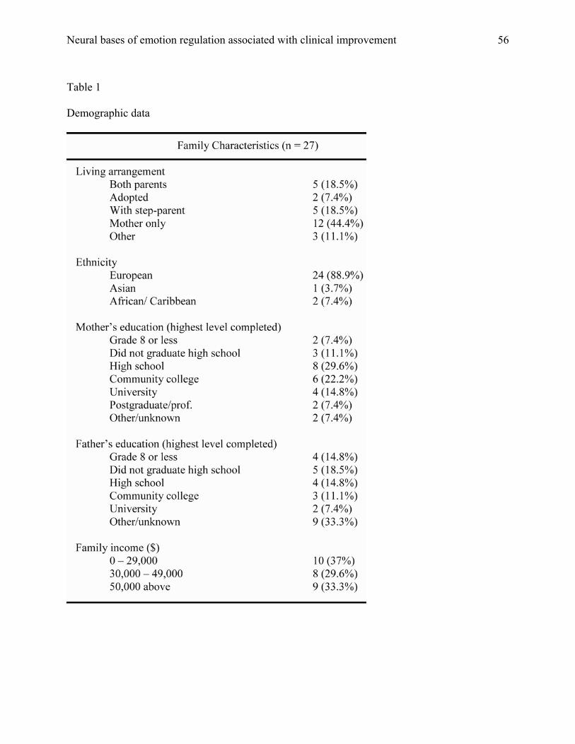
Neural bases of emotion regulation associated with clinical improvement
56
Table 1
Demographic data
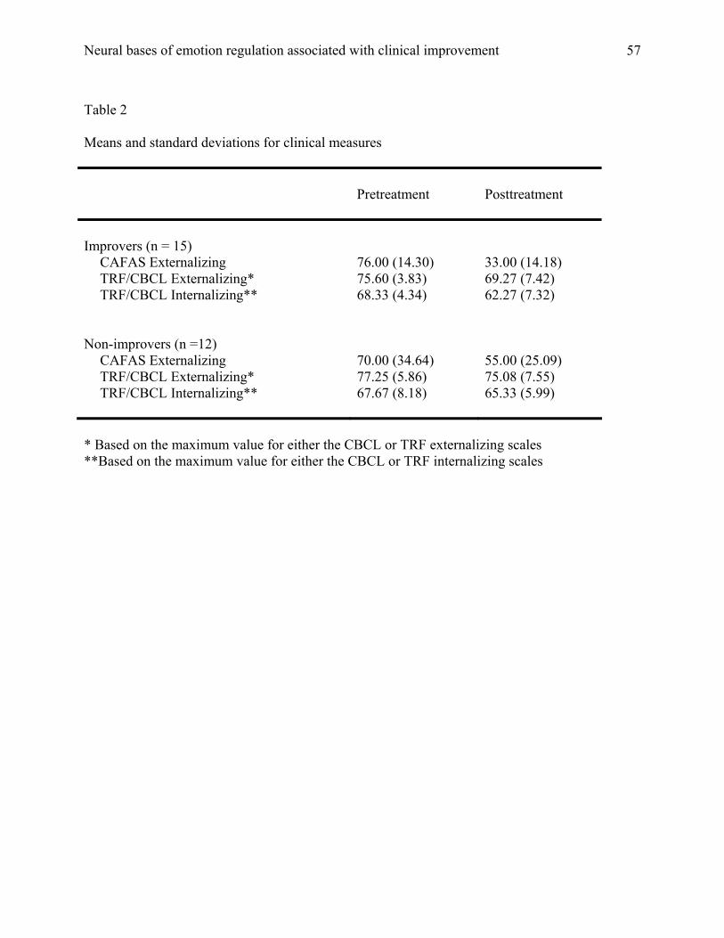
Neural bases of emotion regulation associated with clinical improvement
57
Table 2
Means and standard deviations for clinical measures
Pretreatment
Posttreatment
Improvers (n = 15)
CAFAS Externalizing 76.00 (14.30) 33.00 (14.18) TRF/CBCL Externalizing* 75.60 (3.83) 69.27 (7.42) TRF/CBCL Internalizing** 68.33 (4.34) 62.27 (7.32)
Non-improvers (n =12)
CAFAS Externalizing 70.00 (34.64) 55.00 (25.09) TRF/CBCL Externalizing* 77.25 (5.86) 75.08 (7.55) TRF/CBCL Internalizing** 67.67 (8.18) 65.33 (5.99)
* Based on the maximum value for either the CBCL or TRF externalizing scales **Based on the maximum value for either the CBCL or TRF internalizing scales
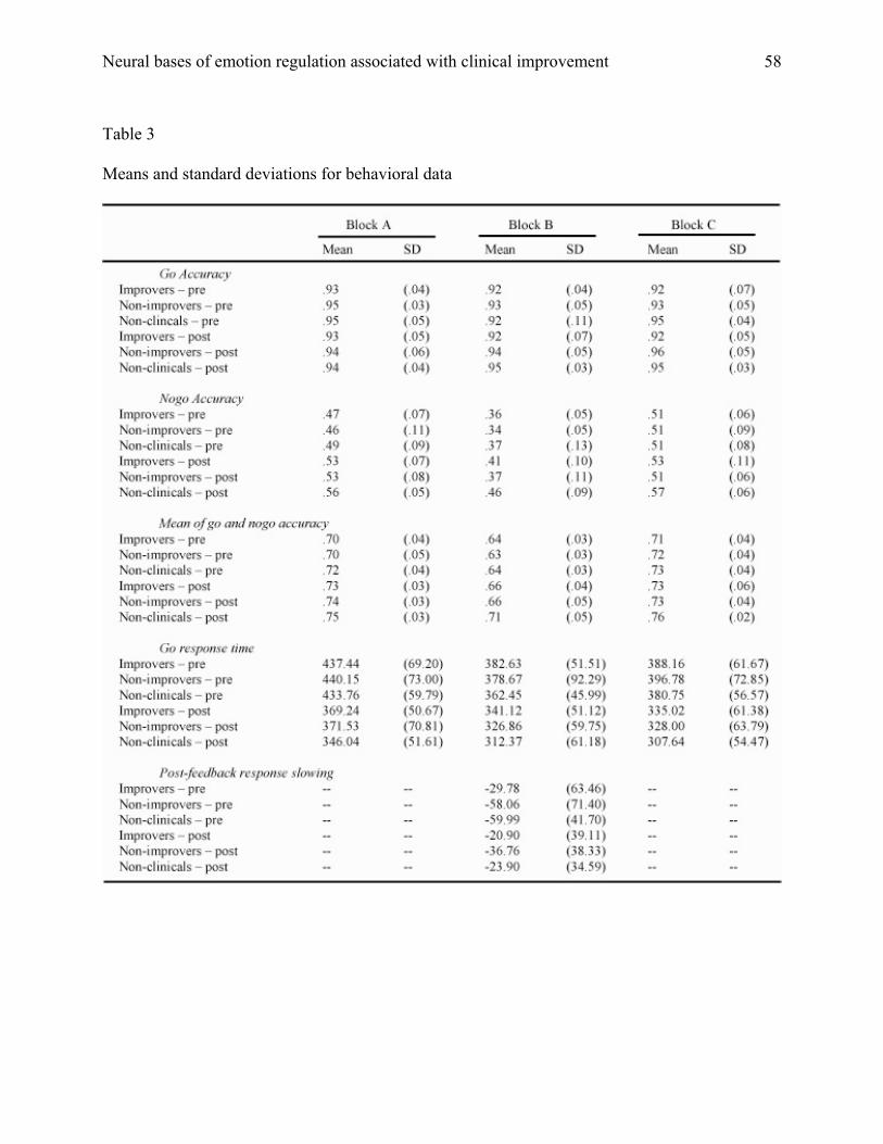
Neural bases of emotion regulation associated with clinical improvement
58
Table 3
Means and standard deviations for behavioral data
Neural bases of emotion regulation associated with clinical improvement
59
Figure Captions
Figure 1. Morphology-based regions of interest (ROIs) generated using the Montreal
Neurological Institute (MNI) average adult MRI scan.
Figure 2. Group differences in performance accuracy before and after treatment.
Figure 3. Grand-averaged ERP waveforms at site FCz for improvers, non-improvers, and non-
clinical participants at pretreatment and posttreatment sessions.
Figure 4. Group differences in ventral (panel A) and dorsal (panel B) activity pre- and
posttreatment for the latency range of 250 to 400 ms. This time range represents a narrowed
window surrounding the average N2 peak for the sample.
Figure 5. Group differences in whole brain activation before and after treatment for the latency
range of 250 to 400 ms.
Figure 6. Group differences in ventral (panel A) and dorsal (panel B) activity pre- and
posttreatment for the latency range of 500 to 650 ms. This time range represents a window that is
not associated with the N2.
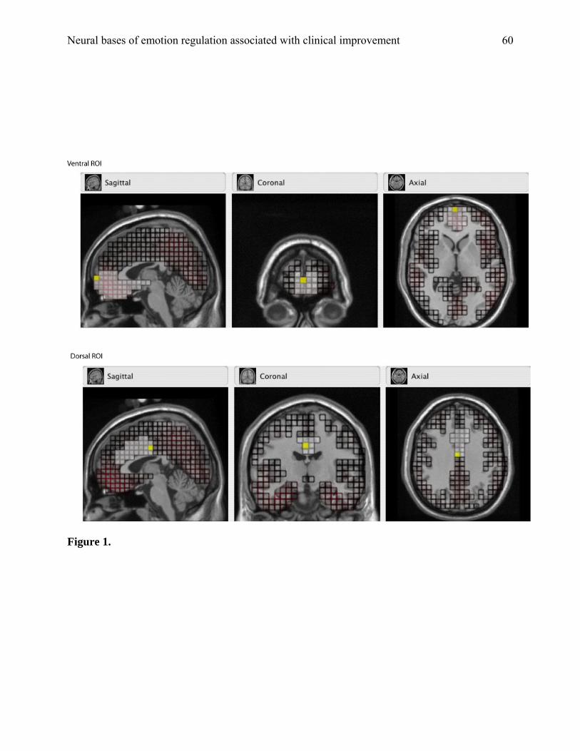
Neural bases of emotion regulation associated with clinical improvement
60
Figure 1.
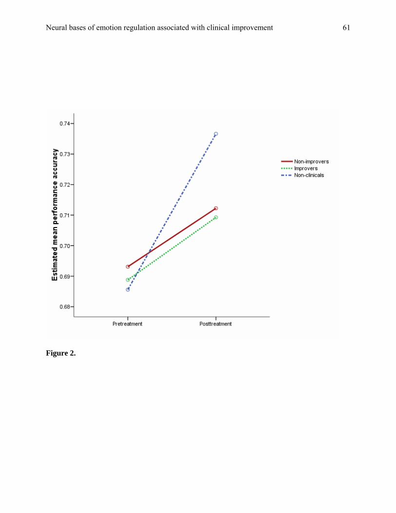
Neural bases of emotion regulation associated with clinical improvement
61
Figure 2.
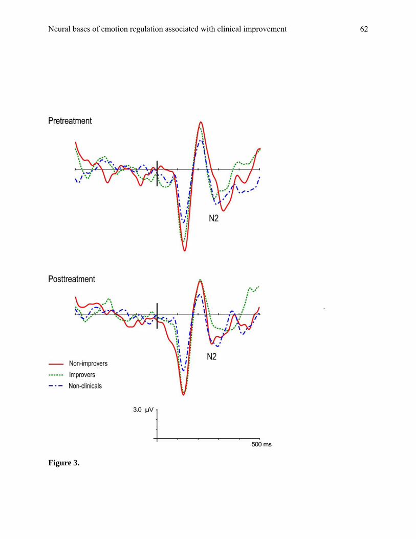
Neural bases of emotion regulation associated with clinical improvement
62
Figure 3.
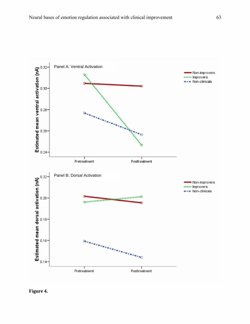
Neural bases of emotion regulation associated with clinical improvement
63
Panel A: Ventral Activation
Panel B: Dorsal Activation
Figure 4.
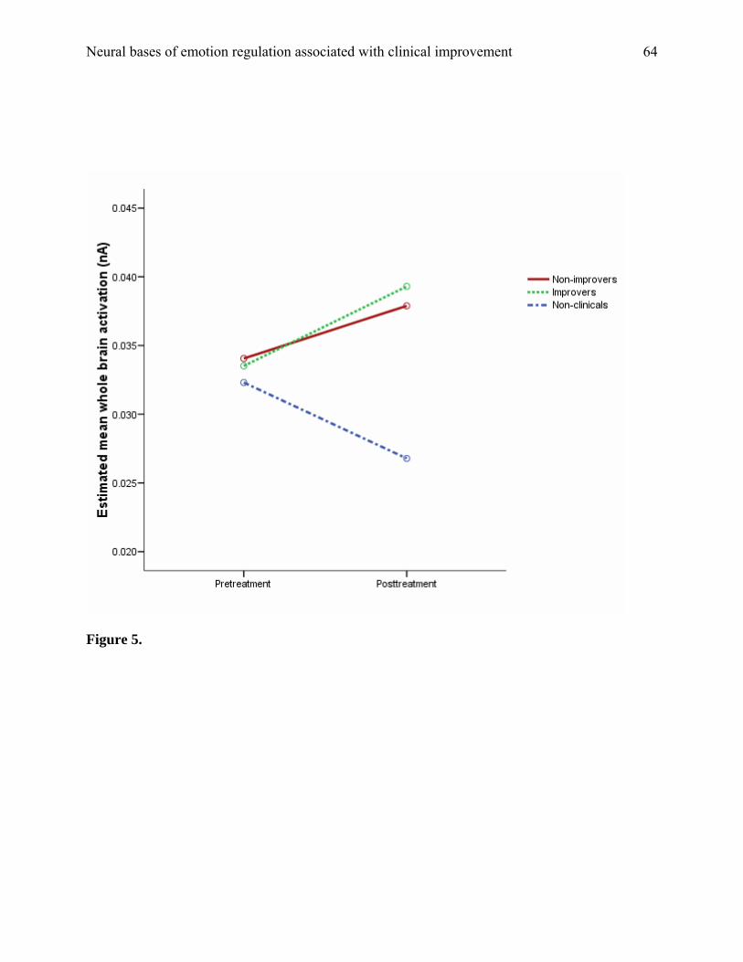
Neural bases of emotion regulation associated with clinical improvement
64
Figure 5.
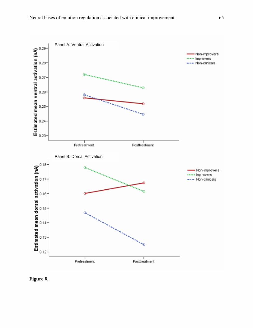
Neural bases of emotion regulation associated with clinical improvement
65
Panel A: Ventral Activation
Panel B: Dorsal Activation
Figure 6.


![Bases Bases Bases Bases Bases Bases Bases Bases Bases ......Hair loss or alopecia is a problem in modern society, which is usually related to hair loss on the scalp [1]. The most common](https://img.pdfslide.us/doc/110x75/5f692ed64ffcd531a566bfdf/bases-bases-bases-bases-bases-bases-bases-bases-bases-hair-loss-or-alopecia.jpg)