Embed Size (px)
Citation preview

JACO Journal of the Academy of Chiropractic Orthopedists
2018
Volume 15
Issue 2
June 2018

JACO Journal of the Academy of
Chiropractic Orthopedists
The Open Access, Peer-Reviewed and Indexed Publication of
the Academy of Chiropractic Orthopedists
June 2018 – Volume 15, Issue 2
Editorial Board Editor-In-Chief
Shawn M. Neff, DC, MAS, FACO
Managing Editor
Tracey A. Littrell, DC, DACBR, DACO, CCSP®
Associate Editors
James Demetrious, DC, FACO
David Swensen, DC, FACO
Alicia M. Yochum, RN, DC, DACBR, RMSK
Current Events Editor
James R. Brandt, DC, MS, FACO
Editorial Advisory Board
James R. Brandt, DC, MS, FACO
Ronald C Evans, DC, FACO
James Demetrious, DC, FACO
Michael Henrie, DO
Robert Morrow, MD
Bruce Gundersen, DC, FACO
Editorial Review Board Scott D. Banks, DC MS Ward Beecher, D.C., FACO
Thomas F. Bergmann, DC Gary Carver, DC, FACO
Jeffrey R. Cates, DC, FACO Rick Corbett, DC, DACBR, FCCO(C)
Donald S. Corenman, MD, DC, FACO Clinton Daniels, DC, MS, DAAPM
Anthony Vincent D'Antoni, MS, DC, PhD James Demetrious, DC, FACO
Daniel P. Dock, DC, FACO Neil L. Erickson, DC, DABCO, CCSP®
Simon John Forster, DC, DABCO Jaroslaw P. Grod, DC, FCCS(C)
Evan M. Gwilliam, DC, MBA Tony Hamm, DC, FACO
Nathan Hinkeldey, DC, DACRB Dale Huntington, DC, FACO
Charmaine Korporaal, M.Tech: Chiropractic Ralph Kruse, DC, FACO
Thomas Mack, DC, FACO Joyce Miller, DC, FACO
Loren C. Miller DC, FACO William E. Morgan, DC, DAAPM
Raymond S Nanko, DC, MD, DAAPM, FACO Deanna O'Dwyer, DC, FACO
Casey Okamoto, DC Joni Owen, DC, FACO
Gregory C. Priest, DC, FACO Christopher Roecker, DC, MS, DACO, DACSP
J Chris Romney, DC, FACO Roger Russell, DC, MS, FACO
Stephen M. Savoie, DC, FACO Alec Schielke, DC
Brandon Steele, DC John Stites, DC, DACBR, DACO
David Swensen, DC, FACO Cliff Tao, DC, DACBR
John M. Ventura, DC, FACO Michelle A Wessely BSc, DC, DACBR
Michael R. Wiles, DC, MEd, MS James A. Wyllie, DC DABCO
Steve Yeomans, DC, FACO Alicia M. Yochum, RN, DC, DACBR, RMSK
Articles, abstracts, opinions and comments appearing in this journal are the work of submitting authors, have been reviewed by
members of the editorial board and do not reflect the positions, opinions, endorsements or consensus of the Academy.
Journal of the Academy of Chiropractic Orthopedists June 2018- Volume 15, Issue 2
Journal of the Academy of Chiropractic Orthopedists
June 2018 – Volume 15, Issue 2
Editor’s Desk
❖ Shawn M. Neff, DC, MAS, FACO
Original Articles
❖ Cupler ZA: Conservative Management of Lumbopelvic and Genital Pain in a
Female Army Veteran: A Case Report: JACO 2018, 15(2):3-15
Abstracts and Literature Review
❖ Douglas AC, Wippold FJ, Broderick DF, et al: ACR Appropriateness Criteria:
Headache; Reviewed by Tao C. JACO 2018, 15(2):16-19
❖ Cassidy JD, et al: Risk of Carotid Stroke after Chiropractic Care: A Population-Based Case-Crossover Study; Reviewed by Grod JP. JACO 2018, 15(2):20-22
❖ Schwalfenberg, GK and Genuis, SJ: The Importance of Magnesium in Clinical
Healthcare; Reviewed by O’Dwyer DL. JACO 2018, 15(2):23-24
Ortho Quiz
❖ Kleinfield SL: Ortho Quiz. JACO 2018, 15(2):25
Current Events
❖ Diplomate Examination Information ❖ Conferences
Answers to Ortho Quiz
❖ Check your knowledge on page 27
Journal of the Academy of Chiropractic Orthopedists
Volume 15, Issue 1
2
The Editor’s Desk
Shawn M. Neff, DC, MAS, FACO
Editor-in-Chief
Welcome to the June 2018 issue of the Journal of the Academy of Chiropractic Orthopedists.
Summer is full swing, and I hope you are all enjoying the weather and some well deserved time
away with those you care about.
This month’s picture is my youngest child Cora.
She will be two years old in July and is enjoying
summer in the pool. It was roughly the time
that Cora was born that I joined the Journal as
editor-in-chief. Both have been exhausting and
beautiful experiences so far. Both have been
full of life and work and empty of sleep.
Cora is my fourth child and although it is not
my first time I still get excited for the new
things, the growth, the firsts, and the milestones.
I am so happy with the growth of the Journal
over this period and I look forward to more
growth and milestones.
I hope you all enjoy this issue.
Sincerely,
-Shawn
3
Original Article
Conservative Management of Lumbopelvic and Genital Pain in a
Female Army Veteran: A Case Report
Zachary A. Cupler, DC, MS1
1Physical Medicine & Rehabilitative Services, VA Butler Healthcare
Published: June 2018 Journal of the Academy of Chiropractic Orthopedists
June 2018, Volume 15, Issue 2
This is an Open Access article which permits unrestricted use, distribution, and reproduction in any medium, provided the original work is
properly cited. The article copyright belongs to the author and the Academy of Chiropractic Orthopedists and is available at:
http://www.dcorthoacademy.com. © 2018 Cupler and the Academy of Chiropractic Orthopedists.
Abstract
Background
To describe the management of a female patient with lumbopelvic and genital pain who
responded to conservative management after ruling out visceral causation.
Case Presentation
A 56-year-old female Army veteran presented with chronic lumbopelvic and genital pain. Her
primary care physician ruled out pelvic visceral origin. The patient was diagnosed with an upper
lumbar derangement.
Management and Outcome
A directional preference, as defined by Mechanical Diagnosis and Therapy, was identified on
evaluation, which guided our home exercise prescription. The patient was treated with
mechanical flexion-distraction spinal manipulation in our office. Outcome measures included
Journal of the Academy of Chiropractic Orthopedists
Volume 15, Issue 1
4
the Oswestry Disability Index (22%) and numeric pain scale (7 out of 10). The patient was
discharged from an active care plan symptom-free with improved Oswestry Disability Index
(2%), and she exhibited confidence in home care to successfully manage potential future
episodes.
Conclusions
A female patient with lumbopelvic and genital pain responded favorably to flexion-distraction
spinal manipulation and home exercise. A follow-up phone call 3 months later found the patient
experienced a single episode of axial lumbar spine pain. She reported she self-managed the
reoccurrence to resolution with the use of her home exercise plan. A musculoskeletal origin for
lumbopelvic and genital pain should be considered when visceral etiology has been ruled out.
Indexing terms
lumbopelvic pain, chiropractic, flexion-distraction spinal manipulation, directional preference,
McKenzie, Veteran, derangement
Background
Groin and lumbopelvic pain experienced by females can be caused by pain-sensitive structures
such as the pelvic viscera or lumbopelvic musculoskeletal tissues, though neuropathic and
psychogenic origins of pelvic pain should also be considered. Excluding endometriosis, the most
common causes of chronic pelvic pain include pelvic varices, post-operative adhesions,
interstitial cystitis and irritable bowel syndrome.1 Chronic pelvic pain is a descriptor of
symptoms rather than a diagnosis and often multiple factors are present. Gyang et al. reported
14-22% of pelvic pain has been correlated with musculoskeletal origin.2 Pregnancy-related
5
lumbopelvic pain management is well reported in the manual therapy literature.3,4,5,6 There is,
however, a paucity of literature describing the management of post-menopausal women with
lumbopelvic and groin pain of musculoskeletal origin not associated with pelvic floor
dysfunction.
After evaluation to rule out red flags and pelvic organ pathology, the musculoskeletal anatomy of
the region should be evaluated. Multiple tissues have been found to refer to the lumbopelvic
region when stimulated. Discogenic referral to the hip and groin is most commonly associated
with L1/L2 or L2/L3 discs.7 Upper lumbar disc lesion symptoms are far less common as
discography reproduced L1/L2 and L2/L3 pain in only 2 of 223 consecutive patients who
presented to a tertiary care center.8 Radicular pain from L1 and L2 is expected to result in
symptoms affecting the hip and/or groin, yet, surgical decompression of L3 or L4 has been found
to alleviate groin pain as well.9 Sacroiliac joint pain referral does not typically radiate to the
anterior thigh or groin.10 Femoroacetabular joint pain referral occurs in the buttock, groin and
distal to the knee 71%, 55%, and 22%, respectively, but never refers to the lumbar spine.11
Travell and Simons described lower quadrant and groin pain due to myofascial trigger points of
either the quadratus lumborum, pectineus or iliopsoas.12 Thoracolumbar (Maigne’s) syndrome is
a pain pattern of the low back, pelvis, groin or upper thighs, and has been associated with
zygapophyseal joints and the posterior rami of the involved segments.13.
Previous cases of lumbopelvic and groin pain have been described in the chiropractic literature,
including management of pregnancy-related lumbopelvic pain,14,15,16 pubic symphysis diastasis,17
paraesthetica meralgia, 18,19 testicular pain, 20, 21 and femoroacetabular impingement 22.
Journal of the Academy of Chiropractic Orthopedists
Volume 15, Issue 1
6
Additional cases of similar presentations have been reported by athletic trainiers23 and physical
therapists.24
Recent conservative low back pain clinical recommendations have focused on patient
subgroupings based on symptom presentation, cluster testing, observance of the centralization
phenomena, motor impairment, and psychosocial co-morbidities.25,26 Centralization is observed
in radiating spine pain that responds to repeated spine loading strategies, resulting in the distal
symptoms moving more proximal or towards the midline. In contrast to centralization,
peripheralization is the distal migration of symptoms.26 Improvement in an obstructed range of
motion may be observed without centralization. The concept of centralization versus
peripheralization can be utilized as a clinical guide for the patient, to monitor his or her own
symptoms outside the clinic. Importantly, centralization has been identified as a clinically
reliable tool.27 In the absence of observed centralization or peripheralization, a direction that
improves the obstructed range of motion is identified as the directional preference.
The purpose of this case is to describe the management of lumbopelvic and groin pain that
responded to mechanical flexion-distraction manipulation and home exercises. There is a paucity
of literature with regards to the conservative management of lumbopelvic and groin pain in
women who are not pregnant.
Case Presentation:
A 56 year-old postmenopausal, female Army veteran was referred by her primary care physician
(PCP) to a chiropractic office at a Veterans Affairs Medical Center for intermittent low back and
pelvic pain in an L1-L2 dermatomal distribution. Her symptoms developed 6 months earlier
without trauma or reported illness and symptoms. Lumbar spine pain radiated to the groin,
7
genitals as well as anterior and medial upper thighs 3-to-4 times per week and could last the
remainder of the day. She noted her symptoms were typically associated with bending and lifting
at work. Thigh, groin and vaginal pain only occurred concurrently with back pain. A revised
Oswestry Disability Index (rODI) was performed during her initial evaluation and her score was
11 out of 50 (22%), while her initial Numeric Pain Rating was 7 out of 10. Dyspareunia was
present since the onset of her symptoms, though she did not experience hematuria or dysuria and
was not concerned about the possibility of a sexually transmitted disease.
A review of systems revealed hypolipoproteinemia, fibrocystic disease of the breast,
gastroesophageal reflux disorder, and osteopenia. She was twice gravida and had healthy
deliveries on both occasions. The patient reported having a levonorgestrel-releasing intrauterine
device from age 45 and to age 56, and she had not had a menstrual cycle for at least 10 years due
to menopause. She did not report constitutional symptoms. Her medication was limited to
naproxen taken as needed since the onset of her symptoms. She also reported being a 42 pack-
year smoker. Her home workout routine included sit-ups and non-specific leg exercises that did
not produce lumbopelvic or genital pain.
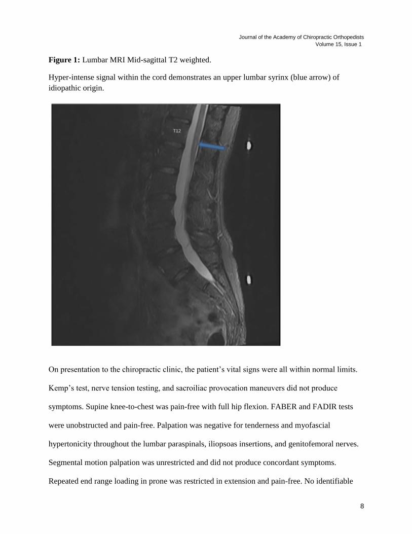
Prior to presentation to our clinic, her PCP performed an internal pelvic exam, which was
unremarkable. The PCP ordered additional testing; a lumbar MRI revealed mild multilevel
degenerative changes and a syrinx at the thoracolumbar junction (figure 1), while a pelvic
ultrasound visualized a normal sized, mildly heterogonous uterus without evidence of fibroid.
There were no abnormalities identified with the transvaginal ultrasound.
Journal of the Academy of Chiropractic Orthopedists
Volume 15, Issue 1
8
Figure 1: Lumbar MRI Mid-sagittal T2 weighted.
Hyper-intense signal within the cord demonstrates an upper lumbar syrinx (blue arrow) of
idiopathic origin.
On presentation to the chiropractic clinic, the patient’s vital signs were all within normal limits.
Kemp’s test, nerve tension testing, and sacroiliac provocation maneuvers did not produce
symptoms. Supine knee-to-chest was pain-free with full hip flexion. FABER and FADIR tests
were unobstructed and pain-free. Palpation was negative for tenderness and myofascial
hypertonicity throughout the lumbar paraspinals, iliopsoas insertions, and genitofemoral nerves.
Segmental motion palpation was unrestricted and did not produce concordant symptoms.
Repeated end range loading in prone was restricted in extension and pain-free. No identifiable
9
centralization or peripheralization was identified. Lumbar active range of motion (AROM)
revealed focal pain-free moderate restriction of movement into extension at the thoracolumbar
spine while flexion was pain-free and unrestricted.
In the absence of hip, sacroiliac, or neural tension findings, and focal obstruction to
thoracolumbar extension without centralization or peripheralization of symptoms, a working
diagnosis of L1/L2 lumbar derangement, as defined by McKenzie, was made with extension
identified as the directional preference.28 Following a detailed discussion related to our findings,
the patient agreed to a course of rehabilitative exercise and mechanical flexion-distraction
manipulation26,29 at a frequency of 1 time per week for 3 weeks. Goals included a reduction of
intermittent lumbopelvic and genital pain by at least 50% in 4 weeks, improvement in rODI by
10 points, and increased functional independence.
The patient was prescribed prone press-ups to be performed at home and work 10 times, 4-5
times per day or when she appreciated onset of symptoms. She was taught to understand the
centralization and peripheralization phenomena, thus allowing her to monitor her symptom
response to exercise.27 Flexion-distraction manipulation was performed in a neutral plane at each
follow-up visit.
The patient did not experience peripheralization of symptoms with her home exercise throughout
the course of care. She reported compliance with the home exercise and was able to demonstrate
the exercise in office. A modified exercise while standing was provided so that she could induce
extension into her thoracolumbar spine when she was at work.
Over the course of 4 visits, she experienced a single episode of axial low back pain that resolved
with home exercise. On the fourth visit, re-assessment of rODI demonstrated a 91% lumbar,
Journal of the Academy of Chiropractic Orthopedists
Volume 15, Issue 1
10
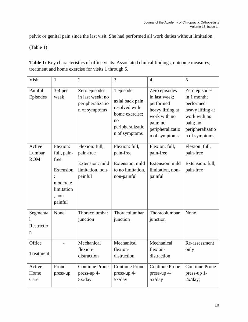
pelvic or genital pain since the last visit. She had performed all work duties without limitation.
(Table 1)
Table 1: Key characteristics of office visits. Associated clinical findings, outcome measures,
treatment and home exercise for visits 1 through 5.
Visit 1 2 3 4 5
Painful
Episodes
3-4 per
week
Zero episodes
in last week; no
peripheralizatio
n of symptoms
1 episode
axial back pain;
resolved with
home exercise;
no
peripheralizatio
n of symptoms
Zero episodes
in last week;
performed
heavy lifting at
work with no
pain; no
peripheralizatio
n of symptoms
Zero episodes
in 1 month;
performed
heavy lifting at
work with no
pain; no
peripheralizatio
n of symptoms
Active
Lumbar
ROM
Flexion:
full, pain-
free
Extension
:
moderate
limitation
, non-
painful
Flexion: full,
pain-free
Extension: mild
limitation, non-
painful
Flexion: full,
pain-free
Extension: mild
to no limitation,
non-painful
Flexion: full,
pain-free
Extension: mild
limitation, non-
painful
Flexion: full,
pain-free
Extension: full,
pain-free
Segmenta
l
Restrictio
n
None Thoracolumbar
junction
Thoracolumbar
junction
Thoracolumbar
junction
None
Office
Treatment
- Mechanical
flexion-
distraction
Mechanical
flexion-
distraction
Mechanical
flexion-
distraction
Re-assessment
only
Active
Home
Care
Prone
press-up
Continue Prone
press-up 4-
5x/day
Continue Prone
press-up 4-
5x/day
Continue Prone
press-up 4-
5x/day
Continue Prone
press-up 1-
2x/day;
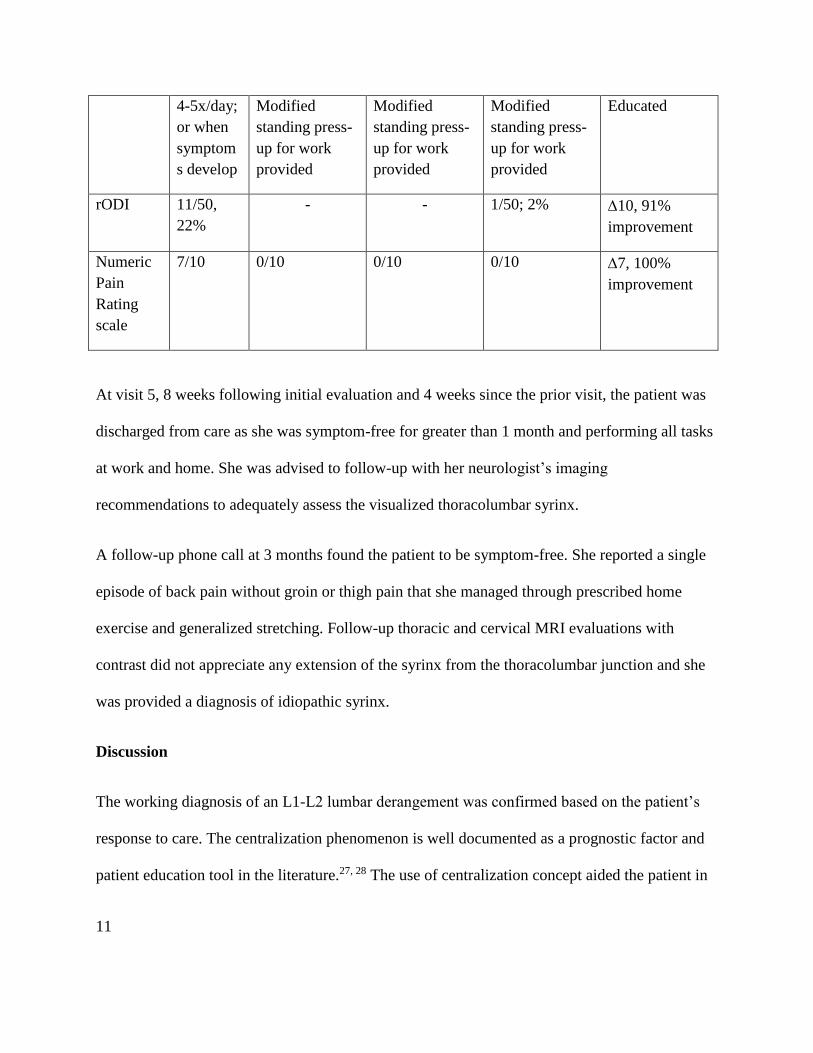
11
4-5x/day;
or when
symptom
s develop
Modified
standing press-
up for work
provided
Modified
standing press-
up for work
provided
Modified
standing press-
up for work
provided
Educated
rODI 11/50,
22%
- - 1/50; 2% 10, 91%
improvement
Numeric
Pain
Rating
scale
7/10 0/10 0/10 0/10 7, 100%
improvement
At visit 5, 8 weeks following initial evaluation and 4 weeks since the prior visit, the patient was
discharged from care as she was symptom-free for greater than 1 month and performing all tasks
at work and home. She was advised to follow-up with her neurologist’s imaging
recommendations to adequately assess the visualized thoracolumbar syrinx.
A follow-up phone call at 3 months found the patient to be symptom-free. She reported a single
episode of back pain without groin or thigh pain that she managed through prescribed home
exercise and generalized stretching. Follow-up thoracic and cervical MRI evaluations with
contrast did not appreciate any extension of the syrinx from the thoracolumbar junction and she
was provided a diagnosis of idiopathic syrinx.
Discussion
The working diagnosis of an L1-L2 lumbar derangement was confirmed based on the patient’s
response to care. The centralization phenomenon is well documented as a prognostic factor and
patient education tool in the literature.27, 28 The use of centralization concept aided the patient in
Journal of the Academy of Chiropractic Orthopedists
Volume 15, Issue 1
12
self-management by guiding her through hurt versus harm conceptualization, thus minimizing
reliance on passive care, and gained the confidence to self-manage a reoccurrence to resolution.
The differential diagnosis for this case includes gynecologic, genitourinary conditions and
musculoskeletal conditions, including but not limited to upper lumbar derangement, upper
lumbar radiculitis due to herniation, sacroiliac joint dysfunction, trigger points of the quadratus
lumborum, meralgia paresthetica, femoroacetabular impingement, and genitofemoral nerve
entrapment. Thoracolumbar (Maigne’s) Syndrome has a similar presentation to an upper lumbar
derangement, and it cannot be necessarily excluded from the diagnosis in this specific case as
Maigne and McKenzie might be describing the same functional lesion. Work-up of the lumbar
syrinx was necessary to rule out additional etiology.
Conclusions
There are few reported cases of conservatively managed lumbopelvic pain and radiating genital
pain in women of non-child bearing age. This case report demonstrates management and
resolution of intermittent lumbopelvic pain with associated bilateral genital pain with patient-
generated end-range loading exercises and mechanical flexion-distraction spinal manipulation.
Integrated medical and chiropractic care led to a resolution of the patient’s symptoms.
Limitations
While interesting, this case report is limited in its scope and cannot be utilized as a generalization
for patient care.
13
Consent:
Written informed consent was obtained from the patient for the publication of this case report
and any accompanying images. A copy of the written consent is available for review by the
Editor-In-Chief of this journal.
List of Abbreviations:
FABER: flexion abduction external rotation
FADIR: flexion adduction internal rotation
MRI: magnetic resonance imaging
PCP: Primary care physician
rODI: revised Oswestry Disability Index
ROM: range of motion
Competing Interests:
No funding sources or conflicts of interest were reported for this report.
Acknowledgments:
This work was conducted at and supported by VA Butler Healthcare. The views expressed in this
article are those of the authors and do not reflect the official policy or position of the Department
of the Army, Department of Defense, Department of Veterans Affairs, or the United States
Government. The author would also like to thank Dr. James Demetrious for his editorial support.
Journal of the Academy of Chiropractic Orthopedists
Volume 15, Issue 1
14
References
1. Vercellini P, Somigliana E, Vigano P, Abbiati A, Barbara G, Fedele L: Chronic Pelvic
Pain in women: etiology, pathogenesis and diagnostic approach. Gynecol Endocrinol
2009, 25(3): 149-158.
2. Gyang, A, Hartman M, Lamvu G: Musculoskeletal causes of Chronic Pelvic Pain
What a Gynecolgoist Should Know. Obset Gynecol 2013, 121(3):645-50.
3. Van Benten E, Pool J, Mens J, Pool-Goudzwaard A: Recommendations for physical
therapists on the treatment of lumbopelvic pain during pregnancy: a systematic
review. J Orthop Sports Phys Ther 2014, 44(7):464-473.
4. Murphy DR, Hurwitz EL, McGovern EE: Outcome of pregnancy-related lumbopelvic
pain treated according to a diagnosis-based decision rule: a prospective
observational cohort study. J Manipulative Physiol Ther 2009, 32(8):616-614.
5. Gausel, AM, Kjærmann, I, Malmqvist, S, Andersen, K, Dalen, I, Larsen, JP, Økland, I:
Chiropractic management of dominating one-sided pelvic girdle pain in pregnant
women; a randomized controlled trial. BMC Pregnancy and Childbirth 2017, 17:331.
6. Peterson, CK, Mühlemann D, Humphreys, BK: Outcomes of pregnant patients with
low back pain undergoing chiropractic treatment: a prospective cohort study with
short term, medium term and 1 year follow-up. Chiropr Man Therap 2014, 22:15.
7. Saifuddin A, Emanuel R, White J, Renton P, Braithwaite I, Taylor BA. An analysis of
radiating pain at lumbar discography. Eur Spine J 1998, 7(5):358-362.
8. Verrills P, Nowesenitz G, Barnard A. Prevalence and characteristics of discogenic
pain in tertiary practice: 223 consecutive cases utilized lumbar discography. Pain
Med 2015. 16:1490-1499.
9. Sasaki M, Aoki M, Matsumoto K, Tsuruzono K, Yonenobu K, Yoshimine T: Groin pain
caused by L3 and L4 radiculopathy. J Spine 2014, 3(3):1-4
10. Fortin JD, Aprill C, Pontieux RT, Pier J: Sacroiliac joint: pain referral maps upon
applying a new injection/arthrography technique. part II: clinical evaluation. Spine
1994, 19:1483-1489.
11. Lesher JM, Dreyfuss P, Hager N, Kaplan M, Furman M: Hip joint pain referral
patterns: a descriptive study. Pain Med 2008, 9(1):22-25.
12. Travell JG, Simons DG: Myofascial Pain and Dysfunction: The Trigger Point Manual.
Vol. 2. Baltimore , MD: Lippincott Williams & Wilkins, 1992.
13. Alptekin K, Örnek NI, Aydın T, Alkan M, Toprak M, A Balcı L, Öncü Alptekin, J:
Effectiveness of exercise and local steroid injections for the thoracolumbar junction
syndrome (the maigne’s syndrome) treatment. Open Orthop J 2017, 11: 467–477.
14. Bernard M, Tuchin, P: Chiropractic Management of Pregnancy-Related Lumbopelvic
Pain: A Case Study. J Chiropr Med 2016, 15(2), 129–133.
15
15. Howell, ER: Pregnancy-related symphysis pubis dysfunction management and
postpartum rehabilitation: two case reports. J Can Chiropr Assoc 2012, 56(2), 102–
111.
16. Ducar D, Skaggs CD: Conservative management of groin pain during pregnancy: a
descriptive case study. J Chiropr Med 2005, 4(4), 195–199.
17. Henry, L: Chiropractic management of postpartum pubic symphysis diastasis: A
case report. J Can Chiropr Assoc 2015, 59(1), 30–36.
18. Houle, S: Chiropractic management of chronic idiopathic meralgia paresthetica: a
case study. Journal of Chiropr Med 2012, 11(1), 36–41.
19. Skaggs, CD, Winchester, BA, Vianin, M, Prather, H: A manual therapy and exercise
approach to meralgia paresthetica in pregnancy: a case report. J Chiropr Med 2006,
5(3), 92–96.
20. Neff S, Warnecke R: Chiropractic management of Low Back Pain and Testicle Pain:
A Case Report: JACO 2017, 14(3):36-41
21. Rowell RM, Rylander SJ: Low back pain, leg pain, and chronic idiopathic testicular
pain treated with chiropractic care. J Altern Complement Med 2012, 18(4):420-422.
22. Stobert JR, Emary PC, Taylor JA: Femoracetabular impingement: a retrospective
case study with 8-year follow-up. J Chiropr Med 2015, 14(4):290-296.
23. Leone JE, Middleton SM: Nontraumatic testicular pain due to sacroiliac-joint
dysfunction: a case report. J Athl Train 2016, 51(8):651-657
24. Horton R: Physical therapy management of chronic testicular pain impacting sexual
function: a case report. Topics in Geriatric Rehabilitation 2016, 32(3):182-187.
25. Alrwaily M, Timko M, Schneider M, Stevans J, Bise C, Hariharan K, Delitto A:
Treatment-based classification system for low back pain: revision and update.
JOSPT 2016, 96(7):1057-1066.
26. Murphy DR: Clinical Reasoning in Spine Pain Volume I Primary Management of Low
Back Disorders. San Bernadino, CA: CRISP Education and Research, LLC.
27. May S, Alessandro A: Centralization and directional preference: a systematic review.
Man Ther 2012, 17(6):497-506.
28. McKenzie RA, May S: The Lumbar Spine: Mechanical Diagnosis and Therapy Volume I
and II. New Zealand: Spinal Publications New Zealand Ltd.
29. Gay, RE, Bronfort G, Evans RL: Distraction Manipulation of the Lumbar Spine: A
Review of the Literature. JMPT 2005, 28(4):266-273.
Journal of the Academy of Chiropractic Orthopedists
Volume 15, Issue 1
16
Editorial Review
ACR Appropriateness Criteria: Headache
Annette C. Douglas MD, Franz J. Wippold II MD, Daniel F. Broderick MD, et al.
https://acsearch.acr.org/docs/69482/Narrative/
Copyright: 2017 Nilsson et al. (Open access article)
JACO Editorial Reviewer: Cliff Tao, DC, DACBR
Published: June 2018
Journal of the Academy of Chiropractic Orthopedists
June 2018, Volume 15, Issue 2
The original article copyright belongs to the original publisher. This review is available from: http://www.dcorthoacademy.com
© 2018 Tao and the Academy of Chiropractic Orthopedists. This is an Open Access article which permits unrestricted use, distribution, and
reproduction in any medium, provided the original work is properly cited.
Author’s Abstract/Introduction:
The cause or type of most headaches can be determined by procuring a careful history and
performing a physical examination while focusing on the warning signals that prompt further
diagnostic testing. In the absence of worrisome features in the history or examination, the task is
then to diagnose the primary headache syndrome based on the clinical features. If atypical
features are present or the patient does not respond to conventional therapy, the possibility of a
secondary headache disorder should be investigated.
Headache is one of the most frequent ailments of the human race. Studies have estimated overall
lifetime prevalence of 0.2%–60% for headache of any kind. In children, prevalence of headache
ranges from 8%–83%. As in the case of migraines, characteristics such as age, gender, and case
definition may largely account for this variance. However, a higher prevalence of headache has
been found by surveys in South America, Europe, and North America than by those of Asian
countries. A survey of the Canadian population showed that only about 20% of people there are
headache free. Prevalence studies on migraine show that genetic factors are related to prevalence
as well as gender differences, as migraines affect approximately 15%–18% of women and 6% of
men. Headaches occur most commonly between the ages of 25–55 years. Muscle contraction or
tension accounts for most of the non-migraine headaches encountered in population surveys.
17
Several studies have confirmed the low yield of imaging procedures for individuals presenting
with isolated headache, ie, headache unaccompanied by other neurological findings. Patients
were referred for imaging because the referring physician suspected imaging-detected pathology
or because patients requested the study to be certain that they did not have a brain tumor. A
prospective review of 293 computed tomography (CT) scans ordered in an ambulatory family
practice setting disclosed that most scans were ordered because the clinician suspected that a
tumor (49%) or a subarachnoid hemorrhage (SAH) (9%) might be present. Fifty-nine (17%)
were
ordered because of patient expectation or medicolegal concerns.
When considering such a common disorder as headache, indications for imaging use become
relevant. This is particularly true in the face of emerging and rapidly evolving technologies in
use today. In frequent conditions, performing low-yield studies is more likely to result in false-
positive results, with the consequent risk of additional and unnecessary procedures. The yield of
positive studies in patients referred with isolated,
nontraumatic headache is approximately 0.4%. Assuming the cost of a CT scan is $400, and a
magnetic resonance imaging (MRI) scan is $900, the cost to detect a lesion is $100,000 with CT
and $225,000 with MRI.
One should not assume, however, that there is no social benefit in negative imaging studies in the
setting of headache. Indeed, headache symptoms can be quite ominous and onerous to those
patients, and there can be tremendous costs with respect to productivity and quality-of-life
issues. Moreover, health-care providers perceive value in imaging headache when the fear of
litigation is taken into account. Although it is beyond the scope of this review to assess the
factors and inherent value of negative imaging tests in headache imaging, it must be emphasized
that the costs of detection or screening in imaging headache are always overstated when the
value of negative results is not factored into the analysis.
JACO Editorial Summary:
• The American College of Radiology (ACR) is a prominent figure in radiology
information and resources, and they have a thorough process for determining the
appropriateness of imaging for various conditions.
• ACR Appropriateness Criteria (ACR AC) are a trademarked, evidence-based set of
guidelines to help physicians and other providers in making the most appropriate imaging
or treatment decision for specific clinical conditions. ACR AC is the most comprehensive
evidence based guidelines for diagnostic imaging, radiotherapy protocols, and image
guided interventional procedures.
• The 15 authors of this article are from the ACR’s Expert Panel on Neurologic Imaging
and are all based in the US, and presumably all neuroradiologists. The lead author is Dr.
Annette C, Douglas from Indiana University Hospital, in Indianapolis, Indiana.
Journal of the Academy of Chiropractic Orthopedists
Volume 15, Issue 1
18
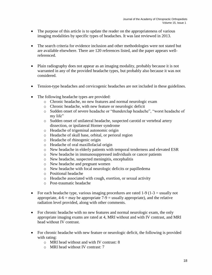
• The purpose of this article is to update the reader on the appropriateness of various
imaging modalities by specific types of headaches. It was last reviewed in 2013.
• The search criteria for evidence inclusion and other methodologies were not stated but
are available elsewhere. There are 120 references listed, and the paper appears well-
referenced.
• Plain radiography does not appear as an imaging modality, probably because it is not
warranted in any of the provided headache types, but probably also because it was not
considered.
• Tension-type headaches and cervicogenic headaches are not included in these guidelines.
• The following headache types are provided:
o Chronic headache, no new features and normal neurologic exam
o Chronic headache, with new feature or neurologic deficit
o Sudden onset of severe headache or “thunderclap headache”, “worst headache of
my life”
o Sudden onset of unilateral headache, suspected carotid or vertebral artery
dissection, or ipsilateral Horner syndrome
o Headache of trigeminal autonomic origin
o Headache of skull base, orbital, or perioral region
o Headache of rhinogenic origin
o Headache of oral maxillofacial origin
o New headache in elderly patients with temporal tenderness and elevated ESR
o New headache in immunosuppressed individuals or cancer patients
o New headache, suspected meningitis, encephalitis
o New headache and pregnant women
o New headache with focal neurologic deficits or papilledema
o Positional headache
o Headache associated with cough, exertion, or sexual activity
o Post-traumatic headache
• For each headache type, various imaging procedures are rated 1-9 (1-3 = usually not
appropriate, 4-6 = may be appropriate 7-9 = usually appropriate), and the relative
radiation level provided, along with other comments.
• For chronic headache with no new features and normal neurologic exam, the only
appropriate imaging exams are rated at 4, MRI without and with IV contrast, and MRI
head without IV contrast.
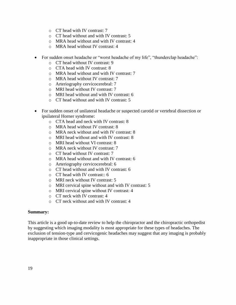
• For chronic headache with new feature or neurologic deficit, the following is provided
with rating:
o MRI head without and with IV contrast: 8
o MRI head without IV contrast: 7
19
o CT head with IV contrast: 7
o CT head without and with IV contrast: 5
o MRA head without and with IV contrast: 4
o MRA head without IV contrast: 4
• For sudden onset headache or “worst headache of my life”, “thunderclap headache”:
o CT head without IV contrast: 9
o CTA head with IV contrast: 8
o MRA head without and with IV contrast: 7
o MRA head without IV contrast: 7
o Arteriography cervicocerebral: 7
o MRI head without IV contrast: 7
o MRI head without and with IV contrast: 6
o CT head without and with IV contrast: 5
• For sudden onset of unilateral headache or suspected carotid or vertebral dissection or
ipsilateral Horner syndrome:
o CTA head and neck with IV contrast: 8
o MRA head without IV contrast: 8
o MRA neck without and with IV contrast: 8
o MRI head without and with IV contrast: 8
o MRI head without VI contrast: 8
o MRA neck without IV contrast: 7
o CT head without IV contrast: 7
o MRA head without and with IV contrast: 6
o Arteriography cervicocerebral: 6
o CT head without and with IV contrast: 6
o CT head with IV contrast:: 6
o MRI neck without IV contrast: 5
o MRI cervical spine without and with IV contrast: 5
o MRI cervical spine without IV contrast: 4
o CT neck with IV contrast: 4
o CT neck without and with IV contrast: 4
Summary:
This article is a good up-to-date review to help the chiropractor and the chiropractic orthopedist
by suggesting which imaging modality is most appropriate for these types of headaches. The
exclusion of tension-type and cervicogenic headaches may suggest that any imaging is probably
inappropriate in those clinical settings.
Journal of the Academy of Chiropractic Orthopedists
Volume 15, Issue 1
20
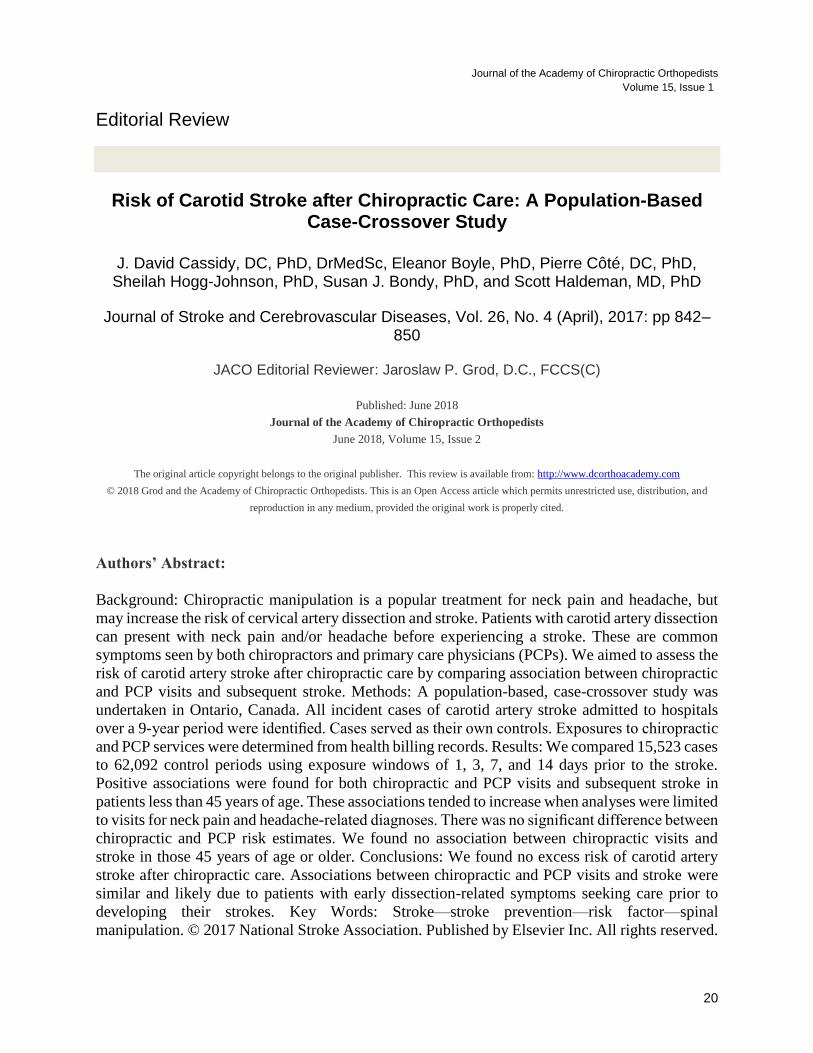
Editorial Review
Risk of Carotid Stroke after Chiropractic Care: A Population-Based Case-Crossover Study
J. David Cassidy, DC, PhD, DrMedSc, Eleanor Boyle, PhD, Pierre Côté, DC, PhD,
Sheilah Hogg-Johnson, PhD, Susan J. Bondy, PhD, and Scott Haldeman, MD, PhD
Journal of Stroke and Cerebrovascular Diseases, Vol. 26, No. 4 (April), 2017: pp 842–850
JACO Editorial Reviewer: Jaroslaw P. Grod, D.C., FCCS(C)
Published: June 2018
Journal of the Academy of Chiropractic Orthopedists
June 2018, Volume 15, Issue 2
The original article copyright belongs to the original publisher. This review is available from: http://www.dcorthoacademy.com
© 2018 Grod and the Academy of Chiropractic Orthopedists. This is an Open Access article which permits unrestricted use, distribution, and
reproduction in any medium, provided the original work is properly cited.
Authors’ Abstract:
Background: Chiropractic manipulation is a popular treatment for neck pain and headache, but
may increase the risk of cervical artery dissection and stroke. Patients with carotid artery dissection
can present with neck pain and/or headache before experiencing a stroke. These are common
symptoms seen by both chiropractors and primary care physicians (PCPs). We aimed to assess the
risk of carotid artery stroke after chiropractic care by comparing association between chiropractic
and PCP visits and subsequent stroke. Methods: A population-based, case-crossover study was
undertaken in Ontario, Canada. All incident cases of carotid artery stroke admitted to hospitals
over a 9-year period were identified. Cases served as their own controls. Exposures to chiropractic
and PCP services were determined from health billing records. Results: We compared 15,523 cases
to 62,092 control periods using exposure windows of 1, 3, 7, and 14 days prior to the stroke.
Positive associations were found for both chiropractic and PCP visits and subsequent stroke in
patients less than 45 years of age. These associations tended to increase when analyses were limited
to visits for neck pain and headache-related diagnoses. There was no significant difference between
chiropractic and PCP risk estimates. We found no association between chiropractic visits and
stroke in those 45 years of age or older. Conclusions: We found no excess risk of carotid artery
stroke after chiropractic care. Associations between chiropractic and PCP visits and stroke were
similar and likely due to patients with early dissection-related symptoms seeking care prior to
developing their strokes. Key Words: Stroke—stroke prevention—risk factor—spinal
manipulation. © 2017 National Stroke Association. Published by Elsevier Inc. All rights reserved.
21
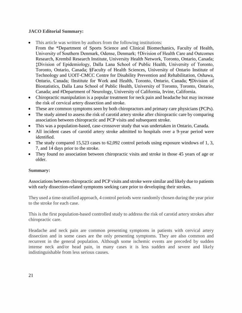
JACO Editorial Summary:
• This article was written by authors from the following institutions:
From the *Department of Sports Science and Clinical Biomechanics, Faculty of Health,
University of Southern Denmark, Odense, Denmark; †Division of Health Care and Outcomes
Research, Krembil Research Institute, University Health Network, Toronto, Ontario, Canada;
‡Division of Epidemiology, Dalla Lana School of Public Health, University of Toronto,
Toronto, Ontario, Canada; §Faculty of Health Sciences, University of Ontario Institute of
Technology and UOIT-CMCC Centre for Disability Prevention and Rehabilitation, Oshawa,
Ontario, Canada; ‖Institute for Work and Health, Toronto, Ontario, Canada; ¶Division of
Biostatistics, Dalla Lana School of Public Health, University of Toronto, Toronto, Ontario,
Canada; and #Department of Neurology, University of California, Irvine, California.
• Chiropractic manipulation is a popular treatment for neck pain and headache but may increase
the risk of cervical artery dissection and stroke.
• These are common symptoms seen by both chiropractors and primary care physicians (PCPs).
• The study aimed to assess the risk of carotid artery stroke after chiropractic care by comparing
association between chiropractic and PCP visits and subsequent stroke.
• This was a population-based, case-crossover study that was undertaken in Ontario, Canada.
• All incident cases of carotid artery stroke admitted to hospitals over a 9-year period were
identified.
• The study compared 15,523 cases to 62,092 control periods using exposure windows of 1, 3,
7, and 14 days prior to the stroke.
• They found no association between chiropractic visits and stroke in those 45 years of age or
older.
Summary:
Associations between chiropractic and PCP visits and stroke were similar and likely due to patients
with early dissection-related symptoms seeking care prior to developing their strokes.
They used a time-stratified approach, 4 control periods were randomly chosen during the year prior
to the stroke for each case.
This is the first population-based controlled study to address the risk of carotid artery strokes after
chiropractic care.
Headache and neck pain are common presenting symptoms in patients with cervical artery
dissection and in some cases are the only presenting symptoms. They are also common and
recurrent in the general population. Although some ischemic events are preceded by sudden
intense neck and/or head pain, in many cases it is less sudden and severe and likely
indistinguishable from less serious causes.

Journal of the Academy of Chiropractic Orthopedists
Volume 15, Issue 1
22
This study base includes the entire population of Ontario, Canada, over a 9-year period,
representing 109,020,875 person-years of observation, and the results should be generalizable to
other populations where chiropractic treatment is offered.
The conclusion of the study suggests that the association between chiropractic care and carotid
artery stroke could be due to care being delivered for dissection-related neck pain and/or headache,
prior to the ischemic event.
23
Editorial Review
The Importance of Magnesium in Clinical Healthcare
Gerry K. Schwalfenberg and Stephen J. Genuis
Scientifica, Volume 2017, Article ID 4179326, https://doi.org/10.1155/2017/4179326
JACO Editorial Reviewer: Deanna L. O’Dwyer, DC
Published: June 2018
Journal of the Academy of Chiropractic Orthopedists
June 2018, Volume 15, Issue 2
The original article copyright belongs to the original publisher. This review is available from: http://www.dcorthoacademy.com ©2010 O’Dwyer
and the Academy of Chiropractic Orthopedists. This is an Open Access article which permits unrestricted use, distribution, and reproduction in
any medium, provided the original work is properly cited.
Authors' Abstract:
Background: Magnesium is an essential element required as a co-factor required in over 300
enzymatic reactions and metabolic pathways. It is estimate that up to two-thirds of the Western
population are not attaining the RDA of magnesium.
Methods: Review of magnesium in the literature was assessed via MEDLINE and PubMed.
Written books, as well as, conference proceedings were also reviewed. A traditional integrated
review format with Level 1 evidence to support the use of magnesium in the prevention or
treatment of many common ailments. These could include, although not limited to: migraine,
metabolic syndrome, diabetes (I and II), hyperlipidemia, asthma, premenstrual syndrome,
preeclampsia, various cardia arrthymias, attention deficit/hyperactivity and other, as yet,
undiscovered ailments. Magnesium may also be influential for the prevention of renal calculi,
cataract formation; as an adjunct to depression. The possibilities are limitless.
Results: Supplementation with as little as 300mg of various forms of magnesium have been
shown to improve health and decrease disease states.
Conclusions: The role of magnesium is more far-reaching than previously considered.
Traditional medical practitioners should consider magnesium supplementation as an adjunct to
well rounded healthcare, especially in certain chronic disease states.
Clinical Relevance: Many chronic illness and disease processes are directly related to
sublimial, chronic hypo -magnesia.
Journal of the Academy of Chiropractic Orthopedists
Volume 15, Issue 1
24
JACO Editorial Summary:
This article is written by authors from the Dept of Family Medicine, Faculty of Medicine,
University of Alberta and the University of Calgary.
The focus of the study was to demonstrate the efficacy of magnesium supplementation in various
disease states to medical practitioners who might otherwise be uninformed to the benefits of
nutritional management of disease.
Magnesium is the fourth most common mineral after calcium, sodium and potassium, in the
human body. It is also the second most common intercellular cation after potassium. Up to 68%
of all Americans are deficient in magnesium. The RDA for magnesium varies from as low as 30
mg in infants to 420 mg for a 51+ year old male.
Processed, preserved and organic foods are generally deficient in magnesium putting the m
majority of Americans who partake in the Standard American Diet, at risk.
Many factors influence the efficiency with which magnesium is absorbed and utilized in the
human body. These factors include, but are not limited to decreased absorption due to Vitamin D
deficiency, certain common medications (antibiotics, antacids, antihypertensive drugs),
pesticides which chelate the minerals. There is excess excretion due to alcohol use and the
presence of diabetes I and II. Decreased plasma concentrations due to smoking. Decreased
absorption due to the normal aging process.
The role of magnesium in cellular function ranges from contributing to the synthesis of ATP,
binding to ATP to yield the bioactive form of Mg-ATP and binding site for up to 3571 human
proteins. Magnesium’s biologic half life is about 42 days (1000 hours).
Disease states, generally, would be improved with improved nutritional protocols.
Summary:
Prudent and further investigation must be conducted to best accommodate the patient and the
disease states. The practitioner should take great care to decide which form of magnesium would
be amenable to the particular disease or deficiency.

25
Ortho Quiz
by Steven L. Kleinfield DC, FACO
1. The most common type of cancer diagnosed in children ages 0-14 is:
a. Leukemia
b. Osteosarcoma
c. Ewing’s Tumor
d. Glioblastoma
2. The most common primary malignant tumor of bone is:
a. Osteosarcoma
b. Multiple Myeloma
c. Paget’s Disease
d. Chondrosarcoma
3. The second most common primary malignant tumor of bone is:
a. Chondrosarcoma
b. Multiple Myeloma
c. Paget’s Disease
d. Osteosarcoma
4. In Adults, an Ivory Vertebrae is classically seen in which condition:
a. Leukemia
b. Glioblastoma
c. Multiple Myeloma
d. Metastatic Disease
5. The “Winking Owl Sign” is a classic finding for which condition:
a. Osteolytic Vertebral Metastases
b. Osteoblastic Vertebral Metastases
c. Paget’s Disease
d. Prostatic Cancer

Journal of the Academy of Chiropractic Orthopedists
Volume 15, Issue 1
26
Current Events
❖ The Part I online examination will be available for candidates to take on either Friday,
July 20th, or Saturday morning July 21st. Apply on the Academy website:
http://dcorthoacademy.org/
❖ Apply for the Lipe Scholarship
Details at http://www.accoweb.org/lipescholarship.html
❖ The full hours of the following conventions have been accepted by the Academy as
qualifying for re-credentialing.
o American College of Chiropractic Orthopedists
2019 Orthopedic Essentials Seminar
April 25-27, 2019
Tropicana Las Vegas in Las Vegas, Nevada

27
Answers to Ortho Quiz
1. The most common type of cancer diagnosed in children ages 0-14 is:
a. Leukemia
https://www.cancer.gov/types/childhood-cancers/child-adolescent-cancers-fact-sheet
2. The most common primary malignant tumor of bone is:
b. Multiple Myeloma
Essentials of Skeletal Radiology by: Terry Yochum and Lindsay Rowe Vol 2 Pg 730
3. The second most common primary malignant tumor of bone is:
d. Osteosarcoma
Essentials of Skeletal Radiology by: Terry Yochum and Lindsay Rowe Vol 2 Pg 743
4. In Adults, an Ivory Vertebrae is classically seen in which condition:
d. Metastatic Disease
https://pubs.rsna.org/doi/abs/10.1148/radiol.2352021743?journalCode=radiology
5. The “Winking Owl Sign” is a classic finding for which condition:
a. Osteolytic Vertebral Metastases
https://lifeinthefastlane.com/the-winking-owl-sign/