Embed Size (px)
Citation preview
JACOJournal of the Academy of Chiropractic Orthopedists
2010
Volume 7
Issue 3
September, 2010
Journal of the Academy of Chiropractic Orthopedists September 2010 - Volume 7, Issue 3
JACO Journal of the Academy of
Chiropractic Orthopedists
The Open Access, Peer-Reviewed and Indexed Publication of the Academy of Chiropractic Orthopedists
September 2010 – Volume 7, Issue 3
Editorial Board Editor-In-Chief
Bruce Gundersen, DC, FACO
Editor James Demetrious, DC, FACO
Contributing Editors Gary Carver, DC, FACO
Wayne Hebert, DC, FACO Dale Huntington, DC, FACO Deanna O'Dwyer, DC, FACO
Current Events Editor James R. Brandt, DC, FACO
Editorial Review Board
Stanley N. Bacso, DC, FACO, FCCO(C) Ralph Kruse, DC, FACO
Scott D. Banks, DC Timothy J. Mick, DC, DACBR, FICC James R. Brandt, MPS, DC, FACO Joyce Miller, DC, FACO
Stephen Capps, DC, FACO Robert E. Morrow, MD Jeffrey R. Cates, DC, FACO Joni Owen, DC, FACO
Rick Corbett, DC, DACBR, FCCO(C) Reed B. Phillips, DC, DACBR, PhD Anthony Vincent D'Antoni, MS, DC, PhD Gregory C. Priest, DC, FACO
Ronald C. Evans, DC, FACO Larry L. Swank, DC, FACO Robert S. Francis, DC Michelle A Wessely BSc, DC, DACBR
Tony Hamm, DC, FACO Michael Wiles, DC, MEd, FCCS(C) A. Michael Henrie, DO Steve Yeomans, DC, FACO
Charmaine Korporaal, M.Tech: Chiropractic, CCFC, CCSP, ICSSD
Articles, abstracts, opinions and comments appearing in this journal are the work of submitting authors, have been
reviewed by members of the editorial board and do not reflect the positions, opinions, endorsements or consensus of the
Academy in any connotation.
JJoouurrnnaall ooff tthhee AAccaaddeemmyy ooff CChhiirroopprraaccttiicc OOrrtthhooppeeddiissttss
SSeepptteemmbbeerr 22001100 –– VVoolluummee 77,, IIssssuuee 33
Letter from the Editor
Letter from the Academy President
Image Gallery
Capps S. Neuschwanstein Castle, Germany. JACO. 2010;7(3):4.
Diagnostic Imaging Corner
Wessely M, Mick T. Case Challenge. JACO. 2010;7(3):6-10.
Abstracts & Literature Review
Huntington, D. Humeral Insertion of the Supraspinatus and Infraspinatus New Anatomical Findings Regarding the Footprint of the Rotator Cuff. JACO. 2010;7(3):11-12.
Priest G. Cervical Radiculopathy vs Parsonage–Turner Syndrome: a Case Report. JACO. 2010;7(3):13-14.
Evans R. The Effect of Backpacks on the Lumbar Spine in Children: A Standing Magnetic Resonance Imaging Study. JACO. 2010;7(3):15-16.
Carver GL. Indications for Computed Tomography In Patients With Minor Head Injury. JACO. 2010;7(3):17-19.
Corbett RP. Current Concepts Review, The Assessment of Fracture Risk. JACO. 2010;7(3):20-21.
Announcements
American College of Chiropractic Orthopedists Annual Convention Update
Council of Chiropractic Orthopedists
Congress of Diplomates Meeting – April 30, 2011
Editorial Review Board Annual Meeting
Academy of Chiropractic Orthopedists – Web Site and Membership Benefits Information
Journal of the Academy of Chiropractic Orthopedists
Volume 7, Issue 3
1
Letter from the Editor
James Demetrious, DC, FACO
Editor
It is my pleasure to announce and welcome three new Editorial Review Board
Members to the Journal of the Academy of Chiropractic Orthopedists:
Scott Banks, DC
Ralph Kruse, DC
Stanley Bacso, DC, FACO
These learned academicians, educators, authors and editors are leaders in
healthcare. The Academy is most appreciative of their willingness to serve the
journal.
The Self Test prototype that was featured in the June, 2010 issue of the journal
has been well received. The Academy board has decided to expand and develop this
concept into a means to achieve re-certification credits. Further information is
forthcoming.
As always, I would encourage our members to submit original articles, reviews of
interesting peer-reviewed research and textbooks reviews for consideration and
publication in the JACO. Please refer to the Author Submission Guidelines posted
on the Academy website at:
http://www.dcorthoacademy.com/guidelines-authors.php
James Demetrious, DC, FACO - Editor Journal of the Academy of Chiropractic Orthopedists
Journal of the Academy of Chiropractic Orthopedists
Volume 7, Issue 3
2
Updates for 2010
James Brandt, DC, MPS, FACO
President, Academy of Chiropractic Orthopedists
The Academy has nearly completed the new and redesigned voluntary re-credentialing for the
chiropractic orthopedist. Here are some reasons to re-certify:
Demonstrate the Diplomate has kept current with new practices, methodologies, equipment,
terminology, procedures and evidence based material.
Demonstrate complying with maintenance of certification (MOC) imposed by governmental
agencies, hospitals, managed care groups, employment with medical groups and the public.
The Institute of Medicine, in their 2003 publication, “Health Professions Education, A
Bridge to Quality” published a recommendation that certification bodies, “…require their
certificate holders to maintain their competence throughout the course of their careers”.
(Demonstrating competency)
Help our Diplomates (Fellows) to better position themselves in a competitive marketplace.
Improve patient care by maintaining re-certification. Obtaining knowledge through the
requirements to maintain your credential.
Medical-legal arena.
Requirement credits are relevant to chiropractic orthopedics; you do not have to sit through
hours that are irrelevant to your practice of orthopedics.
The Congress of Diplomates will be meeting at the American College of Orthopedists annual
symposium in Las Vegas, NV April 28-30. We encourage you to attend the conference and attend
the meeting. Diplomates will be presenting cases. This is a collegial atmosphere and a great
learning experience. Contact Dr. Jerry Wildenauer if you wish to present a paper. These are a 10-
15 minute presentation. Check the website as all the details for the conference are on the home
page.
The Academy has been working to expand the JACO. We have been adding Editorial Board
members to the journal. They have a wide variety of interests that will help the chiropractic
orthopedist. The JACO will play a prominent role in the re-certification process. It's a good idea to
read it when you receive each issue.
James Brandt, DC, MPS, FACO President, Academy of Chiropractic Orthopedists

Journal of the Academy of Chiropractic Orthopedists
Volume 7, Issue 3
3
Journal of the Academy of Chiropractic Orthopedists
Volume 7, Issue 3
4
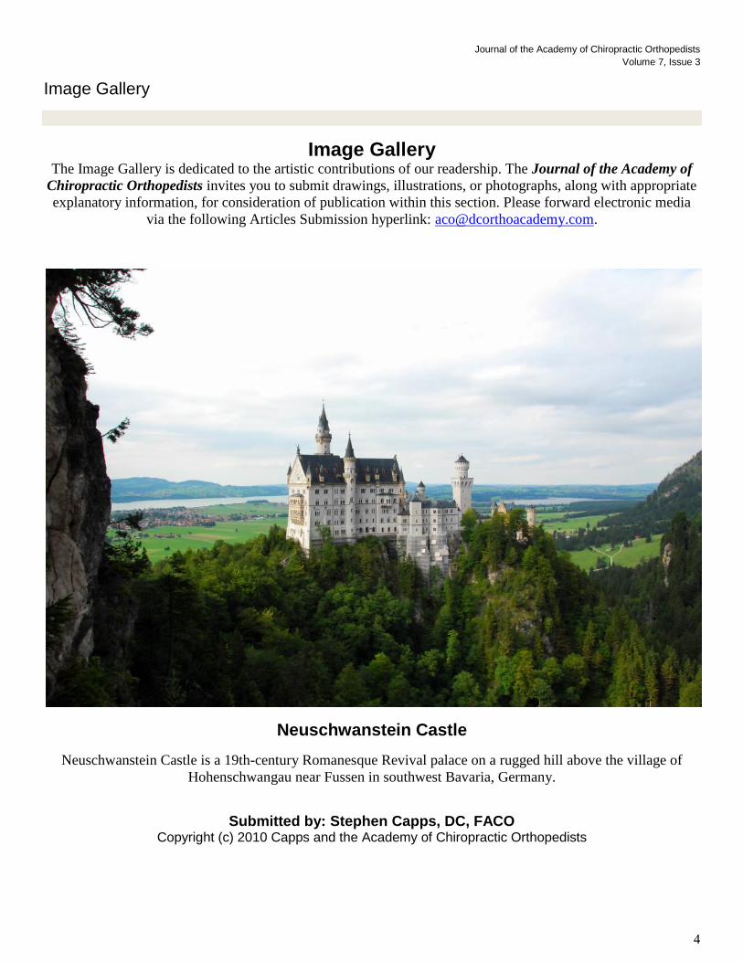
Image Gallery
Image Gallery The Image Gallery is dedicated to the artistic contributions of our readership. The Journal of the Academy of
Chiropractic Orthopedists invites you to submit drawings, illustrations, or photographs, along with appropriate
explanatory information, for consideration of publication within this section. Please forward electronic media
via the following Articles Submission hyperlink: [email protected].
Neuschwanstein Castle
Neuschwanstein Castle is a 19th-century Romanesque Revival palace on a rugged hill above the village of
Hohenschwangau near Fussen in southwest Bavaria, Germany.
Submitted by: Stephen Capps, DC, FACO Copyright (c) 2010 Capps and the Academy of Chiropractic Orthopedists
Journal of the Academy of Chiropractic Orthopedists
Volume 7, Issue 3
5

Journal of the Academy of Chiropractic Orthopedists
Volume 7, Issue 3
6
Diagnostic Imaging Corner
Case challenge
Michelle A Wessely (1) and Timothy J Mick (2)
(1) Director of Radiology (Paris/Toulouse), Department of Radiology, Institut Franco-Europeen de
Chiropratique (IFEC), 24 Boulevard Paul Vaillant Couturier, 94200 Ivry Sur Seine, France
(2) Center for Diagnostic Imaging (CDI), and Imaging Consultants, Inc.565 Arlington Avenue West, St
Paul 55117, Minnesota, USA [email protected]
Published: Journal of the Academy of Chiropractic Orthopedists
September 2010, Volume 7, Issue 3 Received: August, 2010 Accepted: August, 2010
The original article copyright belongs to the original publisher. This review is available from: http://www.dcorthoacademy.com.
© 2010 Wessely/Mick and the Academy of Chiropractic Orthopedists. This is an Open Access article which permits unrestricted use, distribution, and reproduction in any medium, provided the original work is properly cited.
Case History
A 52-year-old male presented with a sit down type
fall recently, resulting in severe back pain, centered
around L1. There is also a history of long-standing
prior trauma related to motorcycle accident, with no
focal upper lumbar pain at that time. The
chiropractor performed imaging (Figures 1-3).
Figure 1.
Figure 2.
Figure 3.
Journal of the Academy of Chiropractic Orthopedists
Volume 7, Issue 3
7
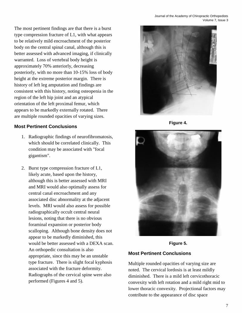
The most pertinent findings are that there is a burst
type compression fracture of L1, with what appears
to be relatively mild encroachment of the posterior
body on the central spinal canal, although this is
better assessed with advanced imaging, if clinically
warranted. Loss of vertebral body height is
approximately 70% anteriorly, decreasing
posteriorly, with no more than 10-15% loss of body
height at the extreme posterior margin. There is
history of left leg amputation and findings are
consistent with this history, noting osteopenia in the
region of the left hip joint and an atypical
orientation of the left proximal femur, which
appears to be markedly externally rotated. There
are multiple rounded opacities of varying sizes.
Most Pertinent Conclusions
1. Radiographic findings of neurofibromatosis,
which should be correlated clinically. This
condition may be associated with "focal
gigantism".
2. Burst type compression fracture of L1,
likely acute, based upon the history,
although this is better assessed with MRI
and MRI would also optimally assess for
central canal encroachment and any
associated disc abnormality at the adjacent
levels. MRI would also assess for possible
radiographically occult central neural
lesions, noting that there is no obvious
foraminal expansion or posterior body
scalloping. Although bone density does not
appear to be markedly diminished, this
would be better assessed with a DEXA scan.
An orthopedic consultation is also
appropriate, since this may be an unstable
type fracture. There is slight focal kyphosis
associated with the fracture deformity.
Radiographs of the cervical spine were also
performed (Figures 4 and 5).
Figure 4.
Figure 5.
Most Pertinent Conclusions
Multiple rounded opacities of varying size are
noted. The cervical lordosis is at least mildly
diminished. There is a mild left cervicothoracic
convexity with left rotation and a mild right mid to
lower thoracic convexity. Projectional factors may
contribute to the appearance of disc space
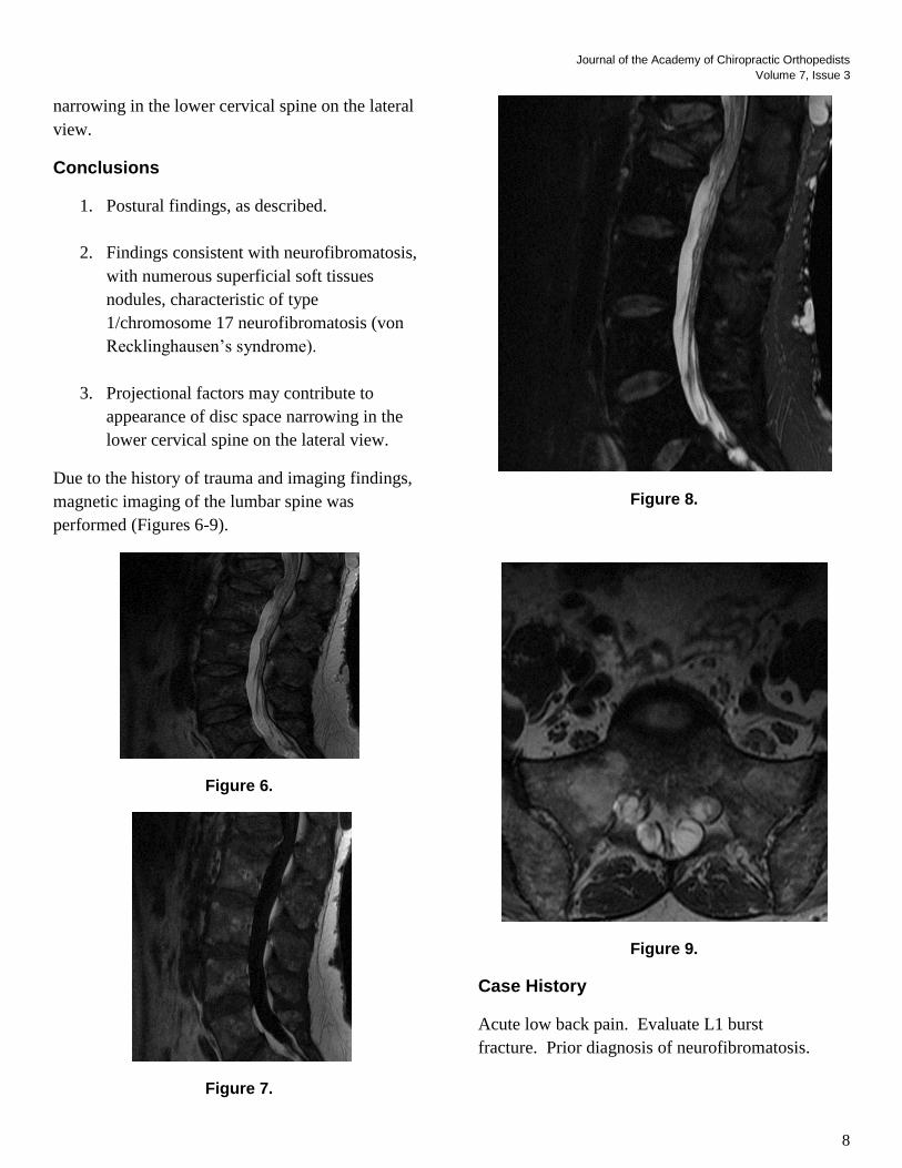
Journal of the Academy of Chiropractic Orthopedists
Volume 7, Issue 3
8
narrowing in the lower cervical spine on the lateral
view.
Conclusions
1. Postural findings, as described.
2. Findings consistent with neurofibromatosis,
with numerous superficial soft tissues
nodules, characteristic of type
1/chromosome 17 neurofibromatosis (von
Recklinghausen’s syndrome).
3. Projectional factors may contribute to
appearance of disc space narrowing in the
lower cervical spine on the lateral view.
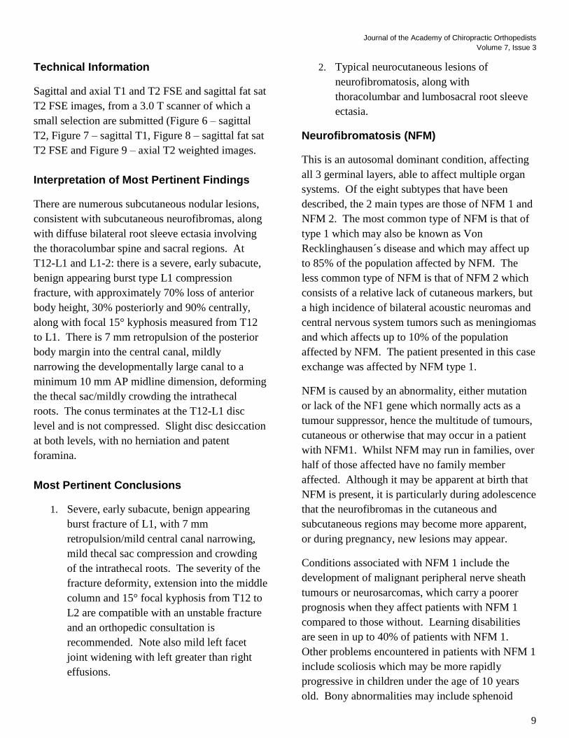
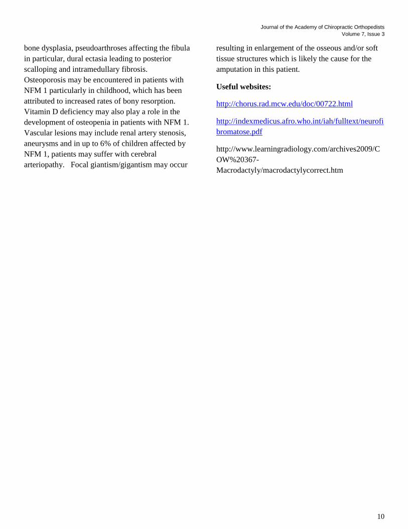
Due to the history of trauma and imaging findings,
magnetic imaging of the lumbar spine was
performed (Figures 6-9).
Figure 6.
Figure 7.
Figure 8.
Figure 9.
Case History
Acute low back pain. Evaluate L1 burst
fracture. Prior diagnosis of neurofibromatosis.
Journal of the Academy of Chiropractic Orthopedists
Volume 7, Issue 3
9
Technical Information
Sagittal and axial T1 and T2 FSE and sagittal fat sat
T2 FSE images, from a 3.0 T scanner of which a
small selection are submitted (Figure 6 – sagittal
T2, Figure 7 – sagittal T1, Figure 8 – sagittal fat sat
T2 FSE and Figure 9 – axial T2 weighted images.
Interpretation of Most Pertinent Findings
There are numerous subcutaneous nodular lesions,
consistent with subcutaneous neurofibromas, along
with diffuse bilateral root sleeve ectasia involving
the thoracolumbar spine and sacral regions. At
T12-L1 and L1-2: there is a severe, early subacute,
benign appearing burst type L1 compression
fracture, with approximately 70% loss of anterior
body height, 30% posteriorly and 90% centrally,
along with focal 15° kyphosis measured from T12
to L1. There is 7 mm retropulsion of the posterior
body margin into the central canal, mildly
narrowing the developmentally large canal to a
minimum 10 mm AP midline dimension, deforming
the thecal sac/mildly crowding the intrathecal
roots. The conus terminates at the T12-L1 disc
level and is not compressed. Slight disc desiccation
at both levels, with no herniation and patent
foramina.
Most Pertinent Conclusions
1. Severe, early subacute, benign appearing
burst fracture of L1, with 7 mm
retropulsion/mild central canal narrowing,
mild thecal sac compression and crowding
of the intrathecal roots. The severity of the
fracture deformity, extension into the middle
column and 15° focal kyphosis from T12 to
L2 are compatible with an unstable fracture
and an orthopedic consultation is
recommended. Note also mild left facet
joint widening with left greater than right
effusions.
2. Typical neurocutaneous lesions of
neurofibromatosis, along with
thoracolumbar and lumbosacral root sleeve
ectasia.
Neurofibromatosis (NFM)
This is an autosomal dominant condition, affecting
all 3 germinal layers, able to affect multiple organ
systems. Of the eight subtypes that have been
described, the 2 main types are those of NFM 1 and
NFM 2. The most common type of NFM is that of
type 1 which may also be known as Von
Recklinghausen´s disease and which may affect up
to 85% of the population affected by NFM. The
less common type of NFM is that of NFM 2 which
consists of a relative lack of cutaneous markers, but
a high incidence of bilateral acoustic neuromas and
central nervous system tumors such as meningiomas
and which affects up to 10% of the population
affected by NFM. The patient presented in this case
exchange was affected by NFM type 1.
NFM is caused by an abnormality, either mutation
or lack of the NF1 gene which normally acts as a
tumour suppressor, hence the multitude of tumours,
cutaneous or otherwise that may occur in a patient
with NFM1. Whilst NFM may run in families, over
half of those affected have no family member
affected. Although it may be apparent at birth that
NFM is present, it is particularly during adolescence
that the neurofibromas in the cutaneous and
subcutaneous regions may become more apparent,
or during pregnancy, new lesions may appear.
Conditions associated with NFM 1 include the
development of malignant peripheral nerve sheath
tumours or neurosarcomas, which carry a poorer
prognosis when they affect patients with NFM 1
compared to those without. Learning disabilities
are seen in up to 40% of patients with NFM 1.
Other problems encountered in patients with NFM 1
include scoliosis which may be more rapidly
progressive in children under the age of 10 years
old. Bony abnormalities may include sphenoid
Journal of the Academy of Chiropractic Orthopedists
Volume 7, Issue 3
10
bone dysplasia, pseudoarthroses affecting the fibula
in particular, dural ectasia leading to posterior
scalloping and intramedullary fibrosis.
Osteoporosis may be encountered in patients with
NFM 1 particularly in childhood, which has been
attributed to increased rates of bony resorption.
Vitamin D deficiency may also play a role in the
development of osteopenia in patients with NFM 1.
Vascular lesions may include renal artery stenosis,
aneurysms and in up to 6% of children affected by
NFM 1, patients may suffer with cerebral
arteriopathy. Focal giantism/gigantism may occur
resulting in enlargement of the osseous and/or soft
tissue structures which is likely the cause for the
amputation in this patient.
Useful websites:
http://chorus.rad.mcw.edu/doc/00722.html
http://indexmedicus.afro.who.int/iah/fulltext/neurofi
bromatose.pdf
http://www.learningradiology.com/archives2009/C
OW%20367-
Macrodactyly/macrodactylycorrect.htm
Journal of the Academy of Chiropractic Orthopedists
Volume 7, Issue 3
11
Abstracts & Literature Review
Humeral Insertion of the Supraspinatus and Infraspinatus
New Anatomical Findings Regarding the Footprint of the Rotator Cuff
Tomoyuki Mochizuki, MD, Hiroyuki Sugaya, MD, Mari Uomizu, MD, KazuhikoMaeda, MD, KeisukeMatsuki,MD
Ichiro Sekiya, MD, TakeshiMuneta, MD, and Keiichi Akita, MD
J Bone Joint Surg Am. 2008;90:962-9
Copyright 2008 By The Journal of Bone and Joint Surgery, Incorporated
JACO Editorial Reviewer: Dale G. Huntington, DC, FACO
Published: Journal of the Academy of Chiropractic Orthopedists
September 2010, Volume 7, Issue 3 Received: August, 2010 Accepted: August, 2010
The original article copyright belongs to the original publisher. This review is available from: http://www.dcorthoacademy.com.
© 2010 Huntington and the Academy of Chiropractic Orthopedists. This is an Open Access article which permits unrestricted use, distribution, and reproduction in any medium, provided the original work is properly cited.
Authors’ Abstract
Background: It is generally believed that the
supraspinatus is the most commonly involved
tendon in rotator cuff tears. Clinically, however,
atrophy of the infraspinatus muscle is frequently
observed in patients with even small to medium-
size rotator cuff tears. This fact cannot be fully
explained by our current understanding of the
anatomical insertions of the supraspinatus and
infraspinatus. The purpose of this study was to
reinvestigate the humeral insertions of these
tendons.
Methods: The study included 113 shoulders from
sixty-four cadavers. The humeral insertion areas of
the supraspinatus and infraspinatus were
investigated in ninety-seven specimens. In sixteen
specimens, all muscular portions of the
supraspinatus and infraspinatus were removed,
leaving the tendinous portions intact, in order to
define the specific characteristics of the tendinous
portion of the muscles. Another twenty-six
shoulders were used to obtain precise measurements
of the footprints of the supraspinatus and
infraspinatus.
Results: The supraspinatus had a long tendinous
portion in the anterior half of the muscle, which
always inserted into the anterior most area of the
highest impression on the greater tuberosity and
which inserted into the superior most area of the
lesser tuberosity in 21% of the specimens. The
footprint of the supraspinatus was triangular in
shape, with an average maximum medial-to-lateral
length of 6.9 mm and an average maximum
anteroposterior width of 12.6 mm. The infraspinatus
had a long tendinous portion in the superior half of
the muscle, which curved anteriorly and extended to
the anterolateral area of the highest impression of
the greater tuberosity. The footprint of the
infraspinatus was trapezoidal in shape, with an
average maximum medial-to-lateral length of 10.2
mm and an average maximum anteroposterior width
of 32.7 mm.
Conclusions: The footprint of the supraspinatus on
the greater tuberosity is much smaller than
Journal of the Academy of Chiropractic Orthopedists
Volume 7, Issue 3
12
previously believed, and this area of the greater
tuberosity is actually occupied by a substantial
amount of the infraspinatus.
Clinical Relevance: The present study suggests that
rotator cuff tears that were previously thought to
involve only the supraspinatus tendon may in fact
have had a substantial infraspinatus component as
well.
JACO Editorial Summary:
The article was written by authors from the
Unit of Clinical Anatomy, Graduate School,
Tokyo Medical and Dental University,
Tokyo, Japan where the research was
conducted.
The purpose of the study was to reevaluate
macroscopically the humeral insertions and
tendinous structures of the supraspinatus
and infraspinatus in cadaver shoulders.
The investigative researchers provide an in
depth anatomical dissection of 113
shoulders and 64 cadavers average of 77.3
years at the time of death in an attempt to
explore and define the footprint of the
supraspinatus and infraspinatus muscles a
part of the rotator cuff group.
It is generally believed the supraspinatus is
the most commonly involved tendon in
rotator cuff tears, however clinically the
infraspinatus has shown atrophy in patients
with even small to medium cuff
tears.Currently tears are assessed
preoperatively with the use of
ultrasonography and magnetic resonance
imaging and are diagnosed on the basis of
intraoperative findings.
Most anatomy textbooks state the
supraspinatus inserts into the highest
impression or the greater tuberosity of the
humerus and the infraspinatus into the
middle impression of the greater tuberosity .
However the difficulty separating these
tendons and delineating their footprints
because of their interdigitated fibers
overlapping one another has been
problematic.. However in all specimens,
after removal of the coraco-humeral
ligament and the loose connective tissues
overlying the supraspinatus and
infraspinatus near their insertions could be
clearly traced. The infraspinatus was found
to occupy about half of the highest
impression and all of the middle impression
of the greater tuberosity of the humeral
head. The supraspinatus was found to be
inserted into the highest impression of the
greater tuberosity as well as the lesser
tuberosity. The infraspinatus was not.
The anatomical findings in the study suggest
that surgeons need to have increased
awareness of pathological conditions of the
infraspinatus tendon especially when
delaminating is observed. It may be
important for surgeons to incorporate these
new anatomical findings in order to properly
restore the geography of the footprint of the
torn anterior rotator cuff.
The supraspinatus has traditionally been
considered to be an important abductor
among the rotator cuff muscles. However
several researchers have reported that the
infraspinatus contributes as much to
abduction as does the supraspinatus.
Summary:
o The results of this investigation
should raise awareness in assisting
the orthopedic surgeons as well as
the treating non-surgical team of
therapists including chiropractic
physicians and/or orthopedic
specialists to better focus on the
conditions/injury to the rotator cuff
and improved rehabilitation methods
allowing for the opportunity of
maximizing outcomes.
Journal of the Academy of Chiropractic Orthopedists
Volume 7, Issue 3
13
Abstracts & Literature Review
Cervical Radiculopathy vs Parsonage–Turner Syndrome: a Case Report
Joseph H. Feinberg, MD, David A. Doward, MD, Alita Gonsalves, MD
HSSJ (2007) 3: 106–111. Published online: 22 December 2006 © Hospital for Special Surgery 2006
JACO Editorial Reviewer: Gregory C. Priest, DC, FACO
Published: Journal of the Academy of Chiropractic Orthopedists
September 2010, Volume 7, Issue 3 Received: September, 2010 Accepted: September, 2010
The original article copyright belongs to the original publisher. This review is available from: http://www.dcorthoacademy.com.
© 2010 Gregory C. Priest, DC, FACO and the Academy of Chiropractic Orthopedists. This is an Open Access article which permits unrestricted use, distribution, and reproduction in any medium, provided the original work is properly cited.
Authors’ Abstract:
No Abstract was provided by the authors.
JACO Editorial Reviewer’s Abstract: This is a
case report of an atraumatic 42-year-old recreational
cyclist that presented to the Physiatry Department
of the Hospital for Special Surgery in New York,
NY with a 3-month history of right-sided neck pain,
right periscapular pain, severe headaches and right
upper extremity numbness and weakness. Her
physical examination was felt to be suggestive of
possible cervical radiculopathy and/or Parsonage-
Turner syndrome, and electrodiagnostic testing was
notable for findings consistent with C7
radiculopathy. Cervical spine and head CT studies
were found to be normal. Two cervical MRI studies
were completed, the first of which was reportedly
normal and the second of which was reported to
have revealed degenerative changes but did not
demonstrate significant nerve root compression.
The patient underwent a course of treatment which
included medications and physical therapy. Her
symptoms completely resolved over the course of a
number of months, and the authors noted that at
one-year follow-up the patient remained symptom-
free with almost full strength and had returned to all
previous activities, including recreational cycling.
Parsonage-Turner syndrome is one of the more
common idiopathic causes of atraumatic brachial
plexopathy and was originally described in 1948
although a patient with similar clinical findings was
reported in 1897. Parsonage-Turner syndrome is
also known as brachial plexitis, brachial neuritis,
acute brachial plexus neuropathy and neuralgic
amyotrophy.
The etiology of Parsonage-Turner syndrome is in
dispute, with 25% of occurrences reported after
viral infection, and 15% after immunization. Other
possible factors are reported to include injury to a
remote area, as well as post-exercise and post-
surgical occurrences.
JACO Editorial Summary:
As chiropractic physicians, we frequently
encounter patients with symptomatology
suggestive of cervical radiculopathy, and we
would do well to consider in our differential
diagnosis the possibility that these patients may
be suffering from Parsonage-Turner syndrome.
The authors acknowledge the similarity
between cervical radiculopathy and Parsonage-
Turner syndrome, and provide an excellent list
of differential diagnoses to consider when we
Journal of the Academy of Chiropractic Orthopedists
Volume 7, Issue 3
14
encounter a patient that presents with these
signs and symptomatology.
It was noted that the authors have evaluated
many patients who were diagnosed with
cervical radiculopathy but were without MRI
evidence of nerve root compression, and in fact
the authors commented that some of these
patients may have had undiagnosed Parsonage-
Turner syndrome. In particular, the authors
suggest that the diagnosis of Parsonage-Turner
syndrome or a variant thereof should be
considered when evaluating patients that
present with radicular symptoms in the
presence of poor anatomic correlation on
imaging studies.
The authors posit that a short course of antiviral
medication may play a role when the diagnosis is
unclear in a patient presenting with radicular
symptoms in the lack of sufficient anatomic
correlation on imaging.
This reviewer is of the opinion that this article,
which demonstrates the diagnostic conundrum
presented by patients with radicular symptoms
without anatomic correlation on imaging
studies, illustrates beautifully the truth of the
aphorism that as prudent physicians, we treat
the patient standing before us, not the imaging
study.
Journal of the Academy of Chiropractic Orthopedists
Volume 7, Issue 3
15
Abstracts & Literature Review
The Effect of Backpacks on the Lumbar Spine in Children: A Standing Magnetic
Resonance Imaging Study
Neuschwander, Timothy B. MD; Cutrone, John MD; Macias, Brandon R. BA; Cutrone, Samantha; Murthy, Gita
PhD; Chambers, Henry MD; Hargens, Alan R. MD
Spine: 1 January 2010 - Volume 35 - Issue 1 - pp 83-88. ©2009, Lippincott Williams & Wilkins
JACO Editorial Reviewer: Ronald Evans, DC, FACO
Published:
Journal of the Academy of Chiropractic Orthopedists September 2010, Volume 7, Issue 3
Received: August, 2010 Accepted: August, 2010
The original article copyright belongs to the original publisher. This review is available from: http://www.dcorthoacademy.com.
© 2010 Ronald Evans, DC, FACO and the Academy of Chiropractic Orthopedists. This is an Open Access article which permits unrestricted use, distribution, and reproduction in any medium, provided the original work is properly cited.
Author’s Abstract
Study Design: This study is a repeated measures
design to measure the lumbar spine response to
typical school backpack loads in healthy children.
The lumbar spine in this setting was measured for
the first time by an upright magnetic resonance
imaging (MRI) scanner.
Objective: The purpose of this study is to measure
the lumbar spine response to typical school
backpack loads in healthy children. We hypothesize
that backpack loads significantly increase disc
compression and lumbar curvature.
Summary of Background Data: Children commonly
carry school backpacks of 10% to 22% bodyweight.
Despite growing concern among parents about
safety, there are no imaging studies which describe
the effect of backpack loads on the spine in
children.
Methods: Three boys and 5 girls, age 11 ± 2 years
(mean ± SD) underwent T2 weighted sagittal and
coronal MRI scans of the lumbar spine while
standing. Scans were repeated with 4, 8, and 12 kg
backpack loads, which represented approximately
10%, 20%, and 30% body weight for our sample.
Main outcome measures were disc compression,
defined as post- minus preloading disc height, and
lumbar asymmetry, defined as the coronal Cobb
angle between the superior endplates of S1 and L1.
Results: Increasing backpack loads significantly
compressed lumbar disc heights measured in the
midline sagittal plane (P < 0.05, repeated-measures
analysis of variance [ANOVA]). Lumbar
asymmetry was: 2.23° ± 1.07° standing, 5.46° ±
2.50° with 4 kg, 9.18° ± 2.25° with 8 kg, and 5.68°
± 1.76° with 12 kg (mean ± SE). Backpack loads
significantly increased lumbar asymmetry (P <
0.03, one-way ANOVA). Four of the 8 subjects had
Cobb angles greater than 10° during 8-kg backpack
loads. Using a visual-analogue scale to rate their
pain (0-no pain, 10-worst pain imaginable), subjects
reported significant increases in back pain
associated with backpack loads of 4, 8, and 12 kg (P
< 0.001, 1-way ANOVA).
Conclusion: Backpack loads are responsible for a
significant amount of back pain in children, which
Journal of the Academy of Chiropractic Orthopedists
Volume 7, Issue 3
16
in part, may be due to changes in lumbar disc height
or curvature. This is the first upright MRI study to
document reduced disc height and greater lumbar
asymmetry for common backpack loads in children.
JACO Editorial Summary:
The article was written by authors from the
Department of Orthopaedic Surgery, University
of California, San Diego, TrueMRI, San Diego,
and the Rady Children's Hospital, San Diego.
The authors revisit lumbar spine response to
typical school backpack loads in healthy
children, however, this time, adding for the first
time, upright MRI imaging. References for this
work span from 1985 to 2008.
The authors hypothesize that backpack loads
significantly increase disc compression and
lumbar curvature. Upright scans viewed the
effects of 4, 8 and 12 kg backpack loads,
representing approximately 10%, 20% and 30%
of body weight of participants.
Increasing backpack loads significantly
compressed lumbar disc heights measured in the
midline sagittal plane; significantly increased
lumbar asymmetry; and are responsible for a
significant amount of back pain in children.
This is the first upright MRI study to document
reduced disc height and greater lumbar
asymmetry for common backpack loads in
children. Kimura S, Steinbach GC,
Watenpaugh DE, Hargens AR previously
explored this measurement in recumbent MRI
studies, reporting their findings in Lumbar
Spine Disc Height And Curvature Responses
To An Axial Load Generated By A
Compression Device Compatible With
Magnetic Resonance Imaging, Spine, Vol 26 #
23, pages 2596-2600, 2001. In this study,
Kimura, et al, used a compression device
compatible with magnetic resonance imaging, to
test two hypotheses: Axial loading of 50% body
weight from shoulder to feet in supine posture
1) simulates the upright lumbar spine alignment
and 2) decreases disc height significantly.
Neuschwander, et al, are the first investigators
to demonstrate loss of disc height in upright
MRI positioning, and under axial loading with
backpacks.
In 1995, Harreby, et al first questioned
adolescent risk factors for adult LBP, in Are
Radiologic Changes In The Thoracic And
Lumbar Spine Of Adolescents Risk Factors
For Low Back Pain In Adults?: A 25-Year
Prospective Cohort Study Of 640 School
Children, Spine Vol 20, #21, 1995, pages 2298-
2302. This study suggests that low back pain in
the growth period is 'a real problem,' with a
trend toward aggravation as time passes. Thus,
implementing preventive measures in schools
may be very important.
While in 1995, adolescent risk factors were
suggested as contributors trending toward adult
LBP, the Neuschwander 2009 study
categorically identifies loading effects on the
lumbar spine, which, as they become chronic,
lead toward the breakdown of spine
biomechanical properties. The ultimate effect is
probable chronic adult low back pain. Harreby
advocated prevention. With the Neuschwander
data, prevention is penultimate.
Journal of the Academy of Chiropractic Orthopedists
Volume 7, Issue 3
17
Abstracts & Literature Review
Indications for Computed Tomography In Patients With Minor Head Injury
Michelle J. Haydel, M.D.; Charles A. Preston, M.D.; Trevor J. Mills, M.D.; Samuel Luber, B.A.; Erick Blaudeau, M.D.; and Peter M..C. DeBlieux, M.D.
The New England Journal of Medicine 2000;343:100-5
© 2000 Massachusetts Medical Society
JACO Editorial Reviewer: Gary L. Carver, DC, FACO
Published: Journal of the Academy of Chiropractic Orthopedists
September 2010, Volume 7, Issue 3 Received: August, 2010 Accepted: August, 2010
The original article copyright belongs to the original publisher. This review is available from: http://www.dcorthoacademy.com.
© 2010 Gary L. Carver and the Academy of Chiropractic Orthopedists. This is an Open Access article which permits unrestricted use, distribution, and reproduction in any medium, provided the original work is properly cited.
Authors’ Abstract:
Background: Computed tomography (CT) is a
widely used screening test in patients with minor
head injuries even though the results are often
normal. A study was performed to develop and
validate clinical criteria’s to identify patients with
minor head injuries who do not need to undergo CT
Methods: The study consists of two phases. The
first phase consisted of 520 consecutive patients
with minor head injury who had normal score on
the Glasgow Coma Scale and normal finings on the
brief neurologic examinations and were given a CT.
Using recursive partitioning the study derived a set
of criteria to identify all patients who had
abnormalities of CT scanning. The second phase ,
the sensitivity and specificity of the criteria for
predicting a positive scan were evaluated in a group
of 909 patients.
Results: Of the 520 patients in the first phase, 36
(6.9 percent) had a positive scan. All patients with
positive CT scans had one or more of the following
seven findings: headache, vomiting, as age over 60
years, drug/alcohol intoxication, deficits in short-
term memory, physical evidence of trauma above
clavicles and seizure. Second phase consisted of
909 patients in which 57 (6.3 percent) patients had
positive scans. This group experienced the
sensitivity of the seven findings combined with 100
percent (95 confidence interval, 95 to 100 percent).
All patients with positive CT scans had at least one
of the seven findings.
Conclusions: Patients with minor head injury, the
use of CT scan can be safely limited to those
patients who have certain clinical findings (NEngl J
Med 2000; 343:100-5).
JACO Editorial Summary:
The authors performed a study involving
1429 patients in two different method phases
to arrive at their conclusion.
The authors basically used consultations,
clinical findings along with Glasgow Coma
Scale as criteria’s to determine minor head
injury and the use of CT scans.
The question regarding the criteria’s for
performing CT on head traumas has been
controversial since the 1970’s when
computed tomography were introduced.
Journal of the Academy of Chiropractic Orthopedists
Volume 7, Issue 3
18
Initially CT was reserved for severely
injured patients.
Between the 1970’s and the 1980’s minor
head injuries CT evaluations became more
common for patients with intracranial
lesions.
In the early 1990’s several retrospective
studies of patients with minor head injury
reported substantial proportions with
intracranial lesions on CT evaluations (17 to
20 percent).
These studies included patients with scores
of 13-15 and a level 3 on the Glasgow Coma
Scale.
Glasgow Coma Scale score of 13-15
indicated little to no impairment in
consciousness. A Glasgow Coma Scale of
15 indicates normal motor and verbal
responses and normal eye opening.
Glasgow Coma Scale score of 3 indicates no
motor or verbal response and no opening of
the eyes.
The authors of the 1990’s retrospective
studies concluded that CT was indicated in
all patients with minor head injury.
Subsequent prospective studies involving a
Glasgow Coma Scale with a score of 15 ,the
rate of intracranial lesions on CT evaluation
was much lower (6-9 percent).
Several studies have been performed in
evaluating clinical finding as a tool in
finding predictors of intracranial lesions in
patients with minor head injury.
In two studies were selective use of CT on
basis of clinical findings indentified
96percent and 98 percent of the patients
with abnormalities’ on CT scanning’s
None of the patients that had CT
abnormalities and who did not have
specified clinical findings required
neurosurgery.
A study was conducted to derive and
validate a set of clinical criteria that could be
use to identify patients with minor head
injury in whom CT could be forgone.
Two phases of studies were performed to
determine criteria’s standards in the use of
CT evaluation of minor head injury.
An independent radiologist randomly
evaluated 50 of the studies which were
performed by the studies radiologists.
There were three clinical findings
significantly associated with a positive CT
scan. 1. Short –term memory deficits 2.
Drug or alcohol intoxication. 3. Physical
evidence of trauma above the clavicles.
Study reveal recursive-partitioning analysis
yielded seven findings which identified all
patients with positive CT scans. 1.
Headache. 2. Over age of 60. 3 Vomiting. 4.
Drug or alcohol intoxication. 5. Deficits in
short-term memory. 6.Physical evidence of
trauma above the clavicles. 7. Seizures.
All patients with a positive CT scan had at
least one of the seven findings.
Physical examination findings that have
been associated with positive CT scan are
linear, basilar, depressed skull fracture, scalp
hematoma and soft tissue injury.
This study will assist the physician in his or
her clinical knowledge and criteria’s of
evaluating and ordering CT scans for the
benefit of their patients.
Discussion: Data shows that approximately two thirds of
patients with head trauma in the United States are
Journal of the Academy of Chiropractic Orthopedists
Volume 7, Issue 3
19
classified as having minor head injury; out of this group
less than ten percent have positive findings on CT scans
and less than one percent requires neurosurgical
intervention. Several studies have concluded that
patients having normal neurological findings and normal
CT scans can be safely discharged form emergency
departments.
Journal of the Academy of Chiropractic Orthopedists
Volume 7, Issue 3
20
Abstracts & Literature Review
Current Concepts Review The Assessment of Fracture Risk
Aasis Unnanuntana, MD, Brian P. Gladnick, BA, Eve Donnelly, PhD, and Joseph M. Lane, MD
J Bone Joint Surg Am. 2010;92:743-53
© 2010 By The Journal Of Bone And Joint Surgery, Incorporated
JACO Editorial Reviewer: Richard P. Corbett, DC, FCCO(C)
Published: Journal of the Academy of Chiropractic Orthopedists
September 2010, Volume 7, Issue 3 Received: August, 2010 Accepted: August, 2010
The original article copyright belongs to the original publisher. This review is available from: http://www.dcorthoacademy.com.
© 2010 Richard P. Corbett and the Academy of Chiropractic Orthopedists. This is an Open Access article which permits unrestricted use, distribution, and reproduction in any medium, provided the original work is properly cited.
Authors’ Abstract:
Background: Bone mineral density is considered to
be the standard measure for the diagnosis of
osteoporosis and the assessment of fracture risk.
The majority of fragility fractures occur in patients
with bone mineral density in the osteopenic range.
Methods: We review the parameters and methods
used to assess fracture risk, which include bone
mineral density as assessed with dual x-ray
absorptiometry, the Fracture Risk Assessment Tool
(FRAX), bone turnover, and biochemical bone
markers.
Results: The Fracture Risk Assessment Tool
(FRAX) can be used as an assessment modality for
the prediction of fractures on the basis of clinical
risk factors, with or without the use of femoral neck
bone mineral density. Treatment of osteoporosis
should be considered for patients with low bone
mineral density (a T-score of between 21.0 and
22.5) as well as a ten-year risk of hip fracture of a
greater than or equal to 3% or a ten-year risk of a
major osteoporosis-related fracture of greater than
or equal to 20% as assessed with the FRAX.
Biochemical bone markers are useful for monitoring
the efficacy of anti-resorptive or anabolic therapy
and may aid in identifying patients who have a high
risk of fracture.
Conclusions: An approach combining the
assessment of bone mineral density, clinical risk
factors for fracture with use of the FRAX, and bone
turnover markers will improve the prediction of
fracture risk and enhance the evaluation of patients
with osteoporosis.
JACO Editorial Summary:
The article was written by authors from
Department of Orthopaedic Surgery, Hospital
for Special Surgery; and Weill Cornell Medical
College, Cornell University.
The National Institutes of Health Consensus
Development Panel on Osteoporosis
Prevention, Diagnosis, and Therapy defines
osteoporosis as a skeletal disorder characterized
by low bone strength and increased risk of
fracture.
The authors provide a review of the factors that
contribute to bone strength, with a review of the
parameters and methods used to assess fracture
risk.
Journal of the Academy of Chiropractic Orthopedists
Volume 7, Issue 3
21
Notably, the authors reflect upon bone mineral
density, the Fracture Risk Assessment Tool
(FRAX), bone turnover, and biochemical bone
markers.
“On the basis of a series of meta-analyses
undertaken to identify clinical risk factors for
osteoporosis, the Fracture Risk Assessment
Tool (FRAX) was developed. FRAX, released
in 2008 by the World Health Organization, was
developed and validated under the direction of
Professor John Kanis with the support of many
individuals and organizations including the
American Society for Bone and Mineral
Research, the National Osteoporosis
Foundation, the International Society for
Clinical Densitometry, and the International
Osteoporosis Foundation.”
FRAX is currently available online at
www.shef.ac.uk/FRAX
The authors report that bone mineral density
reflects only one component of bone strength,
and that FRAX can be used with or without the
use of femoral neck bone mineral density, for
the prediction of fractures on the basis of
clinical risk factors.
In the current article, the authors state that an
improvement in the prediction of fracture risk
might be seen by using a combination of the
assessment of bone mineral density, with use of
FRAX, along with bone turnover markers.
Journal of the Academy of Chiropractic Orthopedists
Volume 7, Issue 3
22
Announcements
American College of Chiropractic Orthopedists Annual Convention to be held in Las Vegas –
April 28-30, 2011
Mark your calendars for the 2011 ACCO convention that will be held April 27th-30th
, 2011. We will be
meeting at the new Tropicana in Las Vegas. The College has lowered the convention price. We have some
great speakers scheduled. Please note there will be a class on Thursday night and no class on Sunday.
The room rate is $125.00 on Friday and Saturday and $95.00 on Sunday through Thursday. There are no resort
parking fees. The hotel is close to the airport. There are no charges for children under 18 staying in rooms with
parents.
Please plan on joining us in Las Vegas in 2011. More information is forthcoming from the ACCO. Contact the
ACCO for information and to register.
Council of Chiropractic Orthopedists
Drs. Dale Huntington and Gary Carver will be attending the ACA/HOD annual meeting in Newport, RI on
September 29-October 2, 2010. They will be representing the Council on Chiropractic Orthopedics at the
meeting. Dr. Carver who will also be representing Missouri as its state delegate. Dr. Huntington will be
representing the orthopedic specialty on the Resolutions committee as well as the American Board of
Chiropractic Specialties.
Congress of Diplomates Meeting – April 30, 2011
The Congress of Diplomates will meet April 30, 2011 at the American College of Chiropractic Orthopedists
(ACCO) symposium in Las Vegas, NV.
The Academy would like to offer the specialty an opportunity to present interesting cases or discuss research
projects with other Diplomates. Diplomates are invited by this notice to present papers to the conference
attendees. These are 10-15 minutes presentations of cases or research work.
Contact Dr. Jerry Wildenauer at ([email protected]) to reserve your spot. This has always been well
received by the conference attendees. Don't wait, contact the Academy.
Editorial Review Board Annual Meeting
The Journal of the Academy of Chiropractic Orthopedists will convene an ERB meeting during the American
College of Chiropractic Orthopedists Annual Convention on April 28, 2011. All ERB members are cordially
invited to this meeting. More information is forthcoming from the Academy.

Journal of the Academy of Chiropractic Orthopedists
Volume 7, Issue 3
23
Visit the New Web Site of the
Academy of Chiropractic Orthopedists
www.DCOrthoAcademy.com
The Academy is working in a proactive manner as an advocate of
chiropractic patients, chiropractic orthopedists and the chiropractic
profession. Many advances and services are being offered by the
Academy including:
Chiropractic Orthopedic Diplomate Examination
Orthopedic Diplomate Re-Credentialing and Re-Certification
Membership among respected leaders of the chiropractic profession
Up to date database to inform Insurance Industry / Legal / Patient /
Consumers of your membership among elite professionals
The Journal of the Academy of Chiropractic Orthopedists (JACO) - A
peer-reviewed, indexed journal that promulgates academic excellence
and higher learning.
To Join or Renew your Membership as a Fellow of the Academy of
Chiropractic Orthopedists, go to:
http://www.dcorthoacademy.com/membership.php

Journal of the Academy of Chiropractic Orthopedists
Volume 7, Issue 3
24
To order the newly designed
Academy of Chiropractic Orthopedists’
Patient Education Brochure, purchase is now available at:
http://www.dcorthoacademy.com/store-pamphlet.php