Embed Size (px)
Citation preview

Geometry acquisition of a car occupant anatomy
SERRE T., BASILE F., BEHR M., DURAND F., BIDAL S., BRUNET C.
INTRODUCTION
INRETS-LBA (Laboratory of Applied Biomechanics) Director : Pr. J. BONNOIT
Faculte de Medecine, Boulevard P. Dramard 1 39 1 6 Marseille Cedex 20, France
The aim of this research was an anatomic study of the seated position in order to reply, in particular, to various questions raised in industry as how to predict the threshold of the main injuries that can be encountered in car accidents. lt shall also produce a basis for further injury mechanism models. This methodology is based on obtaining reference serial sections from an entire subject frozen in the driving position, as well as on computerised three-dimensional visualisation allowing the validation of the anatomic analysis. The Laboratory of Applied Biomechanics was working on this subject for many years<1><2> and this technology was used in the framework of the HUMOS (Human Model for Safety) European project.
METHODOLOGY
The data acquisition process can be divided into three main stages, ie the anatomical subject selection and slicing, the anatomic acquisition ( organs identification on the slices) and a final stage of three-dimension reconstruction.
Subject selection and slicing
l (
Anatomie Acquisition
3D Visualisation
Figu.re 1 : General process define by (JJ
Tue selected cadaver, close to the "50th percentile European male subject", was placed into the cockpit of a car in driving position (in collaboration with ECIA). Then the body was frozen in this position and placed into a foam-filled sarcophagus. In order to have the direction of the section plane as close as possible to conventional anatomic descriptions (ie sagittal, coronal or transversal), the entire body was divided into 5 different blocks (thorax-head, pelvis-abdomen, hands-arms, knees and foot-ankle). Reference slabs were attached to each block in order to ensure the integrity of the blocks and to define a reference system for the data acquisition. Blocks were sectioned each 7.Smm with a 2.5mm thick saw. Each slice were cleaned and photographed.
IRCOBI Conference - Sitges (Spain), September 1999 529
Anatomie acquisition
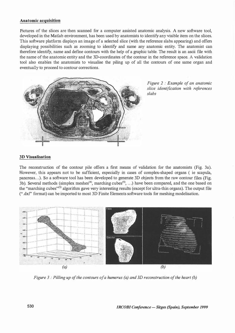
Pictures of the slices are then scanned for a computer assisted anatomic analysis. A new software tool, developed in the Matlab environment, has been used by anatomists to identify any visible item on the slices. This software platform displays an image of a selected slice (with the reference slabs appearing) and offers displaying possibilities such as zooming to identify and name any anatomic entity. The anatomist can therefore identify, name and define contours with the help of a graphic table. The result is an ascii file with the name of the anatomic entity and the 3D-coordinates of the contour in the reference space. A validation tool also enables the anatomists to visualise the piling up of all the contours of one same organ and eventually to proceed to contour corrections.
3D Visualisation
Figure 2 : Example of an anatomic slice identification with references slabs
The reconstruction of the contour pile offers a first means of validation for the anatomists (Fig. 3a). However, this appears not to be sufficient, especially in cases of complex-shaped organs ( ie scapula, pancreas„ .). So a software tool has been developed to generate 3D objects from the raw contour files (Fig. 3b). Several methods (simplex meshes<4l, marching cubes<5), „ .) have been compared, and the one based on the "marching cubes"<5l algorithm gave very interesting results (except for ultra-thin organs). The output file (" .dxf' format) can be imported to most 3D Finite Elements software tools for meshing modelisation .
. „ . ... . . ... .. .... . : " . „„
..
-··';"· -·„1.···-„„ ... 1. ·· ---··· ... .... „ . . :„.„„ ..... „. ___ ..tOO ••••••• ••
··:-•-....... : • ···1 ... 50 · · ····· · .5C() . „ ....
. .
.sso .. ········ .eoo •• •• . • ····: .e.50 •••. -····· . • -�·- •.. -rco . . . . „
.. . :. ·········· · r-·· · ··· , , , .... .... r·· ···· +
····
··· ·j __
··i ·· · · · ·· · ···· ··t ·· ·········· ······j
(a)
. .. :„ .......... 1········· .. � .. ·······� ... „ ..... •:„..
!
. ···················i······:::
··�.::::::·�:1
(b) Figure 3 : Pilling up ofthe contours of a humerus (a) and 3D reconstruction ofthe heart (b)
530 IRCOBI Conference - Sitges (Spain), September 1999

RESULTS
The results of this work is a complete anatomic and geometric database of a seated human body in driving position. The division of the cadaver into 7 .5mm thick slices allowed a software reconstruction of bones and organs with a particularly high resolution. Some anatomic details such as the transverse and spinous
processes (Figure 5) can be underlined. However, the reconstruction tool, based on "marching cube" algorithm, is not we11 adapted for thin organs like diaphragm. This database a11ows to improve knowledge on the spatial arrangement of organs and interaction between them in this particular position. From an anatomic point of view, it is obvious that the lack of pressurisation of the cadaver and his passive musculature induce a decrease of some particular organs dimensions ( cardiovascular and pulmonary system, muscles, . . . ). However, it seems that this deficiency has no effect on orientation and position.
Figu.re 4: Visualisation of thoracic and abdominal organs, and head with cerebellum ,cerebrum and the brain stem.
Figu.re 5: General cu-rve of the spinal column and in detail the 12'h thoracic vertebra.
CONCLUSION
The creation of a geometric database of the human anatomy in the seated position opened up new possibilities in the fields of anatomy teaching and transports security, especially in terms of bio-mechanics. This methodology is used in the framework of the HUMOS European project and contributes to the
elaboration of more accurate numeric models.
IRCOBJ Conference - Sitges (Spain), September 1999 531

AKNOWLEDGEMENTS
We are grateful to C. Cavallero and M. Py from INRETS-LBA and people from ECIA, especially L. Chabert, for their implication in this project and particularly in the experimental work. Thanks go to P. Balandraud and G. Thiery who joined the medical team and got deeply involved in the anatomic analysis process. Tue HUMOS (Human Model for Safety) project is supported by the European Community (DG
XII) and co-ordinated by LAB PSA Peugeot Citroen RENAULT.
REFERENCES
1 S. Ghannouchi , A. Ghorbel, C. Cavallero, J. Bonnoit - Anatomy of the seated position : methodologic approach and initial findings - Surgical and Radiologie Anatomy Vol 1 5 N° 14 Springer-Verlag 1 993 - pp
3 1 5-3 19. 2 S. Ghannouchi - L'etre humain en position assise : etude anatomique et biomecanique, reconstruction tridimensionnelle - Thesis - ENSAM 1998. 3 L. Chabert, S.Ghannouchi, C.Cavalllero, J.Bonnoit - Anatomical study and three-dimensional reconstruction of the belted human body in seated position - 15th ESV Conference, Melbourne, 1 996. Paper n° 96-Sl 0-0-04. 4 Delingette H. - General object reconstruction based on simplex meshes - Febrary 97 - Rapport INRIA n°3 1 1 l . 5 W. Lorensen, H. Cline - Marching cubes : A high resolution 3D surface construction algorithm -
Computer Graphics Vol 2 1 N°4 1987 - pp 163-169.
532 IRCOBJ Conference - Sitges (Spain), September 1999


![IRC-20-82 IRCOBI conference 2020 · 2020. 7. 25. · IRC-20-82 IRCOBI conference 2020 709. pressures [21]. It is unclear whether the introduction of the instrumentation causelocal](https://img.pdfslide.us/doc/110x75/6117914610f9c764771658b0/irc-20-82-ircobi-conference-2020-7-25-irc-20-82-ircobi-conference-2020-709.jpg)


![IRC-20-37 IRCOBI conference 2020 · 2020. 7. 25. · IRC-20-37 IRCOBI conference 2020 231. evaluate ground impact patterns [15–17]. Pedestrian behaviour prior to crash has however](https://img.pdfslide.us/doc/110x75/611b96d4916d69193c362f09/irc-20-37-ircobi-conference-2020-7-25-irc-20-37-ircobi-conference-2020-231.jpg)

![IRC-19-92 IRCOBI conference 2019Martin Östling, Hanna Jeppsson, Nils Lubbe IRC-19-92 IRCOBI conference 2019 626 validated [21] a deterministic analysis method to predict future road](https://img.pdfslide.us/doc/110x75/60a56309ab4a3f476a041ce0/irc-19-92-ircobi-conference-martin-stling-hanna-jeppsson-nils-lubbe-irc-19-92.jpg)