Embed Size (px)
Citation preview
AQUATIC BIOLOGYAquat Biol
Vol. 12: 13–21, 2011doi: 10.3354/ab00312
Published online March 3
INTRODUCTION
In the marine environment, bacteria are the mostcommon colonizers on the surface of macroalgae (Arm-strong et al. 2000). Seaweed-associated bacteriasecrete biologically active, beneficial compounds thatregulate the morphogenesis of marine organisms andhelp them survive under varied environmental condi-tions. The nature of the bacterial–seaweed interactionplays an important role and can influence the growthand various developmental stages (e.g. reproduction)of algae. Bacteria are generally assumed to benefitfrom soluble organic matter (as a source of energy)
produced by host algae (Chandini et al. 2008). Bacter-ial biofilms of certain bacteria influence the settlementof zoospores of green alga of genus Enteromorpha(Patel et al. 2003). Spore release in the red algal genusAcrochaetium has also been reported to be enhancedby marine bacteria through N-acyl homoserine lactonesecretion (Weinberger et al. 2007). The plantletgrowth-promoting nature of bacterial isolates associ-ated with Laminaria japonica was reported by Dimitri-eva et al. (2006).
Members of Ulvaceae lose their typical foliose thal-lus morphology when cultured axenically in definedsynthetic media (Provasoli 1958, Provasoli & Pintner
© Inter-Research 2011 · www.int-res.com*Corresponding author. Email: [email protected]
Isolation of seaweed-associated bacteria and theirmorphogenesis-inducing capability in axenic
cultures of the green alga Ulva fasciata
Ravindra Pal Singh, Vaibhav A. Mantri, C. R. K. Reddy*, Bhavanath Jha
Discipline of Marine Biotechnology and Ecology, Central Salt and Marine Chemicals Research Institute, Council of Scientific and Industrial Research (CSIR), Bhavnagar 364021, India
ABSTRACT: In the marine environment, the bacteria that colonize the surface of a seaweed affect itsgrowth and development. In the laboratory, several seaweeds do not develop normal foliose thalli inaxenic cultures, but adopt an atypical morphology. Adding certain bacteria to the culture, however,can result in reversion to the normal morphology. The aim of the present study was to examine theeffects of various algal-associated bacteria on the growth, morphology, and reproduction of the greenalga Ulva fasciata. In axenic culture without added bacteria, U. fasciata grew into undifferentiatedtissue initially and later developed into a tubular thallus. Of the 53 bacterial isolates we obtained fromdifferent species of Ulva and Gracilaria, only 5 were capable of inducing differentiation and subse-quent growth in U. fasciata cultured in axenic conditions. One bacterial isolate (which we later iden-tified as Marinomonas sp.) was more effective than the others in inducing morphogenesis and growthin U. fasciata. The physical association of bacterial cells with thalli was found to be crucial forthe induction of U. fasciata foliose morphology, growth, and spore release. The culture filtrates fromthe 5 individual bacterial isolates and from a consortium of all 5 of them had less effect on morpho-genesis and growth of U. fasciata than the bacteria themselves had, but more effect than the control(no bacteria added). Analysis of partial 16S rRNA gene sequences from all 5 isolates with morpho-genesis-inducing ability led us to identify them as Marinomonas sp. and Bacillus spp. We found thatthe physical association of bacteria is essential for normal growth of the seaweed, suggesting a sym-biotic interaction between seaweed and bacteria.
KEY WORDS: Bacterial isolates · Culture filtrate · Gracilaria spp. · Morphogenesis · Seaweed ·Ulva fasciata
Resale or republication not permitted without written consent of the publisher
Aquat Biol 12: 13–21, 2011
1980). But their normal foliose morphology is restoredwhen they are co-cultured with bacterial isolates capa-ble of inducing differentiation. The aberrant morpho-logy was successfully reversed to the foliose thallus fol-lowing the inoculation of appropriate morphogenesis-inducing bacterial isolates to the culture media(Nakanishi et al. 1996). Similar findings have beenreported in studies with Ulva pertusa (Nakanishi et al.1996), U. linza (Marshall et al. 2006), U. mutabilis(Wichard & Oertel 2010), Enteromorpha linza (Fries1975), E. compressa (Fries & Aberg 1978), and Mono-stroma oxyspermum (Tatewaki et al. 1983, Matsuo etal. 2003). Direct physical contact between the algaland bacterial cells was shown to be a prerequisite forcomplete morphogenesis in U. pertusa (Nakanishi etal. 1999). Tatewaki et al. (1983) reported that the addi-tion of culture filtrate of morphogenesis-inducing bac-terial isolates was capable of providing the same effectas that caused by the addition of the bacterial cultureitself. Similarly, Marshall et al. (2006) reported that cul-ture filtrate restored the morphology of U. linza. It isnow well established that cytokinin produced bymarine bacteria is effective in regulating the morpho-genesis pattern in seaweeds (Maruyama et al. 1986,1988, Mooney & Van 1986).
Bacterial genera such as Cytophaga, Flavobacterium,Vibrio, Pseudomonas, Halomonas, Escherichia andBacillus have been implicated in promoting morpho-genesis in Ulva pertusa (Nakanishi et al. 1999). Further,it has been shown that morphogenesis in green macro-algae (Ulvaceae and Monostromaceae) is induced byspecies of Cytophaga, Flavobacterium, Bacillus andother species belonging to the phyla Firmicutes andBacteroidetes (Matsuo et al. 2005, Marshall et al. 2006).Along the Indian coast, the occurrence of the generaBacillus, Vibrio, Micrococcus, Flavobacterium, andCytophaga has been reported from the intertidal re-gion, where seaweeds are one of the dominant benthiccommunities (Lakshmanaperumalsamy & Purushotha-man 1982). However, the mechanistic pathways associ-ated with the induction of seaweed morphogenesis byassociated bacterial flora have not yet been investi-gated in detail.
Ulva fasciata is one of the most common green algaeand occurs almost every season on the Indian coast(Jha et al. 2009). The high protein content (26% on adry-weight basis) and nutritionally rich omega-3 fattyacids make this alga a potential source of food and feed(Oza & Rao 1977, Kumari et al. 2010). Previous re-search on this alga has dealt with several fundamentalaspects of biology, including phenology and ontogeny(Subbaramaiah 1970, Mantri et al. in press). Membersof the genus Ulva are popular in Japanese and Koreancuisine and are commercially cultivated in these coun-tries (Hiraoka & Oka 2008). But the effects of associ-
ated bacteria on this alga’s growth, morphology andreproduction have not been examined. The presentstudy describes the isolation and screening of 53 bacte-rial isolates obtained from Ulva and Gracilaria speciesand the bacteria’s morphogenesis-inducing ability inU. fasciata grown in axenic culture. Five potential bac-terial isolates having morphogenesis-inducing activitywere further tested for their induction of growth andreproduction of U. fasciata vegetative tissue in axenicculture. Partial 16S rRNA gene sequences of the 5strains with morphogenesis-inducing ability were usedto determine their correct taxonomic identity and phy-logenetic relationships. Further, the culture filtratesfrom these isolates were also tested for their ability toinduce U. fasciata morphogenesis and subsequentgrowth.
MATERIALS AND METHODS
Isolation of bacteria. Fifty-three bacterial isolateswere isolated from naturally collected thalli of Ulvafasciata, U. taeniata, U. lactuca, Gracilaria corticata, G.dura, and G. salicornia from the coast of Veraval(20° 54.87’ N, 70° 20.83’ E), India (Table 1). These spe-cies were found growing in association with each otherin the intertidal area and thus selected for the presentstudy. All the collections were made from pristine loca-tions, away from anthropogenic activities, during thelowest tide of the chart datum in August 2009. Thesamples were immediately brought to the laboratoryunder cool conditions, and about 100 mg tissue waswashed gently 3 to 4 times in autoclaved seawater on aBioclean bench (Sanyo). The cleaned algal tissue wasinoculated onto marine agar medium (2216; HiMediaLaboratories) to allow growth of associated bacteria.The plates were then incubated at 37°C for 2 d, andindividual colonies were picked off and furtherstreaked on marine agar plates in order to obtain sin-gle colonies. All bacterial isolates were further main-tained on marine agar slants at 4°C and used for thesubsequent experiments.
14
Algal species Abbreviation Number of bacterial isolates
Ulva fasciata UF 16U. lactuca UL 10U. taeniata UT 12Gracilaria dura GD 6G. corticata GC 5G. salicornia GS 4
Table 1. Number of bacteria isolated from Ulva and Gracilariaspp. collected at Veraval, India, in August 2009
Singh et al.: Morphogenesis-inducing bacteria on Ulva fasciata
Preparation of unialgal samples and axenic tissue.Healthy vegetative thalli of Ulva fasciata collectedfrom the intertidal area of the coast of Veraval wereused for obtaining unialgal cultures. Algal fronds werecut into small pieces of about 1 cm2 and were thor-oughly washed to remove adhering debris and otherepiphytic contamination. The cut thallus pieces werefurther cultured in conical flasks with 100 ml ofenriched seawater medium (MP1) supplemented with600 µl GeO2 (1 mg ml–1 stock) to eliminate diatomgrowth. The flasks with algal pieces were then main-tained in a Multi Thermo Incubator (MTI-202; Eyela) at25 ± 1°C temperature under daylight white fluorescentlamps at 15 µmol photon m–2 s–1 irradiance with a12:12 h light:dark photoperiod. After 2 to 3 d of ac-climatization, fronds were given sequential treatmentwith different chemicals to obtain an axenic algal cul-ture as described by Reddy et al. (2006). The axenicityof the algal culture was tested by incubating randomlyselected algal tissue on Zobell agar medium over aweek at 37 ± 1°C in an incubator.
Effect of bacteria on algal morphogenesis andgrowth. Algal zoospores were induced and releasedfrom the axenic algal sample following the method ofMantri et al. (2010). A small aliquot of zoospore sus-pension with density 150 × 103 to 200 × 103 cells cm–2
was dispensed into each well containing 5 ml of en-riched seawater medium in 12-well plates, which werethen incubated in the Multi Thermo Incubator (MTI-202, Eyela) at 25 ± 1°C under daylight white fluores-cent lamps at 15 µmol photon m–2 s–1 irradiance with a12:12 h light:dark photoperiod. After 24 h culture, analiquot of 50 µl bacterial suspension from each of the53 bacterial isolates was inoculated into the Ulva fasci-ata zoospore culture, and this was monitored for devel-opmental morphology of spores. The control culturewas maintained without addition of bacterial isolates.Cultures were grown for 15 d and the medium wasreplenished at 3 d intervals. The experiment wasrepeated 3 times. Visual interpretation of images takenat weekly intervals under an Olympus stereo zoommicroscope (model SZ X16) was adopted as a non-destructive method to study the developmental mor-phogenesis of spores and their subsequent growth.Five putative morphogenesis-inducing bacteria (UF,GC, GS, UL24 and UL, named according to the algalspecies from which they were isolated, see Table 1)were identified for further study. A consortium of all 5isolates was also subsequently assessed in triplicate forits effect on morphogenesis induction and growth. Fur-ther, culture filtrates (0.22 µm, Millipore) obtainedfrom the 5 individual isolates and from the consortiumwere separately incubated to study their effect on mor-phogenesis induction and growth. Growth of frondswas expressed as the relative increase in area. Micro-
scopic features such as the appearance of cells on thesurface, cell area, and presence or absence of marginalspines were also recorded under an Olympus micro-scope (model BX 60). ANOVA (1-way and 2-way) wasused to analyze the morphogenesis-inducing bacteria’seffects on algal growth, cell size, and zoospore induc-tion, and significant differences were determined atp × 0.05. Dunnett’s post hoc analysis was used to ana-lyze bacteria-induced growth in U. fasciata.
To quantify the abundance of associated bacteriaduring algal growth, about 100 µl of algal culture me-dium (10–6 dilution) was spread daily on marine agarmedium (standard plate of 6 cm diameter) separatelyfor all the isolates. The colony-forming units (CFUs)were counted manually after 24 h incubation at 37°C.
Induction of algal spores by bacterial isolates. Disksof Ulva fasciata ~5 mm2 were excised under axenicconditions from the algal stock culture. Ten disks~20 mg were incubated in the multiwell plates contain-ing 3 ml of autoclaved enriched seawater medium andculture maintained in the Multi Thermo Incubator(MTI-202, Eyela) at 25 ± 1°C under daylight white flu-orescent lamps at 15 µmol photon m–2 s–1 irradiancewith a 12:12 h light:dark photoperiod. A small-sizedsterile cover slip was put at the bottom of each well,and the zoospores that were released and that settledafter 24 h were counted under an Olympus invertedmicroscope (model IX70). Data was expressed as thenumber of zoospores per gram of tissue. The bacterialcultures (UF, GC, GS, UL24, and UL) and their culturefiltrates (0.22 µm; Millipore) were inoculated onto sep-arate plates while the control was maintained in theautoclaved medium at 25°C. The experiment wasrepeated 3 times.
Scanning electron and epifluorescence microscopy.The algal fronds grown with the bacterial inoculationwere gently washed 3 to 4 times with autoclaved sea-water to remove loosely associated bacteria. Sampleswere fixed with 2.5% glutaraldehyde in sterile sea-water overnight at 4°C. Then, they were gentlywashed in sterile seawater, and post-fixed in 2% OsO4
at a 1:1 ratio (seawater:Milli-Q) for 2 h. Thereafter,they were washed in sterile seawater and dehydratedin a graded ethanol series from 20% to 100%. Forscanning electron microscopy (LEO 1430 VP), speci-mens were coated with a gold alloy in a Sputter Coater(SC 7620). For epifluorescent microscopic observationof bacterial cells on the algal surface, bacterial DNAwas stained with 5 µg ml–1 DAPI and observed usingan FS 10 (fluorescein isothiocyanate) filter under a CarlZeiss microscope (Zeiss Imager M1 microscope modelAX10).
Genomic DNA isolation and amplification of 16SrDNA gene sequence. Bacterial colonies were firstincubated with lysozyme (10 mg ml–1) for 2 h at 37°C.
15
Aquat Biol 12: 13–21, 2011
Afterwards genomic DNA was extracted using a cetyltrimethylammonium bromide (CTAB) buffer (CTAB2%, NaCl 1.4 mM, EDTA 50 mM, Tris 100 mM, poly-vinyl pyrrolidone [PVP] 20%) method (Chen & Kuo1993). Purification of genomic DNA was confirmedwith 0.8% agarose gel electrophoresis. The universalforward bacterial primer rf (5’-AGA GTT TGATCCTGG CTC AG-3’; Escherichia coli positions 8 to 27) andthe reverse primer rr (5’-AAG GAG GTG ATC CAGCCG CA-3’; E. coli positions 1541 to 1522) (Lapara etal. 2000) were used for PCR amplification of the partial16S rRNA gene sequence. The reaction mixture con-tained 2.5 µl 10 × PCR buffer containing MgCl2, 100 ngof each forward and reverse primer, 25 mM of each de-oxynucleotide triphosphate (dATP, dCTP, dGTP,dTTP), 1 unit of Taq DNA polymerase, and 10 ng ofDNA template. The PCR protocol included a 5 min ini-tial denaturation at 95°C, followed by 30 cycles at 94°Cfor 40 s, 55°C for 40 s, 72°C for 2 min, with a final cycleof 10 min at 72°C, and incubation at 4°C. Partial 16SrDNA gene amplification products were put on 2%agarose gels and run at 50 V for 1 h at 25°C and visu-alized with a UV transilluminator. Bands were excisedand purified using QIAquick PCR purification kit (Qia-gen, no. 28104). Forward and reverse DNA sequencingreactions of PCR amplification were carried out withthe rf and rr primers described above using a BDT v3.1
cycle sequencing kit on an ABI 3730xl Genetic Ana-lyzer. Sequencing of isolates was done at Xcleris Labo-ratories, Ahmedabad (Gujarat, India). A search of theNational Center for Biotechnology Information (NCBI)nucleotide database using the basic local alignmentsearch tool (BLAST; http://blast.ncbi.nlm.nih.gov/Blast.cgi) for similarity of 16S rDNA gene sequences toisolated strains was made using the software MEGA-4(Tamura et al. 2007). Partial sequences of 16S rDNAgenes were generated from forward and reversesequence data using Aligner software (DNA Codon-Code). Partial 16S rRNA gene sequences were usedto carry out BLAST searches in the NCBI GenBankdatabase.
RESULTS
Effect of bacteria on algal morphogenesis andgrowth
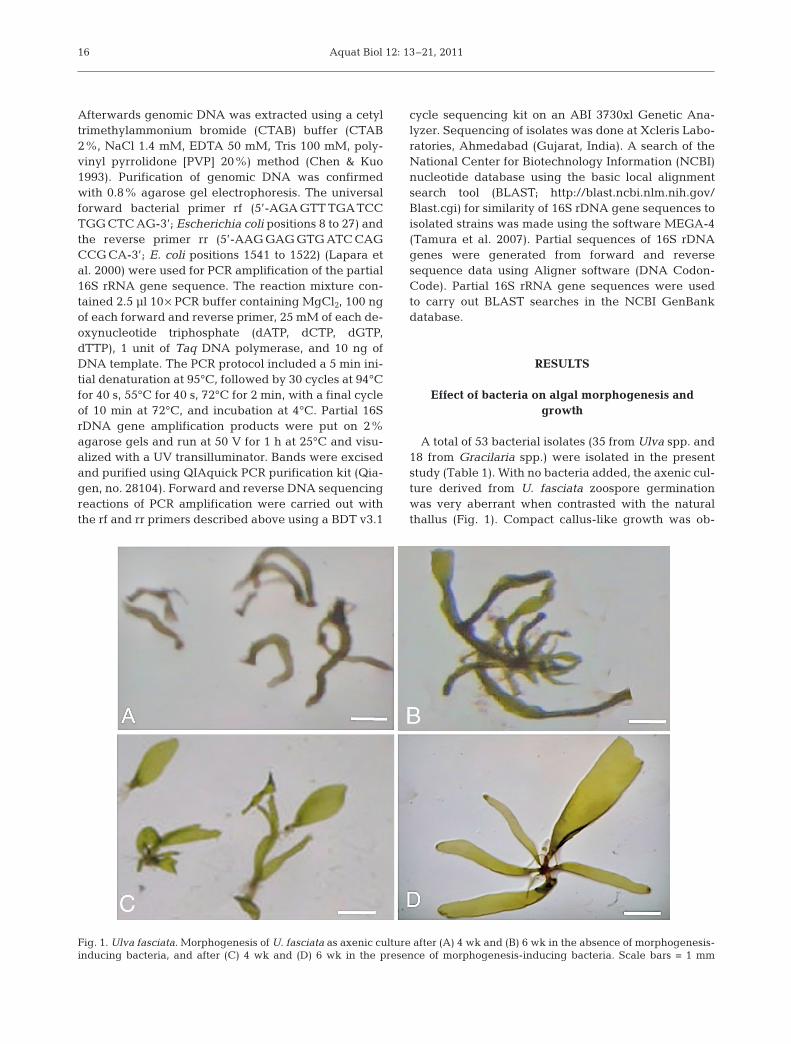
A total of 53 bacterial isolates (35 from Ulva spp. and18 from Gracilaria spp.) were isolated in the presentstudy (Table 1). With no bacteria added, the axenic cul-ture derived from U. fasciata zoospore germinationwas very aberrant when contrasted with the naturalthallus (Fig. 1). Compact callus-like growth was ob-
16
Fig. 1. Ulva fasciata. Morphogenesis of U. fasciata as axenic culture after (A) 4 wk and (B) 6 wk in the absence of morphogenesis-inducing bacteria, and after (C) 4 wk and (D) 6 wk in the presence of morphogenesis-inducing bacteria. Scale bars = 1 mm
Singh et al.: Morphogenesis-inducing bacteria on Ulva fasciata
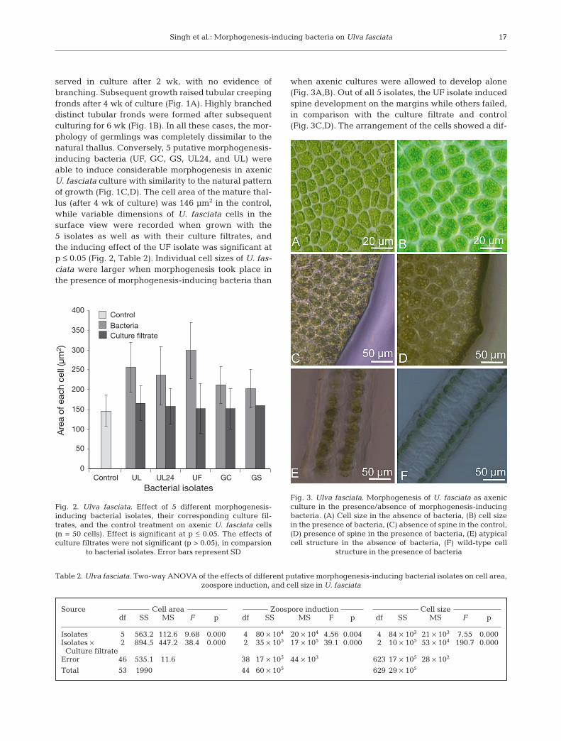
served in culture after 2 wk, with no evidence ofbranching. Subsequent growth raised tubular creepingfronds after 4 wk of culture (Fig. 1A). Highly brancheddistinct tubular fronds were formed after subsequentculturing for 6 wk (Fig. 1B). In all these cases, the mor-phology of germlings was completely dissimilar to thenatural thallus. Conversely, 5 putative morphogenesis-inducing bacteria (UF, GC, GS, UL24, and UL) wereable to induce considerable morphogenesis in axenicU. fasciata culture with similarity to the natural patternof growth (Fig. 1C,D). The cell area of the mature thal-lus (after 4 wk of culture) was 146 µm2 in the control,while variable dimensions of U. fasciata cells in thesurface view were recorded when grown with the5 isolates as well as with their culture filtrates, andthe inducing effect of the UF isolate was significant atp ≤ 0.05 (Fig. 2, Table 2). Individual cell sizes of U. fas-ciata were larger when morphogenesis took place inthe presence of morphogenesis-inducing bacteria than
when axenic cultures were allowed to develop alone(Fig. 3A,B). Out of all 5 isolates, the UF isolate inducedspine development on the margins while others failed,in comparison with the culture filtrate and control(Fig. 3C,D). The arrangement of the cells showed a dif-
17
0
50
100
150
200
250
300
350
400
Control UL UL24 UF GC GS Bacterial isolates
Are
a of
eac
h ce
ll (µ
m2 )
BacteriaControl
Culture filtrate
Fig. 2. Ulva fasciata. Effect of 5 different morphogenesis-inducing bacterial isolates, their corresponding culture fil-trates, and the control treatment on axenic U. fasciata cells(n = 50 cells). Effect is significant at p ≤ 0.05. The effects ofculture filtrates were not significant (p > 0.05), in comparsion
to bacterial isolates. Error bars represent SD
Fig. 3. Ulva fasciata. Morphogenesis of U. fasciata as axenicculture in the presence/absence of morphogenesis-inducingbacteria. (A) Cell size in the absence of bacteria, (B) cell sizein the presence of bacteria, (C) absence of spine in the control,(D) presence of spine in the presence of bacteria, (E) atypicalcell structure in the absence of bacteria, (F) wild-type cell
structure in the presence of bacteria
Source Cell area Zoospore induction Cell sizedf SS MS F p df SS MS F p df SS MS F p
Isolates 5 563.2 112.6 9.68 0.000 4 80 × 104 20 × 104 4.56 0.004 4 84 × 103 21 × 103 7.55 0.000Isolates × 2 894.5 447.2 38.4 0.000 2 35 × 105 17 × 105 39.1 0.000 2 10 × 105 53 × 104 190.7 0.000Culture filtrate
Error 46 535.1 11.6 38 17 × 105 44 × 103 623 17 × 105 28 × 102
Total 53 1990 44 60 × 105 629 29 × 105
Table 2. Ulva fasciata. Two-way ANOVA of the effects of different putative morphogenesis-inducing bacterial isolates on cell area, zoospore induction, and cell size in U. fasciata
Aquat Biol 12: 13–21, 2011
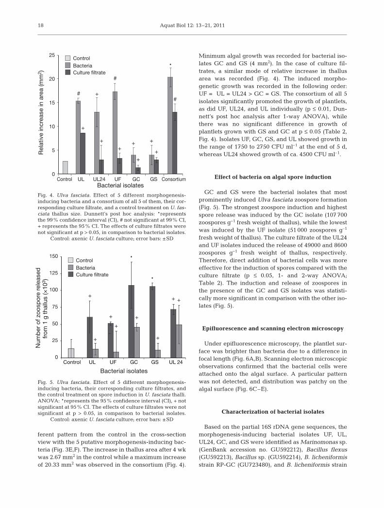
ferent pattern from the control in the cross-sectionview with the 5 putative morphogenesis-inducing bac-teria (Fig. 3E,F). The increase in thallus area after 4 wkwas 2.67 mm2 in the control while a maximum increaseof 20.33 mm2 was observed in the consortium (Fig. 4).
Minimum algal growth was recorded for bacterial iso-lates GC and GS (4 mm2). In the case of culture fil-trates, a similar mode of relative increase in thallusarea was recorded (Fig. 4). The induced morpho-genetic growth was recorded in the following order:UF = UL = UL24 > GC = GS. The consortium of all 5isolates significantly promoted the growth of plantlets,as did UF, UL24, and UL individually (p ≤ 0.01, Dun-nett’s post hoc analysis after 1-way ANOVA), whilethere was no significant difference in growth ofplantlets grown with GS and GC at p ≤ 0.05 (Table 2,Fig. 4). Isolates UF, GC, GS, and UL showed growth inthe range of 1750 to 2750 CFU ml–1 at the end of 5 d,whereas UL24 showed growth of ca. 4500 CFU ml–1.
Effect of bacteria on algal spore induction
GC and GS were the bacterial isolates that mostprominently induced Ulva fasciata zoospore formation(Fig. 5). The strongest zoospore induction and highestspore release was induced by the GC isolate (107 700zoospores g–1 fresh weight of thallus), while the lowestwas induced by the UF isolate (51 000 zoospores g–1
fresh weight of thallus). The culture filtrate of the UL24and UF isolates induced the release of 49000 and 8600zoospores g–1 fresh weight of thallus, respectively.Therefore, direct addition of bacterial cells was moreeffective for the induction of spores compared with theculture filtrate (p ≤ 0.05, 1- and 2-way ANOVA;Table 2). The induction and release of zoospores inthe presence of the GC and GS isolates was statisti-cally more significant in comparison with the other iso-lates (Fig. 5).
Epifluorescence and scanning electron microscopy
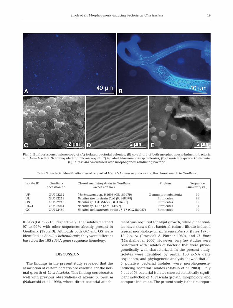
Under epifluorescence microscopy, the plantlet sur-face was brighter than bacteria due to a difference infocal length (Fig. 6A,B). Scanning electron microscopicobservations confirmed that the bacterial cells wereattached onto the algal surface. A particular patternwas not detected, and distribution was patchy on thealgal surface (Fig. 6C–E).
Characterization of bacterial isolates
Based on the partial 16S rDNA gene sequences, themorphogenesis-inducing bacterial isolates UF, UL,UL24, GC, and GS were identified as Marinomonas sp.(GenBank accession no. GU592212), Bacillus flexus(GU592213), Bacillus sp. (GU592214), B. licheniformisstrain RP-GC (GU723480), and B. licheniformis strain
18
*
*
+++
+
++
+
+
0
25
50
75
100
125
150
Num
ber
of z
oosp
ore
rele
ased
from
1 g
tha
llus
(×10
3 )
BacteriaControl
Culture filtrate
Bacterial isolates
Control UL UF GC GS UL 24
Fig. 5. Ulva fasciata. Effect of 5 different morphogenesis-inducing bacteria, their corresponding culture filtrates, andthe control treatment on spore induction in U. fasciata thalli.ANOVA: *represents the 95% confidence interval (CI), + notsignificant at 95% CI. The effects of culture filtrates were notsignificant at p > 0.05, in comparison to bacterial isolates.
Control: axenic U. fasciata culture; error bars: ±SD
0
5
10
15
20
25
UL Control UL24 UF GC GS ConsortiumBacterial isolates
Rel
ativ
e in
crea
se in
are
a (m
m2 )
*
#
+
+ +
+
#
+
+
+
#
+
BacteriaControl
Culture filtrate
Fig. 4. Ulva fasciata. Effect of 5 different morphogenesis-inducing bacteria and a consortium of all 5 of them, their cor-responding culture filtrate, and a control treatment on U. fas-ciata thallus size. Dunnett’s post hoc analysis: *representsthe 99% confidence interval (CI), # not significant at 99% CI,+ represents the 95% CI. The effects of culture filtrates werenot significant at p > 0.05, in comparison to bacterial isolates.
Control: axenic U. fasciata culture; error bars: ±SD
Singh et al.: Morphogenesis-inducing bacteria on Ulva fasciata
RP-GS (GU592215), respectively. The isolates matched97 to 99% with other sequences already present inGenBank (Table 3). Although both GC and GS wereidentified as Bacillus licheniformis, they were differentbased on the 16S rDNA gene sequence homology.
DISCUSSION
The findings in the present study revealed that theassociation of certain bacteria are essential for the nor-mal growth of Ulva fasciata. This finding corroborateswell with previous observations of axenic U. pertusa(Nakanishi et al. 1996), where direct bacterial attach-
ment was required for algal growth, while other stud-ies have shown that bacterial culture filtrate inducedtypical morphology in Enteromorpha sp. (Fries 1975),U. lactuca (Provasoli & Pintner 1980), and U. linza(Marshall et al. 2006). However, very few studies wereperformed with isolates of bacteria that were phylo-genetically well characterized. In the present study,isolates were identified by partial 16S rRNA genesequences, and phylogenetic analysis showed that all5 putative bacterial isolates were morphogenesis-inducing bacterial isolates (Matsuo et al. 2003). Only5 out of 53 bacterial isolates showed statistically signif-icant induction of U. fasciata growth, morphology, andzoospore induction. The present study is the first report
19
Fig. 6. Epifluorescence microscopy of (A) isolated bacterial colonies, (B) co-culture of both morphogenesis-inducing bacteriaand Ulva fasciata. Scanning electron microscopy of (C) isolated Marinomonas sp. colonies, (D) axenically grown U. fasciata,
(E) U. fasciata co-cultured with morphogenesis-inducing bacteria
Isolate ID GenBank Closest matching strain in GenBank Phylum Sequence accession no. (accession no.) similarity (%)
UF GU592212 Marinomonas sp. H1693 (GU183679) Gammaproteobacteria 99UL GU592213 Bacillus flexus strain Twd (FJ948078) Firmicutes 99GS GU592215 Bacillus sp. G1DM-53 (DQ416791) Firmicutes 99UL24 GU592214 Bacillus sp. L157 (AM913927) Firmicutes 97GC GU723480 Bacillus licheniformis strain JS-17 (GQ280087) Firmicutes 99
Table 3. Bacterial identification based on partial 16s rRNA gene sequences and the closest match in GenBank
Aquat Biol 12: 13–21, 2011
that morphogenesis-inducing bacteria are capable ofinducing the differentiation of whole plantlets withrespect to induction of spines on the surface, enlarge-ment of cells, and regaining wild-type cell structure inU. fasciata. On the contrary, the culture filtrate of the 5isolates and the control failed to induce the typicalmorphogenesis of U. fasciata. This suggests that phys-ical contact between bacterial and host cells is neces-sary for the development of the particular cell shape,growth, and differentiation of U. fasciata. In case of theUL isolate and the consortium of all 5 bacterial isolates,the culture filtrate slightly helped in the developmentof morphology of the U. fasciata (Fig. 4), but it mighthave been less significant due to instability of thesecreted substances or the signal. In contrast to the cul-ture filtrate, tightly associated bacteria might be con-tinuously secreting certain substances that enhancethe growth and restore the typical morphology ofU. fasciata.
It has also been reported that morphogenesis inmacrophytic green algae from the families Ulvaceaeand Monostromaceae are controlled by bacteria be-longing to the genera Cytophaga, Pseudomonas, Sta-phylococcus, Vibrio, Bacillus, and Flavobacterium(Duan et al. 1995, Nakanishi et al. 1999, Matsuo et al.2003, Marshall et al. 2006). The present study foundthat Marinomonas sp. and Bacillus spp. induced wild-type morphology and growth in Ulva fasciata. This isthe first study where Marinomonas and Bacillus spp.are shown to be involved in the differentiation andgrowth of U. fasciata. In the present study, Bacillusspp. were also found to affect the morphology andgrowth of U. fasciata to a greater extent than that re-ported by Nakanishi et al. (1996) for U. pertusa. How-ever, the effect of bacteria on the growth rate of macro-algae has not been well quantified. Subjectiveindications of induced growth and development havebeen reported for sporelings of U. pertusa, U. conglo-bata, and U. intestinalis when incubated with bacterialisolates of phylum Bacteroidetes (Matsuo et al. 2005,Marshall et al. 2006). The mechanism by which bacte-ria modulate the morphology of the plantlets is not yetwell understood, although a number of hypotheseshave been suggested. An endosymbiotic bacteriumfrom the Agrobacterium–Rhizobium group, containingthe nifH gene encoding for nitrogenase involved in ni-trogen fixation, was isolated from rhizoids of the greenalga Caulerpa taxifolia (Chisholm et al. 1996); it wassuggested that this isolate might be important for nitro-gen supply to the seaweed. A bacterium of the Roseo-bacter group was responsible for gall formation in thered alga Prionitis lanceolata due to overproduction ofindole-3-acetic acid (Ashen & Goff 2000). Phosphate-solubilizing activity of B. licheniformis has been re-ported to induce growth in mangrove plants (Rojas et
al. 2001). It has also been suggested that secondarymetabolites released by some epibiotic bacteria mayprevent subsequent biofouling by other organisms(Holmstrom et al. 1996, Callow & Callow 1998, Arm-strong et al. 2001), thereby providing some protectionto the host alga. In the present study, no correlationwas found between the bacterial isolates that alteredthe differentiation of U. fasciata and those that en-hanced spore induction. An interesting result is thatthose isolates that induced differentiation did not sig-nificantly induce spore production. Three isolates (UL,UF and UL24) accelerated differentiation in axenic U.fasciata. The consortium of all 5 putative morphogene-sis-inducing isolates significantly induced algal differ-entiation (Table 2). The positive effect shown by theconsortium suggested that normal morphology is notdependent on the presence of a single bacterium, butdifferentiation can be affected by a wide range of dif-ferent bacteria. The results of the present study havefurther confirmed that the wide range of bacteria thatinduce algal differentiation presumably confers eco-logical flexibility to the alga so that it is not dependenton specific bacteria. There might be positive inter-actions between the morphogenesis-inducing bacterialisolates, but the mechanism of their interaction is stillunclear. However, direct attachment of bacteria to theplantlet does appear to be essential, when the culture-filtrate results are considered. In addition, this is thefirst study providing evidence of the effect of bacterialisolates zoospore induction.
Further work on the mechanisms involved in thesebacterial–seaweed interactions is in progress.
Acknowledgements. The financial support received from theCouncil of Scientific and Industrial Research (NWP 018; RSP0016), New Delhi, is gratefully acknowledged. We also thank4 anonymous reviewers for their critical comments whichhave improved the manuscript.
LITERATURE CITED
Armstrong E, Rogerson A, Leftley JW (2000) Utilisation of sea-weed carbon by three surface-associated heterotrophicprotists, Steromyxa ramosa, Nitzschia alba and Labyrin-thula sp. Aquat Microb Ecol 21:49–57
Armstrong E, Tyan L, Boyd KG, Wright PC, Burgess JG (2001)The symbiotic role of marine microbes on living surfaces.Hydrobiologia 461:37–40
Ashen JB, Goff LJ (2000) Molecular and ecological evidencefor species specificity and coevolution in a group of marinealgal–bacterial symbioses. Appl Environ Microbiol 66:3024–3030
Callow ME, Callow JA (1998) Enhance adhesion and chemo-attraction of spore of the fouling alga Enteromorpha to somefoul-release silicone elastomers. Biofouling 13:157–172
Chandini SK, Ganesan P, Suresh PV, Bhaskar N (2008) Sea-weeds as source of nutritionally beneficial compounds - areview. J Food Sci Technol 45:1–13
20
Singh et al.: Morphogenesis-inducing bacteria on Ulva fasciata
Chen W, Kuo T (1993) A simple and rapid method for thepreparation of gram negative bacterial genomic DNA.Nucleic Acids Res 21:2260
Chisholm JRM, Dauga C, Ageron E, Grimont PAD, JaubertJM (1996) ‘Roots’ in mixotrophic algae. Nature 381:382
Dimitrieva GY, Crawford RL, Yuksel GU (2006) The nature ofplant growth-promoting effect of a pseudomonad associ-ated with the marine algae Laminaria japonica and linkedto catalase extraction. J Appl Microbiol 100:1159–1169
Duan D, Xu L, Fei X, Xu H (1995) Marine organism attachedto seaweed surfaces in Jiaozhou Bay, China. WorldJ Microbiol Biotechnol 11:351–352
Fries L (1975) Some observations on the morphology ofEnteromorpha linza (L.) J. Ag. and Enteromorpha com-pressa (L.) Grev. in axenic culture. Bot Mar 18:251–253
Fries L, Aberg S (1978) Morphogenetic effects of phenylaceticacid and p-OH-phenylacetic acid on the green algaEnteromorpha compressa (L.) Grev. in axenic culture. ZPflanzenphysiol 88:383–388
Hiraoka M, Oka N (2008) Tank cultivation of Ulva prolifera indeep seawater using a new ‘germling cluster’ method.J Appl Phycol 20:97–102
Holmstrom C, James S, Egan S, Kjelleberg S (1996) Inhibitionof common fouling organisms by marine bacterial isolateswith special reference to the role of pigmented bacteria.Biofouling 10:251–259
Jha B, Reddy CRK, Thakur MC, Rao MU (2009) Seaweeds ofIndia: the diversity and distribution of seaweeds of Gujaratcoast. Springer, Dordrecht
Kumari P, Kumar M, Gupta V, Reddy CRK, Jha B (2010) Trop-ical marine macroalgae as potential sources of nutrition-ally important PUFAs. Food Chem 120:749–757
Lakshmanaperumalsamy P, Purushothaman A (1982) Hetero-trophic bacteria associated with seaweed. Proc IndianAcad Sci Plant Sci 91:487–493
Lapara TM, Nakatsu CH, Pantea L, Alleman JE (2000) Phylo-genetic analysis of bacterial communities in mesophilicand thermophilic bioreactors treating pharmaceuticalwastewater. Appl Environ Microbiol 66:3951–3959
Mantri VA, Singh RP, Bijo AJ, Kumari P, Reddy CRK, Jha B(2010) Differential response of varying salinity and tem-perature on zoospore induction, regeneration and dailygrowth rate in Ulva fasciata (Chlorophyta, Ulvales). J ApplPhycol doi:10.1007/s10811-010-9544-4
Marshall K, Joint I, Callow ME, Callow JA (2006) Effect ofmarine bacterial isolates on the growth and morphology ofaxenic plantlets of the green alga Ulva linza. Microb Ecol52:302–310
Maruyama A, Maeda M, Simidu U (1986) Occurrence of planthormone (cytokinin)-producing bacteria in the sea. J ApplBacteriol 61:569–574
Maruyama A, Yamaguchi I, Maeda M, Simidu U (1988) Evi-dence of cytokinin production by a marine bacterium andits taxonomic characteristics. Can J Microbiol 34:829–833
Matsuo Y, Suzuli M, Kasai H, Shizuri Y, Harayama S (2003)Isolation and phylogenetic characterization of bacteriacapable of inducing differentiation in the green algaMonostroma oxyspermum. Environ Microbiol 5:25–35
Matsuo Y, Imagawa H, Nishizawa M, Shizuri Y (2005) Isola-tion of an algal morphogenesis inducer from a marine bac-terium. Science 307:1598
Mooney PA, Van SJ (1986) Algae and cytokinins. J PlantPhysiol 123:1–21
Nakanishi K, Nishijima M, Nishimura M, Kuwano K, Saga N(1996) Bacteria that induce morphogenesis in Ulva pertusa(Chlorophyceae) grown under axenic conditions. J Phycol32:479–482
Nakanishi K, Nishijima M, Nomoto AM, Yamazaki A, Saga N(1999) Requisite morphologic interaction for attachmentbetween Ulva pertusa (Chlorophyta) and symbiotic bacte-ria. Mar Biotechnol 1:107–111
Oza RM, Rao PS (1977) Effect of different culture media ongrowth and sporulation of laboratory raised germlings ofUlva fasciata Delile. Bot Mar 20:427–431
Patel P, Callow ME, Joint I, Callow JA (2003) Specificity inthe settlement-modifying response of bacterial biofilmstowards zoospores of the marine alga Enteromorpha.Environ Microbiol 5:338–349
Provasoli L (1958) Effect of plant hormones on Ulva. Biol Bull(Woods Hole) 114:375–384
Provasoli L, Pintner IJ (1980) Bacteria induced polymorphismin an axenic laboratory strain of Ulva lactuca (Chloro-phyceae). J Phycol 32:479–482
Reddy CRK, Dipakkore S, Rajakrishan KG, Jha B, Cheney DP,Fujita Y (2006) An improved enzyme preparation for rapidmass production of protoplasts as seed stock for aquacul-ture of macrophytic marine green algae. Aquaculture 260:290–297
Rojas A, Holguin G, Glick BR, Bashan V (2001) Synergism be-tween Phyllobacterium sp. (N2-fixer) and Bacillus licheni-formis (P-solubilizer), both from a semiarid mangrove rhi-zosphere. FEMS Microbiol Ecol 35:181–187
Subbaramaiah K (1970) Growth and reproduction of Ulva fas-ciata Delile in nature and in culture. Bot Mar 13:25–27
Tamura K, Dudley J, Nei M, Kumar S (2007) MEGA4: Molec-ular Evolutionary Genetics Analysis (MEGA) softwareversion 4.0. Mol Biol Evol 24:1596–1599
Tatewaki M, Provasoli L, Pintner IJ (1983) Morphologenesisof Monostroma oxyspermum (Kutz) Doty (Chlorophyceae)in axenic culture, especially in bialgal culture. J Phycol 19:409–416
Weinberger F, Beltran J, Correa JA, Lion U and others (2007)Spore release in Acrochaetium sp. (Rhodophyta) is bacte-rially controlled. J Phycol 43:235–241
Wichard T, Oertel W (2010) Gametogenesis and gameterelease of Ulva mutabilis and Ulva lactuca (Chlorophyta):regulatory effects and chemical characterization of the‘swarming inhibitor’. J Phycol 46:248–259
21
Editorial responsibility: Hans Heinrich Janssen,Oldendorf/Luhe, Germany
Submitted: June 29, 2010; Accepted: December 8, 2010Proofs received from author(s): February 18, 2011