Embed Size (px)
Citation preview
IsdC from Staphylococcus lugdunensis Induces Biofilm Formationunder Low-Iron Growth Conditions
Antonino Missineo,a* Antonella Di Poto,a Joan A. Geoghegan,b Simonetta Rindi,a Simon Heilbronner,b Valentina Gianotti,a
Carla Renata Arciola,c Timothy J. Foster,b Pietro Speziale,a Giampiero Pietrocolaa
Department of Molecular Medicine, Unit of Biochemistry, Pavia, Italya; Department of Microbiology, Moyne Institute of Preventive Medicine, Trinity College, Dublin,Irelandb; Research Unit on Implant Infections, Rizzoli Orthopaedic Institute, and DIMES, University of Bologna, Bologna, Italyc
Staphylococcus lugdunensis is a coagulase-negative staphylococcus that is a commensal of humans and an opportunistic patho-gen. It can cause a spectrum of infections, including those that are associated with the ability to form biofilm, such as occurs withendocarditis or indwelling medical devices. The genome sequences of two strains revealed the presence of orthologues of the icagenes that are responsible for synthesis of poly-N-acetylglucosamine (PNAG) that is commonly associated with biofilm in otherstaphylococci. However, we discovered that biofilm formed by a panel of S. lugdunensis isolates growing in iron-restricted me-dium was susceptible to degradation by proteases and not by metaperiodate, suggesting that the biofilm matrix comprised pro-teins and not PNAG. When the iron concentration was raised to 1 mM biofilm formation by all strains tested was greatly re-duced. A mutant of strain N920143 lacking the entire locus that encodes iron-regulated surface determinant (Isd) proteins wasdefective in biofilm formation under iron-limited conditions. An IsdC-null mutant was defective, whereas IsdK, IsdJ, and IsdBmutants formed biofilm to the same level as the parental strain. Expression of IsdC was required both for the primary attach-ment to unconditioned polystyrene and for the accumulation phase of biofilm involving cell-cell interactions. Purified recombi-nant IsdC protein formed dimers in solution and Lactococcus lactis cells expressing only IsdC adhered to immobilized recombi-nant IsdC but not to IsdJ, IsdK, or IsdB. This is consistent with a specific homophilic interaction between IsdC molecules onneighboring cells contributing to accumulation of S. lugdunensis biofilm in vivo.
Coagulase-negative staphylococci (CoNS) and Staphylococcusaureus are the predominant etiological agents of medical de-
vice-related infections, largely owing to their ability to form bio-film. Biofilms are defined as communities of bacteria encased in aself-synthesized extracellular polymeric matrix (1) growing at-tached to biological or abiotic surfaces. Staphylococci in biofilmsare resistant to antibiotics (2) and host immune responses (3),reducing the efficacy of available antimicrobials. The formation ofbiofilm is a complex, multifactorial process. Initially, bacteria ad-here directly to the surface of implanted device or to devicescoated with the host matrix components. In S. aureus biofilm themajor autolysin, Atl, mediates primary attachment to plastic sur-faces by promoting release of DNA from bacterial cells (4, 5),while adherence to surfaces conditioned by host plasma proteinsis promoted by surface protein adhesins such as the fibrinogen-binding clumping factor A or fibronectin-binding proteins (6).This process is followed by proliferation, accumulation, and in-tercellular interactions mediated by the icaADBC-encoded poly-saccharide intercellular adhesins (PIA) (7) or surface proteinssuch as Bap (8), SasG (9), SasC (10), protein A (11), or fibronec-tin-binding proteins (FnBPs) (12, 13). Likewise, biofilm forma-tion by S. epidermidis is dependent on PIA or proteinaceous com-ponents such as Aap (14, 15) or SesC (16).
Staphylococcus lugdunensis is a coagulase-negative species withenhanced virulence compared to the other CoNS (17). S. lug-dunensis causes a severe form of native valve endocarditis (18, 19)and infections of prosthetic heart valves (20), intravascular cath-eters (21), prosthetic joints (22), and ventriculoperitoneal shunts(23). This pathogenic potential is largely attributed to the ability ofthis bacterium to form biofilm. A previous study by Frank andPatel (24) demonstrated that despite the presence of icaADBCorthologues in S. lugdunensis, PIA is not the major component of
the extracellular matrix of biofilms formed in vitro by this species.Rather, S. lugdunensis biofilms appear to be composed of proteins.S. lugdunensis expresses a fibrinogen-binding protein (Fbl) (25), amember of the family of MSCRAMM (Microbial Surface Compo-nents Recognizing Adhesive Matrix Molecules) family that isclosely related to ClfA, which does not appear to have any role inbiofilm formation (26), and a yet-uncharacterized von Wille-brand factor binding protein (27).
Uniquely for CoNS, S. lugdunensis contains a cluster of geneswith similarity both in terms of organization and sequence to theiron-regulated surface determinant (isd) locus of S. aureus (28).Both systems are expressed under iron-restricted conditions (28,29). Four of the S. lugdunensis Isd proteins are anchored to the cellwall peptidoglycan by sortases. In S. aureus and S. lugdunensis, theIsd proteins cooperate to capture heme and transfer it acrossthe wall to a membrane-bound transporter, which delivers it tothe cytoplasm, where heme is degraded to recover iron (30). There
Received 3 February 2014 Returned for modification 9 March 2014Accepted 22 March 2014
Published ahead of print 31 March 2014
Editor: A. Camilli
Address correspondence to Pietro Speziale, [email protected].
* Present address: Antonino Missineo, IRBM Science Park SpA, Pomezia, Rome,Italy.
A.M. and A.D.P. contributed equally to this article.
Supplemental material for this article may be found at http://dx.doi.org/10.1128/IAI.01542-14.
Copyright © 2014, American Society for Microbiology. All Rights Reserved.
doi:10.1128/IAI.01542-14
2448 iai.asm.org Infection and Immunity p. 2448 –2459 June 2014 Volume 82 Number 6
on May 3, 2018 by guest
http://iai.asm.org/
Dow
nloaded from
is evidence that surface-exposed Isd proteins may have additionalroles in colonization and pathogenesis of both species. For exam-ple, IsdJ from S. lugdunensis (29) and IsdA from S. aureus (31) aremultifunctional proteins that recognize and bind several host pro-teins and can confer resistance to skin fatty acids. In the presentstudy, we investigated biofilm formation in vitro by a collection ofS. lugdunensis isolates grown under low-iron conditions and as-sessed the role of isd locus in biofilm formation by this importantpathogen.
MATERIALS AND METHODSBacterial strains and culture conditions. The microorganisms used inthe present study are reported in Table 1. S. aureus V329 (8) and SA113(32) were kindly donated by J. R. Penades (Universidad Cardenal Her-
rera-CEU, Moncada, Valencia, Spain). The clinical isolate of S. epidermi-dis 5179R (14) was provided by H. Rohde (University Medical CentreHamburg-Eppendorf, Hamburg, Germany). S. epidermidis RP62A wasoriginally isolated from a patient with intravascular catheter-associatedsepsis by Muller et al. (33). Staphylococci were grown in Trypticase soybroth (TSB; Difco, Detroit, MI) or in RPMI 1640 (RPMI; Biowest, Nu-aillé, France) supplemented with 2 mM glutamine (Lonza Srl, Bergamo,Italy) at 37°C for 16 to 18 h with intense shaking (200 rpm). L. lactistransformants were grown in M17 medium (Difco) containing 0.5% glu-cose and 10% lactose at 30°C.
Construction of S. lugdunensis mutants. S. lugdunensis N920143 mu-tants deficient in individual genes isdB, isdC, isdJ, and isdK and the mutantdeficient in the entire isd locus were as reported earlier (29) (Table 1).
In order to reverse the deletion mutation in N920143, the programSILENT (emboss.bioinformatics.nl/cgi-bin/emboss/silent) was used to
TABLE 1 Bacterial strains and plasmids
Strain or plasmid Relevant characteristicsa Source or reference
StrainsStaphylococcus lugdunensis
N920143 Human breast abscess isolate 28N930432 Human endocarditis isolate F. Vandenesch (unpublished data)N940025 Human endocarditis isolate F. Vandenesch (unpublished data)N940084 Finger pulp infection isolate This studyN940113 Vertebral infection isolate This studyN940135 Perineal infection isolate This studyN940164 Perineal infection isolate This study1871 Orthopedic infection with internal fixation system This study2050 Orthopedic infection with knee implant This studyN920143�isdB isdB-null mutation in N920143 29N920143�isdC isdC-null mutation in N920143 29N920143�isdCr Reversion strain expressing isdCr in the native context This studyN920143�isdJ isdJ-null mutation in N920143 29N920143�isdK isdK-null mutation in N920143 29N920143�isd isd-null mutation in N920143 29N920143�atlI atlI-null mutation in N920143 This studyN920143�srtB srtB-null mutation in N920143 This study
Staphylococcus aureusSA113 ATCC 35556, restriction deficient 31V329 Isolate obtained from a bovine subclinical phenotype 8
Staphylococcus epidermidisRP62A ATCC 35984, isolated from a patient with sepsis 325179R Clinical isolate obtained from cerebrospinal fluid infection 14
Lactococcus lactis NZ9000 L. lactis subsp. cremoris MG1363 carrying nisin resistance cassette 33Escherichia coli
TOPP3 E. coli cloning and protein purification host Stratagene
PlasmidspQE30 E. coli cloning and expression vector; Apr StratagenepQE30isdB pQE30 encoding residues 45 to 655 of IsdB 29pQE30isdC pQE30 encoding residues 30 to 190 of IsdC 29pQE30isdJ(45-610) pQE30 encoding residues 45 to 610 of IsdJ 29pQE30isdK pQE30 encoding residues 35 to 426 of IsdK 29pNZ8048 L. lactis shuttle vector containing the PnisA promoter and start codon in
NcoI site; Cmr allowing nisin-inducible expression of insert33
pNZ8048isdC-LPQTG pNZ8048 encoding isdC, for controlled expression of sortase A-anchored IsdC in L. lactis
This study
pIMAY Themosensitive vector for allelic exchange; Cmr 39pIMAYisdCr Construct for restoration of the �isdC mutation This studypIMAY�atl and pIMAY�srtB Constructs for deleting atl and srt�, respectively This studypRMC2 Inducible expression vector for staphylococci; Apr Cmr 35pRMC2isdC-LPQTG pRMC2 encoding isdC, for controlled expression of sortase A-anchored
IsdC in S. lugdunensis �isdThis study
a Apr, ampicillin resistance; Cmr, chloramphenicol resistance. isdCr, recombined gene isdC.
S. lugdunensis IsdC Induces Biofilm Formation
June 2014 Volume 82 Number 6 iai.asm.org 2449
on May 3, 2018 by guest
http://iai.asm.org/
Dow
nloaded from
identify single nucleotides within isdC that can be mutated to create anovel restriction site without causing translational changes in the proteinsequence. The novel restriction sites allow the discrimination of the wildtype and the restored allele. Primers isdC-E and isdC-F exchanged nucle-otide 459 of isdC (A to C), creating a novel SmaI site. Primers isdC-A andisdC-E were used to amplify a 500-bp upstream sequence and the 5=-fragment of isdC, and primers isdC-F and isdC-D were used to amplify a500-bp downstream region, together with the 3= fragment of isdC. PCRproducts were gel-purified and used for the spliced overlap extension PCRusing primers A and D. The cassette was gel purified, cleaved at endonu-clease cleavage sites introduced in primers A and D, and cloned intopIMAY by using SLIC (sequence and ligase independent cloning), form-ing pIMAYisdCr. The isdCr gene was recombined into the chromosome ofS. lugdunensis N920143�isdC by allelic exchange.
Additional mutants deficient in srtB and the autolysin atlI were createdusing the thermosensitive vector pIMAY and allelic exchange. A detailedprotocol of the procedure is described elsewhere (34). Primers used forthe construction of deletion cassettes are summarized in Table S1 in thesupplemental material.
Construction and expression of isdC in S. lugdunensis �isd mutant.In order to create a S. lugdunensis strain expressing IsdC in the absence ofany other Isd proteins, the isdC gene was cloned into the anhydrotetracy-cline-inducible vector pRMC2 (35). The gene was PCR amplified with theprimers isdC-pRF and isdC-pRR using pNZ8048isdC-LPQTG as the tem-plate. The S. lugdunensis �isd mutant is deficient in sortase B, which willprevent cell wall sorting of the wild-type IsdC protein harboring anNPQTS motif. The IsdC-LPQTG protein will be sorted by sortase A.
The isdC LPQTG gene was cloned into pRMC2 using SLIC (36, 37).This method involves PCR amplification of the vector backbone and ofthe insert. Amplification of the vector backbone generates a linear productwith blunt ends at the site required for the cloning. Primers incorporateshort identical DNA sequences in vector and insert. Treatment of bothPCR products with T4 polymerase (3=-5= exonuclease activity) createssingle-stranded, 5= overhangs in both vector and insert. The DNA frag-ments are assembled in vitro (without ligation) and used to transformEscherichia coli.
Primers pRA and pRB were used to amplify the pRMC2 backbone (seeTable S1 in the supplemental material). Identical sequences (20 to 25nucleotides) were integrated in the primers for the amplification of theinsert (isdC-pRF/isdC-pRR; see Table S1 in the supplemental material).Next, 10 ng of plasmid DNA was used as the template for the amplificationof the plasmid backbone with Phusion polymerase (Keilaranta 16A;Finnzymes, Espoo, Finland). The PCR products were purified, and thevector product was digested with DpnI to remove methylated templateDNA. Then, 1 �g of vector and insert DNA was treated with T4 polymer-ase in a final volume of 40 �l of NEB buffer 2 (New England BioLabs,Ipswich, MA) with highly purified bovine serum albumin at 100 �g/ml(New England BioLabs), 5 mM dithiothreitol, 200 mM urea, and 3 U of T4DNA polymerase (New England BioLabs), followed by incubation for 20min at 23°C, and the reaction was stopped by the addition of 25 mMEDTA and subsequent incubation for 20 min at 73°C.
We then mixed 5 �l of vector DNA and 5 �l of insert DNA, and thesingle-stranded overhangs were allowed to anneal. The tube was placed ina PCR machine for 10 min at 65°C, followed by a slow decrease in tem-perature from 65 to 25°C with a 1-min hold for each degree. A 2.5-�lportion of the reaction mixture was used to transform E. coli to isolatepRMC2isdC-LPQTG, which was confirmed by DNA sequencing. Theplasmid was transformed into the S. lugdunensis �isd strain (34). Induc-tion of IsdC-LPXTG expression from pRMC2 in S. lugdunensis �isd mu-tant was carried out by adding anhydrotetracycline (0.125 �g/ml; Sigma,St. Louis, MO) to exponentially growing cultures.
Construction of Lactococcus lactis expressing IsdC. To express IsdCin the surrogate host L. lactis, the isdC gene was amplified (IsdC-F/IsdC-R) and cloned into the nisin-inducible expression vector pNZ8048
(38). L. lactis was transformed with the recombinant plasmid as describedearlier (39).
IsdC possesses a NPQTS sorting signal at its C terminus and is there-fore anchored to the cell wall by the transpeptidase sortase B, which isencoded within the isd operon. L. lactis does not encode sortase B, whichwill prevent sorting of IsdC to the cell wall. To allow sorting, pNZ8048isdCwas isolated, and DNA encoding the NPQTS signal was exchanged toLPQTG using 5=-phosphorylated primers (LPXTG-A/LPXTG-B) and in-verse PCR. The primers allowed the amplification of the entire plasmidand introduced the required nucleotide substitutions in the cell wall an-choring region of isdC. The PCR product was treated with T4 ligase toallow circularization of the plasmid and transformed into L. lactis. Theresulting plasmid (pNZ8048isdC-LPQTG) was confirmed by DNA se-quencing. Primers are summarized in Table S1 in the supplemental ma-terial. Induction of IsdC-LPXTG expression from pNZ8048 in L. lactiswas carried out by adding nisin (0.4 ng/ml) to exponentially growingcultures.
Biofilm formation. Overnight cultures of staphylococci were diluted1:200 in TSB containing 0.3% glucose (TSB0.3%glucose) or RPMI supple-mented with 0.3% glucose (RPMI0.3%glucose) and 2 mM glutamine. Ali-quots (200 �l) of the diluted bacterial suspensions were added to 96-wellflat-bottom sterile polystyrene microplates (Costar; Corning, New York,NY) and incubated statically for 24 h at 37°C. Biofilm formation wasdetected by the method of Christensen et al. (40). Briefly, biofilms formedon the plates were gently washed twice with phosphate-buffered saline(PBS) (137 mM NaCl, 2.7 mM KCl, 4.3 mM Na2HPO4 [pH 7.4]) toremove planktonic and loosely adhering bacteria. Adherent cells werefixed with 96% ethanol for 10 min, stained with 0.1% crystal violet for 15min, and—after several washings—the wells were air dried. For a quanti-tative estimation of biofilm density, bound crystal violet was solubilizedwith 10% glacial acetic acid, and the absorbance of the solubilized dye wasread at 595 nm in a microplate reader (model 680; Bio-Rad Laboratories,Inc., Hercules, CA). To test the role of iron on biofilm formation, S.lugdunensis strains N920143, N940025, N940113, and N940135 were cul-tured in RPMI0.3%glucose–2 mM glutamine supplemented with 1,000 �MFeCl3 in 96-well flat-bottom sterile polystyrene microplates and treated asdescribed above. Iron-depleted growth medium was obtained by treat-ment of RPMI with divalent metal chelator Chelex 100 according to themanufacturer’s instructions (Bio-Rad).
Enzymatic and chemical treatment of biofilms. Chemical and enzy-matic treatments of biofilms were carried out as described previously (41,42). Briefly, the biofilms grown in microtiter plates were rinsed with 200�l of 0.9% NaCl and then treated for 2 h at 37°C with 100 �l of 10 mMsodium metaperiodate (Sigma) in 50 mM sodium acetate buffer (pH 4.5).Alternatively, biofilms were incubated with 100 �l of proteinase K(Sigma) at 1 mg/ml in 20 mM Tris buffer containing 100 mM NaCl (pH7.5) or 100 �l of DNase I (Sigma) at 2 mg/ml in PBS. Enzymes or sodiummetaperiodate were replaced with the appropriate amounts of buffer inthe controls. To rule out the possibility that DNase I could be contami-nated with proteases, the enzyme was incubated with albumin for 2 h, andthe mixture was subjected to SDS-PAGE. Under these conditions, nodifference in the electrophoretic mobility of DNase I-treated and un-treated samples of albumin was observed.
Expression and purification of recombinant proteins. RecombinantHis-tagged proteins were expressed and purified by Ni2� chelate chroma-tography as described previously (29) (Table 1). E. coli strain TOPP3(Stratagene, La Jolla, CA), used for the expression of recombinant His-tagged proteins, was grown in Luria-Bertani (LB) broth (Difco) supple-mented with ampicillin (100 �g/ml; Sigma) at 37°C for 18 h with shaking(150 rpm). Overnight cultures were diluted 1:100 in LB medium andgrown at 37°C, with shaking, until the optical density at 600 nm (OD600)reached 0.5 to 0.6. Expression was induced by adding IPTG (isopropyl-�-D-thiogalactopyranoside; Inalco, Milan, Italy) to a final concentration of 1mM. Bacteria were harvested by centrifugation at 1,700 � g for 20 minand lysed by passage through a French press. The cell debris was removed
Missineo et al.
2450 iai.asm.org Infection and Immunity
on May 3, 2018 by guest
http://iai.asm.org/
Dow
nloaded from
by centrifugation (20,000 � g), and the filtered supernatant (0.45-�m-pore-size membrane) was applied to a 1-ml Ni2�-Sepharose His-Trap HPcolumn (GE Healthcare, Buckinghamshire, United Kingdom). Fusion pro-teins were eluted with 20 column volumes of a 0.00 to 500 mM imidazole(Sigma) gradient in 20 mM sodium phosphate–0.5 M NaCl buffer (pH 7.4).Fractions corresponding to the recombinant protein were pooled and exten-sively dialyzed against PBS. Protein concentrations were determined with abicinchoninic acid protein assay kit (Pierce, Rockford, IL).
Primary attachment assay. The attachment assay was performed asreported by Geoghegan et al. (9). Briefly, bacteria were grown overnight inRPMI–2 mM glutamine and diluted in RPMI0.3%glucose–2 mM glutamine,and �300 CFU in 100 �l was then spread onto the bases of empty petridishes. Dishes were incubated upright at 37°C for 30 min, washed threetimes with 5 ml of sterile PBS, and covered with TSB agar. Bacterial platecounts were run in parallel, and the percent attachment was calculated.Each experiment was repeated three times. The statistical significance wasdetermined with Student t test using GraphPad software (GraphPad Soft-ware, Inc., San Diego, CA).
Aggregation assays. The aggregation assay was based on the methoddescribed by Geoghegan et al. (9). Bacteria were grown overnight inRPMI–2 mM glutamine and diluted in RPMI0.3%glucose–2 mM glutamineto an OD600 of 1.0. Tubes were incubated statically at 37°C for up to 24 h.A 1-ml portion of broth was removed from the top of the tube at theindicated times, and the OD600 was measured. The remaining culture wasvortexed to resuspend the cells, and the OD600 was measured again. Thepercent aggregation was calculated using the following formula: 100 �[(OD600 of vortexed sample OD600 before vortexing)/(OD600 of vor-texed sample)]. The statistical significance was determined with a Studentt test using GraphPad software.
Gel filtration chromatography. The size exclusion chromatographyexperiment was performed using a Superose 12 HR 10/30 column (GEHealthcare) connected to an AKTA design chromatography system (GEHealthcare). A 100-�l portion of 100 �M IsdC was loaded onto a gelfiltration column equilibrated in PBS with or without 100 �M FeCl3 andeluted with one column volume (24 ml) at a flow rate of 0.5 ml/min.Recorded data were analyzed using UNICORN 5.10 control software (GEHealthcare). The Mr of IsdC was determined from the calibration curve(plot of the partition coefficient [Kav] versus log Mr) once its Kav valuewas calculated from the measured elution volume.
Preparation of bacterial lysates. The lysates from S. lugdunensis andL. lactis strains were prepared as previously described (29) with minormodifications. Briefly, bacterial cultures were harvested by centrifugation,washed in PBS, and adjusted to an OD600 of 10. A 1-ml portion of thebacterial suspension was pelleted and resuspended in 250 �l of digestionbuffer (50 mM Tris-HCl, 20 mM MgCl2, 30% [wt/vol] raffinose; pH 7.5)containing Complete Mini-Protease inhibitors (Roche). Cell wall pro-teins were solubilized by digestion with lysostaphin (500 �g/ml) (S. lug-dunensis) or a combination of mutanolysin (1,000 U/ml) and lysozyme(900 �g/ml) (L. lactis) at 37°C for 30 or 15 min, respectively. Protoplastswere harvested by centrifugation (5,000 � g, 15 min), and the superna-tants were subjected to SDS-PAGE and Western blotting.
Western immunoblotting. Whole-cell lysates of transformants of L.lactis were subjected to 12.5% polyacrylamide gel electrophoresis andthen electroblotted onto a nitrocellulose membrane (GE Healthcare). Themembrane was treated with a solution containing 5% (wt/vol) dried milkin PBS, washed, and incubated with anti-IsdC rabbit IgG for 1 h at 22°C.After additional washings with 0.5% (vol/vol) Tween 20 in PBS, the mem-brane was incubated for 1 h with horseradish peroxidase (HRP)-conju-gated goat anti-rabbit IgG. The membrane was treated with enhancedchemiluminescence detection reagents 1 and 2, as recommended by themanufacturer (GE Healthcare), and exposed to an X-ray film for 30 to60 s.
Attachment of L. lactis transformants to Isd proteins. Isd proteins (1�g/well) were coated in microtiter wells in bicarbonate buffer overnight.L. lactis(pNZ8048isdC) and L. lactis(pNZ8048) (5 � 108 cells/well) were
added to the wells, followed by incubation for 1 h at 22°C. After extensivewashing with PBS, adhering cells were fixed with 25% formaldehyde(Sigma) and stained with 2.5% crystal violet, and the A595 was measured.
Statistical methods. Continuous data were expressed as means andstandard deviations. Two-group comparisons were performed by Studentt test. One-way analysis of variance, followed by Bonferroni’s post hoctests, was exploited for comparison of three or more groups. Analyseswere performed using Prism 4.0 (GraphPad). Two-tailed P values of0.05 were considered statistically significant.
RESULTSS. lugdunensis clinical isolates form proteinaceous biofilm iniron-restricted conditions. It was reported previously that S. lug-dunensis strains growing in rich broth formed biofilm that waspredominantly proteinaceous in that preformed biofilm could bedisrupted by proteases but not by periodate (24). In order to in-vestigate the nature of S. lugdunensis biofilm in more detail, wetested a panel of clinical isolates along with S. aureus and S. epi-dermidis controls where the composition of the biofilm matrix isknown to be composed predominantly of protein (V329, 5179R)or polysaccharide (SA113, RP62A) growing in iron-replete TSB orin iron-deficient RPMI. The controls formed protein or polysac-charide dependent biofilms as previously reported.
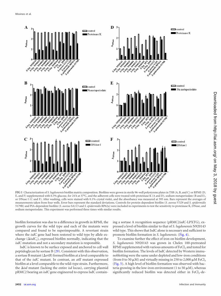
All S. lugdunensis strains tested formed biofilm when growingin both media. Glucose concentrations of �0.25% supportedhigher levels of biofilm formation compared to lower concentra-tions (P 0.001, data not shown). We confirmed the previousreport that biofilms formed by S. lugdunensis growing in TSB-glucose were susceptible to detachment by proteinase K but not bysodium metaperiodate (Fig. 1). We also found that biofilmsformed by S. lugdunensis growing in RPMI were susceptible toprotease and not to periodate. Furthermore, DNase caused signif-icant detachment of S. lugdunensis biofilm formed under bothgrowth conditions. DNase also detached the control protein-de-pendent biofilm but not that involving a polysaccharide matrix(Fig. 1).
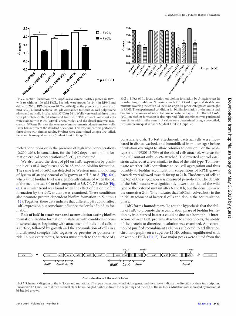
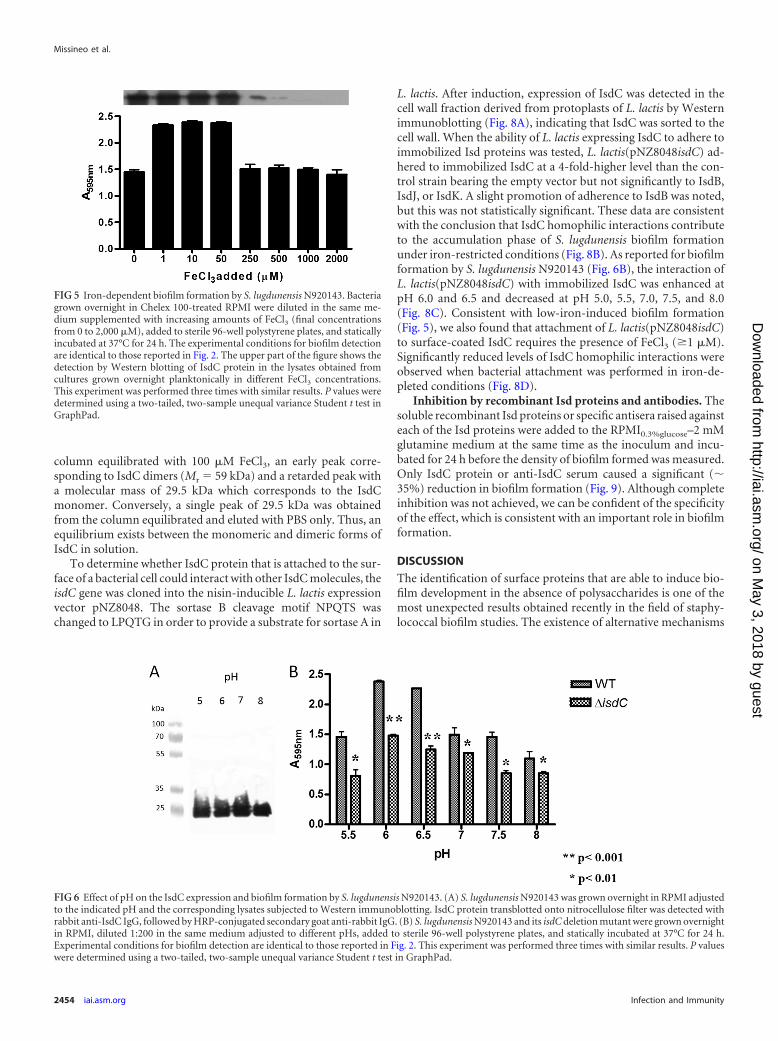
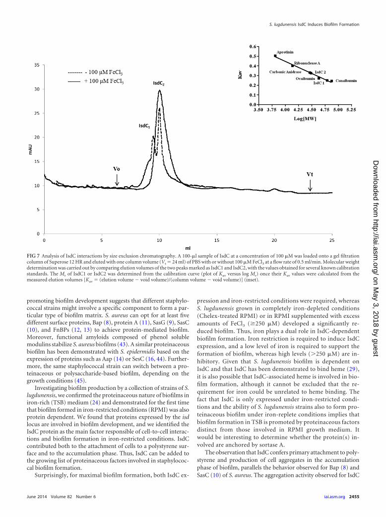
Involvement of the IsdC protein in S. lugdunensis biofilm.To determine whether S. lugdunensis biofilm formed duringgrowth in RPMI was influenced by the availability of iron, threestrains were tested in RPMI supplemented with 1 mM FeCl3. In-terestingly, the addition of iron reduced the biofilm density, sug-gesting that the involvement of proteins whose expression wasregulated by iron (Fig. 2). S. lugdunensis is the only species ofcoagulase-negative staphylococcus that harbors an iron-regulatedsurface determinant (Isd) locus. It is only expressed under iron-limited conditions and is responsible for the acquisition of ironfrom hemoglobin and heme in vivo (28, 29). To determinewhether the isd locus is involved in biofilm formation, S. lug-dunensis N920143 wild type and a mutant in which the entire isdlocus is deleted (�isd) were compared. A schematic representationof the isd locus and mutations is shown in Fig. 3. The level ofbiofilm formed by the isd mutant was the same as that formed bythe wild-type strain in the presence of FeCl3. This indicates thatone or more proteins expressed by the isd locus are involved inbiofilm formation in iron-limited conditions (Fig. 4). The S. lug-dunensis isd locus expresses four cell envelope-associated proteinsIsdC, IsdB, IsdJ, and IsdK (29). Mutants lacking each of the Isdproteins were tested for biofilm formation in RPMI. Notably, onlythe IsdC mutant was defective and showed the same low level ofbiofilm formation as the wild type supplemented with 1 mM FeCl3or the �isd mutant. To exclude the possibility that the reduction in
S. lugdunensis IsdC Induces Biofilm Formation
June 2014 Volume 82 Number 6 iai.asm.org 2451
on May 3, 2018 by guest
http://iai.asm.org/
Dow
nloaded from
biofilm formation was due to a difference in growth in RPMI, thegrowth curves for the wild type and each of the mutants werecompared and found to be superimposable. A revertant strainwhere the isdC gene had been restored to wild type by allele ex-change (�isdCr), expressed biofilm normally, indicating that theisdC mutation and not a secondary mutation is responsible.
IsdC is known to be surface exposed and anchored to cell wallpeptidoglycan by sortase B (29). Consistent with this observation,a sortase B mutant (�srtB) formed biofilm at a level comparable tothat of the isdC mutant. In contrast, an atlI mutant expressedbiofilm at a level comparable to the wild-type strain. Furthermore,the �isd mutant (lacking the entire isd locus), carrying plasmidpRMC2 bearing an isdC gene engineered to express IsdC contain-
ing a sortase A recognition sequence (pRMC2isdC-LPXTG), ex-pressed a level of biofilm similar to that of S. lugdunensis N920143wild type. This shows that IsdC alone is necessary and sufficient topromote biofilm formation in S. lugdunensis. (Fig. 4).
To examine further the effect of iron on biofilm development,S. lugdunensis N920143 was grown in Chelex 100-pretreatedRPMI supplemented with various amounts of FeCl3 and tested forbiofilm formation. The levels of IsdC detected by Western immu-noblotting were the same under depleted and low-iron conditions(from 0 to 50 �M) and virtually missing in 250 to 2,000 �M FeCl3(Fig. 5). A high level of biofilm formation was observed with bac-teria growing in the low-iron environment (1 to 50 �M), whereassignificantly reduced biofilm was detected either in FeCl3-de-
FIG 1 Characterization of S. lugdunensis biofilm matrix composition. Biofilms were grown in sterile 96-well polystyrene plates in TSB (A, B, and C) or RPMI (D,E, and F) supplemented with 0.3% glucose, for 24 h at 37°C, and the adherent cells were treated with proteinase K (A and D), sodium metaperiodate (B and E),or DNase I (C and F). After washing, cells were stained with 0.1% crystal violet, and the absorbance was measured at 595 nm. Bars represent the averages ofmeasurements taken from four wells. Error bars represent the standard deviations. Controls for protein-dependent biofilm (S. aureus V329 and S. epidermidis5179R) and PIA-dependent biofilm (S. aureus SA113 and S. epidermidis RP62a) were included in experiments to test the sensitivity to proteinase K, DNase, andsodium metaperiodate. This experiment was performed three times with similar results.
Missineo et al.
2452 iai.asm.org Infection and Immunity
on May 3, 2018 by guest
http://iai.asm.org/
Dow
nloaded from
pleted conditions or in the presence of high iron concentrations(�250 �M). In conclusion, for the IsdC-dependent biofilm for-mation critical concentrations of FeCl3 are required.
We also tested the effect of pH on IsdC expression by plank-tonic cells of S. lugdunensis N920143 and on biofilm formation.The same level of IsdC was detected by Western immunoblottingof lysates of staphylococcal cells grown at pH 5 to 8 (Fig. 6A),whereas the biofilm level was significantly enhanced when the pHof the medium was 6.0 or 6.5 compared to 5.5, 7.0, 7.5, or 8.0 (Fig.6B). A similar trend was found when the effect of pH on biofilmformation by the isdC mutant was examined. These conditionsalso promote protein-dependent biofilm formation in S. aureus(12). Together, these data indicate that different pHs do not affectIsdC expression but somehow influence the levels of biofilm for-mation.
Role of IsdC in attachment and accumulation during biofilmformation. Biofilm formation in static growth conditions occursin several stages, beginning with attachment of individual cells toa surface, followed by growth and the accumulation of cells in amultilayered complex held together by proteins or polysaccha-ride. In our experiments, bacteria must attach to the surface of a
polystyrene dish. To test attachment, bacterial cells were incu-bated in dishes, washed, and immobilized in molten agar beforeincubation overnight to allow colonies to develop. For the wild-type strain N920143 73% of the added cells attached, whereas forthe isdC mutant only 36.7% attached. The reverted control isdCr
strain adhered at a level similar to that of the wild type. To inves-tigate whether IsdC contributes to cell-cell aggregation and thuspossibly to biofilm accumulation, suspensions of RPMI-grownbacteria were allowed to settle for up to 24 h. The density of cells atthe top of the suspension was measured periodically. The densityof the isdC mutant was significantly lower than that of the wildtype or the restored mutant after 6 and 8 h, but the densities werethe same after 24 h. This indicates that IsdC is involved both in theinitial attachment of bacterial cells and also in the accumulationphase.
IsdC forms homodimers. To test the hypothesis that the abil-ity of IsdC to promote the accumulation phase of biofilm forma-tion by iron-starved bacteria could be due to a homophilic inter-action between IsdC proteins attached to adjacent cells, the abilityof the protein to dimerize in solution was examined. A prepara-tion of purified recombinant IsdC was subjected to gel filtrationchromatography on a Superose 12 HR column equilibrated withor without FeCl3 (Fig. 7). Two major peaks were eluted from the
FIG 2 Biofilm formation by S. lugdunensis clinical isolates grown in RPMIwith or without 100 �M FeCl3. Bacteria were grown for 24 h in RPMI anddiluted 1:200 in RPMI-glucose (0.3% [wt/vol]) in the presence or absence of 1mM FeCl3. Diluted bacteria (200 �l) were added to sterile 96-well polystyreneplates and statically incubated at 37°C for 24 h. Wells were washed three timeswith phosphate-buffered saline and fixed with 96% ethanol. Adherent cellswere stained with 0.1% (wt/vol) crystal violet, and the absorbance was mea-sured at 595 nm. Bars are the averages of measurements taken from four wells.Error bars represent the standard deviations. This experiment was performedthree times with similar results. P values were determined using a two-tailed,two-sample unequal variance Student t test in GraphPad.
FIG 3 Schematic diagram of the isd locus and mutations. The open boxes denote individual genes, and the arrows indicate the direction of their transcription.Encoded NEAT motifs are shown as small black boxes. Angled dashes indicate the beginning and the end of the isd locus. Mutations are indicated by horizontalbi-headed arrows.
FIG 4 Effect of isd locus deletion on biofilm formation by S. lugdunensis iniron-limiting conditions. S. lugdunensis N920143 wild type and its deletionmutants covering the entire isd locus or single isd genes were grown overnightin RPMI. The experimental conditions for biofilm formation by the strains andbiofilm detection are identical to those reported in Fig. 2. The effect of 1 mMFeCl3 on biofilm formation is also reported. This experiment was performedfour times with similar results. P values were determined using a two-tailed,two-sample unequal variance Student t test in GraphPad.
S. lugdunensis IsdC Induces Biofilm Formation
June 2014 Volume 82 Number 6 iai.asm.org 2453
on May 3, 2018 by guest
http://iai.asm.org/
Dow
nloaded from
column equilibrated with 100 �M FeCl3, an early peak corre-sponding to IsdC dimers (Mr � 59 kDa) and a retarded peak witha molecular mass of 29.5 kDa which corresponds to the IsdCmonomer. Conversely, a single peak of 29.5 kDa was obtainedfrom the column equilibrated and eluted with PBS only. Thus, anequilibrium exists between the monomeric and dimeric forms ofIsdC in solution.
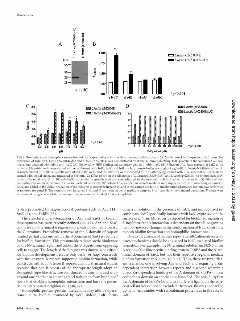
To determine whether IsdC protein that is attached to the sur-face of a bacterial cell could interact with other IsdC molecules, theisdC gene was cloned into the nisin-inducible L. lactis expressionvector pNZ8048. The sortase B cleavage motif NPQTS waschanged to LPQTG in order to provide a substrate for sortase A in
L. lactis. After induction, expression of IsdC was detected in thecell wall fraction derived from protoplasts of L. lactis by Westernimmunoblotting (Fig. 8A), indicating that IsdC was sorted to thecell wall. When the ability of L. lactis expressing IsdC to adhere toimmobilized Isd proteins was tested, L. lactis(pNZ8048isdC) ad-hered to immobilized IsdC at a 4-fold-higher level than the con-trol strain bearing the empty vector but not significantly to IsdB,IsdJ, or IsdK. A slight promotion of adherence to IsdB was noted,but this was not statistically significant. These data are consistentwith the conclusion that IsdC homophilic interactions contributeto the accumulation phase of S. lugdunensis biofilm formationunder iron-restricted conditions (Fig. 8B). As reported for biofilmformation by S. lugdunensis N920143 (Fig. 6B), the interaction ofL. lactis(pNZ8048isdC) with immobilized IsdC was enhanced atpH 6.0 and 6.5 and decreased at pH 5.0, 5.5, 7.0, 7.5, and 8.0(Fig. 8C). Consistent with low-iron-induced biofilm formation(Fig. 5), we also found that attachment of L. lactis(pNZ8048isdC)to surface-coated IsdC requires the presence of FeCl3 (�1 �M).Significantly reduced levels of IsdC homophilic interactions wereobserved when bacterial attachment was performed in iron-de-pleted conditions (Fig. 8D).
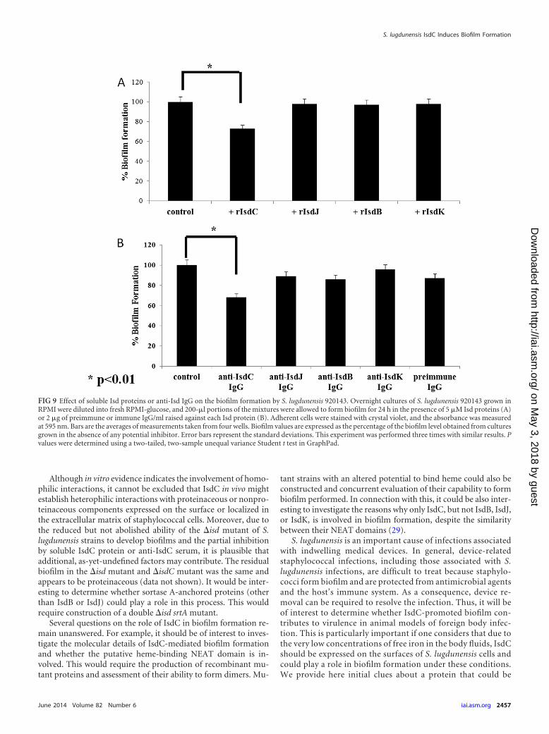
Inhibition by recombinant Isd proteins and antibodies. Thesoluble recombinant Isd proteins or specific antisera raised againsteach of the Isd proteins were added to the RPMI0.3%glucose–2 mMglutamine medium at the same time as the inoculum and incu-bated for 24 h before the density of biofilm formed was measured.Only IsdC protein or anti-IsdC serum caused a significant (�35%) reduction in biofilm formation (Fig. 9). Although completeinhibition was not achieved, we can be confident of the specificityof the effect, which is consistent with an important role in biofilmformation.
DISCUSSION
The identification of surface proteins that are able to induce bio-film development in the absence of polysaccharides is one of themost unexpected results obtained recently in the field of staphy-lococcal biofilm studies. The existence of alternative mechanisms
FIG 5 Iron-dependent biofilm formation by S. lugdunensis N920143. Bacteriagrown overnight in Chelex 100-treated RPMI were diluted in the same me-dium supplemented with increasing amounts of FeCl3 (final concentrationsfrom 0 to 2,000 �M), added to sterile 96-well polystyrene plates, and staticallyincubated at 37°C for 24 h. The experimental conditions for biofilm detectionare identical to those reported in Fig. 2. The upper part of the figure shows thedetection by Western blotting of IsdC protein in the lysates obtained fromcultures grown overnight planktonically in different FeCl3 concentrations.This experiment was performed three times with similar results. P values weredetermined using a two-tailed, two-sample unequal variance Student t test inGraphPad.
FIG 6 Effect of pH on the IsdC expression and biofilm formation by S. lugdunensis N920143. (A) S. lugdunensis N920143 was grown overnight in RPMI adjustedto the indicated pH and the corresponding lysates subjected to Western immunoblotting. IsdC protein transblotted onto nitrocellulose filter was detected withrabbit anti-IsdC IgG, followed by HRP-conjugated secondary goat anti-rabbit IgG. (B) S. lugdunensis N920143 and its isdC deletion mutant were grown overnightin RPMI, diluted 1:200 in the same medium adjusted to different pHs, added to sterile 96-well polystyrene plates, and statically incubated at 37°C for 24 h.Experimental conditions for biofilm detection are identical to those reported in Fig. 2. This experiment was performed three times with similar results. P valueswere determined using a two-tailed, two-sample unequal variance Student t test in GraphPad.
Missineo et al.
2454 iai.asm.org Infection and Immunity
on May 3, 2018 by guest
http://iai.asm.org/
Dow
nloaded from
promoting biofilm development suggests that different staphylo-coccal strains might involve a specific component to form a par-ticular type of biofilm matrix. S. aureus can opt for at least fivedifferent surface proteins, Bap (8), protein A (11), SasG (9), SasC(10), and FnBPs (12, 13) to achieve protein-mediated biofilm.Moreover, functional amyloids composed of phenol solublemodulins stabilize S. aureus biofilms (43). A similar proteinaceousbiofilm has been demonstrated with S. epidermidis based on theexpression of proteins such as Aap (14) or SesC (16, 44). Further-more, the same staphylococcal strain can switch between a pro-teinaceous or polysaccharide-based biofilm, depending on thegrowth conditions (45).
Investigating biofilm production by a collection of strains of S.lugdunensis, we confirmed the proteinaceous nature of biofilms iniron-rich (TSB) medium (24) and demonstrated for the first timethat biofilm formed in iron-restricted conditions (RPMI) was alsoprotein dependent. We found that proteins expressed by the isdlocus are involved in biofilm development, and we identified theIsdC protein as the main factor responsible of cell-to-cell interac-tions and biofilm formation in iron-restricted conditions. IsdCcontributed both to the attachment of cells to a polystyrene sur-face and to the accumulation phase. Thus, IsdC can be added tothe growing list of proteinaceous factors involved in staphylococ-cal biofilm formation.
Surprisingly, for maximal biofilm formation, both IsdC ex-
pression and iron-restricted conditions were required, whereasS. lugdunensis grown in completely iron-depleted conditions(Chelex-treated RPMI) or in RPMI supplemented with excessamounts of FeCl3 (�250 �M) developed a significantly re-duced biofilm. Thus, iron plays a dual role in IsdC-dependentbiofilm formation. Iron restriction is required to induce IsdCexpression, and a low level of iron is required to support theformation of biofilm, whereas high levels (�250 �M) are in-hibitory. Given that S. lugdunensis biofilm is dependent onIsdC and that IsdC has been demonstrated to bind heme (29),it is also possible that IsdC-associated heme is involved in bio-film formation, although it cannot be excluded that the re-quirement for iron could be unrelated to heme binding. Thefact that IsdC is only expressed under iron-restricted condi-tions and the ability of S. lugdunensis strains also to form pro-teinaceous biofilm under iron-replete conditions implies thatbiofilm formation in TSB is promoted by proteinaceous factorsdistinct from those involved in RPMI growth medium. Itwould be interesting to determine whether the protein(s) in-volved are anchored by sortase A.
The observation that IsdC confers primary attachment to poly-styrene and production of cell aggregates in the accumulationphase of biofilm, parallels the behavior observed for Bap (8) andSasC (10) of S. aureus. The aggregation activity observed for IsdC
FIG 7 Analysis of IsdC interactions by size exclusion chromatography. A 100-�l sample of IsdC at a concentration of 100 �M was loaded onto a gel filtrationcolumn of Superose 12 HR and eluted with one column volume (Vt � 24 ml) of PBS with or without 100 �M FeCl3 at a flow rate of 0.5 ml/min. Molecular weightdetermination was carried out by comparing elution volumes of the two peaks marked as IsdC1 and IsdC2, with the values obtained for several known calibrationstandards. The Mr of IsdC1 or IsdC2 was determined from the calibration curve (plot of Kav versus log Mr) once their Kav values were calculated from themeasured elution volumes [Kav � (elution volume void volume)/(column volume void volume)] (inset).
S. lugdunensis IsdC Induces Biofilm Formation
June 2014 Volume 82 Number 6 iai.asm.org 2455
on May 3, 2018 by guest
http://iai.asm.org/
Dow
nloaded from
is also promoted by staphylococcal proteins such as Aap (14),SasG (9), and FnBPs (13).
The structural characterization of Aap and SasG in biofilmdevelopment has been recently defined (46, 47). Aap and SasGcomprise an N-terminal A region and repeated B domains towardthe C terminus. Proteolytic removal of the A domain of Aap orlimited partial cleavage within the B domains of SasG is requiredfor biofilm formation. This presumably reduces steric hindranceby the N-terminal region and allows the B repeats from opposingcells to engage. The length of the B region was shown to be criticalfor biofilm development because only SasG (or Aap) constructswith five or more B-repeats supported biofilm formation, whileconstructs with four or fewer B-repeats did not. Structural studiesrevealed that Aap B-repeats of the appropriate length adopt anelongated, rope-like structure coordinated by zinc ions and wraparound one another in an antiparallel fashion to form bundles offibers that establish homophilic interactions and have the poten-tial to interconnect neighbor cells (46, 47).
Homophilic protein-protein interactions may also be opera-tional in the biofilm promoted by IsdC. Indeed, IsdC forms
dimers in solution in the presence of FeCl3 and immobilized re-combinant IsdC specifically interacts with IsdC expressed on thesurface of L. lactis. Moreover, as reported for biofilm formation byS. lugdunensis, this interaction is dependent on the pH, suggestingthat pH-induced changes in the conformation of IsdC contributeto both biofilm formation and homophilic interactions.
Due to the absence of tandem repeats in IsdC, alternative struc-tures/mechanisms should be envisaged in IsdC-mediated biofilmformation. For example, the N-terminal subdomain N2N3 of theA region of the fibronectin-binding protein FnBPA and the N-ter-minal domain of SasC, but not their repetitive regions, mediatebiofilm formation in S. aureus (10, 13). Thus, there are two differ-ent scenarios: one involving Aap and SasG and requiring a Zn-dependent interaction between repeats and a second wherein adirect Zn-dependent binding of the A domain of FnBPA on onecell to the A domain on another one is needed. The possibility thatthe A domain of FnBPA bound to a different ligand on the adja-cent cell surface cannot be excluded. However, this was not backedup by in vitro studies with recombinant proteins as in the case ofIsdC.
FIG 8 Homophilic and heterophilic interactions of IsdC expressed by L. lactis with surface-coated Isd proteins. (A) Validation of IsdC expression by L. lactis. Theexpression of IsdC by L. lactis(pNZ8084isdC) and L. lactis(pNZ8084) was demonstrated by Western immunoblotting. IsdC protein in the solubilized cell walllysates was detected with rabbit anti-IsdC IgG, followed by HRP-conjugated secondary goat anti-rabbit IgG. (B) Adhesion of L. lactis expressing IsdC to Isdproteins. Microtiter wells were coated with recombinant IsdB, IsdC, IsdK, and IsdJ in a bicarbonate buffer overnight (1 �g/well). L. lactis(pNZ8084isdC) and L.lactis(pNZ8084) (5 � 108 cells/well) were added to the wells, and the mixtures were incubated for 1 h. After being washed with PBS, adherent cells were fixed,stained with crystal violet, and measured at 595 nm. (C) Effect of pH on the adherence of L. lactis(pNZ8084isdC) and L. lactis(pNZ8084) to immobilized IsdCprotein. Bacterial cells (5 � 108 cells/well) suspended in growth medium were adjusted to the indicated pHs and added to the wells. (D) Effect of ironconcentrations on the adherence of L. lactis. Bacterial cells (5 � 108 cells/well) suspended in growth medium were supplemented with increasing amounts ofFeCl3 and added to the wells. Incubation of the mixtures as described in panels C and D was carried out for 1 h, and detection of attached bacteria was performedas reported for panel B. The results shown in panels B, C, and D are mean values of triplicate samples. Error bars show the standard deviations. P values weredetermined using a two-tailed, two-sample unequal variance Student t test in GraphPad.
Missineo et al.
2456 iai.asm.org Infection and Immunity
on May 3, 2018 by guest
http://iai.asm.org/
Dow
nloaded from
Although in vitro evidence indicates the involvement of homo-philic interactions, it cannot be excluded that IsdC in vivo mightestablish heterophilic interactions with proteinaceous or nonpro-teinaceous components expressed on the surface or localized inthe extracellular matrix of staphylococcal cells. Moreover, due tothe reduced but not abolished ability of the �isd mutant of S.lugdunensis strains to develop biofilms and the partial inhibitionby soluble IsdC protein or anti-IsdC serum, it is plausible thatadditional, as-yet-undefined factors may contribute. The residualbiofilm in the �isd mutant and �isdC mutant was the same andappears to be proteinaceous (data not shown). It would be inter-esting to determine whether sortase A-anchored proteins (otherthan IsdB or IsdJ) could play a role in this process. This wouldrequire construction of a double �isd srtA mutant.
Several questions on the role of IsdC in biofilm formation re-main unanswered. For example, it should be of interest to inves-tigate the molecular details of IsdC-mediated biofilm formationand whether the putative heme-binding NEAT domain is in-volved. This would require the production of recombinant mu-tant proteins and assessment of their ability to form dimers. Mu-
tant strains with an altered potential to bind heme could also beconstructed and concurrent evaluation of their capability to formbiofilm performed. In connection with this, it could be also inter-esting to investigate the reasons why only IsdC, but not IsdB, IsdJ,or IsdK, is involved in biofilm formation, despite the similaritybetween their NEAT domains (29).
S. lugdunensis is an important cause of infections associatedwith indwelling medical devices. In general, device-relatedstaphylococcal infections, including those associated with S.lugdunensis infections, are difficult to treat because staphylo-cocci form biofilm and are protected from antimicrobial agentsand the host’s immune system. As a consequence, device re-moval can be required to resolve the infection. Thus, it will beof interest to determine whether IsdC-promoted biofilm con-tributes to virulence in animal models of foreign body infec-tion. This is particularly important if one considers that due tothe very low concentrations of free iron in the body fluids, IsdCshould be expressed on the surfaces of S. lugdunensis cells andcould play a role in biofilm formation under these conditions.We provide here initial clues about a protein that could be
FIG 9 Effect of soluble Isd proteins or anti-Isd IgG on the biofilm formation by S. lugdunensis 920143. Overnight cultures of S. lugdunensis 920143 grown inRPMI were diluted into fresh RPMI-glucose, and 200-�l portions of the mixtures were allowed to form biofilm for 24 h in the presence of 5 �M Isd proteins (A)or 2 �g of preimmune or immune IgG/ml raised against each Isd protein (B). Adherent cells were stained with crystal violet, and the absorbance was measuredat 595 nm. Bars are the averages of measurements taken from four wells. Biofilm values are expressed as the percentage of the biofilm level obtained from culturesgrown in the absence of any potential inhibitor. Error bars represent the standard deviations. This experiment was performed three times with similar results. Pvalues were determined using a two-tailed, two-sample unequal variance Student t test in GraphPad.
S. lugdunensis IsdC Induces Biofilm Formation
June 2014 Volume 82 Number 6 iai.asm.org 2457
on May 3, 2018 by guest
http://iai.asm.org/
Dow
nloaded from
targeted to prevent developing of biofilm under conditions re-sembling those present in the body fluids.
ACKNOWLEDGMENTS
We acknowledge funding from Fondazione CARIPLO (Grant Vaccines2009-3546) to P.S. Research in Dublin was supported by the IRCSETEmbark Scholarship and by Science Foundation Ireland (Programme In-vestigator grant 08/IN.1/B1854).
REFERENCES1. Götz F. 2002. Staphylococcus and biofilms. Mol. Microbiol. 43:1367–1378.
http://dx.doi.org/10.1046/j.1365-2958.2002.02827.x.2. Stewart PS, Costerton JW. 2001. Antibiotic resistance of bacteria in biofilms.
Lancet 358:135–138. http://dx.doi.org/10.1016/S0140-6736(01)05321-1.3. Vuong C, Voyich JM, Fischer ER, Braughton KR, Whitney AR, DeLeo
FR, Otto M. 2004. Polysaccharide intercellular adhesin (PIA) protectsStaphylococcus epidermidis against major components of the human in-nate immune system. Cell. Microbiol. 6:269 –275. http://dx.doi.org/10.1046/j.1462-5822.2004.00367.x.
4. Bose JL, Lehman MK, Fey PD, Bayles KW. 2012. Contribution of theStaphylococcus aureus Atl AM and GL murein hydrolase activities in celldivision, autolysis, and biofilm formation. PLoS One 7:e42244. http://dx.doi.org/10.1371/journal.pone.0042244.
5. Houston P, Rowe SE, Pozzi C, Waters EM, O’Gara JP. 2011. Essentialrole for the major autolysin in the fibronectin-binding protein-mediatedStaphylococcus aureus biofilm phenotype. Infect. Immun. 79:1153–1165.http://dx.doi.org/10.1128/IAI.00364-10.
6. Vaudaux PE, François P, Proctor RA, McDevitt D, Foster TJ, AlbrechtRM, Lew DP, Wabers H, Cooper SL. 1995. Use of adhesion-defectivemutants of Staphylococcus aureus to define the role of specific plasmaproteins in promoting bacterial adhesion to canine arteriovenous shunts.Infect. Immun. 63:585–590.
7. Heilmann C, Schweitzer O, Gerke C, Vanittanakom N, Mack D, GötzF. 1996. Molecular basis of intercellular adhesion in the biofilm-formingStaphylococcus epidermidis. Mol. Microbiol. 20:1083–1091. http://dx.doi.org/10.1111/j.1365-2958.1996.tb02548.x.
8. Cucarella C, Solano C, Valle J, Amorena B, Lasa I, Penadés JR. 2001. Bap, aStaphylococcus aureus surface protein involved in biofilm formation. J.Bacteriol. 183:2888 –2896. http://dx.doi.org/10.1128/JB.183.9.2888-2896.2001.
9. Geoghegan JA, Corrigan RM, Gruszka DT, Speziale P, O’Gara JP, PottsJR, Foster TJ. 2010. Role of surface protein SasG in biofilm formation byStaphylococcus aureus. J. Bacteriol. 192:5663–5673. http://dx.doi.org/10.1128/JB.00628-10.
10. Schroeder K, Jularic M, Horsburgh SM, Hirschhausen N, Neumann C,Bertling A, Schulte A, Foster S, Kehrel BE, Peters G, Heilmann C. 2009.Molecular characterization of a novel Staphylococcus aureus surface pro-tein (SasC) involved in cell aggregation and biofilm accumulation. PLoSOne 4:e7567. http://dx.doi.org/10.1371/journal.pone.0007567.
11. Merino N, Toledo-Arana A, Vergara-Irigaray M, Valle J, Solano C,Calvo E, Lopez JA, Foster TJ, Penadés JR, Lasa I. 2009. Protein A-me-diated multicellular behavior in Staphylococcus aureus. J. Bacteriol. 191:832– 843. http://dx.doi.org/10.1128/JB.01222-08.
12. O’Neill E, Pozzi C, Houston P, Humphreys H, Robinson DA, Lough-man A, Foster TJ, O’Gara JP. 2008. A novel Staphylococcus aureus biofilmphenotype mediated by the fibronectin-binding proteins, FnBPA and Fn-BPB. J. Bacteriol. 190:3835–3850. http://dx.doi.org/10.1128/JB.00167-08.
13. Geoghegan JA, Monk IR, O’Gara JP, Foster TJ. 2013. Subdomains N2N3of fibronectin binding protein A mediate Staphylococcus aureus biofilmformation and adherence to fibrinogen using distinct mechanisms. J. Bac-teriol. 195:2675–2683. http://dx.doi.org/10.1128/JB.02128-12.
14. Rohde H, Burdelski C, Bartscht K, Hussain M, Buck F, Horstkotte MA,Knobloch JK, Heilmann C, Herrmann M, Mack D. 2005. Induction ofStaphylococcus epidermidis biofilm formation via proteolytic processing ofthe accumulation-associated protein by staphylococcal and host pro-teases. Mol. Microbiol. 55:1883–1895. http://dx.doi.org/10.1111/j.1365-2958.2005.04515.x.
15. Banner MA, Cunniffe JG, Macintosh RL, Foster TJ, Rohde H, Mack D,Hoyes E, Derrick J, Upton M, Handley PS. 2007. Localized tufts of fibrilson Staphylococcus epidermidis NCTC 11047 are comprised of the accumu-lation-associated protein. J. Bacteriol. 189:2793–2804. http://dx.doi.org/10.1128/JB.00952-06.
16. Shahrooei M, Hira V, Stijlemans B, Merckx R, Hermans PW, VanEldere J. 2009. Inhibition of Staphylococcus epidermidis biofilm formationby rabbit polyclonal antibodies against the SesC protein. Infect. Immun.77:3670 –3678. http://dx.doi.org/10.1128/IAI.01464-08.
17. Freney J, Brun Y, Bes M, Meugnier H, Grimont F, Grimont PAD, Nervi C,Fleurette J. 1988. Staphylococcus lugdunensis sp. nov. and Staphylococcus schleiferisp.nov.,twospeciesfromhumanclinicalspecimens.Int.J.Syst.Bacteriol.38:168–172. http://dx.doi.org/10.1099/00207713-38-2-168.
18. Vandenesch F, Etienne J, Reverdy ME, Eykyn SJ. 1993. Endocarditis dueto Staphylococcus lugdunensis: report of 11 cases and review. Clin. Infect.Dis. 17:871– 876.
19. Patel R, Piper KE, Rouse MS, Uhl JR, Cockerill FR, III, Steckelberg JM.2000. Frequency of isolation of Staphylococcus lugdunensis among staph-ylococcal isolates causing endocarditis: a 20-year experience. J. Clin. Mi-crobiol. 38:4262– 4263.
20. Anguera I, Del Río A, Miró JM, Matínez-Lacasa X, Marco F, Gumá JR,Quaglio G, Claramonte X, Moreno A, Mestres CA, Mauri E, AzquetaM, Benito N, García-de la María C, Almela M, Jiménez-Expósito, SuedMJO, De Lazzari E, Gatell JM. 2005. Staphylococcus lugdunensis infectiveendocarditis: description of 10 cases and analysis of native valve, pros-thetic valve, and pacemaker lead endocarditis clinical profiles. Heart 91:e10. http://dx.doi.org/10.1136/hrt.2004.040659.
21. Ebright JR, Penugonda N, Brown W. 2004. Clinical experience with Staphylo-coccus lugdunensis bacteremia: a retrospective analysis. Diagn. Microbiol. Infect.Dis. 48:17–21. http://dx.doi.org/10.1016/j.diagmicrobio.2003.08.008.
22. Sampathkumar P, Osmon DR, Cockerill FR. 2000. Prosthetic jointinfection due to Staphylococcus lugdunensis. Mayo Clin. Proc. 75:511–512.http://dx.doi.org/10.1016/S0025-6196(11)64220-1.
23. Sandoe JA, Longshaw CM. 2001. Ventriculoperitoneal shunt infectioncaused by Staphylococcus lugdunensis. Clin. Microbiol. Infect. 7:385–387.http://dx.doi.org/10.1046/j.1198-743x.2001.00268.x.
24. Frank KL, Patel R. 2007. Poly-N-acetylglucosamine is not a major com-ponent of the extracellular matrix in biofilms formed by icaADBC-positive Staphylococcus lugdunensis isolates. Infect. Immun. 75:4728 –4742. http://dx.doi.org/10.1128/IAI.00640-07.
25. Geoghegan JA, Ganesh VK, Smeds E, Liang X, Höök M, Foster TJ. 2010.Molecular characterization of the interaction of staphylococcal microbialsurface components recognizing adhesive matrix molecules(MSCRAMM) ClfA and Fbl with fibrinogen. J. Biol. Chem. 285:6208 –6216. http://dx.doi.org/10.1074/jbc.M109.062208.
26. Szabados F, Marlinghaus L, Korte M, Neumann S, Kaase M, Gater-mann SG. 2011. Fbl is not involved in the invasion of eukaryotic epithelialand endothelial cells by Staphylococcus lugdunensis. FEMS Microbiol. Lett.324:48 –55. http://dx.doi.org/10.1111/j.1574-6968.2011.02382.x.
27. Nilsson M, Bjerketorp J, Wiebensjö A, Ljungh A, Frykberg L, Guss B.2004. A von Willebrand factor-binding protein from Staphylococcus lug-dunensis. FEMS Microbiol. Lett. 234:155–161. http://dx.doi.org/10.1111/j.1574-6968.2004.tb09527.x.
28. Heilbronner S, Holden MT, van Tonder A, Geoghegan JA, Foster TJ,Parkhill J, Bentley SD. 2011. Genome sequence of Staphylococcus lug-dunensis N920143 allows identification of putative colonization and viru-lence factors. FEMS Microbiol. Lett. 322:60 – 67. http://dx.doi.org/10.1111/j.1574-6968.2011.02339.x.
29. Zapotoczna M, Heilbronner S, Speziale P, Foster TJ. 2012. Iron-regulated surface determinant (Isd) proteins of Staphylococcus lugdunen-sis. J. Bacteriol. 194:6453– 6467. http://dx.doi.org/10.1128/JB.01195-12.
30. Haley KP, Janson EM, Heilbronner S, Foster TJ, Skaar EP. 2011.Staphylococcus lugdunensis IsdG liberates iron from host heme. J. Bacte-riol. 193:4749 – 4757. http://dx.doi.org/10.1128/JB.00436-11.
31. Clarke SR, Mohamed R, Bian L, Routh AF, Kokai-Kun JF, Mond JJ,Tarkowski A, Foster SJ. 2007. The Staphylococcus aureus surface proteinIsdA mediates resistance to innate defenses of human skin. Cell Host Mi-crobe 1:199 –212. http://dx.doi.org/10.1016/j.chom.2007.04.005.
32. Cramton SE, Gerke C, Schnell NF, Nichols WW, Götz F. 1999. Theintercellular adhesion (ica) locus is present in Staphylococcus aureus and isrequired for biofilm formation. Infect. Immun. 67:5427–5433.
33. Muller E, Takeda S, Shiro H, Goldmann D, Pier GB. 1993. Occurrenceof capsular polysaccharide/adhesin among clinical isolates of coagulase-negative staphylococci. J. Infect. Dis. 168:1211–1218. http://dx.doi.org/10.1093/infdis/168.5.1211.
34. Heilbronner S, Hanses F, Monk IR, Speziale P, Foster TJ. 2013. SortaseA promotes virulence in experimental Staphylococcus lugdunensis endo-carditis. Microbiology 159:2141–2152.
Missineo et al.
2458 iai.asm.org Infection and Immunity
on May 3, 2018 by guest
http://iai.asm.org/
Dow
nloaded from
35. Corrigan RM, Foster TJ. 2009. An improved tetracycline-inducible ex-pression vector for Staphylococcus aureus. Plasmid 61:126 –129. http://dx.doi.org/10.1016/j.plasmid.2008.10.001.
36. Li MZ, Elledge SJ. 2007. Harnessing homologous recombination in vitroto generate recombinant DNA via SLIC. Nat. Methods 4:251–256. http://dx.doi.org/10.1038/nmeth1010.
37. Li MZ, Elledge SJ. 2012. SLIC: a method for sequence- and ligation-independent cloning. Methods Mol. Biol. 852:51–59. http://dx.doi.org/10.1007/978-1-61779-564-0_5.
38. Bahey-El-Din M, Griffin BT, Gahan CG. 2008. Nisin inducible produc-tion of listeriolysin O in Lactococcus lactis NZ9000. Microb. Cell Fact 7:24.http://dx.doi.org/10.1186/1475-2859-7-24.
39. Monk IR, Casey PG, Hill C, Gahan CG. 2010. Directed evolution andtargeted mutagenesis to murinize Listeria monocytogenes internalin A forenhanced infectivity in the murine oral infection model. BMC Microbiol.10:318. http://dx.doi.org/10.1186/1471-2180-10-318.
40. Christensen GD, Simpson WA, Younger JJ, Baddour LM, Barrett FF,Melton DM, Beachey EH. 1985. Adherence of coagulase-negative staph-ylococci to plastic tissue culture plates: a quantitative model for the adher-ence of staphylococci to medical devices. J. Clin. Microbiol. 22:996 –1006.
41. Chaignon P, Sadovskaya I, Ragunah Ch Ramasubbu N, Kaplan JB,Jabbouri S. 2007. Susceptibility of staphylococcal biofilms to enzymatictreatments depends on their chemical composition. Appl. Microbiol. Bio-technol. 75:125–132. http://dx.doi.org/10.1007/s00253-006-0790-y.
42. Izano EA, Amarante MA, Kher WB, Kaplan JB. 2008. Differential roles
of poly-N-acetylglucosamine surface polysaccharide and extracellularDNA in Staphylococcus aureus and Staphylococcus epidermidis biofilms.Appl. Environ. Microbiol. 74:470 – 476. http://dx.doi.org/10.1128/AEM.02073-07.
43. Schwartz K, Syed AK, Stephenson RE, Rickard AH, Boles BR. 2012.Functional amyloids composed of phenol soluble modulins stabilizeStaphylococcus aureus biofilms. PLoS Pathog. 8:e1002744. http://dx.doi.org/10.1371/journal.ppat.1002744.
44. Shahrooei M, Hira V, Khodaparast L, Khodaparast L, Stijlemans B,Kucharíková S, Burghout P, Hermans PW, Van Eldere J. 2012. Vacci-nation with SesC decreases Staphylococcus epidermidis biofilm formation.Infect. Immun. 80:3660 –3668. http://dx.doi.org/10.1128/IAI.00104-12.
45. Vergara-Irigaray M, Valle J, Merino N, Latasa C, García B, Ruiz de LosMozos I, Solano C, Toledo-Arana A, Penadés JR, Lasa I. 2009. Relevantrole of fibronectin-binding proteins in Staphylococcus aureus biofilm-associated foreign-body infections. Infect. Immun. 77:3978 –3991. http://dx.doi.org/10.1128/IAI.00616-09.
46. Conrady DG, Wilson JJ, Herr AB. 2013. Structural basis for Zn2�-dependentintercellular adhesion in staphylococcal biofilms. Proc. Natl. Acad. Sci. U. S. A.110:E202–E211. http://dx.doi.org/10.1073/pnas.1208134110.
47. Gruszka DT, Wojdyla JA, Bingham RJ, Turkenburg JP, Manfield IW,Steward A, Leech AP, Geoghegan JA, Foster TJ, Clarke J, Potts JR. 2012.Staphylococcal biofilm-forming protein has a contiguous rod-like struc-ture. Proc. Natl. Acad. Sci. U. S. A. 109:E1011–E1018. http://dx.doi.org/10.1073/pnas.1119456109.
S. lugdunensis IsdC Induces Biofilm Formation
June 2014 Volume 82 Number 6 iai.asm.org 2459
on May 3, 2018 by guest
http://iai.asm.org/
Dow
nloaded from
![Staphylococcus lugdunensis Infections of the Skin and Soft … · 2017. 11. 21. · skin and soft tissue [1]. We describe five patients who developed significant skin and soft tissue](https://img.pdfslide.us/doc/110x75/5ffa3471c482fc7f276c61e5/staphylococcus-lugdunensis-infections-of-the-skin-and-soft-2017-11-21-skin.jpg)


![SOCIORUM ET OFFICIORlill PROVINCIA] LUGDUNENSIS · sociorum et officiorlill provincia] lugdunensis ... elias briguet, credent., ... p. josephus andrÉ, prre¡:](https://img.pdfslide.us/doc/110x75/5b9d498d09d3f2df1f8ca31b/sociorum-et-officiorlill-provincia-sociorum-et-officiorlill-provincia-lugdunensis.jpg)