Embed Size (px)
Citation preview

Injury and Intestinal Barrier Dysfunction: Past,
Present, and Future
Raul Coimbra MD, PhD, FACSProfessor of Surgery
Chief, Division of Trauma, Surgical Critical Care, and Burns
Department of Surgery
University of California San Diego School of Medicine
Western Trauma Association
1st Founder’s Basic Science Lecture

Background
• The intestine plays a significant role in the systemic inflammatory response (SIRS)
• SIRS can lead to distant organ injury, multi-organ failure,
and death
• Our understanding of the gut’s role in causing SIRS has evolved over the past several decades
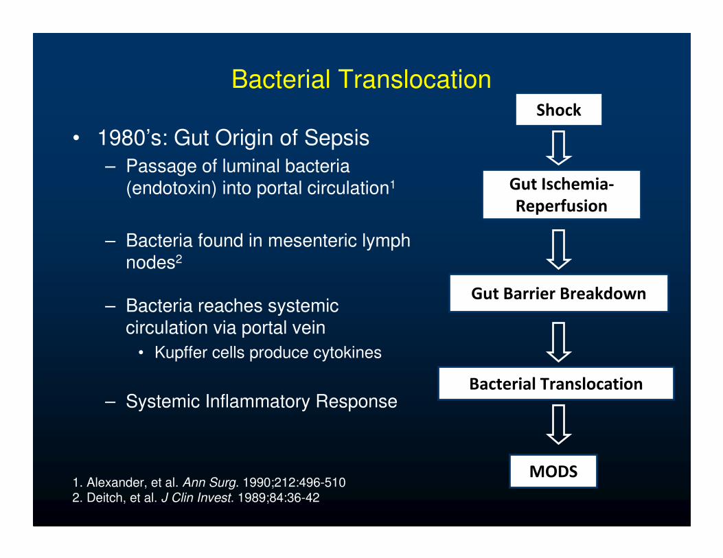
Bacterial Translocation
• 1980’s: Gut Origin of Sepsis
– Passage of luminal bacteria (endotoxin) into portal circulation1
– Bacteria found in mesenteric lymph nodes2
– Bacteria reaches systemic circulation via portal vein
• Kupffer cells produce cytokines
– Systemic Inflammatory Response
Shock
Gut Ischemia-
Reperfusion
Gut Barrier Breakdown
Bacterial Translocation
MODS1. Alexander, et al. Ann Surg. 1990;212:496-510 2. Deitch, et al. J Clin Invest. 1989;84:36-42

Bacterial Translocation
• Early 1990’s: Bacterial Translocation in question
• Moore, et al: Is there enteric bacteria in the portal blood of severely injured trauma patients?
– 20 injured patients requiring emergent laparotomy
– Portal vein catheters inserted
– Blood drawn up to 5 days post-operatively
– 8/212 (2%) of blood cultures positive
• 7 presumed contaminants
• 1 S. Aureus in patient with known S. Aureus pneumonia
– Conclusion: No portal or systemic bacteremia despite 30% incidence of MOF in these patients
Moore FA, et al. J Trauma. 1991;31:629-636.

"We look hard at what is routinely done in
the Shock Trauma ICU and ask, 'How does
this treatment affect the gut function?' We
are finding that when a person is critically
ill, the gastrointestinal (GI) tract doesn't
work. If we can make the gut work better,
then we can prevent a lot of infection,"

Gut Inflammation
• 1990’s-Present: Gut Inflammation
– Gut barrier breakdown causes
intestinal inflammatory response
• Intestinal cytokine production
– Gut-derived inflammatory
mediators carried in intestinal
lymph
– Activated intestinal lymph causes SIRS, distant organ injury
Gut Ischemia-
Reperfusion
Gut Barrier Breakdown
Intestinal Inflammatory
Response
MODS
Activated Mesenteric
Lymph
1. Deitch, EA. Surgery. 2002;131:241-2442. Magnotti, et al. Ann Surg. 1998;228:518-27


• Mesenteric lymph from burned animals:
– Activate PMNs
– Activate endothelial cells
• Portal vein plasma did not activate PMNs
Deitch, et al. Crit Care Med. 2004;32:533-38

Senthil, et al. Ann Surg. 2007;246:822-830
ShamSham
Injected with HS Lymph Hem. Shock

Watkins, et al. J Trauma. 2008;64:126-130.
• Lymphatic Duct Ligation (LDL)
– Decreases histologic lung injury
– Decreases lung permeability
– Decreases neutrophil CD11b expression
Lung Injury Score

• Mesenteric lymph flow depends on depth of shock
• Maximal PMN priming by
mesenteric lymph occurs in
the 3rd hour post-shock
• Activity of mesenteric lymph depends on depth
and duration of shock
Masuno, et al. Shock. 2006;26:285-289
Time Course of PMN Priming byMesenteric Lymph


RINGERRINGER’’S LACTATES LACTATE
�Current standard
resuscitation regimen
• Potentiates neutrophil activation
• Rhee et al. 1998
• Contributes to end organ injury
• Savage et al. 2005
Sham
RL

Pentoxifylline (PTX)
• Non-specific
Phosphodiesterase Inhibitor
– Increases cyclic AMP
– PKA activation
• Clinical Applications:
– Intermittent Claudication
– Alcoholic Hepatitis
• Animal Models:
– Decreases pro-inflammatory
cytokine activation
– Attenuates neutrophil oxidative
burst
– Decreases distant organ injury

Hemorrhagic shock
HTS LR0
20
40
60
80
%
LR+ PTX
*
Bacterial Translocation
0
2
4
6
8
10
0 60 120TIME (min)
mm
ol/
L
HTSLR
LR + PTX
Lactate
Coimbra R, et al, Shock 2000

Classic treatment
HemorrhagicShock
Ringer’s Lactate
Proposed treatment
HemorrhagicShock
Hypertonic Saline + Pentoxifylline
•Improves microcirculation
•Attenuates oxidative stress
•Downregulates neutrophil function
•Reduces host organ injury

Hemorrhagic Shock
Ringer’s Lactate (RL)
• Potentiate neutrophil
activation
Resuscitation 2004
• Promote endothelial
dysfunction
J Trauma 2005
• Contribute to end
organ injury
J Trauma 2006
HSPTX• Reduce oxidative stress
• Downregulate PMN function
J Trauma 2005
• Attenuate Post-shock Lung Injury
J Trauma 2006

Coimbra, et al. J Trauma 2006;60:41-51
Sham Ringers Lactate HSPTX

Nitric oxide and Ischemia Reperfusion
• iNOS induction and production of sustained quantities of NO occur in the gut after I/R injury.
• Nitric oxideDirect effects on cell signaling: Transcription
factor activation (NF-κB and STAT3) and cytokine production (TNF-α and IL-6)
J Exp Med 1998
Indirect cytotoxic effects: Peroxynitrite formation

Intestinal I/R Injury
iNOS
NO
NF-κB/STAT3 Peroxynitrite
TNF-α, CINC, IL-6 Organ Injury

Hypothesis
• The attenuation in gut injury observed with HSPTX after hemorrhagic shock is associated with a decrease in intestinal iNOS activity and NO-mediated events including local pro-inflammatory cytokine production when compared to RL in vivo.

Methods
• RL: 32 mL/kg racemic RL (n=7)
• HSPTX: 4 mL/kg 7.5% NaCl + PTX 25 mg/kg (n=7)
• Sham group (n=5)
0 1h 5h
Resuscitation Sacrifice andDistal Ileum
Procurement
Shock35 ± 5mm
Hg

iNOS Content
Sham RL HSPTX
Ilea
l iN
OS
Co
nte
nt
(Pix
el
To
tal
+ S
EM
)
0
20000
40000
60000
80000
100000
120000
*
* P < 0.05130 kD

Nitrite
Sham RL HSPTX
Nit
rite
Co
ncen
trati
on
(µµ µµ
mo
l/L
+ S
EM
0
200
400
600
800
1000
1200
1400
1600
1800
*
* P < 0.05

Cytoplasmic I-κBα Phosphorylation
Sham RL HSPTXI-κκ κκB
αα αα P
ho
sp
ho
ryla
tio
n (
Pix
el
To
tal
+ S
EM
)
0
20000
40000
60000
80000
*
* P < 0.0141 kD

Nuclear NF-κB Phosphorylation
Sham RL HSPTXNF
- κκ κκB
p65
Ph
os
ph
ory
lati
on
(P
ixe
l T
ota
l +
SE
M)
0
20000
40000
60000
80000
*
* P < 0.0165 kD

STAT-3

TNF-α
Sham RL HSPTX
TN
F- αα αα
Co
nce
ntr
ati
on
(p
g/m
L +
SE
M)
0
20
40
60
80
100
120
*
* P < 0.01

Interleukin-6
Sham RL HSPTX
IL-6
Co
ncen
trati
on
(p
g/m
L +
SE
M)
0
100
200
300
400
500
*
* P < 0.01

Intestinal I/R Injury
iNOS
NO
NF-κB Peroxynitrite
TNF-α, CINC, IL-6 Organ Injury
HSPTX
HSPTX
HSPTX
HSPTX
HSPTX

Deree, et al. J Trauma. 2007;62:818-28.

TNF-α and Intestinal Barrier
The pivotal role of tumor necrosis factor-alpha
in signaling apoptosis in intestinal epithelial cells under shock conditions.
Diebel LN, Liberati DM, Baylor AE 3rd,
Brown WJ, Diglio CA
J Trauma. 2005 May;58(5):995-100
L. Diebel, MD

Gut Barrier Breakdown
• 2008: Intestinal Barrier Injury
– Can we prevent the intestinal inflammatory response and subsequent SIRS by limiting intestinal barrier breakdown?
• Intestinal Tight Junction
– Creates physical barrier that seals the space between adjacent epithelial cells
– Regulates intestinal permeability
– Modulation of tight junction proteins alters epithelial barrier function
Gut Ischemia-
Reperfusion
Gut Barrier Breakdown
Intestinal Inflammatory
Response
MODS
X
Turner JR. Am J Pathol. 2006;169:1901-1909

Normal Intestinal Barrier
Leaphart CL, Tepas III, JJ;

Intestinal Tight Junction
• Occludin
– Four transmembrane domains
– Attaches adjacent cells at tight junction
• ZO-1
– Attaches occludin to perijunctional actin cytoskeleton
• Myosin light chain kinase (MLCK)
– Increases phosphorylation of myosin light chain (MLC)
– Modulates contraction of the actin cytoskeleton

ZO-1ZO-1
OccludinOccludin
Actin
Actin

ZO-1ZO-1
Occludin
Occludin
TNF-αIL1-βIFN-γ
TNF-αIL1-βIFN-γ
Actin
Actin

Caco-2 cells + Cytomix
Costantini T, et al., Life Sciences, 2009

Caco-2 cells + Cytomix
Costantini T, et al., Life Sciences, 2009

Costantini T, et al., Life Sciences, 2009

Burn-induced Histologic Gut Injury
Costantini, et al. J Trauma. 2009

β-actin
Occludin
6 hour 24 hour
*
†
‡
§
Intestinal Occludin
Costantini, et al. Shock. 2009
* p < 0.01 vs. Sham
† p < 0.05 vs. Burn‡ p < 0.01 vs. Sham§ p < 0.05 vs. Burn

β-actin
ZO-1
6 hour 24 hour
*
†
‡
Intestinal ZO-1
Costantini, et al. Shock. 2009
* p < 0.01 vs. Sham † p < 0.05 vs. Sham‡ p < 0.05 vs. Burn
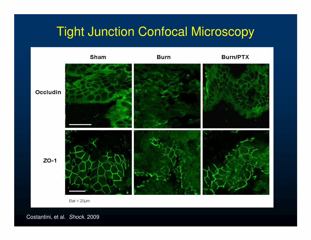
Tight Junction Confocal Microscopy
Costantini, et al. Shock. 2009

Burn-induced Intestinal Permeability to
4kDa FITC-Dextan
* p < 0.001 vs. Sham
† p < 0.001 vs. BurnCostantini, et al. Shock. 2009
Intestinal Permeability
Intestinal Barrier Breakdown
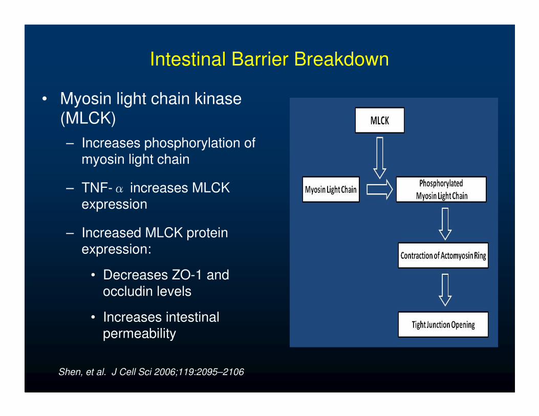
• Myosin light chain kinase (MLCK)
– Increases phosphorylation of myosin light chain
– TNF-α increases MLCK expression
– Increased MLCK protein expression:
• Decreases ZO-1 and
occludin levels
• Increases intestinal permeability
Shen, et al. J Cell Sci 2006;119:2095–2106

Intestinal Barrier Breakdown
• Intestinal NF-κB
– NF-κB mediates
activation of MLCK by binding to MLCK
promoter
– Inhibition of NF-κB p65
decreases MLCK activation
Ye, et al. Am J Physiol Gastrointest Liver Physiol 2006;290:496-504

Methods
30% TBSA steam burn
for 7 seconds
2hr 4hr
IP injection:
12.5mg/kg PTX in
500μl Normal Saline
vs.
500μl Normal Saline
balb/c mice
Harvest Distal Ileum:
Histology
TNF-α ELISA
Confocal Microscopy
- Phosphorylated MLC
Western blot
- MLCK
- Cytoplasmic IKK, IkBa
- Nuclear NF-ΚB p65
Intestinal Permeability:
4 kDa FITC-Dextran

MLCK
Βeta-actin
**
Intestinal Myosin Light Chain Kinase
* p < 0.02 vs. Burn

Cytoplasmic Phosphorylated IKKα/β
P-IKKα/β
IKKα/β
* *
* p < 0.05 vs. Burn

Cytoplasmic Phosphorylated IkBα
P-IkBα
IkBα
**
* p < 0.01 vs. Burn

P-NF-κB p65
Βeta-Laminin
**
Nuclear NF-κBp65
* p < 0.03 vs. Burn

*
†
Intestinal TNF-α
* p < 0.03 vs. Sham
† p < 0.05 vs. Burn

Phosphorylated MLC Confocal Microscopy
Costantini, et al. J Trauma 2009Bar = 20 µm

Intestinal Permeability 4 Hours Post-Burn
J Trauma. 2009;66:17-24

What is in the Future?

Future #1:
Novel Imaging of Intestinal Injury
• Intraluminal placement of near-infrared dye
– Alexa Fluor 680
• Imaging using Xenogen
IVIS Lumina
• Quantification of fluorescence
– Correlates with “classic”assays of intestinal injury and intestinal permeability

Near-infrared Imaging of Intestinal Injury
Sham 4hr Burn 24hr Burn0hr Burn 6hr Burn 48hr Burn

Quantification of Near-infrared Imaging
Sham 4hr Burn Abdominal Quantification
Intestinal Quantification
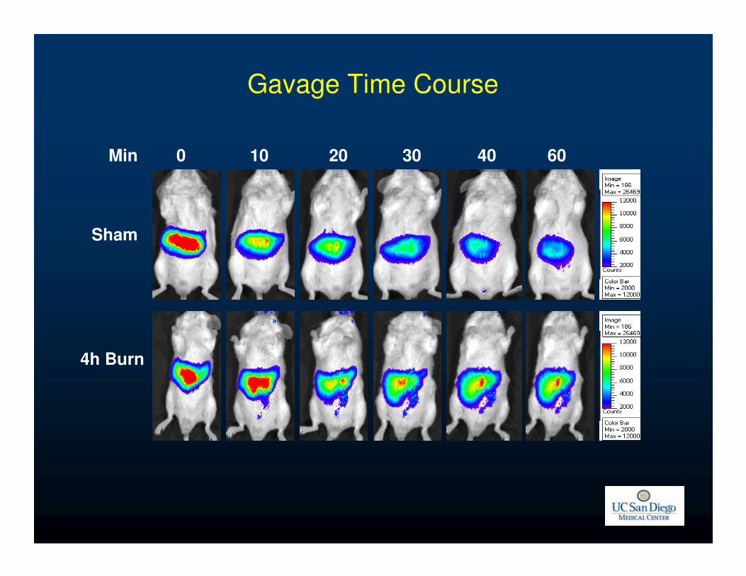
Gavage Time Course
0 10 20 30 40 60Min
Sham
4h Burn
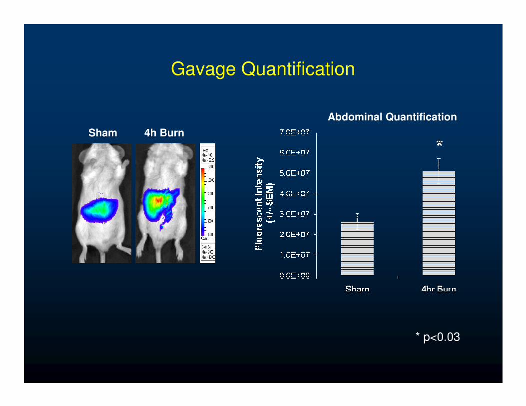
Gavage Quantification
Sham 4h Burn
Abdominal Quantification
* p<0.03

Utilizing Phage Display Technology to Identify
Peptide Sequences Targeting the Burn Injured
Intestinal Barrier
Todd W. Costantini MD, Carrie Y. Peterson MD, James G. Putnam BS, Ritsuko Sawada PhD, William H. Loomis BS, Brian P. Eliceiri PhD,
Andrew Baird PhD, Vishal Bansal MD, Raul Coimbra, MD, PhD
Future #2

Background
• Intestinal injury is known to result from several clinical conditions resulting in significant morbidity and mortality
– Severe trauma, burn
– Inflammatory bowel disease
– Necrotizing enterocolitis
• The ability to effectively target the intestinal mucosa to deliver biotherapies could be of powerful clinical utility
– Prevent gut injury
– Speed intestinal barrier healing

Drug Delivery
• Delivery of therapeutics to the intestinal mucosa remains a difficult problem
• Must be delivered to the cells of the intestinal wall in
sufficient quantities to achieve the desired effect
– Issues of clearance
– Timing of drug delivery
– Alterations in perfusion to the gut following injury
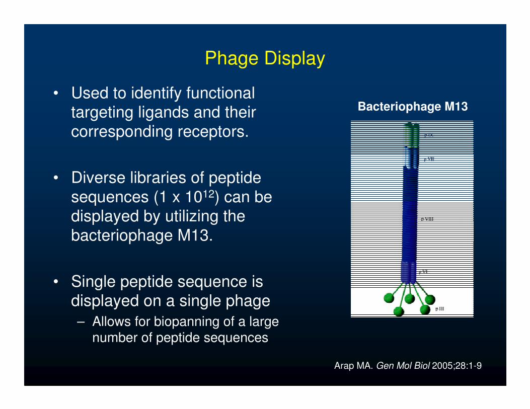
Phage Display
• Used to identify functional targeting ligands and their
corresponding receptors.
• Diverse libraries of peptide
sequences (1 x 10¹²) can be displayed by utilizing the
bacteriophage M13.
• Single peptide sequence is displayed on a single phage
– Allows for biopanning of a large number of peptide sequences
Bacteriophage M13
Arap MA. Gen Mol Biol 2005;28:1-9

Phage Display
• Phage-based vectors can be used to identify peptides which can perform targeted delivery of biotherapeutics
– Genes, antibiotics, growth factors
• Screen for peptides that home to specific tissues
• Wide-ranging applications
– Cancer Therapies:
• Targeting tumor vasculature with TNF-α1
• Screening for antigens overexpressed by carcinomas2
1. Tandle A, et al. Cancer 2009;115:128-392. Kurosawa G, et al. Nat Med 2008;105:7287-92
Phage Display
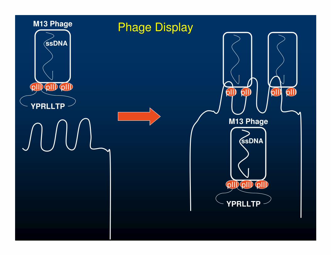
YPRLLTP
M13 Phage
ssDNA
pIII pIIIpIII
YPRLLTP
M13 Phage
ssDNA
pIII pIIIpIII
pIIIpIII pIIIpIII

Hypothesis
• We postulated that by utilizing in vivo phage
display, we would identify peptide sequences
which internalize into the intestinal epithelial
following severe injury
• We could bind this newly discovered peptide
sequence to fluorescent nanoparticles in order
to image its delivery into the gut barrier

Methods- Phage Screening
• Intestinal mucosa isolated 2 hours following burn
• Mucosa incubated with Phage library containing 1012
different peptide sequence
• Selected Phage amplified using E. coli
• Process repeated 3 times to select for gut-targeting peptide sequence
30% TBSA steam burn
for 7 seconds
2hr
balb/c mice

Methods- Intraluminal Delivery of Phage
30% TBSA steam burn
for 7 seconds
balb/c mice
• Harvest each segment
of distal small intestine
•Bowel segments washed
with PBS, Trypsin using a
peristaltic pump
•Phage DNA isolated
from specimens for PCR
30 min
• Perform Laparotomy
• Isolate 3 segments of distal
small intestine between silk ties
• Inject 200μl containing 1 x 10⁹
phage or control (PBS or “empty
phage”)
• Close Abdomen
2 hr

Candidate Peptide Sequences
• T-18 identified as candidate gut-targeting sequence• Isolated in several rounds of screening

Ex Vivo Staining of Intestine

Intestinal qPCR
Sham Animal

Intestinal qPCR
2 Hour Burn

DNA Sequencing of PCR Product
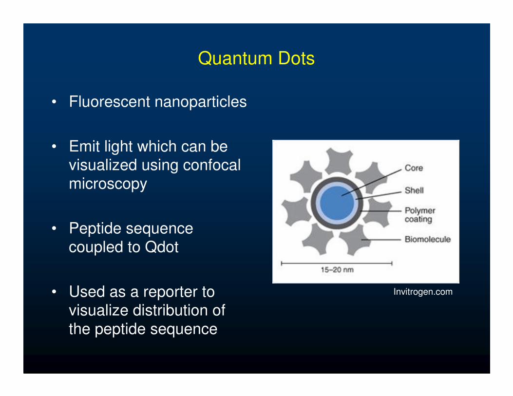
Quantum Dots
• Fluorescent nanoparticles
• Emit light which can be visualized using confocal
microscopy
• Peptide sequence
coupled to Qdot
• Used as a reporter to visualize distribution of
the peptide sequence
Invitrogen.com

Qdot Imaging of T18 Sequence

Summary
• Utilized phage display to screen for peptides that target the intestinal barrier
• Identification of a 12 amino acid peptide sequence
that binds and internalizes into intestinal epithelial
cells after burn injury
• Demonstrated delivery of fluorescent nanoparticles bound to the peptide sequence

Conclusion
• This sequence may allow for targeted
therapies designed to attenuate intestinal
dysfunction following severe injury,
inflammation, or other pathologic conditions of
the small bowel

Future #3:The Neuro-Enteric Axis
• Enteric Nervous System
– Gastrointestinal tissues innervated by complex component of the peripheral nervous system
• Enteric Glia
– Similar to astrocytes of the CNS
– Express glial fibrillary acidic protein (GFAP) when activated
– Promote intestinal barrier function
• Secretion of S-nitrosoglutathioine (GSNO)

Savidge TC, et al. Lab Invest 2007;87:731-36

Bush TG, et al. Cell 1998;93:189-201
Sham vs. GFAP Conditional knockout
GFAP-HSV-Tk Mice • Fatal by 19 days• Severe inflammation• Hemorrhagic necrosis
Sham GFAP Ablation
GFAP is required to maintain gut architecture

Control IL1-β
Von Boynen, et al. Gut 2004;53:222-28
Pro-inflammatory cytokines increase percentage of GFAP positive staining (red) neurons
Inflammation activates enteric glia cells

Savidge TC, et al. Gastroenterology 2007;132:1344-58
Addition of enteric glia cells (EGC) to Caco2 cell culture:• Increases occludin and ZO-1 levels• Improves barrier function (TER and FITC-Dextran)
Addition of enteric glia cells to Caco-2 culture improves barrier function and tight junction protein expression

GSNO improves barrier function at low concentrations and increases Permeability at high concentrations
Enteric glia cells secrete GSNO when activated, which
improves intestinal barrier function at low concentrations

Intestinal GFAP qPCR Time Course

Sham 2h Burn 6h Burn
Green = S100
Red = GFAP
Intestinal GFAP- Confocal Time Course
LaminaPropria
VillousTips

Sham
Burn
2hr 6hr Gut Brain
GFAP-luc Transgenic Mice

Quantification of Luminescence from GFAP-luc
Mice

Vagal Stim / Burn4h BurnShamVagotomy /
Vagal Stim / Burn
Histology

Intestinal Permeability 4 hrs Post-burn
n=4 n=5 n=5 n=5 n=5

Occludin Western blot 4hrs post-burn

Intestinal GFAP qPCR 4 hours post-burn
N=5 N=6 N=2 N=3

Burn +
Vagotomy +
Vagal Stim
(408)
Burn
(373)
Sham
(368)
Burn +
Vagal Stim
(370)
60X Magnification Comparison
GFAP Confocal- 4 hrs post-burn
LaminaPropria
VillousTips
LaminaPropria
VillousTips

Conclusions
• Past: Translocation through the portal vein to liver.
• Present: Lymph route more important
• Future: Already here
– Non-invasive method of monitoring organ injury. One animal – multiple measurements
– Drug delivery to target cells. Specific, more effective, perhaps cheaper
– Manipulation of PNS and enteric glia – promising therapeutic strategy.
Sham 4hr Burn

The UCSD Team
• Faculty– R. Coimbra MD, PhD– B. Potenza MD– J. Doucet MD– V. Bansal MD– J. Lee MD– B. Eliceiri PhD– A. Baird PhD
• TPM– S. Pacyna RN
• Clinical Fellows– L Nwakanma MD– P Bosarge MD
• Research Fellows– T Costantini MD– C Peterson MD
• Programmer / Analyst– Dale Fortlage BA
• Trauma Registrar– P. Stout RN– C. Mohrle RN
• Data Entry and Maintenance– E. Hernandez
• Administrative Assistant– R. Velez

Christine Cocanour, MD
Program Committee Chair


Grace Rozycki, MD & David Feliciano, MD

Thank you
Downtown San Diego
http://trauma.ucsd.edu