Embed Size (px)
Citation preview
Hindawi Publishing CorporationAnemiaVolume 2012, Article ID 582018, 6 pagesdoi:10.1155/2012/582018
Research Article
Spatiotemporal Dysfunction of the Vascular Permeability Barrierin Transgenic Mice with Sickle Cell Disease
Samit Ghosh,1 Fang Tan,1 and Solomon F. Ofori-Acquah1, 2
1 Aflac Cancer and Blood Disorders Center, Division of Hematology/Oncology/BMT, Department of Pediatrics,Emory University School of Medicine, Atlanta, GA 30322, USA
2 Department of Pediatrics, Children’s Healthcare of Atlanta, Atlanta, GA 30322, USA
Correspondence should be addressed to Solomon F. Ofori-Acquah, [email protected]
Received 16 December 2011; Accepted 15 February 2012
Academic Editor: Kenneth R. Peterson
Copyright © 2012 Samit Ghosh et al. This is an open access article distributed under the Creative Commons Attribution License,which permits unrestricted use, distribution, and reproduction in any medium, provided the original work is properly cited.
Sickle cell disease (SCD) is characterized by chronic intravascular hemolysis that generates excess cell-free hemoglobin in the bloodcirculation. Hemoglobin causes multiple endothelial dysfunctions including increased vascular permeability, impaired reactivityto vasoactive agonists, and increased adhesion of leukocytes to the endothelium. While the adhesive and vasomotor defects ofSCD associated with cell-free hemoglobin are well defined, the vascular permeability phenotype remains poorly appreciated. Weaddressed this issue in two widely used and clinically relevant mouse models of SCD. We discovered that the endothelial barrier isnormal in most organs in the young but deteriorates with aging particularly in the lung. Indeed, middle-aged sickle mice developedpulmonary edema revealing for the first time similarities in the chronic permeability phenotypes of the lung in mice and humanswith SCD. Intravenous administration of lysed red blood cells into the circulation of sickle mice increased vascular permeabilitysignificantly in the lung without impacting permeability in other organs. Thus, increased vascular permeability is an endothelialdysfunction of SCD with the barrier in the lung likely the most vulnerable to acute inflammation.
1. Introduction
Sickle cell disease (SCD) is characterized by the productionof red blood cells with increased propensity for lysis andadhesion [1]. Its clinical manifestations fall broadly into twosubphenotypes defined by hyperhemolysis and vasoocclu-sion [2]. At least 30% of the hemolysis in SCD is intravas-cular [3], which means that the endothelial wall in thisdisease is persistently exposed to cell-free hemoglobin. Theendothelium is a semipermeable barrier that regulates theresponse of the vascular wall to inflammatory agonists. Thisresponse involves activation of adhesion molecule expres-sion, increased permeability of the endothelium, and extra-vasations of fluid from the blood into interstitial tissuecompartments [4]. Increased vascular permeability resultsfrom opening of gaps at sites of endothelial cell-cell contacts.There are multiple indicators of systemic inflammation inSCD [5]. In addition, markers of vascular inflammation havealso been documented [6–8]. There is increased expression
of adhesion molecules in the pulmonary endothelium of theBerkeley sickle mice [9], although the histology of these samemice shows less severe inflammatory and ischemic changesand no evidence of pneumonia [10]. Nonetheless, theyspontaneously develop pulmonary hypertension [11], whichis a major problem in SCD [12]. Pulmonary edema and theacute chest syndrome implicate increased vascular perme-ability in both chronic and acute complications of SCD [13,14]. Despite this significance, there is currently no knowledgeof the vascular permeability phenotypes of major organs thatare impacted by SCD.
2. Materials and Methods
2.1. Transgenic Sickle Mice. Experiments were performedusing protocols approved by the Institutional Animal Careand Use Committee (IACUC) of Emory University. TheBerkeley [15] and Townes [16] transgenic SCD mousemodels used have previously been described.
2 Anemia
hβA/hβS hβS/hβS
Spleen
hβS/hβS
Lung
hβA/hβS hβS/hβS
Brain
hβA/hβS
hβA/hβS
hβS/hβS
Heart
hβA/hβS hβS/hβS
Kidney
hβS /hβA hβS/hβS
Liver
(a)
0.8
1.2
0.4
0Adult AdultMA MA
0.8
1.2
0.4
0Adult AdultMA MA
0.8
1.2
0.4
0Adult AdultMA MA
Brain
0.8
1.2
0.4
0Adult AdultMA MA
Liver
Vas
cula
r le
akag
e (O
D a
t 62
0 n
m)
Vas
cula
r le
akag
e (O
D a
t 62
0 n
m)
Townes model
Heart Kidney
∗∗ ∗
0.8
1.2
0.4
0Adult AdultMA MA
Lung
∗∗
∗∗∗∗∗
0.8
1.2
0.4
0Adult AdultMA MA
Spleen
∗
hβA/hβS
hβS /hβS
(b)
Figure 1: Continued.
Anemia 3
Vas
cula
r le
akag
e (O
D a
t 62
0 n
m)
Vas
cula
r le
akag
e (O
D a
t 62
0 n
m)
HemiBerkeley model
Sickle
KidneyHeartBrain
Liver Lung Spleen
0.4
0.6
0.2
0Adult AdultMA MA
∗∗
0.4
0.6
0.2
0Adult AdultMA MA
∗
∗∗
0.4
0.6
0.2
0Adult AdultMA MA
∗∗∗
∗∗
0.4
0.6
0.2
0Adult AdultMA MA
∗∗
∗∗
0.4
0.6
0.2
0Adult AdultMA MA
∗∗∗
∗∗∗∗
0.4
0.6
0.2
0Adult AdultMA MA
∗∗∗∗∗
(c)
00 4 8 12 16 20
Vas
cula
r le
akag
e (O
D a
t 62
0 n
m)
0.2
0.4
0.6
0.8
r = −0.7639
P ≤ 0.0001
Hb (g/dL)
(d)
Brain Heart Kidney Lung
(e)
0Brain Heart Kidney Lung
0.05
0.01
0.015
0.02
Vas
cula
r le
akag
e (O
D a
t 62
0 n
m)
(f)
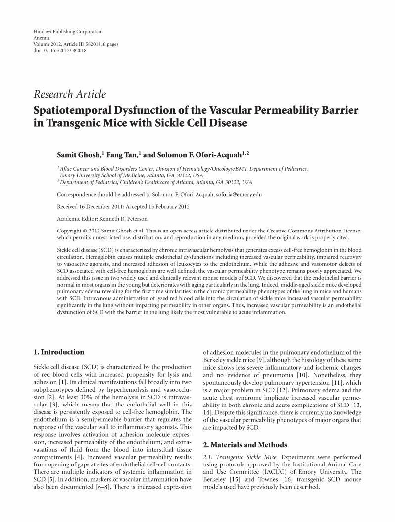
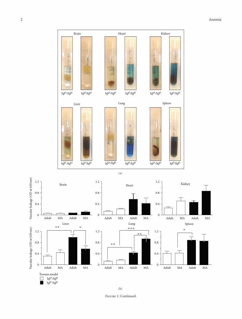
Figure 1: Vascular barrier dysfunction in sickle mice. (a) Representative images of organs isolated from sickle mice after injection with Evansblue dye and incubation in formamide for three days. (b, c) Vascular leakage in the indicated organs in adult (3–6 months) and middle-aged(10–13 months) mice of the Townes (heterozygotes-HbAS (hβA/hβS) and homozygote sickle-HbSS (hβS/hβS)) and Berkeley (hemizygotesand sickle) models. The number of mice studied was as follows: Townes: n = 3 for each genotype and age group; Berkeley: Sickle adult,n = 9; sickle middle-aged n = 6; hemizygote adult, n = 10, hemizygote middle-aged n = 5. (d) Vascular leakage correlates with hemoglobin.Data shown is the vascular leakage in the lung for a total of 30 mice (15 sickle and 15 hemizygotes) of the Berkeley model. (e) Typical imagesfor the indicated organs isolated from young (4–6 weeks old) Berkeley mice injected with Evans blue dye and incubated in formamide for3 days. (f) Histogram showing the quantification of Evans blue extravasation of major organs in young sickle mice (n = 4). ∗P < 0.05,∗∗P < 0.01, and ∗∗∗P < 0.001.
2.2. Vascular Leakage and Lung Edema. Vascular leakage wasstudied by intravenous injection of cell-impermeable Evan’sBlue dye as widely described by several investigators. Micewere injected with 100 μL of 1% cell-impermeable Evans Bluedye (Sigma-Aldrich, St. Louis, MO) in PBS intravenouslythrough tail vein. After 40 min, mice were anesthetized by i.p.injection of avertin (300 mg/kg body weight). To remove the
dye from circulation, mice were perfused by injecting 40 mLof PBS containing 2 mM EDTA through left ventricle of theheart allowing the blood to flow out by puncturing renalartery. Organs were harvested and incubated in formamidefor 3 days to extract the dye and OD determined at 620 nm.For edema analysis mice were euthanized and the rightlobe removed and weighed immediately using an isometric

4 Anemia
Hemi
Sickle
Lun
g w
et/d
ry w
eigh
t ra
tio
Young Middle age
7
6
5
4
3
∗∗
(a)
1
0.8
0.6
0.4
0.2
0
Vas
cula
r le
akag
e (O
D a
t 62
0 n
m)
SalineLysed RBCs
Brain Heart Kidney Lung
∗∗
(b)
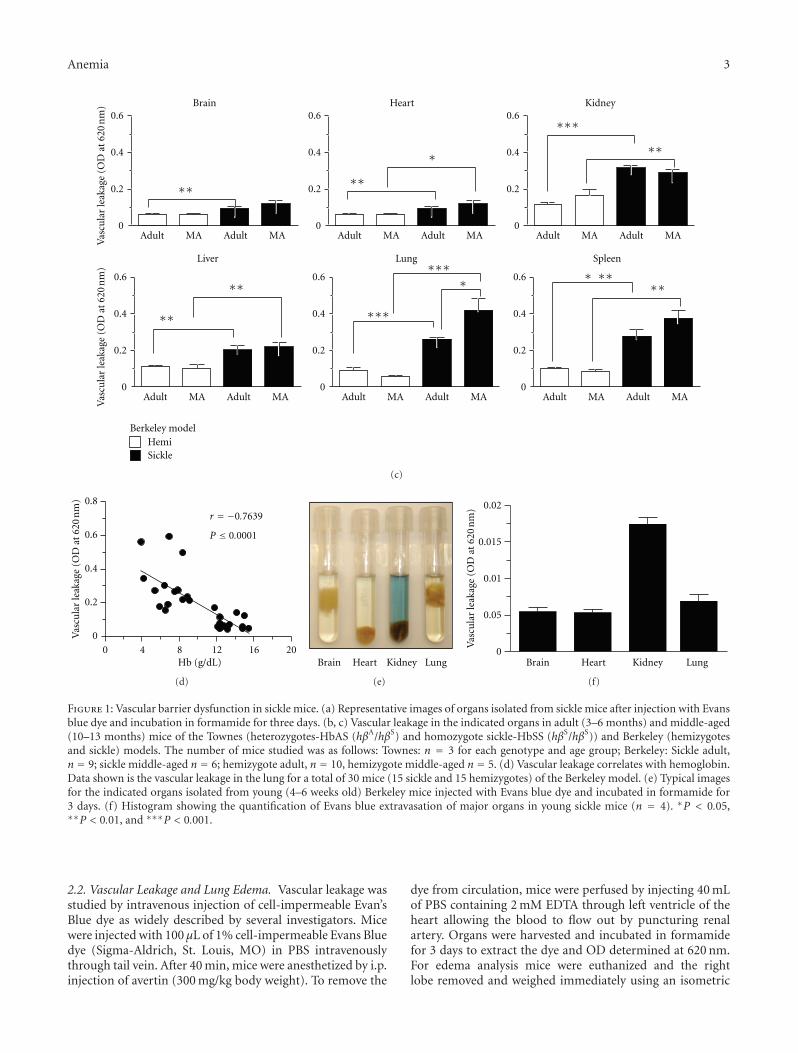
Figure 2: Chronic and acute changes in vascular permeability in sickle mouse lungs. (a) Lung edema in middle-aged Berkeley sickle mice asdetermined by wet/dry weight ratios (n = 6). Control groups include young sickle mice (n = 6) and young (n = 6) and middle-aged (n = 6)hemizygotes. (b) Vascular leakage in the indicated organs in Berkeley sickle mice intravenously injected with lysed red blood cells. Note thatpermeability is significantly increased by lysed red blood cells in the lung but not in other organs. ∗∗P < 0.01.
transducer (Harvard Apparatus, Holliston, MA). Lungs weredried in an oven at 80◦C containing desiccant crystals for24 h, dry weight determined, and ratios calculated.
2.3. Statistical Analyses. GraphPad software version 5.0 wasused. Differences in vascular leakage and weights were ana-lyzed using t-test and correlation studies performed usingPearson’s test.
3. Results and Discussion
In SCD, the adhesive and vasomotor defects of the vascula-ture are well defined [17–20], while the vascular permeabilityremains poorly appreciated. To address this knowledge gap,adult (3–6 months) and middle-aged (10–13 months) micewere injected with 1% Evans blue via the tail vein andthe amount of dye that leaked from the circulation intothe parenchyma of individual organs examined. Figure 1(a)shows virtually no leakage in the brain contrary to theclear evidence of endothelial barrier breakdown in the otherorgans. Quantification of vascular leakage revealed that theendothelial barrier is generally more permeable in the sicklemice than in control littermates (Figures 1(b) and 1(c)),despite some differences in the two transgenic models.Indeed, there was a significant correlation between steady-state hemoglobin concentration and lung permeability(Figure 1(d)) (r = −0.7639, P < 0.0001), indicating thatendothelial barrier dysfunction is related to an aspect of SCD.Unlike most organs, vascular permeability in the lung inmiddle-aged sickle mice was significantly higher than inadult mice, highlighting a role for age in this disease process.This was investigated by extending our study to include
younger mice aged 5-6 weeks. Remarkably, permeability inthe heart and lung at this early stage of the disease wasidentical to that of the brain, which is widely known to havea highly restrictive barrier (Figures 1(e) and 1(f)). Thus, theendothelial barrier in SCD is normal in most organs in theyoung, becomes abnormal during adulthood, and deterio-rates further with aging particularly in the lung.
Pulmonary edema is a common postmortem finding inSCD and yet it has not been appreciated as a chronic lungcomplication of SCD, probably because of the confoundingeffect of death [21, 22]. While histology does not revealevidence of edema in the Berkeley sickle mice [10], we haveused a more sensitive approach to clearly demonstrate thatvascular permeability in the lungs of both the Berkeley andTownes sickle mice is increased. Our result is in agreementwith a recent study that investigated permeability exclusivelyin the lung of 12–16-weeks-old NY1DD sickle mouse usingthe same approach [23]. In agreement with these permeabil-ity findings, we discovered that the average wet lung weight ofmiddle-aged sickle mice is significantly heavier than that ofage-matched hemizygotes (0.64 mg ± 0.04 versus 0.54 mg ±0.02; P = 0.03), and, accordingly, the wet/dry weight ratio,widely used to confirm edema in lungs, was significantlyhigher (P = 0.002) (Figure 2(a)). Taken together, theseresults show for the first time that middle-aged sickle micedevelop pulmonary edema. We cannot exclude the possibilitythat chronic heart failure contributed to the lung edemareported here; however, the Berkeley mice used in the lungweight measurements were at least three months youngerthan those reported to have heart failure [11]. Importantly,we show that lung edema correlates with higher vascular per-meability in sickle mouse lungs. This concordance advancesboth conceptual and practical research objectives. It suggests
Anemia 5
that the permeability phenotypes of the lung in mice andhumans with SCD are similar, and it validates, for the firsttime, the use of the Evans blue extravasation approach tostudy vascular permeability in transgenic sickle mice.
The permeability phenotypes identified here likely reflectthe intrinsic properties of individual organs, as well as ofendothelial cell types. For instance, it is well establishedthat heterogeneity of endothelial cell junction contributes tounique permeability attributes [24], and this may account forsome of the dramatic differences in permeability phenotypesreported here (e.g., brain and lung). However, endothelialcells in individual organs may also respond differently to bar-rier disrupting factors found in SCD. Among these factors,cell-free hemoglobin is unique because it is released inabundance during acute intravascular hemolysis, which is adaily event in SCD. We assessed the response of major vas-cular beds in the sickle mice to acute hemolysis. Leuko-depleted packed red blood cells were lysed by repeated freeze-thaw cycles and intravenously administered to sickle micevia tail vein. Compared to saline, the lysed red blood cellsincreased vascular permeability by 2-fold in the lungs ofadult sickle mice but had a modest or negligible impact onthe kidney, brain, and heart (Figure 2(b)). That lysed redblood cells caused morbidity and significantly altered barrierfunction in the Berkeley sickle mice indicates this and othersevere models of SCD can be used to unlock mechanisms ofacute hemolysis in SCD. In particular the vulnerability of thelung endothelial barrier to lysed red blood cells highlightsacute intravascular hemolysis as a potential trigger of theacute chest syndrome since a decreasing concentration ofhemoglobin is invariably associated with this condition [14].
In conclusion, SCD appears to be characterized by weak-ening of the endothelial barrier, which predisposes someorgans, such as the lung to acute loss of barrier function,reminiscent of the acute chest syndrome. Ongoing studies arefocused on unraveling the relationship between acute hemol-ysis and the endothelial barrier in SCD.
Authors’ Contribution
S. Ghosh designed and performed most of the experiments.F. Tan characterized the transgenic mice and performedresearch. S. F. Ofori-Acquah designed the study and providedoverall oversight of the projects.
Acknowledgments
The authors are grateful to Dr. Townes of the Universityof Alabama at Birmingham for the knock-in transgenicmice with SCD and to Dr. Archer of Emory Universityfor the Berkeley mice. This work was supported by GrantsR01HL077769 awarded to SFOA.
References
[1] S. Embury, R. P. Hebbel, N. Mohandas, and M. H. Steinberg,Sickle Cell Disease: Basic Principles and Clinical Practice, RavenPress, New York, NY, USA, 1995.
[2] G. J. Kato, M. T. Gladwin, and M. H. Steinberg, “Deconstruct-ing sickle cell disease: reappraisal of the role of hemolysis in thedevelopment of clinical subphenotypes,” Blood Reviews, vol.21, no. 1, pp. 37–47, 2007.
[3] T. A. Bensinger and P. N. Gillette, “Hemolysis in sickle cell dis-ease,” Archives of Internal Medicine, vol. 133, no. 4, pp. 624–631, 1974.
[4] W. Aird, Endothelial Biomedicine, Cambridge University Press,2007.
[5] M. H. Steinberg, “Sickle cell anemia, the first molecular dis-ease: overview of molecular etiology, pathophysiology, andtherapeutic approaches,” TheScientificWorldJournal, vol. 8, pp.1295–1324, 2008.
[6] A. J. Duits, R. C. Pieters, A. W. Saleh et al., “Enhanced levelsof soluble VCAM-1 in sickle cell patients and their specificincrement during vasoocclusive crisis,” Clinical Immunologyand Immunopathology, vol. 81, no. 1, pp. 96–98, 1996.
[7] A. Solovey, Y. Lin, P. Browne, S. Choong, E. Wayner, and R.P. Hebbel, “Circulating activated endothelial cells in sickle cellanemia,” New England Journal of Medicine, vol. 337, no. 22, pp.1584–1590, 1997.
[8] A. Solovey, L. Gui, N. S. Key, and R. P. Hebbel, “Tissue factorexpression by endothelial cells in sickle cell anemia,” Journal ofClinical Investigation, vol. 101, no. 9, pp. 1899–1904, 1998.
[9] J. D. Belcher, C. J. Bryant, J. Nguyen et al., “Transgenic sicklemice have vascular inflammation,” Blood, vol. 101, no. 10, pp.3953–3959, 2003.
[10] E. A. Manci, C. A. Hillery, C. A. Bodian, Z. G. Zhang, G. A.Lutty, and B. S. Coller, “Pathology of Berkeley sickle cell mice:similarities and differences with human sickle cell disease,”Blood, vol. 107, no. 4, pp. 1651–1658, 2006.
[11] L. L. Hsu, H. C. Champion, S. A. Campbell-Lee et al., “Hemo-lysis in sickle cell mice causes pulmonary hypertension due toglobal impairment in nitric oxide bioavailability,” Blood, vol.109, no. 7, pp. 3088–3098, 2007.
[12] M. T. Gladwin, V. Sachdev, M. L. Jison et al., “PulmonaryHypertension as a Risk Factor for Death in Patients with SickleCell Disease,” New England Journal of Medicine, vol. 350, no. 9,pp. 886–895, 2004.
[13] J. K. Graham, M. Mosunjac, R. L. Hanzlick, and M. Mosunjac,“Sickle cell lung disease and sudden death: a retrospective/prospective study of 21 autopsy cases and literature review,”American Journal of Forensic Medicine and Pathology, vol. 28,no. 2, pp. 168–172, 2007.
[14] E. P. Vichinsky, L. D. Neumayr, A. N. Earles et al., “Causes andoutcomes of the acute chest syndrome in sickle cell disease,”New England Journal of Medicine, vol. 342, no. 25, pp. 1855–1865, 2000.
[15] C. Paszty, C. M. Brion, E. Manci et al., “Transgenic knockoutmice with exclusively human sickle hemoglobin and sickle celldisease,” Science, vol. 278, no. 5339, pp. 876–878, 1997.
[16] L. C. Wu, C. W. Sun, T. M. Ryan, K. M. Pawlik, J. Ren, and T.M. Townes, “Correction of sickle cell disease by homologousrecombination in embryonic stem cells,” Blood, vol. 108, no. 4,pp. 1183–1188, 2006.
[17] C. D. Reiter, X. Wang, J. E. Tanus-Santos et al., “Cell-freehemoglobin limits nitric oxide bioavailability in sickle-celldisease,” Nature Medicine, vol. 8, no. 12, pp. 1383–1389, 2002.
[18] M. T. Gladwin, V. Sachdev, M. L. Jison et al., “Pulmonaryhypertension as a risk factor for death in patients with sicklecell disease,” New England Journal of Medicine, vol. 350, no. 9,pp. 886–895, 2004.
[19] R. P. Hebbel, R. Osarogiagbon, and D. Kaul, “The endothelialbiology of sickle cell disease: inflammation and a chronic
6 Anemia
vasculopathy,” Microcirculation, vol. 11, no. 2, pp. 129–151,2004.
[20] B. N. Y. Setty and M. J. Stuart, “Vascular cell adhesion mol-ecule-1 is involved in mediating hypoxia- induced sickle redblood cell adherence to endothelium: potential role in sicklecell disease,” Blood, vol. 88, no. 6, pp. 2311–2320, 1996.
[21] J. K. Graham, M. Mosunjac, R. L. Hanzlick, and M. Mosunjac,“Sickle cell lung disease and sudden death: a retrospective/prospective study of 21 autopsy cases and literature review,”American Journal of Forensic Medicine and Pathology, vol. 28,no. 2, pp. 168–172, 2007.
[22] W. Girard, “Case report: postoperative pulmonary edema andsickle cell crisis,” Clinical Notes on Respiratory Diseases, vol. 17,no. 4, pp. 13–14, 1979.
[23] K. L. Wallace, M. A. Marshall, S. I. Ramos et al., “NKT cellsmediate pulmonary inflammation and dysfunction in murinesickle cell disease through production of IFN-γ and CXCR3chemokines,” Blood, vol. 114, no. 3, pp. 667–676, 2009.
[24] A. Masedunskas, J. A. King, F. Tan et al., “Activated leukocytecell adhesion molecule is a component of the endothelial junc-tion involved in transendothelial monocyte migration,” FEBSLetters, vol. 580, no. 11, pp. 2637–2645, 2006.

Submit your manuscripts athttp://www.hindawi.com
Stem CellsInternational
Hindawi Publishing Corporationhttp://www.hindawi.com Volume 2014
Hindawi Publishing Corporationhttp://www.hindawi.com Volume 2014
MEDIATORSINFLAMMATION
of
Hindawi Publishing Corporationhttp://www.hindawi.com Volume 2014
Behavioural Neurology
EndocrinologyInternational Journal of
Hindawi Publishing Corporationhttp://www.hindawi.com Volume 2014
Hindawi Publishing Corporationhttp://www.hindawi.com Volume 2014
Disease Markers
Hindawi Publishing Corporationhttp://www.hindawi.com Volume 2014
BioMed Research International
OncologyJournal of
Hindawi Publishing Corporationhttp://www.hindawi.com Volume 2014
Hindawi Publishing Corporationhttp://www.hindawi.com Volume 2014
Oxidative Medicine and Cellular Longevity
Hindawi Publishing Corporationhttp://www.hindawi.com Volume 2014
PPAR Research
The Scientific World JournalHindawi Publishing Corporation http://www.hindawi.com Volume 2014
Immunology ResearchHindawi Publishing Corporationhttp://www.hindawi.com Volume 2014
Journal of
ObesityJournal of
Hindawi Publishing Corporationhttp://www.hindawi.com Volume 2014
Hindawi Publishing Corporationhttp://www.hindawi.com Volume 2014
Computational and Mathematical Methods in Medicine
OphthalmologyJournal of
Hindawi Publishing Corporationhttp://www.hindawi.com Volume 2014
Diabetes ResearchJournal of
Hindawi Publishing Corporationhttp://www.hindawi.com Volume 2014
Hindawi Publishing Corporationhttp://www.hindawi.com Volume 2014
Research and TreatmentAIDS
Hindawi Publishing Corporationhttp://www.hindawi.com Volume 2014
Gastroenterology Research and Practice
Hindawi Publishing Corporationhttp://www.hindawi.com Volume 2014
Parkinson’s Disease
Evidence-Based Complementary and Alternative Medicine
Volume 2014Hindawi Publishing Corporationhttp://www.hindawi.com