Embed Size (px)
Citation preview
Environmental Health PerspectivesVol. 12, pp. 81-88, 1975
Blood-Brain Barrier Dysfunction inAcute Lead Encephalopathy: AReappraisal
by Thomas W. Bouldin,* Paul Mushak,* Lorcan A.O'Tuama, and Martin R. Krigman*
Acute lead encephalopathy was induced in adult guinea pigs by administering dailyoral doses of lead carbonate. During the development of the encephalopathy, the structuraland functional integrity of the blood-brain barrier was evaluated with electronmicroscopy and tracer probes. Blood, cerebral gray matter, liver, and kidney wereanalyzed for lead, calcium, and magnesium content.
The animals regularly developed an encephalopathy after four doses of lead. Therewere no discernible pathomorphologic alterations in the cerebral capillaries or perivascu-lar glial sheaths. Furthermore, no evidence of blood-brain barrier dysfunction wasdemonstrated with Evans blue-albumin complex or horseradish peroxidase. Blood-brainbarrier permeability to radiolead was not increased in the intoxicated animals. During thedevelopment of the encephalopathy there was a progressive rise in the lead concentrationin all tissues. Concurrently, there was a significant rise in brain calcium.
These results suggest that the encephalopathic effects of lead may be mediated directlyat the neuronal level.
Introduction
The vulnerability of the nervous system to leadis well known; however, the pathogenesis of leadencephalopathy remains a vexing problem. Basedupon clinical and experimental studies, a numberof investigators (1-6) have concluded that the en-
cephalopathic effects of lead are mediated througha primary vascular lesion and a concomitantblood-brain barrier dysfunction. This conclusionis based upon the presence of electron-dense
*Department of Pathology, University of North Carolina,School of Medicine, Chapel Hill, N.C. 27514.
tDepartment of Neurology, University of North Carolina,School of Medicine, Chapel Hill, N. C. 27514.
vacuoles in perivascular astrocytic end-feet and en-dothelial hyperplasia in experimental and clinicalcases of lead encephalopathy (3, 5). Moreover,perivascular hemorrhages, cerebral edema, andblood-brain barrier dysfunction as evaluated bymacromolecular tracers are prominent features ofthe encephalopathy in the suckling rat model ofPentschew and Garro (2, 3). Despite the existenceof these cerebral vascular alterations in lead intox-ication, the direct neural effects of lead must alsobe considered as a factor in the pathogenesis of leadencephalopathy. Evidence is accumulating thatlead ions can directly affect synaptic transmission(7). Moreover, it has been previously shown thatwhen lead ions are directly instilled into thecerebrospinal fluid of dogs, the animals develop anencephalopathy and ultimately die (8, 9).
December 1975 81
We report here studies designed to evaluate therole of blood-brain barrier dysfunction in thegenesis of acute lead encephalopathy in the adultguinea pig. The model used was initially describedby Popow (10) in 1885 and more fully by Weller(11) in 1927. Four consecutive daily oral doses oflead carbonate (155 mg) produce an encephalopa-thy in the adult guinea pig characterized by tonic-clonic seizures. When five consecutive daily dosesof lead are given, all of the animals developseizures and die. Despite the development of thissevere encephalopathy, these animals show nomorphological changes in the structures of theblood-brain barrier, and the permeability of thebarrier to macromolecules is not increased.
Materials and Methods
Experimental Model
Random-bred, adult, male guinea pigs, weighing475-800 g, were maintained in individual plasticcages with an ambient temperature of 68°F, 12 hrof artificial light per day, and fed guinea pig chowand tap water, ad libitum. A single, daily, oral doseof 155 mg of lead carbonate in a gelatin capsulewas administered to the experimental animals.[The dosage is arbitrary and was used by Weller(11) because it produced an encephalopathy.]
Morphologic Evaluation ofBlood-Brain Barrier with Evans Blueand Horseradish Peroxidase
The guinea pigs were sacrificed 24 hr after two(three animals), three (four animals), five (sixanimals), or six (two animals) consecutive dailydoses. Two of the five-dose animals also receivedEvans blue (5 mg/ml saline), intraperitoneally, 24hr prior to sacrifice. After pentobarbital anesthesia(3.6 mg/100 g body weight, intramuscularly), theanimals received an intravascular injection of Sig-ma type II horseradish peroxidase (25 mg/100 gbody weight, dissolved in 3 ml physiologic saline)and were sacrificed 10-15 min later by decapita-tion or intravascular perfusion. For perfusion, theanimals were mechanically ventilated while thethorax was opened and the ascending aorta wascannulated via the heart. The brains were perfusedwith Karnovsky's dialdehyde fixative (12) diluted(1:3) with 0.1M sodium cacodylate buffer (pH 7.4)for 15 min at a pressure of 100 cm of fixative. Theperfused brains and the brains from the decapi-tated animals were removed, coronally sectioned
through the neostriatum, mamillary bodies, andmidbrain and cerebellum, and fixed by immersionfor 2-4 hr at room temperature in full strengthKarnovsky's fixative. These fixed tissues were thenwashed overnight in O.1M sodium cacodylatebuffer, pH 7.2, at 4°C. For light microscopy, 20 ,umcoronal sections of brain were cut with a freezingmicrotome; for electron microscopy, 70 um slices ofcerebral cortex and cerebellum were cut with aSmith-Farquhar tissue chopper. Peroxidase ac-tivity in the frozen sections and tissue slices wasidentified by using the method ofGraham and Kar-novsky (13). Frozen sections were also mounted inglycerol for the identification of Evans blue byfluorescence microscopy. Following incubation inthe Graham-Karnovsky medium, the 70 Am sliceswere postfixed in 1% osmium tetroxide in O.1Mcacodylate buffer for 1 hr, stained en bloc with 1%uranyl acetate in 0.1 M maleate at pH 5.4 for 1 hr,dehydrated with graded ethanol and finally withpropylene oxide, and embedded in Epon 812.Survey thick sections were stained with toluidineblue. Ultrathin sections, either unstained or doublystained with uranyl acetate and lead citrate, wereexamined in a JEM-T7 electron microscope. Braintissue was also prepared for conventional lightmicroscopy by embedding in paraffin, sectioning at6-8 ,m, and staining with hematoxylin and eosinor Luxol fast blue-periodic acid-Schiff stains.
Control guinea pigs did not receive lead carbo-nate, but did receive intravascular horseradishperoxidase 15 min prior to vascular brain perfu-sion. Tissue was processed for light and electronmicroscopy as previously described.
Penetration of Tracer Amounts ofRadioactive Lead (21OPb) into Brain
The methodology is as previously describedelsewhere (14). In brief, control and experimentalgroups of animals were prepared as describedabove under experimental methods. 21OPb (in ap-proximately 3N HNO3; 60 mCi/mg of lead), 0.01mCi/kg, was injected into a cannula inserted in theinternal jugular vein. Blood samples were taken at5, 60, 120, and 240 min after the tracer injection.The samples were diluted with saline to 1 ml andcounted for 210Pb. Blood samples of 0.5 ml weretaken for total lead estimation.
Tissue Analyses for Lead, Calcium,and Magnesium
Lead-treated guinea pigs were sacrificed bydecapitation 24 hr after one (four animals), three
Environmental Health Perspectives82
(four animals), and five (three animals) doses oflead; four control animals were also studied.Blood, cerebral gray matter, liver, and kidney wereanalyzed for lead, calcium, and magnesium. Tissuehomogenates and whole blood were analyzed forlead by the Delves cup microabsorption spectro-photometric technique (15) as modified by Edigerand Coleman (16). The samples were analyzeddirectly without predigestion in a Perkin-Elmeratomic absorption spectrophotometer equippedwith a deuterium arc. Calcium and magnesiumwere also measured by atomic absorptionspectroscopy. Each of the values reported is theaverage of three sample determinations.
ResultsThe guinea pigs began showing symptoms of in-
toxication after two to three doses of lead. Thesesymptoms included an increased startle responseand decreased food consumption. By the fifth dosethe animals were obtunded, with a greatly in-creased startle response, and occasional seizures.Tonic-clonic seizures occurred in three of theanimals studied for morphologic alterations in theblood-brain barrier. Examination of the histologicpreparations by light microscopy revealed nodemonstrable changes in the central nervoussystem. There were also no signs of hemorrhage.
Macromolecular Tracer Studies
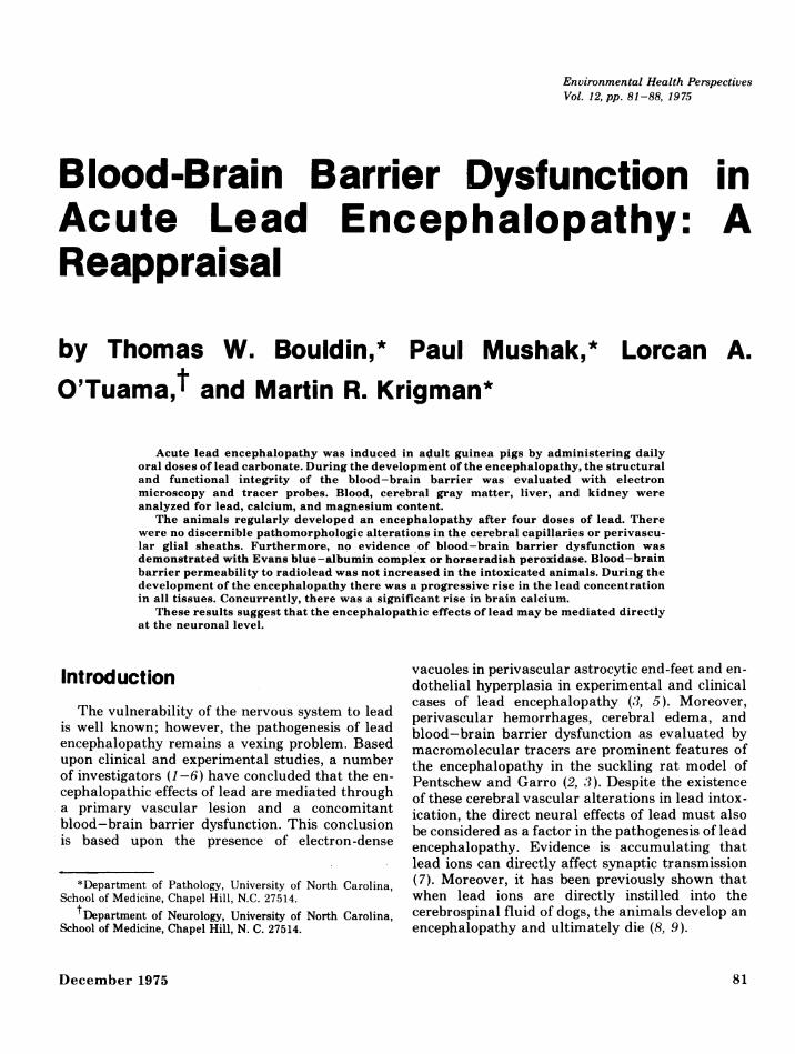
Macroscopic examination of the brains of lead-poisoned animals revealed no abnormal staining ofthe neural tissue by Evans blue. By fluorescenceand light microscopy, the red fluorescence of theEvans blue and the brown reaction product of thehorseradish peroxidase were limited to the vascu-lar lumina except for the choroid plexus and tubercinereum-areas which normally have noblood-brain barrier to these macromolecules (Fig.1.).By electron microscopy, the capillary en-
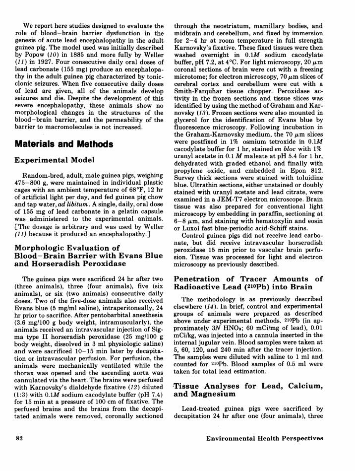
dothelium of lead-poisoned animals (Fig. 2) was in-distinguishable from that of control animals. Theendothelial cells had a normal complement oforganelles and no increased numbers of vacuoles orpinocytotic vesicles. The perithelial cells, basementmembranes, and perivascular glial end-feet werenormal in the lead-poisoned animals. Intracellularand extracellular edema were not apparent at anytime in the intoxicated animals. The electron-densereaction product was limited to capillary lumina(Fig. 3), and did not extend beyond interen-
FIGURE 1. Macrophotograph of coronal section of brain fromanimal receiving 5 daily doses of lead and a single in-travascular injection of horseradish peroxidase (HRP) 15min prior to sacrifice. Reaction product of HRP is limited tothe neuropil of the tuber cinereum (arrow). Nissl stain. x 14.
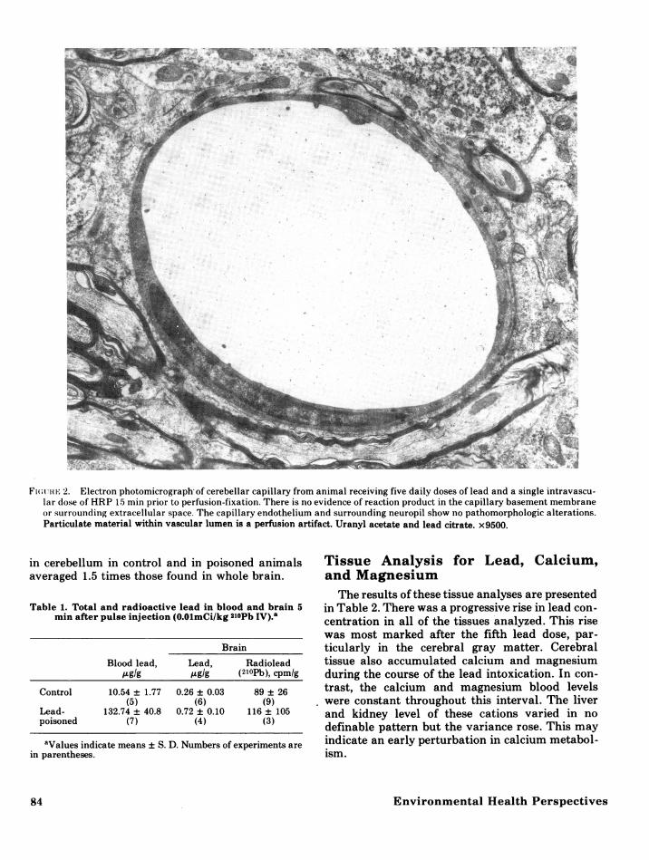
dothelial cell zonulae occludentes. Reaction pro-duct was not observed within pericapillary base-ment membranes or the extracellular space of thebrain. Examination of the choroid plexus revealedreaction product within capillary lumina,perivascular stroma, and in the lateral intercellu-lar spaces between choroidal epithelial cells (Fig.4). Reaction product did not pass through choroi-dal epithelial zonulae occludentes to the ventricu-lar surface.
Radiolead Levels in Blood and Brain
Constant levels of 2lOPb were achieved in bloodand brain within 5 min of intravenous injection ofradiolead. Despite a 13-fold increase in total bloodlead and a 3-fold increase in total brain lead levelsin the encephalopathic animals, the concentrationof 2lOPb in whole brain was not significantlydifferent in the two groups (Table 1). Levels of 2lOPb
December 1975 83
Fw(uiti- 2. Electron photomicrograph of cerebellar capillary from animal receiving five daily doses of lead and a single intravascu-lar dose of HRP 15 min prior to perfusion-fixation. There is no evidence of reaction product in the capillary basement membraneor surrounding extracellular space. The capillary endothelium and surrounding neuropil show no pathomorphologic alterations.Particulate material within vascular lumen is a perfusion artifact. Uranyl acetate and lead citrate. x9500.
in cerebellum in control and in poisoned animalsaveraged 1.5 times those found in whole brain.
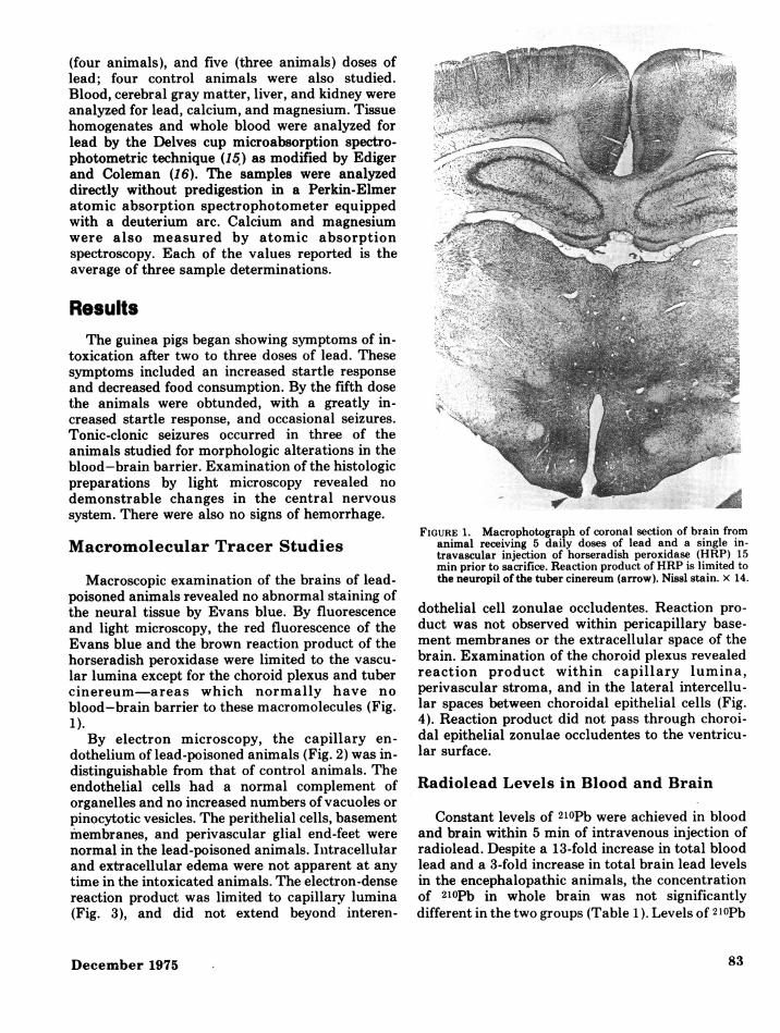
Table 1. Total and radioactive lead in blood and brain 5min after pulse injection (O.OlmCi/kg 2lOPb IV).'
Brain
Blood lead, Lead, RadioleadtgIg pgIg (210Pb), cpm/g
Control 10.54 ± 1.77 0.26 ± 0.03 89 ± 26(5) (6) (9)
Lead- 132.74 ± 40.8 0.72 ± 0.10 116 ± 105poisoned (7) (4) (3)
aValues indicate means ± S. D. Numbers of experiments arein parentheses.
Tissue Analysis for Lead, Calcium,and MagnesiumThe results of these tissue analyses are presented
in Table 2. There was a progressive rise in lead con-
centration in all of the tissues analyzed. This risewas most marked after the fifth lead dose, par-
ticularly in the cerebral gray matter. Cerebraltissue also accumulated calcium and magnesiumduring the course of the lead intoxication. In con-
trast, the calcium and magnesium blood levelswere constant throughout this interval. The liverand kidney level of these cations varied in no
definable pattern but the variance rose. This mayindicate an early perturbation in calcium metabol-ism.
Environmental Health Perspectives84
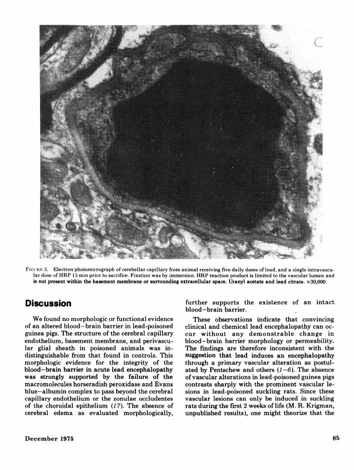
Fiu(;111E 3. Electron photomicrograph of cerebellar capillary from animal receiving five daily doses of lead, and a single intravascu-lar dose of HRP 15 min prior to sacrifice. Fixation was by immersion. HRP reaction product is limited to the vascular lumen andis not present within the basement membrane or surrounding extracellular space. Uranyl acetate and lead citrate. x30,000.
Discussion
We found no morphologic or functional evidenceof an altered blood-brain barrier in lead-poisonedguinea pigs. The structure of the cerebral capillaryendothelium, basement membrane, and perivascu-lar glial sheath in poisoned animals was in-distinguishable from that found in controls. Thismorphologic evidence for the integrity of theblood-brain barrier in acute lead encephalopathywas strongly supported by the failure of themacromolecules horseradish peroxidase and Evansblue-albumin complex to pass beyond the cerebralcapillary endothelium or the zonulae occludentesof the choroidal epithelium (17). The absence ofcerebral edema as evaluated morphologically,
further supports the existence of an intactblood-brain barrier.
These observations indicate that convincingclinical and chemical lead encephalopathy can oc-cur without any demonstrable change inblood-brain barrier morphology or permeability.The findings are therefore inconsistent with thesuggestion that lead induces an encephalopathythrough a primary vascular alteration as postul-ated by Pentschew and others (1-6). The absenceof vascular alterations in lead-poisoned guinea pigscontrasts sharply with the prominent vascular le-sions in lead-poisoned suckling rats. Since thesevascular lesions can only be induced in sucklingrats during the first 2 weeks of life (M. R. Krigman,unpublished results), one might theorize that the
December 1975
ok
85
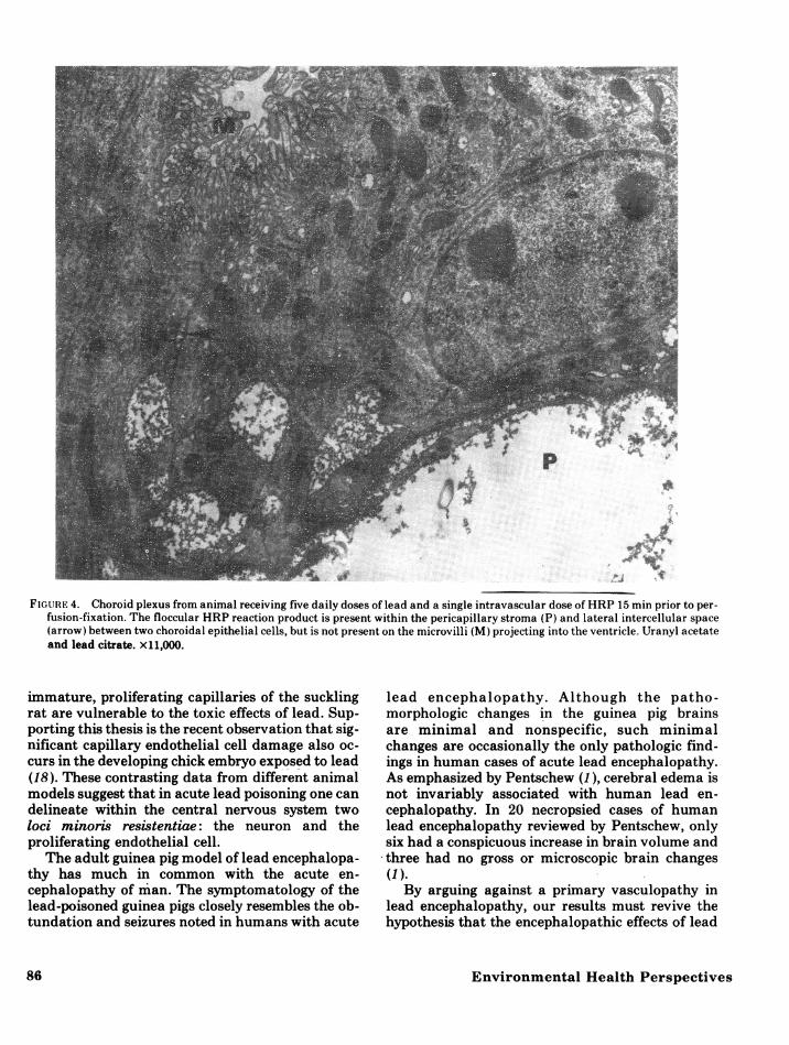
FIGURE 4. Choroid plexus from animal receiving five daily doses of lead and a single intravascular dose of HRP 15 min prior to per-fusion-fixation. The floccular HRP reaction product is present within the pericapillary stroma (P) and lateral intercellular space(arrow) between two choroidal epithelial cells, but is not present on the microvilli (M) projecting into the ventricle. Uranyl acetateand lead citrate. x11,000.
immature, proliferating capillaries of the sucklingrat are vulnerable to the toxic effects of lead. Sup-porting this thesis is the recent observation that sig-nificant capillary endothelial cell damage also oc-curs in the developing chick embryo exposed to lead(18). These contrasting data from different animalmodels suggest that in acute lead poisoning one candelineate within the central nervous system twoloci minoris resistentiae: the neuron and theproliferating endothelial cell.
The adult guinea pig model of lead encephalopa-thy has much in common with the acute en-cephalopathy of man. The symptomatology of thelead-poisoned guinea pigs closely resembles the ob-tundation and seizures noted in humans with acute
lead encephalopathy. Although the patho-morphologic changes in the guinea pig brainsare minimal and nonspecific, such minimalchanges are occasionally the only pathologic find-ings in human cases of acute lead encephalopathy.As emphasized by Pentschew (1), cerebral edema isnot invariably associated with human lead en-cephalopathy. In 20 necropsied cases of humanlead encephalopathy reviewed by Pentschew, onlysix had a conspicuous increase in brain volume andthree had no gross or microscopic brain changes(1).By arguing against a primary vasculopathy in
lead encephalopathy, our results must revive thehypothesis that the encephalopathic effects of lead
Environmental Health Perspectives

Table 2. Tissue concentration of metals in selected organs during the course of lead intoxication.
No. of doses Metal level, /.tg/g wet weightbTissue of leada Lead Caldum Magnesium
Cerebral gray matter 0 0.17 ± 0.12 0.91 ± 0.55 1.8 ± 0.611 0.59 + 0.23 1.3 ± 0.43 2.0 ± 0.523 1.3 ± 0.62 1.5 ± 0.51 2.4 ± 0.715 3.8 ± 1.8 1.6 ± 0.60 2.3 ± 0.56
Kidney 0 5.8 + 2.1 1.8 ± 0.20 1.6 ± 0.041 73 14 1.3 0.30 1.3 0.133 137 ± 48 1.3 ± 0.24 1.3 ± 0.225 556 ± 51 1.2 ± 0.20 1.8 ± 0.62
Liver 0 2.1 ± 1.8 1.2 ± 0.03 1.2 + 0.061 49 ± 2.1 1.2 ± 0.33 1.0 + 0.223 65 41 1.2 0.13 1.3 0.105 132 34 2.1 1.2 1.6 0.61
Blood 0 0.08 ± 0.02 42.0 ± 0.94 6.2 ± 0.401 0.61 ± 0.16 43 ± 0.64 6.0 ± 0.283 1.3 0.74 43 0.55 5.7 0.465 1.6 ± 0.62 43 ± 0.44 5.8 ± 0.24
aLead carbonate, 155 mg oral dose.bMetal level in blood, ,ug/ml.
are mediated at the neuronal level. Recent studiesdemonstrate fundamental neural alterationsdirectly induced by lead. Bull et al. (19) reportedthat lead chloride interferes with the potassiumn-stimulated respiration of rat cerebral cortex slices,and Nathanson et al. (20) found that even low con-centrations of lead inhibit brain adenyl cyclase invitro. Lead has also been found (7) to inhibit com-petitively the calcium-mediated release ofneurotransmitters from presynaptic nerve ter-minals of the bullfrog sympathetic ganglia studiedin vitro.
The radiolead data show that, although the netflux of lead across the blood-brain barrier is in-creased in lead encephalopathy, the permeabilitycoefficient of 21OPb is approximately the same incontrol and poisoned animals. This indicates thatacute lead poisoning does not reveal a saturableprocess which might serve to limit lead uptake inthe face of acute overdosage. This conclusion issupported by the failure of lead encephalopathy toproduce structural changes in the cerebralcapillary-glial complex which is the putative siteof carrier-dependent passage of small moleculesfrom blood into brain. Our findings leave open forfurther study two important and related problemareas: (a) the forces governing unidirectional up-take of tracer amounts of lead in the normal brainand (b) the possible effects of lead poisoning on themetabolism and transport of other physiologicallyimportant cations such as calcium and magnesium.
Some information on the latter issue is providedby the elevated brain calcium and magnesium
levels found in the poisoned animals. Hoffman andWeber (21) have also reported elevated braincalcium levels in lead-poisoned rats, even after asingle dose of lead and after parathyroidectomy.Unfortunately, these investigators did not measuremagnesium levels. While the mechanisms for theprogressive rise in these cations are unknown, theaccumulation of calcium may be related to analtered transport system. From in vitro experi-ments it appears that calcium enters the centralnervous system passively and is transported out ac-tively (22). The progressive accumulation of braincalcium observed in this study in the presence of aconstant blood level suggests the efflux of this ca-tion may be reduced due to a disturbance of an ac-tive efflux mechanism for calcium. An alternativepossibility is competition by lead for binding siteson molecules involved in the active efflux ofcalcium from the brain. The recent report of lead-induced inhibition of adenyl cyclase is of special in-terest because of the close interrelationship be-tween cyclic adenosine 3',5'-monophosphate andcalcium (23).
In summary, we have demonstrated that leadcan enter the central nervous system and produce asevere and fatal encephalopathy in the adultguinea pig without morphologic alterations of theblood-brain barrier or demonstrable dysfunctionof this barrier to macromolecules. Thus in this ex-perimental model the encephalopathic effects oflead may well be directly mediated at the cellularlevel. This does not mean that lead does not pro-duce a vasculopathy in susceptible species. We do,
December 1975 87
however, raise the question as to how much suchvascular . lesions contribute to the acute en-cephalopathy associated with lead intoxication.
AcknowledgementThis study was supported by U.S. Public Health
Service Grant ES01104-01.
REFERENCES
1. Pentschew, A. Morphology and morphogenesis of lead en-cephalopathy. Acta Neuropathol. (Berl.) 5: 133 (1965).
2. Pentschew, A., and Garro, F. Lead encephalo-myelopathyof the suckling rat and its implications on theporphyrinopathic nervous diseases. Acta Neuropathol.(Berl.) 6: 266 (1966).
3. Lampert, P., Garro, F., and Pentschew, A. Lead en-cephalopathy in suckling rats. In: Symposium on BrainEdema, Vienna, 1965. I. Klatzo and F. Seitelberger, eds.Springer Verlag, Berlin-Heidelberg-New York, 1967.
4. Thomas, J. A., Dallenbach, F. D., and Thomas, M. Con-siderations on the development of experimental lead en-cephalopathy. Virchows Arch. (Pathol. Anat.) 352: 61(1971).
5. Clasen, R. A., et al. Electron microscopic and chemicalstudies of the vascular changes and edema of lead en-cephalopathy. Am. J. Pathol. 74: 215 (1974).
6. Goldstein, G. W., Asbury, A. K., and Diamond, I.Pathogenesis of lead encephalopathy: uptake of lead andreaction of brain capillaries. Arch. Neurol. 31: 382 (1974).
7. Kober, T. E., and Cooper, G. P. Competitive action of leadand calcium on transmitter release in bullfrog sympatheticganglia. In: Annual Report, Center for the Study of theHuman Environment, Dept. of Environmental Health,College of Medicine, Univ. of Cincinnati, Cincinnati, Ohio(1974-1975).
8. Camus, J. Toxicite des sels de plomb sur les centres ner-veux. Leur periode d'incubation C. R. Soc. Biol. 68: 509(1910).
9. Camus, J. Meningite et intoxication saturnine. C. R. Soc.Biol. 72. 861 (1912).
10. Popow, N. Ueber die Verinderungen im Riickemnarkenach Vergiftung mit Arsen, Blei, und Quecksilber. Arch.Pathol. Anat. 93: 351 (1885).
11. Weller, C. V. Tolerance in respect to the meningocerebralmanifestations of actue and subacute lead poisoning. Arch.Intern. Med. 39: 45 (1927).
12. Karnovsky, M.J. A formaldehyde-glutaraldehyde fixativeof high osmolality for use in electron microscopy. J. CellBiol. 27: 137A (1965).
13. Graham, R. C., and Karnovsky, M. J. The early stages ofabsorption of injected horseradish peroxidase in the prox-imal tubules of mouse kidney: ultrastructural cytochemis-try by a new technique. J. Histochem. Cytochem. 14: 291(1966).
14. O'Tuama, L. A., et al. The distribution of inorganic lead inguinea pig brain and neural barrier tissues. Observationsin control and lead poisoned animals. Toxicol. Appl. Phar-macol. (in press).
15. Delves, H. T. A micro-sampling method for the rapiddetermination of lead in blood by atomic absorptionspectrophotometry. Analyst 95: 431 (1970).
16. Ediger, R. D., and Coleman, R. L. Modified Delves cupatomic absorption procedure for the determination of leadin blood. Atomic Absorption Newsletter 11: 33 (1972).
17. Brightman, M. W., and Reese, T. S. Junctions between in-timately apposed cell membranes in the vertebrate brain.J. Cell Biol. 40: 648 (1969).
18. Roy, S., et al. Ultrastructure of cerebral vessels in chickembryo in lead intoxication. Acta Neuropathol. (Berl.) 30:287 (1974).
19. Bull, R. J., et al. Specificity of the effects of lead on brainenergy metabolism for substrates donating a cytoplasmicreducing equivalent. Environ. Health Perspect. 12: 89(1975).
20. Nathanson, J. A., and Bloom F. E. Lead-induced inhibi-tion of brain adenyl cyclase. Nature 255: 419 (1975).
21. Hoffman, N. E., and Weber, L. J. Increased brain calciumlevels induced by an acute dose of lead in the rat. Fed.Proc. 32: 262 Abstr. (1973).
22. Cooke, W. J., and Robinson, J. D. Factors influencingcalcium movements in rat brain slices. Am. J. Physiol. 221:218 (1971).
23. Rasmussen, H. Cell communication, calcium ion, andcyclic adenosine monophosphate. Science 170: 404 (1970).
88 Environmental Health Perspectives