Embed Size (px)
Citation preview
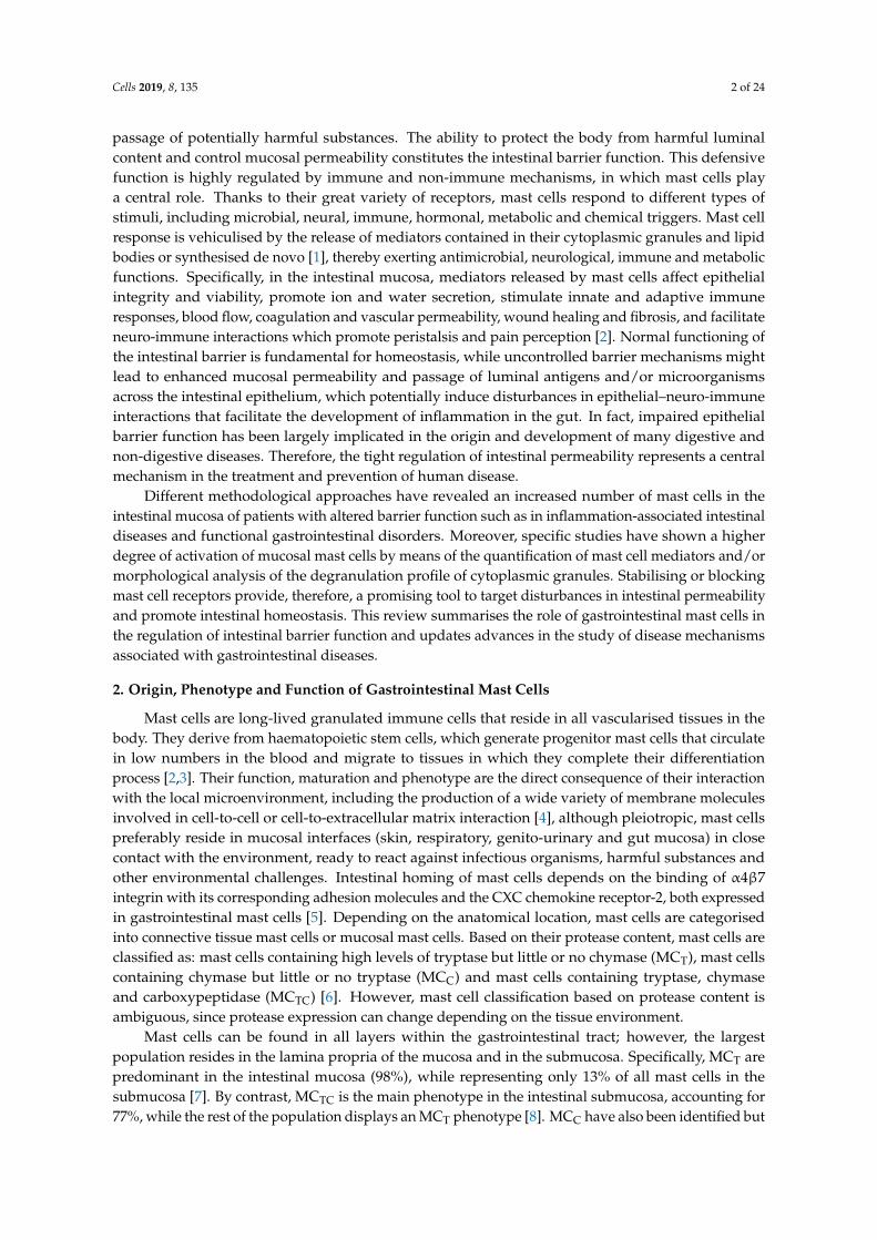
cells
Review
Intestinal Mucosal Mast Cells: Key Modulatorsof Barrier Function and Homeostasis
Mercé Albert-Bayo 1,† , Irene Paracuellos 1,† , Ana M. González-Castro 1,* ,Amanda Rodríguez-Urrutia 2,3 , María J. Rodríguez-Lagunas 4 , Carmen Alonso-Cotoner 5,6 ,Javier Santos 5,6,‡ and María Vicario 1,6,*,‡
1 Laboratory of Translational Mucosal Immunology, Digestive Diseases Research Unit, Vall d’Hebron Institutde Recerca, Department of Gastroenterology, Hospital Universitario Vall d’Hebron, 08035 Barcelona, Spain;[email protected] (M.A.-B.); [email protected] (I.P.)
2 Department of Psychiatry, Hospital Universitari Vall d’Hebron & Facultat de Medicina, UniversitatAutònoma de Barcelona, 08035 Barcelona, Spain; [email protected]
3 Centro de Investigación Biomédica en Red de Salud Mental (CIBERSAM), 28029 Madrid, Spain4 Department of Biochemistry and Physiology, Faculty of Pharmacy and Food Sciences,
University of Barcelona, 08028 Barcelona, Spain; [email protected] Laboratory of Neuro-Immuno-Gastroenterology, Digestive Diseases Research Unit, Vall d’Hebron Institut de
Recerca, Department of Gastroenterology, Hospital Universitario Vall d’Hebron, 08035 Barcelona, Spain;[email protected] (C.A.-C.); [email protected] (J.S.)
6 Centro de Investigación Biomédica en Enfermedades Hepáticas y Digestivas (CIBEREHD),28029 Madrid, Spain
* Correspondence: [email protected] (A.M.G.-C.); [email protected] (M.V.)† Contributed equally to this work.‡ Share co-senior authorship.
Received: 29 December 2018; Accepted: 2 February 2019; Published: 8 February 2019�����������������
Abstract: The gastrointestinal tract harbours the largest population of mast cells in the body;this highly specialised leukocyte cell type is able to adapt its phenotype and function to themicroenvironment in which it resides. Mast cells react to external and internal stimuli thanksto the variety of receptors they express, and carry out effector and regulatory tasks by meansof the mediators of different natures they produce. Mast cells are fundamental elements of theintestinal barrier as they regulate epithelial function and integrity, modulate both innate and adaptivemucosal immunity, and maintain neuro-immune interactions, which are key to functioning of thegut. Disruption of the intestinal barrier is associated with increased passage of luminal antigensinto the mucosa, which further facilitates mucosal mast cell activation, inflammatory responses,and altered mast cell–enteric nerve interaction. Despite intensive research showing gut dysfunctionto be associated with increased intestinal permeability and mucosal mast cell activation, the specificmechanisms linking mast cell activity with altered intestinal barrier in human disease remain unclear.This review describes the role played by mast cells in control of the intestinal mucosal barrier andtheir contribution to digestive diseases.
Keywords: intestinal barrier function; mucosal mast cells
1. Introduction
Mast cells develop a fundamental defensive and immuno-regulatory function, particularly atthe mucosal border between the body and the environment. The intestinal mucosa is the largestinterface that separates the inner and outer environments constantly exposed to luminal content.It allows only small amounts of antigens and bacteria to cross the epithelium, while preventing the
Cells 2019, 8, 135; doi:10.3390/cells8020135 www.mdpi.com/journal/cells
Cells 2019, 8, 135 2 of 24
passage of potentially harmful substances. The ability to protect the body from harmful luminalcontent and control mucosal permeability constitutes the intestinal barrier function. This defensivefunction is highly regulated by immune and non-immune mechanisms, in which mast cells playa central role. Thanks to their great variety of receptors, mast cells respond to different types ofstimuli, including microbial, neural, immune, hormonal, metabolic and chemical triggers. Mast cellresponse is vehiculised by the release of mediators contained in their cytoplasmic granules and lipidbodies or synthesised de novo [1], thereby exerting antimicrobial, neurological, immune and metabolicfunctions. Specifically, in the intestinal mucosa, mediators released by mast cells affect epithelialintegrity and viability, promote ion and water secretion, stimulate innate and adaptive immuneresponses, blood flow, coagulation and vascular permeability, wound healing and fibrosis, and facilitateneuro-immune interactions which promote peristalsis and pain perception [2]. Normal functioning ofthe intestinal barrier is fundamental for homeostasis, while uncontrolled barrier mechanisms mightlead to enhanced mucosal permeability and passage of luminal antigens and/or microorganismsacross the intestinal epithelium, which potentially induce disturbances in epithelial–neuro-immuneinteractions that facilitate the development of inflammation in the gut. In fact, impaired epithelialbarrier function has been largely implicated in the origin and development of many digestive andnon-digestive diseases. Therefore, the tight regulation of intestinal permeability represents a centralmechanism in the treatment and prevention of human disease.
Different methodological approaches have revealed an increased number of mast cells in theintestinal mucosa of patients with altered barrier function such as in inflammation-associated intestinaldiseases and functional gastrointestinal disorders. Moreover, specific studies have shown a higherdegree of activation of mucosal mast cells by means of the quantification of mast cell mediators and/ormorphological analysis of the degranulation profile of cytoplasmic granules. Stabilising or blockingmast cell receptors provide, therefore, a promising tool to target disturbances in intestinal permeabilityand promote intestinal homeostasis. This review summarises the role of gastrointestinal mast cells inthe regulation of intestinal barrier function and updates advances in the study of disease mechanismsassociated with gastrointestinal diseases.
2. Origin, Phenotype and Function of Gastrointestinal Mast Cells
Mast cells are long-lived granulated immune cells that reside in all vascularised tissues in thebody. They derive from haematopoietic stem cells, which generate progenitor mast cells that circulatein low numbers in the blood and migrate to tissues in which they complete their differentiationprocess [2,3]. Their function, maturation and phenotype are the direct consequence of their interactionwith the local microenvironment, including the production of a wide variety of membrane moleculesinvolved in cell-to-cell or cell-to-extracellular matrix interaction [4], although pleiotropic, mast cellspreferably reside in mucosal interfaces (skin, respiratory, genito-urinary and gut mucosa) in closecontact with the environment, ready to react against infectious organisms, harmful substances andother environmental challenges. Intestinal homing of mast cells depends on the binding of α4β7integrin with its corresponding adhesion molecules and the CXC chemokine receptor-2, both expressedin gastrointestinal mast cells [5]. Depending on the anatomical location, mast cells are categorisedinto connective tissue mast cells or mucosal mast cells. Based on their protease content, mast cells areclassified as: mast cells containing high levels of tryptase but little or no chymase (MCT), mast cellscontaining chymase but little or no tryptase (MCC) and mast cells containing tryptase, chymaseand carboxypeptidase (MCTC) [6]. However, mast cell classification based on protease content isambiguous, since protease expression can change depending on the tissue environment.
Mast cells can be found in all layers within the gastrointestinal tract; however, the largestpopulation resides in the lamina propria of the mucosa and in the submucosa. Specifically, MCT arepredominant in the intestinal mucosa (98%), while representing only 13% of all mast cells in thesubmucosa [7]. By contrast, MCTC is the main phenotype in the intestinal submucosa, accounting for77%, while the rest of the population displays an MCT phenotype [8]. MCC have also been identified but
Cells 2019, 8, 135 3 of 24
appear to be uncommon [6]. A new phenotype of mast cells expressing tryptase and carboxypeptidaseA3, but not chymase, has recently been defined in the bronchial and oesophageal mucosa associatedwith the pathophysiology of asthma and eosinophilic oesophagitis, respectively [9,10]. However,exactly how this phenotype contributes to human disease remains unknown.
Mast cells are currently recognised as regulatory and effector cells in both innate and adaptiveimmunity. Their broad functions rely on their ability to react to a great variety of stimuli and secretebiologically-active products with pro-inflammatory, anti-inflammatory and/or immunosuppressiveproperties. Mast cells play a prominent role in immunoglobulin(Ig)-mediated allergic inflammation,and are also involved in a variety of intestinal and non-intestinal diseases such as gastrointestinalinflammation, functional gastrointestinal disorders, infections, autoimmune diseases, atherosclerosisand carcinogenesis [11] as well as in neuropsychiatric conditions [12]. Of importance for intestinalhomeostasis, mast cells are fundamental for diverse intestinal physiological processes such as theregulation of mucosal integrity and epithelial barrier activity, and the maintenance of neuro-immunointeraction that supports the brain-gut axis. The fact that mast cells have persisted throughoutvertebrate evolution, with an ancient origin even before the development of adaptive immunity,reinforces their importance in innate immunity as well as their remarkable role in such a variety ofdiseases [13].
3. Mast Cell Activation
The surface of mast cells is covered with a variety of receptors specific for immune ligands(Igs, complement fragments and cytokines) and for non-immune mediators, which includeneurotransmitters, neuropeptides, hormones, growth factors and other biological and physicochemicalstimuli [14]. Mast cell versatility implies they can be activated by different mechanisms,with cross-linking of IgE high-affinity receptor (FcεRI) to cell surface-bound IgE being the traditionaland best studied stimulus in sensitised individuals [15]. A response is then triggered by a serie ofphosphorylation cascades and activation motifs that lead to intracellular calcium flux, activationof transcription factors (such as AP-1, MITF and STAT-5), mast cell degranulation and cytokineproduction [16]. Mast cells can additionally be stimulated by IgG since they also express its receptor(FcγRI) [17] and other Ig-associated receptors. Of importance for the control of gastrointestinaldisease is the recently identified IgG signalling via FcγRIIb, which suppresses a hypersensitivityreaction [18]. As innate immune sentinels, mast cells recognise microbial agents (bacterial, viral,parasitic and fungal) and endogenous factors derived from cell damage by germline-encoded patternrecognition receptors, which include toll-like receptors (TLRs), C-type lectin-like receptors (CLRs),retinoic acid-inducible gene I (RIG-I)-like receptors (RLRs) and nucleotide-binding oligomerisationdomain (NOD)-like receptors (NLRs) [19]. Importantly for homeostasis and for ensuring an appropriateresponse to injury, mast cells also respond to different endogenous stimuli since they express receptorsfor neurotransmitters (such as acetylcholine and serotonin), neuropeptides (such as substance P, SP andvasoactive intestinal peptide, VIP), neurotrophins (such as nerve growth factor, NGF) and gaseousneurotransmitters (such as nitric oxide, NO).
Upon activation, mast cells release biologically-active products (Table 1), newly synthesised oralready contained in their cytoplasmic granules and lipid bodies [1]. The storage of these molecules inmast cell granules is possible, thanks to the anionic gel matrix composed of heparin and chondroitinsulphate, in which the mediators become trapped [20]. Pre-formed mediators include proteases,biogenic amines, proteoglycans, lysosomal enzymes, certain cytokines and growth factors andgranule membrane-associated proteins. Newly-synthesised mediators include lipidic compounds,neuropeptides and a huge variety of cytokines, chemokines and growth factors [1,21]. This wide varietyof molecules produced by mast cells supports their pleiotropic functions during both homeostasisand disease.
Cells 2019, 8, 135 4 of 24
Table 1. Mast cell mediators.
Pre-Formed Mediators
Proteases Mast cell-specific: tryptase, chymase, carboxypeptidase ANon-mast cell-specific: cathepsin G, granzyme B, active caspase 3, ADAMTS5, renin
Biogenic amines Histamine, serotonin, dopamine, polyamines
Proteoglycans Serglycin, chondroitin sulphates, heparin
Lysosomal enzymes β-hexosaminidase, β-glucuronidase, arylsulphatase, cathepsins
Cytokines/growth factors TNF, IL-4, GMCSF, bFGF, VEGF, NGF
Granule membrane-associated proteins VAMPs, syntaxin 3, synaptotagmins, MUNCs, SCAMPs, CD63, RABs, LC3-II, MHC class II
Others Heparanase, CAP-18, secretogranin-III and chromogranin A
Newly-Synthesised Mediators
Lipid mediators Leukotriene C4/B4, prostaglandin D2, platelet-activating factor
Cytokines IL-1, IL-3, IL-6, IL-18, TNF, SCF, TGF-β
Chemokines MCP-1, RANTES, eotaxin, TARC
Growth factors GMCSF, MCSF, bFGF, PDGF, NGF, VEGF, GnRH
ADAMTS5, a disintegrin and metalloproteinase with thrombospondin motifs 5; TNF,tumour necrosis factor; IL, interleukin; GMCSF, granulocyte-macrophage colony-stimulating factor;bFGF, basic fibroblast growth factor; VEGF, vascular endothelial growth factor; NGF, nerve growthfactor; VAMP, vesicle-associated membrane protein; MUNC, mammalian uncoordinated-18protein; SCAMP, secretory carrier-associated membrane protein; LC3, lipidated light chain 3;MHC, major histocompatibility complex; CAP, cathelicidin antimicrobial peptide; SCF, stem cell factor;TGF, transforming growth factor; MCP, monocyte chemoattractant protein; RANTES, regulated uponactivation normal T-cell expressed and secreted chemokine; TARC, thymus and activation-regulatedchemokine; MCSF, macrophage colony–stimulating factor; PDGF, platelet-derived growth factor; GnRH,gonadotropin-releasing hormone.
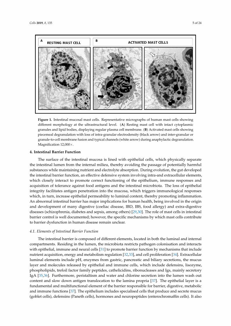
The secretion of mast cell mediators is carried out mainly by two mechanisms: piecemeal andanaphylactic degranulation (Figure 1). Piecemeal degranulation leads to partial or total granuleemptying, causing a selective release of the content without inter-granule or granule-to-plasmamembrane fusions. Ultrastructural analysis showed granule morphology to be quite conservedafter the piecemeal process [22]. This type of secretion is promoted by neuropeptides, cytokines andmicrobial products that interact with mast cells, as described in physiological conditions and a variety ofdigestive diseases, among which inflammatory bowel disease (IBD), irritable bowel syndrome (IBS) andfunctional dyspepsia (FD) have been the most studied [23–25]. By contrast, anaphylactic degranulationis the explosive release of mast cell content by granule or granule-to-plasma membrane fusionsfollowed by extrusion [26] and is associated with hypersensitivity reactions. Mast cell degranulation ismediated by the soluble N-ethylmaleimide-sensitive factor attachment protein receptors (SNARES),of which VAMP-7, VAMP-8, SNAP-23 and STX-4 have been reported to be significant SNARE moleculesin human intestinal mast cell granule fusion and exocytosis [27,28]. The degree and type of activationof mast cells, as a consequence of their interaction with the microenvironment, determines themaintenance of homeostasis or disruption of essential defensive functions in the mucosa. This isof key importance for intestinal physiological activity, since the intestine represents the largest surfaceof the body in contact with the external environment and mast cells interact with virtually all cells in themucosa as well as with microorganisms and harmful molecules that reach the intestine. Cell plasticityand responsiveness define mast cells as a fundamental component of intestinal barrier function duringboth homeostasis and disease.
Cells 2019, 8, 135 5 of 24
Cells 2019, 8, 135 5 of 24
Figure 1. Intestinal mucosal mast cells. Representative micrographs of human mast cells showing different morphology at the ultrastructural level. (A) Resting mast cell with intact cytoplasmic granules and lipid bodies, displaying regular plasma cell membrane. (B) Activated mast cells showing piecemeal degranulation with loss of intra-granular electrodensity (black arrow) and inter-granular or granule-to-cell membrane fusion and typical channels (white arrow) during anaphylactic degranulation. Magnification 12,000×.
4. Intestinal Barrier Function
The surface of the intestinal mucosa is lined with epithelial cells, which physically separate the intestinal lumen from the internal milieu, thereby avoiding the passage of potentially harmful substances while maintaining nutrient and electrolyte absorption. During evolution, the gut developed the intestinal barrier function, an effective defensive system involving intra-and extracellular elements, which closely interact to promote correct functioning of the epithelium, immune responses and acquisition of tolerance against food antigens and the intestinal microbiota. The loss of epithelial integrity facilitates antigen penetration into the mucosa, which triggers immunological responses which, in turn, increase epithelial permeability to luminal content, thereby promoting inflammation. An abnormal intestinal barrier has major implications for human health, being involved in the origin and development of many digestive (coeliac disease, IBD, IBS, food allergy) and extra-digestive diseases (schizophrenia, diabetes and sepsis, among others) [29,30]. The role of mast cells in intestinal barrier control is well documented; however, the specific mechanisms by which mast cells contribute to barrier dysfunction in human disease remain unclear.
4.1. Elements of Intestinal Barrier Function
The intestinal barrier is composed of different elements, located in both the luminal and internal compartments. Residing in the lumen, the microbiota restricts pathogen colonisation and interacts with epithelial, immune and neural cells [31] to promote barrier function by mechanisms that include nutrient acquisition, energy and metabolism regulation [32,33], and cell proliferation [34]. Extracellular luminal elements include pH, enzymes from gastric, pancreatic and biliary secretions, the mucus layer and molecules released by epithelial and immune cells, which include defensins, lisozyme, phospholipids, trefoil factor family peptides, cathelicidins, ribonucleases and Igs, mainly secretory IgA [35,36]. Furthermore, peristaltism and water and chlorine secretion into the lumen wash out content and slow down antigen translocation to the lamina propria [37]. The epithelial layer is a fundamental and multifunctional element of the barrier responsible for barrier, digestive, metabolic and immune functions [37]. The epithelium includes specialised cells that produce and secrete mucus (goblet cells), defensins (Paneth cells), hormones and neuropeptides (enterochromaffin cells). It also includes a unique cell type specialised in antigen uptake from the lumen (M cells) [38].
Figure 1. Intestinal mucosal mast cells. Representative micrographs of human mast cells showingdifferent morphology at the ultrastructural level. (A) Resting mast cell with intact cytoplasmicgranules and lipid bodies, displaying regular plasma cell membrane. (B) Activated mast cells showingpiecemeal degranulation with loss of intra-granular electrodensity (black arrow) and inter-granular orgranule-to-cell membrane fusion and typical channels (white arrow) during anaphylactic degranulation.Magnification 12,000×.
4. Intestinal Barrier Function
The surface of the intestinal mucosa is lined with epithelial cells, which physically separatethe intestinal lumen from the internal milieu, thereby avoiding the passage of potentially harmfulsubstances while maintaining nutrient and electrolyte absorption. During evolution, the gut developedthe intestinal barrier function, an effective defensive system involving intra-and extracellular elements,which closely interact to promote correct functioning of the epithelium, immune responses andacquisition of tolerance against food antigens and the intestinal microbiota. The loss of epithelialintegrity facilitates antigen penetration into the mucosa, which triggers immunological responseswhich, in turn, increase epithelial permeability to luminal content, thereby promoting inflammation.An abnormal intestinal barrier has major implications for human health, being involved in the originand development of many digestive (coeliac disease, IBD, IBS, food allergy) and extra-digestivediseases (schizophrenia, diabetes and sepsis, among others) [29,30]. The role of mast cells in intestinalbarrier control is well documented; however, the specific mechanisms by which mast cells contributeto barrier dysfunction in human disease remain unclear.
4.1. Elements of Intestinal Barrier Function
The intestinal barrier is composed of different elements, located in both the luminal and internalcompartments. Residing in the lumen, the microbiota restricts pathogen colonisation and interactswith epithelial, immune and neural cells [31] to promote barrier function by mechanisms that includenutrient acquisition, energy and metabolism regulation [32,33], and cell proliferation [34]. Extracellularluminal elements include pH, enzymes from gastric, pancreatic and biliary secretions, the mucuslayer and molecules released by epithelial and immune cells, which include defensins, lisozyme,phospholipids, trefoil factor family peptides, cathelicidins, ribonucleases and Igs, mainly secretoryIgA [35,36]. Furthermore, peristaltism and water and chlorine secretion into the lumen wash outcontent and slow down antigen translocation to the lamina propria [37]. The epithelial layer is afundamental and multifunctional element of the barrier responsible for barrier, digestive, metabolicand immune functions [37]. The epithelium includes specialised cells that produce and secrete mucus(goblet cells), defensins (Paneth cells), hormones and neuropeptides (enterochromaffin cells). It also
Cells 2019, 8, 135 6 of 24
includes a unique cell type specialised in antigen uptake from the lumen (M cells) [38]. To ensure aneffective physical barrier, epithelial cells are tightly bonded to each other by intercellular junctions(tight junctions, TJ, at the apical junctional complex, followed by adherent junctions, and desmosomes).
The immune contribution to the mucosal barrier is carried out by the gut-associated lymphoidtissue (GALT) distributed in organised lymphoid structures such as lymphoid follicles, Peyer’s patchesand mesenteric lymph nodes, in which immune responses are initiated. GALT also includes a diffusedistribution of effector cells throughout the epithelium and the lamina propria of the intestinalmucosa [39], composed mainly of plasma cells, macrophages, mast cells, lymphocytes, eosinophilsand dendritic cells [40]. Furthermore, connective tissue, blood and lymph vessels and fibroblaststhat reside in the lamina propria also contribute to barrier function maintenance. An integratednetwork of neural cells from both the central and enteric nervous systems coordinates digestivefunctions and intestinal homeostasis maintenance via the release of neurotransmitters and, indirectly,neuro-immune interactions.
4.2. Mast Cells as Neuro-Immune Players in the Regulation of Intestinal Barrier Function
Control of the intestinal barrier results from a network of interactions among the microbiota,epithelial cells and immune and nervous systems. The communication between the central and entericnervous systems, the so-called brain-gut axis, permits regulation of the intestinal barrier by monitoringion secretion, epithelial tightness, immune function and peristalsis. The functional unit established bymast cell-nerve interaction [41] is a fundamental component in such interplay via paracrine signalling(the most common), transgranulation (granule fragments and mediators are delivered directly to theneuronal body) and integrin signalling (physical synapses through integrins) [21]. Enteric neurons,as well as vagal and spinal afferents, express receptors for molecules (mainly proteases, neuropeptides,hormones and growth factors) released by mast cells, which stimulate nerve terminals, therebymodulating the firing threshold. Similarly, neuropeptides and neurotransmitters released by neuronsstimulate mast cell secretion of mediators, which further activate neuronal receptors [21], supportingthe maintenance of this neuro-immune interplay. This interaction contributes to monitoring of gutfunction by the central nervous system; however, if overstimulated, it can exert harmful effectsassociated with disease [42].
The contribution of mast cells to barrier function through neuro-immune mechanisms hasbeen evidenced in different experimental settings. Diverse stressors (physical and psychological,acute and chronic) have been shown to disturb barrier homeostasis by increasing ion secretion andepithelial permeability [43,44], effects avoided in mast cell knock-out rats and also in humans afteroral pretreatment with a mast cell stabiliser [45,46]. The stress response, which includes endocrineand behavioural changes, is centrally mediated by the release of corticotropin-releasing factor (CRF).Additionally, intestinal mucosal cells, including immunocytes, nerves and enterochromaffin cells,produce and release CRF upon activation. Local CRF interacts with its receptors (CRF-R1, CRF-R2) onsubepithelial mast cells [47] to induce mucin release, ion and water secretion, and increase epithelialpermeability [46,48,49]. Moreover, other neuropeptides such as SP and NGF induce the release ofvasoactive mediators from mast cells, thereby contributing to chloride secretion, barrier dysfunction,hyperalgesia, diarrhoea, inflammation and motility changes [50]. In fact, epithelial permeability toluminal bacteria seems to be modulated by VIP and mast cell activity, as suggested by the VIP–mastcell-dependent regulation of commensal and pathogenic bacteria passage in the human colon [51].
5. Regulation of Intestinal Mucosal Barrier by Mast Cells
Mast cells are unique due to their ability to modulate their phenotype, a process calledtransdifferentiation [52] by which they synthesise and release a specific mediator profile dependingon the microenvironment [53]. The anatomical complexity of the intestine and the ever-changingenvironment contribute to mast cell phenotype which, based on the nature of mediators released intothe extracellular milieu, determines organ function. In this way, intestinal mast cells perform multiple
Cells 2019, 8, 135 7 of 24
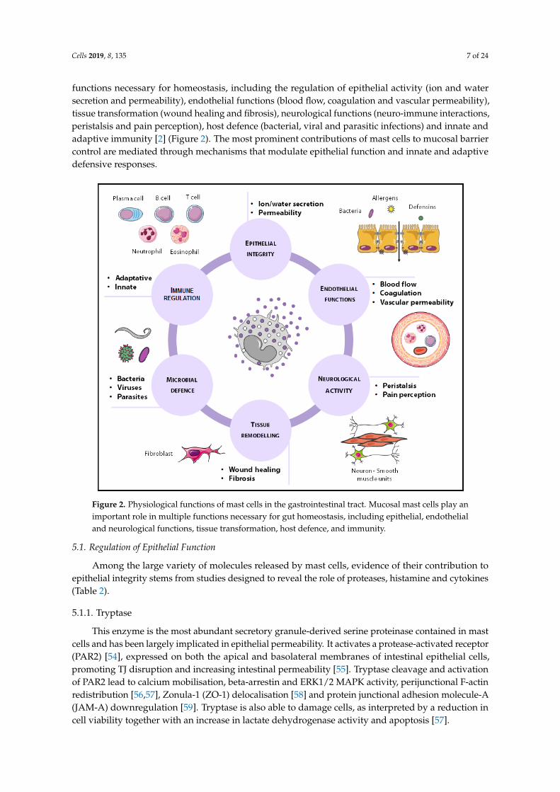
functions necessary for homeostasis, including the regulation of epithelial activity (ion and watersecretion and permeability), endothelial functions (blood flow, coagulation and vascular permeability),tissue transformation (wound healing and fibrosis), neurological functions (neuro-immune interactions,peristalsis and pain perception), host defence (bacterial, viral and parasitic infections) and innate andadaptive immunity [2] (Figure 2). The most prominent contributions of mast cells to mucosal barriercontrol are mediated through mechanisms that modulate epithelial function and innate and adaptivedefensive responses.
Cells 2019, 8, 135 7 of 24
secretion and permeability), endothelial functions (blood flow, coagulation and vascular permeability), tissue transformation (wound healing and fibrosis), neurological functions (neuro-immune interactions, peristalsis and pain perception), host defence (bacterial, viral and parasitic infections) and innate and adaptive immunity [2] (Figure 2). The most prominent contributions of mast cells to mucosal barrier control are mediated through mechanisms that modulate epithelial function and innate and adaptive defensive responses.
Figure 2. Physiological functions of mast cells in the gastrointestinal tract. Mucosal mast cells play an important role in multiple functions necessary for gut homeostasis, including epithelial, endothelial and neurological functions, tissue transformation, host defence, and immunity.
5.1. Regulation of Epithelial Function
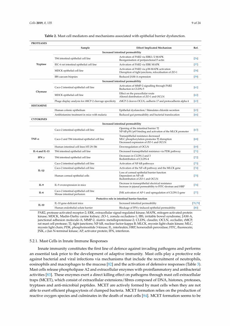
Among the large variety of molecules released by mast cells, evidence of their contribution to epithelial integrity stems from studies designed to reveal the role of proteases, histamine and cytokines (Table 2).
5.1.1. Tryptase
This enzyme is the most abundant secretory granule-derived serine proteinase contained in mast cells and has been largely implicated in epithelial permeability. It activates a protease-activated receptor (PAR2) [54], expressed on both the apical and basolateral membranes of intestinal epithelial cells, promoting TJ disruption and increasing intestinal permeability [55]. Tryptase cleavage and activation of PAR2 lead to calcium mobilisation, beta-arrestin and ERK1/2 MAPK activity, perijunctional F-actin redistribution [56,57], Zonula-1 (ZO-1) delocalisation [58] and protein junctional adhesion molecule-A (JAM-A) downregulation [59]. Tryptase is also able to damage cells, as interpreted by a reduction in cell viability together with an increase in lactate dehydrogenase activity and apoptosis [57].
Figure 2. Physiological functions of mast cells in the gastrointestinal tract. Mucosal mast cells play animportant role in multiple functions necessary for gut homeostasis, including epithelial, endothelialand neurological functions, tissue transformation, host defence, and immunity.
5.1. Regulation of Epithelial Function
Among the large variety of molecules released by mast cells, evidence of their contribution toepithelial integrity stems from studies designed to reveal the role of proteases, histamine and cytokines(Table 2).
5.1.1. Tryptase
This enzyme is the most abundant secretory granule-derived serine proteinase contained in mastcells and has been largely implicated in epithelial permeability. It activates a protease-activated receptor(PAR2) [54], expressed on both the apical and basolateral membranes of intestinal epithelial cells,promoting TJ disruption and increasing intestinal permeability [55]. Tryptase cleavage and activationof PAR2 lead to calcium mobilisation, beta-arrestin and ERK1/2 MAPK activity, perijunctional F-actinredistribution [56,57], Zonula-1 (ZO-1) delocalisation [58] and protein junctional adhesion molecule-A(JAM-A) downregulation [59]. Tryptase is also able to damage cells, as interpreted by a reduction incell viability together with an increase in lactate dehydrogenase activity and apoptosis [57].
Cells 2019, 8, 135 8 of 24
5.1.2. Chymase
This chymotrypsin-type serine protease is implicated mainly in extracelluar matrix degradationand also targets epithelial integrity through activation of the PAR2 receptor. The biological function ofchymase has been studied through the generation of mouse strains deficient in different murine mastcell proteases (mMCP), functional homologues to human chymase: mMCP-1 mMCP-2 mMCP-4 ormMCP-5 [60]. Experimental studies have shown that the activation of PAR2 receptors by chymaseinduces p38 phosphorylation and p44/42 (ERK1/2) signalling pathway activation, which increasemetalloprotease-2 (MMP-2) and reduce claudin-5, resulting in epithelial barrier dysfunction [61].Specifically, the rat mucosal mast cell chymase, rMCP-2, increases epithelial permeability throughalteration of ZO-1 and occludin distribution in epithelial cells [62] and cleavage of occludin, cadherin17 and protocadherin alpha [63].
5.1.3. Histamine
This amine is a preformed mast cell mediator involved in a variety of physiological andpathological processes throughout the gastrointestinal tract. It mediates immunological responses,visceral nociception, modulation of intestinal motility and gastric acid secretion through activation ofits receptors (H1-H4) [64]. The contribution of histamine to epithelial dysfunction is mediated by H1receptors directly stimulating chloride secretion, as demonstrated in the human colonic epithelium [65].Recent evidence also revealed a role of histamine in increasing epithelial intestinal permeability andbacterial translocation in malaria-infected mice [66]; however, the specific mechanisms leading tobarrier deregulation remain unknown.
5.1.4. Cytokines
These small molecules are released mainly by lymphocytes, macrophages, eosinophils, dendriticcells and mast cells that mediate inter-cell communication. Mast cells produce a large varietyof cytokines, many of which have a direct impact on the intestinal epithelial barrier (Table 2).TNF-α directly disrupts TJ via myosin light-chain kinase (MLCK)-mediated phosphorylation ofthe myosin light chain (MLC) [67,68] and ZO-1 and occludin downregulation [68,69]. Interleukin(IL)-4 stimulates mast cell IL-13 production [70], with both molecules sharing the IL-4Rα chainreceptor, which elicits phosphatidylinositol 3-kinase (PI3K) activation and modulation of epithelialparacellular permeability [71]. IFN-γ also alters paracellular permeability in the intestine throughthe reduction of claudin 2 and 3 and reorganisation of claudin 4 [72]. IL-1β regulates intestinalfunction mainly via activation of the NF-κB pathway and the MLCK gene [73–75]. IL-9 also increasesintestinal permeability associated with a genetic profile identified in intestinal anaphylaxis [76],and IL-6 promotes JNK activation of AP-1 and upregulation of the claudin 2 gene, leading to TJdisruption [77]. On the other hand, the anti-inflammatory cytokine IL-10 has also been shownto develop a protective role in intestinal barrier function, since IL-10-gene-deficient mice showedincreased intestinal permeability [78,79] and the administration of IL-10 prevented IFN-γ-inducedbarrier dysfunction [80]. However, IL-10 has been shown to enhance IgE-mediated mast cell activity,which suggests a potential contribution to barrier dysfunction during food allergy response [81].
5.2. Regulation of Mucosal Immunity
A multivalent capacity to recognise and respond to both internal and external dangers,together with the ability to cross-talk with other immune cells, render mast cells a unique playerin linking innate and adaptive immunity.
Cells 2019, 8, 135 9 of 24
Table 2. Mast cell mediators and mechanisms associated with epithelial barrier dysfunction.
PROTEASES
Sample Effect/ Implicated Mechanism Ref.
Tryptase
Increased intestinal permeability
T84 intestinal epithelial cell line Activation of PAR2 via ERK1/2 MAPKReorganisation of perijunctional F-actin [56]
IEC-6 rat intestinal epithelial cell line Activation of PAR2 via ERK MAPK [57]
MDCK epithelial cell line Activation of PAR2 via p38-MAPK activationDisruption of tight junctions, relocalisation of ZO-1 [58]
IBS caecum biopsies Reduced JAM-A expression [59]
Chymase
Increased intestinal permeability
Caco-2 intestinal epithelial cell line Activation of MMP-2 signalling through PAR2Reduction in CLDN-5 [61]
MDCK epithelial cell line Effect on the paracellular routeAltered distribution of ZO-1 and OCLN [62]
Phage display analysis for rMCP-2 cleavage specificity rMCP-2 cleaves OCLN, cadherin 17 and protocadherin alpha 4 [63]
HISTAMINE
Human colonic epithelium Epithelial dysfunction/ Stimulates chloride secretion [65]
Antihistamine treatment in mice with malaria Reduced gut permeability and bacterial translocation [66]
CYTOKINES
Increased intestinal permeability
TNF-α
Caco-2 intestinal epithelial cell line Opening of the intestinal barrier TJNF-kB p50/p65 binding and activation of the MLCK promoter [67]
Caco-2 and T84 intestinal epithelial cell lineTransepithelial resistance decreasedMLC phosphorylation promotes TJ disruptionDecreased expression of ZO-1 and OCLN
[68]
Human intestinal cell lines HT-29/B6 Downregulation of OCLN [69]
IL-4 and IL-13 T84 intestinal epithelial cell line Decreased transepithelial resistance via PI3K pathway [71]
IFN γ T84 intestinal epithelial cell line Decreases in CLDN-2 and 3Redistribution of CLDN-4 [72]
IL-1β
Caco-2 intestinal epithelial cell line Activation of NF-kB pathways [73]
Caco-2 intestinal epithelial cell line Activation of the NF-κB pathway and the MLCK gene [74]
Human corneal epithelial cellsLoss of corneal epithelial barrier functionDependent on NF-κBRedistribution of ZO-1 and OCLN
[75]
IL-9 IL-9 overexpression in mice Decrease in transepithelial electrical resistanceIncrease in jejunal permeability to FITC-dextran and HRP [76]
IL-6 Caco-2 intestinal epithelial cell lineMouse intestinal perfusion JNK activation of AP-1 and upregulation of CLDN-2-gene [77]
Protective role in intestinal barrier function
IL-10IL-10 gene-deficient mice Increased intestinal permeability [78,79]
Human endothelial solute barrier Blockage of IFNγ-induced epithelial permeability [80]
PAR2, protease-activated receptor-2; ERK, extracellular signal-regulated kinase; MAPK, mitogen-activated proteinkinase; MDCK, Madin-Darby canine kidney; ZO-1, zonula occludens-1; IBS, irritable bowel syndrome; JAM-A,junctional adhesion molecule-A; MMP-2, matrix metalloproteinase-2; CLDN, claudin; OCLN, occludin; rMCP,rat mast cell protease; TJ, tight junctions; NF-kB, nuclear factor-kappa B; MLCK, myosin light-chain kinase; MLC,myosin light chain; PI3K, phosphoinositide 3-kinase; IL, interleukin; HRP, horseradish peroxidase; FITC, fluorescein;JNK, c-Jun N-terminal kinase; AP, activator protein; IFN, interferon.
5.2.1. Mast Cells in Innate Immune Responses
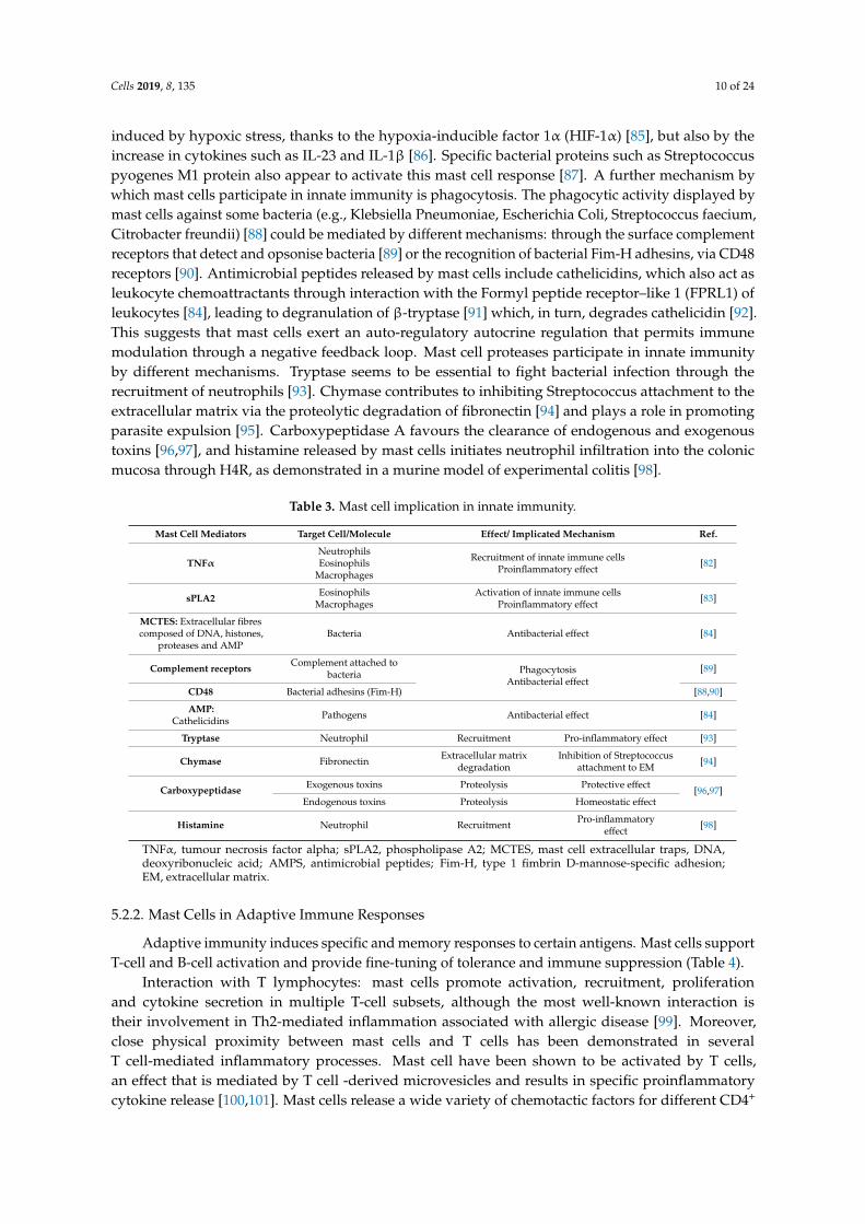
Innate immunity constitutes the first line of defence against invading pathogens and performsan essential task prior to the development of adaptive immunity. Mast cells play a protective roleagainst bacterial and viral infections via mechanisms that include the recruitment of neutrophils,eosinophils and macrophages to the mucosa [82] and the activation of defensive responses (Table 3).Mast cells release phospholipase A2 and extracellular enzymes with proinflammatory and antibacterialactivities [83]. These enzymes exert a direct killing effect on pathogens through mast cell extracellulartraps (MCET), which consist of extracellular extensions/fibres composed of DNA, histones, proteases,tryptases and anti-microbial peptides. MCET are actively formed by mast cells when they are notable to exert efficient phagocytosis of clumped bacteria. MCET formation relies on the production ofreactive oxygen species and culminates in the death of mast cells [84]. MCET formation seems to be
Cells 2019, 8, 135 10 of 24
induced by hypoxic stress, thanks to the hypoxia-inducible factor 1α (HIF-1α) [85], but also by theincrease in cytokines such as IL-23 and IL-1β [86]. Specific bacterial proteins such as Streptococcuspyogenes M1 protein also appear to activate this mast cell response [87]. A further mechanism bywhich mast cells participate in innate immunity is phagocytosis. The phagocytic activity displayed bymast cells against some bacteria (e.g., Klebsiella Pneumoniae, Escherichia Coli, Streptococcus faecium,Citrobacter freundii) [88] could be mediated by different mechanisms: through the surface complementreceptors that detect and opsonise bacteria [89] or the recognition of bacterial Fim-H adhesins, via CD48receptors [90]. Antimicrobial peptides released by mast cells include cathelicidins, which also act asleukocyte chemoattractants through interaction with the Formyl peptide receptor–like 1 (FPRL1) ofleukocytes [84], leading to degranulation of β-tryptase [91] which, in turn, degrades cathelicidin [92].This suggests that mast cells exert an auto-regulatory autocrine regulation that permits immunemodulation through a negative feedback loop. Mast cell proteases participate in innate immunityby different mechanisms. Tryptase seems to be essential to fight bacterial infection through therecruitment of neutrophils [93]. Chymase contributes to inhibiting Streptococcus attachment to theextracellular matrix via the proteolytic degradation of fibronectin [94] and plays a role in promotingparasite expulsion [95]. Carboxypeptidase A favours the clearance of endogenous and exogenoustoxins [96,97], and histamine released by mast cells initiates neutrophil infiltration into the colonicmucosa through H4R, as demonstrated in a murine model of experimental colitis [98].
Table 3. Mast cell implication in innate immunity.
Mast Cell Mediators Target Cell/Molecule Effect/ Implicated Mechanism Ref.
TNFαNeutrophilsEosinophils
Macrophages
Recruitment of innate immune cellsProinflammatory effect [82]
sPLA2 EosinophilsMacrophages
Activation of innate immune cellsProinflammatory effect [83]
MCTES: Extracellular fibrescomposed of DNA, histones,
proteases and AMPBacteria Antibacterial effect [84]
Complement receptors Complement attached tobacteria Phagocytosis
Antibacterial effect[89]
CD48 Bacterial adhesins (Fim-H) [88,90]
AMP:Cathelicidins Pathogens Antibacterial effect [84]
Tryptase Neutrophil Recruitment Pro-inflammatory effect [93]
Chymase Fibronectin Extracellular matrixdegradation
Inhibition of Streptococcusattachment to EM [94]
Carboxypeptidase Exogenous toxins Proteolysis Protective effect[96,97]
Endogenous toxins Proteolysis Homeostatic effect
Histamine Neutrophil Recruitment Pro-inflammatoryeffect [98]
TNFα, tumour necrosis factor alpha; sPLA2, phospholipase A2; MCTES, mast cell extracellular traps, DNA,deoxyribonucleic acid; AMPS, antimicrobial peptides; Fim-H, type 1 fimbrin D-mannose-specific adhesion;EM, extracellular matrix.
5.2.2. Mast Cells in Adaptive Immune Responses
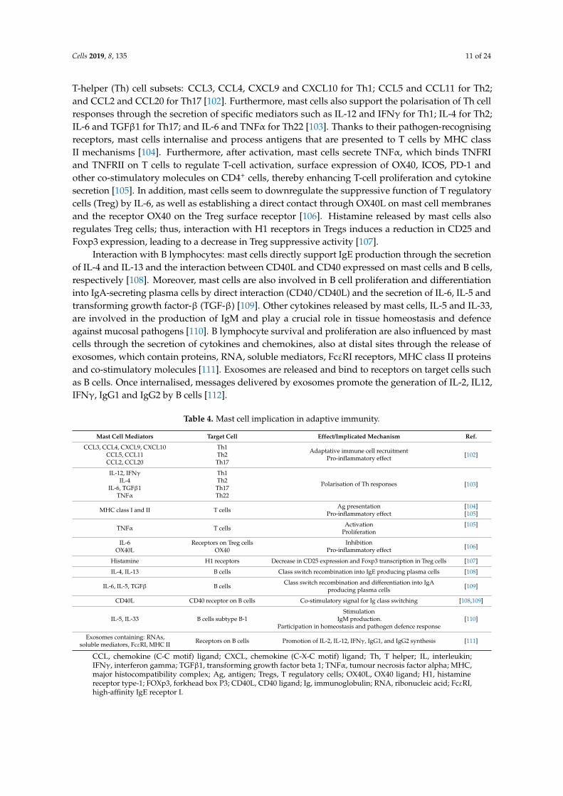
Adaptive immunity induces specific and memory responses to certain antigens. Mast cells supportT-cell and B-cell activation and provide fine-tuning of tolerance and immune suppression (Table 4).
Interaction with T lymphocytes: mast cells promote activation, recruitment, proliferationand cytokine secretion in multiple T-cell subsets, although the most well-known interaction istheir involvement in Th2-mediated inflammation associated with allergic disease [99]. Moreover,close physical proximity between mast cells and T cells has been demonstrated in severalT cell-mediated inflammatory processes. Mast cell have been shown to be activated by T cells,an effect that is mediated by T cell -derived microvesicles and results in specific proinflammatorycytokine release [100,101]. Mast cells release a wide variety of chemotactic factors for different CD4+
Cells 2019, 8, 135 11 of 24
T-helper (Th) cell subsets: CCL3, CCL4, CXCL9 and CXCL10 for Th1; CCL5 and CCL11 for Th2;and CCL2 and CCL20 for Th17 [102]. Furthermore, mast cells also support the polarisation of Th cellresponses through the secretion of specific mediators such as IL-12 and IFNγ for Th1; IL-4 for Th2;IL-6 and TGFβ1 for Th17; and IL-6 and TNFα for Th22 [103]. Thanks to their pathogen-recognisingreceptors, mast cells internalise and process antigens that are presented to T cells by MHC classII mechanisms [104]. Furthermore, after activation, mast cells secrete TNFα, which binds TNFRIand TNFRII on T cells to regulate T-cell activation, surface expression of OX40, ICOS, PD-1 andother co-stimulatory molecules on CD4+ cells, thereby enhancing T-cell proliferation and cytokinesecretion [105]. In addition, mast cells seem to downregulate the suppressive function of T regulatorycells (Treg) by IL-6, as well as establishing a direct contact through OX40L on mast cell membranesand the receptor OX40 on the Treg surface receptor [106]. Histamine released by mast cells alsoregulates Treg cells; thus, interaction with H1 receptors in Tregs induces a reduction in CD25 andFoxp3 expression, leading to a decrease in Treg suppressive activity [107].
Interaction with B lymphocytes: mast cells directly support IgE production through the secretionof IL-4 and IL-13 and the interaction between CD40L and CD40 expressed on mast cells and B cells,respectively [108]. Moreover, mast cells are also involved in B cell proliferation and differentiationinto IgA-secreting plasma cells by direct interaction (CD40/CD40L) and the secretion of IL-6, IL-5 andtransforming growth factor-β (TGF-β) [109]. Other cytokines released by mast cells, IL-5 and IL-33,are involved in the production of IgM and play a crucial role in tissue homeostasis and defenceagainst mucosal pathogens [110]. B lymphocyte survival and proliferation are also influenced by mastcells through the secretion of cytokines and chemokines, also at distal sites through the release ofexosomes, which contain proteins, RNA, soluble mediators, FcεRI receptors, MHC class II proteinsand co-stimulatory molecules [111]. Exosomes are released and bind to receptors on target cells suchas B cells. Once internalised, messages delivered by exosomes promote the generation of IL-2, IL12,IFNγ, IgG1 and IgG2 by B cells [112].
Table 4. Mast cell implication in adaptive immunity.
Mast Cell Mediators Target Cell Effect/Implicated Mechanism Ref.
CCL3, CCL4, CXCL9, CXCL10 Th1 Adaptative immune cell recruitmentPro-inflammatory effect [102]CCL5, CCL11 Th2
CCL2, CCL20 Th17
IL-12, IFNγ Th1
Polarisation of Th responses [103]IL-4 Th2
IL-6, TGFβ1 Th17TNFα Th22
MHC class I and II T cells Ag presentationPro-inflammatory effect
[104][105]
TNFα T cells ActivationProliferation
[105]
IL-6 Receptors on Treg cells InhibitionPro-inflammatory effect [106]OX40L OX40
Histamine H1 receptors Decrease in CD25 expression and Foxp3 transcription in Treg cells [107]
IL-4, IL-13 B cells Class switch recombination into IgE producing plasma cells [108]
IL-6, IL-5, TGFβ B cells Class switch recombination and differentiation into IgAproducing plasma cells [109]
CD40L CD40 receptor on B cells Co-stimulatory signal for Ig class switching [108,109]
IL-5, IL-33 B cells subtype B-1Stimulation
IgM production.Participation in homeostasis and pathogen defence response
[110]
Exosomes containing: RNAs,soluble mediators, FcεRI, MHC II Receptors on B cells Promotion of IL-2, IL-12, IFNγ, IgG1, and IgG2 synthesis [111]
CCL, chemokine (C-C motif) ligand; CXCL, chemokine (C-X-C motif) ligand; Th, T helper; IL, interleukin;IFNγ, interferon gamma; TGFβ1, transforming growth factor beta 1; TNFα, tumour necrosis factor alpha; MHC,major histocompatibility complex; Ag, antigen; Tregs, T regulatory cells; OX40L, OX40 ligand; H1, histaminereceptor type-1; FOXp3, forkhead box P3; CD40L, CD40 ligand; Ig, immunoglobulin; RNA, ribonucleic acid; FcεRI,high-affinity IgE receptor I.
Cells 2019, 8, 135 12 of 24
6. Experimental Procedures to Evaluate Intestinal Mast Cells
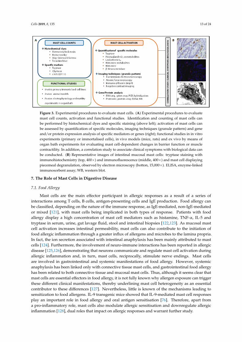
Mucosal mast cell infiltration and activation can be assessed by several methods (Figure 3).Morphological analysis in tissue specimens is recommended for identifying cell architecture andmast cell location through simple histochemical dyes (with toluidine blue being the most specific)or by using antibodies anti-mast cell proteins via immunohistochemistry or immunofluorescencetechniques (tryptase, chymase, c-kit, FcεR1) [113,114]. Mast cell activation can be assessed by analyticalmethods quantifying mast cell mediators or metabolites in different biological samples (urine, blood,luminal content, biopsy, cell suspension, cell culture supernatant). For example, tryptase content inthe intestinal lumen as well as tryptase gene expression in mucosal biopsies have been identifiedin activated mucosal mast cells in IBS patients and associated with symptom severity [115,116].Urinary prostaglandin D2 and leukotriene E4 levels are also raised after mast cell activation, althoughthe use of the former as a single marker is not recommended and the latter has not been fullyvalidated. Urinary histamine has been widely used as a specific marker; however, its metabolitesare influenced by diet or bacterial contamination and its concentration in blood can also derive frombasophils and be affected by several factors such as blood sample handling [117]. Since histochemicalstaining does not provide information on granule secretion at subcellular level, ultrastructural analysisis necessary to establish the type and degree of degranulation. Different imaging techniques areavailable, with electron microscopy being the most commonly used, since high magnification revealscellular and subcellular structure. Transmission electron microscopy allows us to determine whethermast cells display a secretory profile, type of degranulation, granular and plasma cell membranemorphology and the proximity to other cells or structures within the tissue. Nevertheless, it has somelimitations since the dehydration of samples or imaging under vacuum can affect cell membranes.A complementary technique is atomic force microscopy, which preserves structural integrity andpermits cell analysis of high-resolution 3D-generated images. Degranulation of mast cells can bestudied in vivo using a fluorescent protein-based indicator of degranulation named immune-pHluorin(impH) that identifies changes in fluorescence according to the pH value. The study of secretorygranule biogenesis, maintenance, regulation and recycling can also be analysed using negative contrastimaging [113]. Gene and protein expression (after sample fixation for RNA/protein stabilisation) hasbeen analysed to identify a mast cell-associated gene/protein profile or to assess the expression ofspecific molecules.
Functional assays can also be performed in different experimental settings to revealactivation/inhibition mechanisms of mast cells. Different immortalised mast cell lines (LAD-2, HMC-1,LUVA) or isolated primary cells (blood progenitors or tissue mast cells) are currently being usedindividually or in co-culture with other leukocytes or intestinal epithelial cell lines. Besides, in vitromast cell degranulation can be detected and quantified by measuring histamine or β-hexosaminidaserelease to culture medium [118]. On the other hand, in vivo studies using animal models such as IL-9transgenic mice [76], Cre/loxP mast cell-deficient mice [119] or mast cell knock-out rats [45] can alsoreveal the role of mast cells in the intestinal mucosa. Furthermore, mast cell-dependent changes inbarrier function or motility can be evaluated by electrophysiological measurements in ex vivo tissuespecimens mounted in Ussing chambers or in an organ bath for the assessment of muscle contractility.In this setting, a pharmacological approach can be designed in which activation/blockade can help toreveal the effect of mast cells on molecule transport across the epithelial barrier or muscle contractility.Finally, a correlation study is essential for linking clinical manifestations, digestive dysfunction andmast cell activation [120].
Cells 2019, 8, 135 13 of 24
Cells 2019, 8, 135 13 of 24
release to culture medium [118]. On the other hand, in vivo studies using animal models such as IL-9 transgenic mice [76], Cre/loxP mast cell-deficient mice [119] or mast cell knock-out rats [45] can also reveal the role of mast cells in the intestinal mucosa. Furthermore, mast cell-dependent changes in barrier function or motility can be evaluated by electrophysiological measurements in ex vivo tissue specimens mounted in Ussing chambers or in an organ bath for the assessment of muscle contractility. In this setting, a pharmacological approach can be designed in which activation/blockade can help to reveal the effect of mast cells on molecule transport across the epithelial barrier or muscle contractility. Finally, a correlation study is essential for linking clinical manifestations, digestive dysfunction and mast cell activation [120].
Figure 3. Experimental procedures to evaluate mast cells. (A) Experimental procedures to evaluate mast cell counts, activation and functional studies. Identification and counting of mast cells can be performed by histochemical dyes and specific staining (above left); activation of mast cells can be assessed by quantification of specific molecules, imaging techniques (granule pattern) and gene and/or protein expression analysis of specific mediators or genes (right); functional studies in in vitro experiments (primary or immortalised cells), in vivo models (mice, rats) and ex vivo by means of organ bath experiments for evaluating mast cell-dependent changes in barrier function or muscle contractility. In addition, a correlation study to associate clinical symptoms with biological data can be conducted. (B) Representative images of intestinal mucosal mast cells: tryptase staining with immunohistochemistry (top, 400×) and immunofluorescence (middle, 400×) and mast cell displaying piecemeal degranulation, observed by electron microscopy (botton, 15,000×). ELISA, enzyme-linked immunosorbent assay; WB, western blot.
7. The Role of Mast Cells in Digestive Disease
7.1. Food Allergy
Mast cells are the main effector participant in allergic responses as a result of a series of interactions among T cells, B cells, antigen-presenting cells and IgE production. Food allergy can be classified, depending on the nature of the immune response, as IgE-mediated, non-IgE-mediated or mixed [121], with mast cells being implicated in both types of response. Patients with food allergy display a high concentration of mast cell mediators such as histamine, TNF-α, IL-5 and tryptase in serum, urine, gut lavage fluid, stool and intestinal biopsies [122,123]. As mucosal mast cell activation increases intestinal permeability, mast cells can also contribute to the initiation of food allergic inflammation through a greater influx of allergens and microbes to the lamina propria. In fact, the ion secretion associated with intestinal anaphylaxis has been mainly attributed to mast cells [124]. Furthermore, the involvement of neuro-immune interactions has been reported in allergic disease [125,126], demonstrating that neurons communicate and regulate mast cell activation during allergic inflammation and, in turn, mast cells, reciprocally, stimulate nerve endings. Mast cells are involved in gastrointestinal and systemic manifestations of food allergy. However, systemic anaphylaxis has
Figure 3. Experimental procedures to evaluate mast cells. (A) Experimental procedures to evaluatemast cell counts, activation and functional studies. Identification and counting of mast cells canbe performed by histochemical dyes and specific staining (above left); activation of mast cells canbe assessed by quantification of specific molecules, imaging techniques (granule pattern) and geneand/or protein expression analysis of specific mediators or genes (right); functional studies in in vitroexperiments (primary or immortalised cells), in vivo models (mice, rats) and ex vivo by means oforgan bath experiments for evaluating mast cell-dependent changes in barrier function or musclecontractility. In addition, a correlation study to associate clinical symptoms with biological data canbe conducted. (B) Representative images of intestinal mucosal mast cells: tryptase staining withimmunohistochemistry (top, 400×) and immunofluorescence (middle, 400×) and mast cell displayingpiecemeal degranulation, observed by electron microscopy (botton, 15,000×). ELISA, enzyme-linkedimmunosorbent assay; WB, western blot.
7. The Role of Mast Cells in Digestive Disease
7.1. Food Allergy
Mast cells are the main effector participant in allergic responses as a result of a series ofinteractions among T cells, B cells, antigen-presenting cells and IgE production. Food allergy canbe classified, depending on the nature of the immune response, as IgE-mediated, non-IgE-mediatedor mixed [121], with mast cells being implicated in both types of response. Patients with foodallergy display a high concentration of mast cell mediators such as histamine, TNF-α, IL-5 andtryptase in serum, urine, gut lavage fluid, stool and intestinal biopsies [122,123]. As mucosal mastcell activation increases intestinal permeability, mast cells can also contribute to the initiation offood allergic inflammation through a greater influx of allergens and microbes to the lamina propria.In fact, the ion secretion associated with intestinal anaphylaxis has been mainly attributed to mastcells [124]. Furthermore, the involvement of neuro-immune interactions has been reported in allergicdisease [125,126], demonstrating that neurons communicate and regulate mast cell activation duringallergic inflammation and, in turn, mast cells, reciprocally, stimulate nerve endings. Mast cellsare involved in gastrointestinal and systemic manifestations of food allergy. However, systemicanaphylaxis has been linked only with connective tissue mast cells, and gastrointestinal food allergyhas been related to both connective tissue and mucosal mast cells. Thus, although it seems clear thatmast cells are essential effectors in food allergy, it is not fully known why allergen exposure can triggerthese different clinical manifestations, thereby underlining mast cell heterogeneity as an essentialcontributor to these differences [127]. Nevertheless, little is known of the mechanisms leading tosensitization to food allergens. IL-9 transgenic mice showed that IL-9-mediated mast cell responsesplay an important role in food allergy and oral antigen sensitisation [76]. Therefore, apart froma pro-inflammatory role, mast cells also modulate allergic sensitisation and downregulate allergicinflammation [128], dual roles that impact on allergic responses and warrant further study.
Cells 2019, 8, 135 14 of 24
7.2. Inflammatory Bowel Disease
IBD includes two main entities, ulcerative colitis (UC) and Crohn’s disease (CD), in whichchronic gut inflammation results from altered host-microbial interactions in genetically-susceptibleindividuals [129]. The contribution of mast cells to IBD has been demonstrated in human andexperimental studies [130] in which increased numbers of mast cells were found in tissue specimensfrom both UC and CD patients [131–133], showing ultrastructural changes with evidence of piecemealand anaphylactic degranulation. Different types of mast cell mediators involved in IBD pathogenesisinclude TNF-α, IL-6, SP, histamine, prostaglandins and leukotrienes [134–137]. Altered brain-gutinteractions have also been detected in IBD [138] and mast cells are thought to play a part in theneural inflammation present in these patients [139]. In fact, in the DSS experimental model of colitis,the number of mucosal mast cells in close proximity to VIP nerves was significantly increased [140].Moreover, alterations in intestinal ion transport [141], the fibrotic response in CD [142,143], microbiotadysbiosis [144], fibroblast proliferation, collagen production and contractile activity have also beenassociated with intestinal mast cell activation [145]. The participation of mast cells in IBD is undeniable,since they play a role in several aspects of the disease, among which intestinal permeability, initiationand maintenance of inflammatory processes (with ensuing tissue remodelling) and transmittance ofsignals during neuropathological stress [146] are noteworthy. Therefore, mast cell-stabilising drugs ordrugs interfering with mast cell mediators are considered an additional therapeutic possibility in thetreatment of IBD [147].
7.3. Coeliac Disease
Coeliac disease is a chronic inflammatory disorder in the small intestine caused by intoleranceto gluten. It implies remodelling of the intestinal mucosa where immune cells accumulate as aconsequence of both adaptive and innate immune responses to undigested gliadin peptides [148].A relationship between mast cells and coeliac disease has been reported, showing increased numbersof mast cells and their mediator histamine in the small intestine [149]. Moreover, the jejunum ofcoeliac disease patients shows inflammation caused by histamine, together with albumin secretion,resulting from endothelial disruption, which facilitates mucosal leakage [150]. Mast cells have beenfound to directly react to gliadin fragments by releasing proinflammatory mediators, and have beenassociated with increased neutrophil accumulation, prevalence of M1 macrophages, and severity oftissue damage during onset and progression of the disease [151]. Hence, mucosal mast cell count hasbeen suggested as a marker for monitoring coeliac disease severity and a target for re-establishing guttolerance to gluten.
7.4. Irritable Bowel Syndrome
IBS is a functional gastrointestinal disorder for which, despite intensive research, no biomarkerhas been identified to date. In the intestinal mucosa of IBS patients, a low-grade inflammatory infiltrate,characterised by an increased number of mucosal mast cells and T lymphocytes, has been reportedin all clinical IBS subtypes in the small and large intestine [152]; however, not all studies reportedsimilar results [153]. Despite disparities in immune cell counts among studies, altered intestinal barrierwith increased epithelial permeability and disruption of TJ appeared to be a common finding [154].The loss of functional integrity may facilitate the uncontrolled flux of antigens (food, microorganisms,toxins, etc) across the epithelium and stimulation of immunological responses in the laminapropria [155], thereby further increasing paracellular epithelial permeability and promoting low-grademucosal inflammation. As tryptase is implicated in intestinal barrier deregulation, the generation ofgastrointestinal motor abnormalities and visceral pain [116,156], mast cell activation may be of greaterimportance than number, as tryptase production, and not cell counts, correlates with TJ disruptionand clinical symptoms, at least in the small intestine of IBS-D [116]. Moreover, the number of colonicmucosal mast cells in proximity to nerves positively correlates with abdominal pain severity in
Cells 2019, 8, 135 15 of 24
IBS [157]. Since mast cells form a link between the brain and the gut by local neuro-immune interaction,they mediate mucosal responses to central stimuli such as psychological stress, thanks to their locationnear nerve fibres and the presence of receptors for CRF or SP. Remarkably, the high prevalence ofpsychiatric comorbidities in patients with gastrointestinal disorders [158] highlights the significance ofstress in the aetiopathogenesis of IBS. On these lines, recent research revealed associations betweenimmune activation (humoral activity) and psychiatric comorbidities [159] and between stress episodesand the initiation/exacerbation of functional gastrointestinal disorders [160]. Evidence of mast cellimplication in IBS pathophysiology is also supported by studies identifying an improvement in gutsymptoms after administration of the stabiliser disodium chromoglycate [24,161,162] or ketotifen,a histamine H1-receptor antagonist and mast cell stabiliser, which led to reduced visceral perception,particularly in hypersensitive IBS patients [163].
7.5. Functional Dyspepsia
FD, one of the most common functional gastrointestinal disorders, is characterised by a diversityof symptoms occurring in the epigastric region. As with IBS, no biomarker has been identified,despite an increased number of mast cells and eosinphils being observed in the duodenal [164] andgastric [165,166] mucosa together with epithelial barrier dysfunction. Moreover, a recent meta-analysisconfirmed this previous evidence [167]. Notably, in FD, there is a higher number of mucosal mast cellswith an activated phenotype; however, this finding does not appear to correlate with the impairedbarrier integrity observed in duodenal mucosa [25]. Despite these results, evidence still suggeststhat mast cell activation may play a role in the pathophysiology of FD, since granule morphologysignificantly differed when FD and control mucosal mast cells were compared, suggesting a differentialsynthesis and storage of mediators in mast cell granules [25]. Therefore, further studies are required toelucidate the role of mast cells in this disorder.
7.6. Mast Cell Activation Disorder
Mast cell activation disorders cover a wide range of entities, from relatively common IgE-mediateddisease and chronic urticaria to rarer conditions such as mastocytosis or monoclonal mast cell activationdisorder. Patients with symptoms stemming from a mast cell activation disorder, which do not meet thecriteria for anaphylaxis, are considered for the diagnosis of mast cell activation syndrome, a conditionfor which gastrointestinal symptoms are well documented [168]. In fact, the symptoms have beenconsidered as secondary to mast cell infiltration of the gut, in addition to deregulation of the localrelease of mast cell mediators such as histamine, prostaglandin, gastrin, SP and VIP [169–174]. The mostcommon symptoms are abdominal pain, nausea/vomiting, diarrhoea, gastrointestinal bleeding andvisceromegaly [174]. However, it remains unclear whether these symptoms arise from locally activatedmast cells or mediators derived from sites distant from the gastrointestinal tract. Further studies areneeded to improve understanding in this field.
8. Concluding Remarks
In summary, mucosal mast cells contribute to homeostasis and are actively involved in a variety ofgastrointestinal diseases. Considering the heterogeneity of digestive entities to which mast cellscontribute, it is indisputable that mast cells are able to influence and regulate gastrointestinalfunction through different mechanisms. In this context, the role mast cells play in epithelialbarrier function maintenance, neuro-immune interaction and the regulation of mucosal immunity isremarkable. Therefore, considering the available data, future therapy approaches to stabilising mastcells constitute a promising tool for the improvement in gastrointestinal disorders associated withaltered barrier function.
Funding: Supported in part by Fondo Europeo de Desarrollo Regional (FEDER), Fondo de InvestigaciónSanitaria and Centro de Investigación Biomédica en Red de Enfermedades Hepáticas y Digestivas (CIBEREHD)y de Salud Mental (CIBERSAM), Instituto de Salud Carlos III, Subdirección General de Investigación Sanitaria,
Cells 2019, 8, 135 16 of 24
Ministerio de Ciencia Innovación y Universidades PI15/00301 (C.A.-C.); PI17/00190 (J.S.); CPII16/00031,PI16/00583 (M.V.); CB06/04/0021 (C.A.-C.; J.S.; M.V.), CIBERSAM (A.R.-U.).
Acknowledgments: The authors thank Christine O’Hara for editing of the manuscript.
Conflicts of Interest: M.A.-B., I.P., A.M.G.-C., M.J.R.-L., A.R.-U., C.A.-C. and M.V. have no conflict of interest todeclare. J.S. is consultant for Ipsen and Noventure.
References
1. Wernersson, S.; Pejler, G. Mast cell secretory granules: Armed for battle. Nat. Rev. Immunol. 2014, 14, 478.[CrossRef]
2. Bischoff, S.C. Role of mast cells in allergic and non-allergic immune responses: Comparison of human andmurine data. Nat. Rev. Immunol. 2007, 7, 93. [CrossRef] [PubMed]
3. Kirshenbaum, A.S.; Kessler, S.W.; Goff, J.P.; Metcalfe, D.D. Demonstration of the origin of human mast cellsfrom CD34+ bone marrow progenitor cells. J. Immunol. 1991, 146, 1410–1415. [PubMed]
4. Galli, S.; Borregaard, N.; Wynn, T. Phenotypic and functional plasticity of cells of innate immunity:Macrophages, mast cells and neutrophils. Nat. Immunol. 2011, 12, 1035–1044. [CrossRef] [PubMed]
5. Abonia, J.P.; Austen, K.F.; Rollins, B.J.; Joshi, S.K.; Flavell, R.A.; Kuziel, W.A.; Koni, P.A.; Gurish, M.F.Constitutive homing of mast cell progenitors to the intestine depends on autologous expression of thechemokine receptor CXCR2. Blood 2005, 105, 4308–4313. [CrossRef]
6. Weidner, N.; Austen, K.F. Heterogeneity of Mast Cells at Multiple Body Sites. Fluorescent determination ofavidin binding and immunofluorescent determination of chymase, tryptase, and carboxypeptidase content.Pathol. Res. Pract. 1993, 189, 156–162. [CrossRef]
7. Irani, A.; Schechter, N.; Craig, S.; DeBlois, G.; Schwartz, L. Two types of human mast cells that have distinctneutral protease compositions. Proc. Natl. Acad. Sci. USA 1986, 83, 4464–4468. [CrossRef]
8. Irani, A.M.; Bradford, T.R.; Kepley, C.L.; Schechter, N.M.; Schwartz, L.B. Detection of MCT and MCTCtypes of human mast cells by immunohistochemistry using new monoclonal anti-tryptase and anti-chymaseantibodies. J. Histochem. Cytochem. 1989, 37, 1509–1515. [CrossRef]
9. Abonia, J.P.; Blanchard, C.; Butz, B.B.; Rainey, H.F.; Collins, M.H.; Stringer, K.; Putnam, P.E.; Rothenberg, M.E.Involvement of mast cells in eosinophilic esophagitis. J. Allergy Clin. Immunol. 2010, 126, 140–149. [CrossRef]
10. Dougherty, R.H.; Sidhu, S.S.; Raman, K.; Solon, M.; Solberg, O.D.; Caughey, G.H.; Woodruff, P.G.; Fahy, J.V.Accumulation of intraepithelial mast cells with a unique protease phenotype in TH2-high asthma. J. AllergyClin. Immunol. 2010, 125, 1046–1053. [CrossRef]
11. Vliagoftis, H.; Befus, A.D. Rapidly changing perspectives about mast cells at mucosal surfaces. Immunol. Rev.2005, 206, 190–203. [CrossRef] [PubMed]
12. Kempuraj, D.; Selvakumar, G.P.; Thangavel, R.; Ahmed, M.E.; Zaheer, S.; Raikwar, S.P.; Iyer, S.S.;Bhagavan, S.M.; Beladakere-Ramaswamy, S.; Zaheer, A. Mast cell activation in brain injury, stress, andpost-traumatic stress disorder and Alzheimer’s disease pathogenesis. Front. Neurosci. 2017, 11, 703.[CrossRef] [PubMed]
13. Wong, G.W.; Zhuo, L.; Kimata, K.; Lam, B.K.; Satoh, N.; Stevens, R.L. Ancient origin of mast cells.Biochem. Biophys. Res. Commun. 2014, 451, 314–318. [CrossRef] [PubMed]
14. Zhang, L.; Song, J.; Hou, X. Mast cells and irritable bowel syndrome: From the bench to the bedside.J. Neurogastroenterol. Motil. 2016, 22, 181–192. [CrossRef] [PubMed]
15. Kitaura, J.; Song, J.; Tsai, M.; Asai, K.; Maeda-Yamamoto, M.; Mocsai, A.; Kawakami, Y.; Liu, F.-T.;Lowell, C.A.; Barisas, B.G.; et al. Evidence that IgE molecules mediate a spectrum of effects on mastcell survival and activation via aggregation of the FcepsilonRI. Proc. Natl. Acad. Sci. USA 2003, 100,12911–12916. [CrossRef]
16. Rivera, J.; Gilfillan, A. Molecular regulation of mast cell activation. J. Allergy Clin. Immunol. 2006, 117,1214–1225. [CrossRef] [PubMed]
17. Daëron, M.; Prouvost-Danon, A.; Voisin, G.A. Mast cell membrane antigens and Fc receptors in anaphylaxis.II. Functionally distinct receptors for IgG and for IgE on mouse mast cells. Cell. Immunol. 1980, 49, 178–189.[CrossRef]
Cells 2019, 8, 135 17 of 24
18. Burton, O.T.; Epp, A.; Fanny, M.E.; Miller, S.J.; Stranks, A.J.; Teague, J.E.; Clark, R.A.; van de Rijn, M.;Oettgen, H.C. Tissue-specific expression of the low-affinity IgG receptor, FcγRIIb, on human mast cells.Front. Immunol. 2018, 9, 1244. [CrossRef]
19. Agier, J.; Pastwinska, J.; Brzezinska-Błaszczyk, E. An overview of mast cell pattern recognition receptors.Inflamm. Res. 2018, 67, 737–746. [CrossRef]
20. Uvnäs, B. Recent observations on mechanisms of storage and release of mast cell histamine. Applicability toother biogenic amines. Agents Actions. Suppl. 1992, 36, 23–33.
21. Forsythe, P. Mast cells in neuroimmune interactions. Trends Neurosci. 2018, 42, 43–55. [CrossRef] [PubMed]22. Dvorak, A.M.; Morgan, E.S. Diamine oxidase-gold enzyme-affinity ultrastructural demonstration that human
gut mucosal mast cells secrete histamine by piecemeal degranulation in vivo. J. Allergy Clin. Immunol. 1997,99, 812–820. [CrossRef]
23. Dvorak, A.M.; McLeod, R.S.; Onderdonk, A.; Monahan-Earley, R.A.; Cullen, J.B.; Antonioli, D.A.; Morgan, E.;Blair, J.E.; Estrella, P.; Cisneros, R.L.; et al. Ultrastructural evidence for piecemeal and anaphylacticdegranulation of human gut mucosal mast cells in vivo. Int. Arch. Allergy Immunol. 1992, 99, 74–83.[CrossRef] [PubMed]
24. Lobo, B.; Ramos, L.; Martínez, C.; Guilarte, M.; González-Castro, A.M.; Alonso-Cotoner, C.; Pigrau, M.;de Torres, I.; Rodiño-Janeiro, B.K.; Salvo-Romero, E.; et al. Downregulation of mucosal mast cell activationand immune response in diarrhoea-irritable bowel syndrome by oral disodium cromoglycate: A pilot study.United Eur. Gastroenterol. J. 2017, 5, 887–897. [CrossRef] [PubMed]
25. Vanheel, H.; Vicario, M.; Boesmans, W.; Vanuytsel, T.; Salvo-Romero, E.; Tack, J.; Farré, R. Activation ofEosinophils and Mast Cells in Functional Dyspepsia: An Ultrastructural Evaluation. Sci. Rep. 2018, 8, 5383.[CrossRef] [PubMed]
26. Dvorak, A.M.; Massey, W.; Warner, J.; Kissell, S.; Kagey-Sobotka, A.; Lichtenstein, L.M. IgE-mediatedanaphylactic degranulation of isolated human skin mast cells. Blood 1991, 77, 569–578. [PubMed]
27. Xu, H.; Arnold, M.G.; Kumar, S.V. Differential effects of munc18s on multiple degranulation-relevantTrans-SNARE complexes. PLoS ONE 2015, 10, e0138683. [CrossRef]
28. Vukman, K.V.; Försönits, A.; Oszvald, Á.; Tóth, E.Á.; Buzás, E.I. Mast cell secretome: Soluble and vesicularcomponents. Semin. Cell Dev. Biol. 2017, 67, 65–73. [CrossRef]
29. Pascual, S.; Martínez, J.; Pérez-Mateo, M. The intestinal barrier: Functional disorders in digestive andnon-digestive diseases. Gastroenterol. Hepatol. 2001, 24, 256–267. [CrossRef]
30. Thaiss, C.A.; Levy, M.; Grosheva, I.; Zheng, D.; Soffer, E.; Blacher, E.; Braverman, S.; Tengeler, A.C.; Barak, O.;Elazar, M.; et al. Hyperglycemia drives intestinal barrier dysfunction and risk for enteric infection. Science2018, 359, 1376–1383. [CrossRef]
31. De Palma, G.; Collins, S.M.; Bercik, P.; Verdu, E.F. The microbiota-gut-brain axis in gastrointestinal disorders:Stressed bugs, stressed brain or both? J. Physiol. 2014, 592, 2989–2997. [CrossRef] [PubMed]
32. Palmer, C.; Bik, E.M.; DiGiulio, D.B.; Relman, D.A.; Brown, P.O. Development of the human infant intestinalmicrobiota. PLoS Biol. 2007, 5, e177. [CrossRef] [PubMed]
33. Tappenden, K.A.; Deutsch, A.S. The physiological relevance of the intestinal microbiota–contributions tohuman health. J. Am. Col.l. Nutr. 2007, 26, 679S–683S. [CrossRef]
34. Neish, A.S. Microbes in gastrointestinal health and disease. Gastroenterology 2009, 136, 65–80. [CrossRef][PubMed]
35. Qin, X.; Caputo, F.J.; Xu, D.-Z.; Deitch, E.A. Hydrophobicity of mucosal surface and its relationship to gutbarrier function. Shock 2008, 29, 372–376. [CrossRef] [PubMed]
36. Bevins, C.L.; Salzman, N.H. Paneth cells, antimicrobial peptides and maintenance of intestinal homeostasis.Nat. Rev. Microbiol. 2011, 9, 356–368. [CrossRef] [PubMed]
37. Pardo-Camacho, C.; González-Castro, A.M.; Rodiño-Janeiro, B.K.; Pigrau, M.; Vicario, M. Epithelialimmunity: Priming defensive responses in the intestinal mucosa. Am. J. Physiol. Liver Physiol. 2018,314, G247–G255. [CrossRef] [PubMed]
38. Van der Flier, L.G.; Clevers, H. Stem cells, self-renewal, and differentiation in the intestinal epithelium.Annu. Rev. Physiol. 2009, 71, 241–260. [CrossRef]
39. Brandtzaeg, P.; Kiyono, H.; Pabst, R.; Russell, M.W. Terminology: Nomenclature of mucosa-associatedlymphoid tissue. Mucosal. Immunol. 2008, 1, 31–37. [CrossRef]
Cells 2019, 8, 135 18 of 24
40. Kato, L.M.; Kawamoto, S.; Maruya, M.; Fagarasan, S. The role of the adaptive immune system in regulationof gut microbiota. Immunol. Rev. 2014, 260, 67–75. [CrossRef]
41. Forsythe, P.; Bienenstock, J. The mast cell-nerve functional unit: A key component of physiologic andpathophysiologic responses. In Chemical Immunology and Allergy; French, L.E., Ed.; S. KARGER AG: Basel,Switzerlands, 2012; Volume 98, pp. 196–221.
42. Wood, J.D. Neuropathophysiology of functional gastrointestinal disorders. World J. Gastroenterol. 2007, 13,1313–1332. [CrossRef] [PubMed]
43. Vicario, M.; Guilarte, M.; Alonso, C.; Yang, P.; Martínez, C.; Ramos, L.; Lobo, B.; González, A.; Guilà, M.;Pigrau, M.; et al. Chronological assessment of mast cell-mediated gut dysfunction and mucosal inflammationin a rat model of chronic psychosocial stress. Brain. Behav. Immun. 2010, 24, 1166–1175. [CrossRef] [PubMed]
44. Barreau, F.; Ferrier, L.; Fioramonti, J.; Bueno, L. Neonatal maternal deprivation triggers long term alterationsin colonic epithelial barrier and mucosal immunity in rats. Gut 2004, 53, 501–506. [CrossRef] [PubMed]
45. Santos, J.; Yang, P.C.; Söderholm, J.D.; Benjamin, M.; Perdue, M.H. Role of mast cells in chronic stress inducedcolonic epithelial barrier dysfunction in the rat. Gut 2001, 48, 630–636. [CrossRef] [PubMed]
46. Vanuytsel, T.; van Wanrooy, S.; Vanheel, H.; Vanormelingen, C.; Verschueren, S.; Houben, E.; Salim Rasoel, S.;Tóth, J.; Holvoet, L.; Farré, R.; et al. Psychological stress and corticotropin-releasing hormone increaseintestinal permeability in humans by a mast cell-dependent mechanism. Gut 2014, 63, 1293–1299. [CrossRef][PubMed]
47. Wallon, C.; Yang, P.-C.; Keita, A.V.; Ericson, A.-C.; McKay, D.M.; Sherman, P.M.; Perdue, M.H.; Söderholm, J.D.Corticotropin-releasing hormone (CRH) regulates macromolecular permeability via mast cells in normalhuman colonic biopsies in vitro. Gut 2008, 57, 50–58. [CrossRef] [PubMed]
48. Castagliuolo, I.; Lamont, J.T.; Qiu, B.; Fleming, S.M.; Bhaskar, K.R.; Nikulasson, S.T.; Kornetsky, C.;Pothoulakis, C. Acute stress causes mucin release from rat colon: Role of corticotropin releasing factorand mast cells. Am. J. Physiol. Liver Physiol. 1996, 271, G884–G892. [CrossRef] [PubMed]
49. Saunders, P.R.; Maillot, C.; Million, M.; Taché, Y. Peripheral corticotropin-releasing factor induces diarrhea inrats: Role of CRF1 receptor in fecal watery excretion. Eur. J. Pharmacol. 2002, 435, 231–235. [CrossRef]
50. Wang, L.; Stanisz, A.M.; Wershil, B.K.; Galli, S.J.; Perdue, M.H. Substance P induces ion secretion in mousesmall intestine through effects on enteric nerves and mast cells. Am. J. Physiol. 1995, 269, G85–G92. [CrossRef]
51. Bednarska, O.; Walter, S.A.; Casado-Bedmar, M.; Ström, M.; Salvo-Romero, E.; Vicario, M.; Mayer, E.A.;Keita, Å.V. Vasoactive Intestinal Polypeptide and Mast Cells Regulate Increased Passage of Colonic Bacteriain Patients With Irritable Bowel Syndrome. Gastroenterology 2017, 153, 948–960. [CrossRef]
52. Kitamura, Y.; Kanakura, Y.; Fujita, J.; Nakano, T. Differentiation and transdifferentiation of mast cells;a unique member of the hematopoietic cell family. Int. J. Cell Cloning 1987, 5, 108–121. [CrossRef] [PubMed]
53. Frossi, B.; De Carli, M.; Pucillo, C. The mast cell: An antenna of the microenvironment that directs theimmune response. J. Leukoc. Biol. 2004, 75, 579–585. [CrossRef] [PubMed]
54. Compton, S.J.; Renaux, B.; Wijesuriya, S.J.; Hollenberg, M.D. Glycosylation and the activation ofproteinase-activated receptor 2 (PAR2) by human mast cell tryptase. Br. J. Pharmacol. 2001, 134, 705–718.[CrossRef]
55. Bueno, L.; Fioramonti, J. Protease-activated receptor 2 and gut permeability: A review. Neurogastroenterol.Motil. 2008, 20, 580–587. [CrossRef]
56. Jacob, C.; Yang, P.-C.; Darmoul, D.; Amadesi, S.; Saito, T.; Cottrell, G.S.; Coelho, A.-M.; Singh, P.;Grady, E.F.; Perdue, M.; et al. Mast cell tryptase controls paracellular permeability of the intestine. Role ofprotease-activated receptor 2 and beta-arrestins. J. Biol. Chem. 2005, 280, 31936–31948. [CrossRef] [PubMed]
57. Li, S.; Guan, J.; Ge, M.; Huang, P.; Lin, Y.; Gan, X. Intestinal mucosal injury induced by tryptase-activatedprotease-activated receptor 2 requires β-arrestin-2 in vitro. Mol. Med. Rep. 2015, 12, 7181–7187. [CrossRef]
58. Enjoji, S.; Ohama, T.; Sato, K. Regulation of epithelial cell tight junctions by protease-activated receptor 2.J. Vet. Med. Sci. 2014, 76, 1225–1229. [CrossRef]
59. Wilcz-Villega, E.M.; McClean, S.; O’Sullivan, M.A. Mast cell tryptase reduces junctional adhesion molecule-A(JAM-A) expression in intestinal epithelial cells: Implications for the mechanisms of barrier dysfunction inirritable bowel syndrome. Am. J. Gastroenterol. 2013, 108, 1140–1151. [CrossRef] [PubMed]
60. Pejler, G.; Ronnberg, E.; Waern, I.; Wernersson, S. Mast cell proteases: Multifaceted regulators of inflammatorydisease. Blood 2010, 115, 4981–4990. [CrossRef]
Cells 2019, 8, 135 19 of 24
61. Groschwitz, K.R.; Wu, D.; Osterfeld, H.; Ahrens, R.; Hogan, S.P. Chymase-mediated intestinalepithelial permeability is regulated by a protease-activating receptor/matrix metalloproteinase-2-dependentmechanism. Am. J. Physiol. Liver Physiol. 2013, 304, G479–G489. [CrossRef]
62. Scudamore, C.L.; Jepson, M.A.; Hirst, B.H.; Miller, H.R. The rat mucosal mast cell chymase, RMCP-II,alters epithelial cell monolayer permeability in association with altered distribution of the tight junctionproteins ZO-1 and occludin. Eur. J. Cell Biol. 1998, 75, 321–330. [CrossRef]
63. Fu, Z.; Thorpe, M.; Hellman, L. rMCP-2, the major rat mucosal mast cell protease, an analysis of its extendedcleavage specificity and its potential role in regulating intestinal permeability by the cleavage of cell adhesionand junction proteins. PLoS ONE 2015, 10, e0131720. [CrossRef]
64. Deiteren, A.; De Man, J.G.; Pelckmans, P.A.; De Winter, B.Y. Histamine H4 receptors in the gastrointestinaltract. Br. J. Pharmacol. 2015, 172, 1165–1178. [CrossRef]
65. Keely, S.J.; Stack, W.A.; O’Donoghue, D.P.; Baird, A.W. Regulation of ion transport by histamine in humancolon. Eur. J. Pharmacol. 1995, 279, 203–209. [CrossRef]
66. Potts, R.A.; Tiffany, C.M.; Pakpour, N.; Lokken, K.L.; Tiffany, C.R.; Cheung, K.; Tsolis, R.M.; Luckhart, S.Mast cells and histamine alter intestinal permeability during malaria parasite infection. Immunobiology 2016,221, 468–474. [CrossRef]
67. Ye, D.; Ma, I.; Ma, T.Y. Molecular mechanism of tumor necrosis factor-alpha modulation of intestinal epithelialtight junction barrier. Am. J. Physiol. Liver Physiol. 2006, 290, G496–G504.
68. Zolotarevsky, Y.; Hecht, G.; Koutsouris, A.; Gonzalez, D.E.; Quan, C.; Tom, J.; Mrsny, R.J.; Turner, J.R.A membrane-permeant peptide that inhibits MLC kinase restores barrier function in in vitro models ofintestinal disease. Gastroenterology 2002, 123, 163–172. [CrossRef]
69. Mankertz, J.; Tavalali, S.; Schmitz, H.; Mankertz, A.; Riecken, E.O.; Fromm, M.; Schulzke, J.D. Expression fromthe human occludin promoter is affected by tumor necrosis factor alpha and interferon gamma. J. Cell Sci.2000, 113, 2085–2090.
70. McLeod, J.J.A.; Baker, B.; Ryan, J.J. Mast cell production and response to IL-4 and IL-13. Cytokine 2015, 75,57–61. [CrossRef]
71. Ceponis, P.J.M.; Botelho, F.; Richards, C.D.; McKay, D.M. Interleukins 4 and 13 Increase Intestinal EpithelialPermeability by a Phosphatidylinositol 3-Kinase Pathway. J. Biol. Chem. 2000, 275, 29132–29137. [CrossRef]
72. Prasad, S.; Mingrino, R.; Kaukinen, K.; Hayes, K.L.; Powell, R.M.; MacDonald, T.T.; Collins, J.E. Inflammatoryprocesses have differential effects on claudins 2, 3 and 4 in colonic epithelial cells. Lab. Investig. 2005, 85,1139–1162. [CrossRef]
73. Al-Sadi, R.M.; Ma, T.Y. IL-1beta causes an increase in intestinal epithelial tight junction permeability.J. Immunol. 2007, 178, 4641–4649. [CrossRef]
74. Al-Sadi, R.; Ye, D.; Said, H.M.; Ma, T.Y. IL-1beta-induced increase in intestinal epithelial tight junctionpermeability is mediated by MEKK-1 activation of canonical NF-kappaB pathway. Am. J. Pathol. 2010, 177,2310–2322. [CrossRef]
75. Kimura, K.; Teranishi, S.; Nishida, T. Interleukin-1β–Induced Disruption of Barrier Function in CulturedHuman Corneal Epithelial Cells. Investig. Opthalmol. Vis. Sci. 2009, 50, 597. [CrossRef]
76. Forbes, E.E.; Groschwitz, K.; Abonia, J.P.; Brandt, E.B.; Cohen, E.; Blanchard, C.; Ahrens, R.; Seidu, L.;McKenzie, A.; Strait, R.; et al. IL-9– and mast cell–mediated intestinal permeability predisposes to oralantigen hypersensitivity. J. Exp. Med. 2008, 205, 897–913. [CrossRef]
77. Al-Sadi, R.; Ye, D.; Boivin, M.; Guo, S.; Hashimi, M.; Ereifej, L.; Ma, T.Y. Interleukin-6 modulation of intestinalepithelial tight junction permeability is mediated by JNK pathway activation of claudin-2 gene. PLoS ONE2014, 9, e85345. [CrossRef]
78. Madsen, K.L.; Malfair, D.; Gray, D.; Doyle, J.S.; Jewell, L.D.; Fedorak, R.N. Interleukin-10 gene-deficient micedevelop a primary intestinal permeability defect in response to enteric microflora. Inflamm. Bowel Dis. 1999,5, 262–270. [CrossRef]
79. Chichlowski, M.; Westwood, G.S.; Abraham, S.N.; Hale, L.P. Role of mast cells in inflammatory boweldisease and inflammation-associated colorectal neoplasia in IL-10-deficient mice. PLoS ONE 2010, 5, e12220.[CrossRef]
80. Oshima, T.; Laroux, F.S.; Coe, L.L.; Morise, Z.; Kawachi, S.; Bauer, P.; Grisham, M.B.; Specian, R.D.; Carter, P.;Jennings, S.; et al. Interferon-gamma and interleukin-10 reciprocally regulate endothelial junction integrityand barrier function. Microvasc. Res. 2001, 61, 130–143. [CrossRef]
Cells 2019, 8, 135 20 of 24
81. Polukort, S.H.; Rovatti, J.; Carlson, L.; Thompson, C.; Ser-Dolansky, J.; Kinney, S.R.M.; Schneider, S.S.;Mathias, C.B. IL-10 enhances IgE-mediated mast cell responses and is essential for the development ofexperimental food allergy in IL-10-deficient mice. J. Immunol. 2016, 196, 4865–4876. [CrossRef]
82. Malaviya, R.; Ikeda, T.; Ross, E.; Abraham, S.N. Mast cell modulation of neutrophil influx and bacterialclearance at sites of infection through TNF-alpha. Nature 1996, 381, 77–80. [CrossRef]
83. Triggiani, M.; Giannattasio, G.; Calabrese, C.; Loffredo, S.; Granata, F.; Fiorello, A.; Santini, M.; Gelb, M.H.;Marone, G. Lung mast cells are a source of secreted phospholipases A2. J. Allergy Clin. Immunol. 2009, 124,558–565. [CrossRef]
84. Möllerherm, H.; von Köckritz-Blickwede, M.; Branitzki-Heinemann, K. Antimicrobial activity of mast cells:Role and relevance of extracellular DNA traps. Front. Immunol. 2016, 7, 265. [CrossRef]
85. Branitzki-Heinemann, K.; Okumura, C.Y.; Völlger, L.; Kawakami, Y.; Kawakami, T.; Naim, H.Y.; Nizet, V.;Von Köckritz-Blickwede, M. A novel role for the transcription factor HIF-1α in the formation of mast cellextracellular traps. Biochem. J. 2012, 446, 159–163. [CrossRef]
86. Lin, A.M.; Rubin, C.J.; Khandpur, R.; Wang, J.Y.; Riblett, M.; Yalavarthi, S.; Villanueva, E.C.; Shah, P.;Kaplan, M.J.; Bruce, A.T. Mast cells and neutrophils release IL-17 through extracellular trap formation inpsoriasis. J. Immunol. 2011, 187, 490–500. [CrossRef]
87. Lauth, X.; von Köckritz-Blickwede, M.; McNamara, C.W.; Myskowski, S.; Zinkernagel, A.S.; Beall, B.;Ghosh, P.; Gallo, R.L.; Nizet, V. M1 protein allows Group A streptococcal survival in phagocyte extracellulartraps through cathelicidin inhibition. J. Innate Immun. 2009, 1, 202–214. [CrossRef]
88. Arock, M.; Ross, E.; Lai-Kuen, R.; Averlant, G.; Gao, Z.; Abraham, S.N. Phagocytic and tumor necrosisfactor alpha response of human mast cells following exposure to gram-negative and gram-positive bacteria.Infect. Immun. 1998, 66, 6030–6034.
89. Sher, A.; Hein, A.; Moser, G.; Caulfield, J.P. Complement receptors promote the phagocytosis of bacteria byrat peritoneal mast cells. Lab. Invest. 1979, 41, 490–499.
90. Shin, J.-S.; Gao, Z.; Abraham, S.N. Involvement of cellular caveolae in bacterial entry into mast cells. Science2000, 289, 785–788. [CrossRef]
91. Pundir, P.; Catalli, A.; Leggiadro, C.; Douglas, S.E.; Kulka, M. Pleurocidin, a novel antimicrobial peptide,induces human mast cell activation through the FPRL1 receptor. Mucosal. Immunol. 2013, 7, 177. [CrossRef]
92. Schiemann, F.; Brandt, E.; Gross, R.; Lindner, B.; Mittelstädt, J.; Sommerhoff, C.P.; Schulmistrat, J.;Petersen, F. The Cathelicidin LL-37 Activates Human Mast Cells and Is Degraded by Mast Cell Tryptase:Counter-Regulation by CXCL4. J. Immunol. 2009, 183, 2223–2231. [CrossRef]
93. Shin, K.; Watts, G.F.M.; Oettgen, H.C.; Friend, D.S.; Pemberton, A.D.; Gurish, M.F.; Lee, D.M. Mouse mast celltryptase mMCP-6 is a critical link between adaptive and innate immunity in the chronic phase of Trichinellaspiralis infection. J. Immunol. 2008, 180, 4885–4891. [CrossRef]
94. Gendrin, C.; Shubin, N.J.; Boldenow, E.; Merillat, S.; Clauson, M.; Power, D.; Doran, K.S.; Abrink, M.;Pejler, G.; Rajagopal, L.; et al. Mast cell chymase decreases the severity of group B Streptococcus infections.J. Allergy Clin. Immunol. 2018, 142, 120–129. [CrossRef]
95. Ha, T.Y.; Reed, N.D.; Crowle, P.K. Delayed expulsion of adult Trichinella spiralis by mast cell-deficientW/Wv mice. Infect. Immun. 1983, 41, 445–447.
96. Schneider, L.A.; Schlenner, S.M.; Feyerabend, T.B.; Wunderlin, M.; Rodewald, H.-R. Molecular mechanism ofmast cell mediated innate defense against endothelin and snake venom sarafotoxin. J. Exp. Med. 2007, 204,2629–2639. [CrossRef]
97. Maurer, M.; Wedemeyer, J.; Metz, M.; Piliponsky, A.M.; Weller, K.; Chatterjea, D.; Clouthier, D.E.;Yanagisawa, M.M.; Tsai, M.; Galli, S.J. Mast cells promote homeostasis by limiting endothelin-1-inducedtoxicity. Nature 2004, 432, 512. [CrossRef]
98. Wechsler, J.B.; Szabo, A.; Hsu, C.L.; Krier-Burris, R.A.; Schroeder, H.A.; Wang, M.Y.; Carter, R.G.; Velez, T.E.;Aguiniga, L.M.; Brown, J.B.; et al. Histamine drives severity of innate inflammation via histamine 4 receptorin murine experimental colitis. Mucosal. Immunol. 2018, 11, 861–870. [CrossRef]
99. Bulfone-Paus, S.; Bahri, R. Mast cells as regulators of T cell responses. Front. Immunol. 2015, 6, 394. [CrossRef]100. Shefler, I.; Salamon, P.; Reshef, T.; Mor, A.; Mekori, Y.A. T cell-induced mast cell activation: A role for
microparticles released from activated T cells. J. Immunol. 2010, 185, 4206–4212. [CrossRef]
Cells 2019, 8, 135 21 of 24
101. Shefler, I.; Pasmanik-Chor, M.; Kidron, D.; Mekori, Y.A.; Hershko, A.Y. T cell-derived microvesicles inducemast cell production of IL-24: Relevance to inflammatory skin diseases. J. Allergy Clin. Immunol. 2014, 133,217–224. [CrossRef]
102. Marshall, J.S. Mast-cell responses to pathogens. Nat. Rev. Immunol. 2004, 4, 787. [CrossRef]103. Galli, S.J.; Nakae, S.; Tsai, M. Mast cells in the development of adaptive immune responses. Nat. Immunol.
2005, 6, 135. [CrossRef]104. Valitutti, S.; Espinosa, E. Cognate interactions between mast cells and helper T lymphocytes. Self Nonself
2010, 1, 114–122. [CrossRef]105. Nakae, S.; Suto, H.; Kakurai, M.; Sedgwick, J.D.; Tsai, M.; Galli, S.J. Mast cells enhance T cell activation:
Importance of mast cell-derived TNF. Proc. Natl. Acad. Sci. USA 2005, 102, 6467–6472. [CrossRef]106. Mekori, Y.A.; Hershko, A.Y.; Frossi, B.; Mion, F.; Pucillo, C.E. Integrating innate and adaptive immune cells:
Mast cells as crossroads between regulatory and effector B and T cells. Eur. J. Pharmacol. 2016, 778, 84–89.[CrossRef]
107. Forward, N.A.; Furlong, S.J.; Yang, Y.; Lin, T.-J.; Hoskin, D.W. Mast cells down-regulate CD4+CD25+ Tregulatory cell suppressor function via histamine H1 receptor interaction. J. Immunol. 2009, 183, 3014–3022.[CrossRef]
108. Pawankar, R.; Okuda, M.; Yssel, H.; Okumura, K.; Ra, C. Nasal mast cells in perennial allergic rhiniticsexhibit increased expression of the Fc epsilonRI, CD40L, IL-4, and IL-13, and can induce IgE synthesis in Bcells. J. Clin. Invest. 1997, 99, 1492–1499. [CrossRef]
109. Merluzzi, S.; Frossi, B.; Gri, G.; Parusso, S.; Tripodo, C.; Pucillo, C. Mast cells enhance proliferation of Blymphocytes and drive their differentiation toward IgA-secreting plasma cells. Blood 2010, 115, 2810–2817.[CrossRef]
110. Baumgarth, N. The double life of a B-1 cell: Self-reactivity selects for protective effector functions.Nat. Rev. Immunol. 2010, 11, 34. [CrossRef]
111. Camussi, G.; Deregibus, M.C.; Bruno, S.; Cantaluppi, V.; Biancone, L. Exosomes/microvesicles as amechanism of cell-to-cell communication. Kidney Int. 2010, 78, 838–848. [CrossRef]
112. Skokos, D.; Le Panse, S.; Villa, I.; Rousselle, J.-C.; Peronet, R.; David, B.; Namane, A.; Mécheri, S.Mast cell-dependent B and T lymphocyte activation is mediated by the secretion of immunologicallyactive exosomes. J. Immunol. 2001, 166, 868–876. [CrossRef]
113. Tikoo, S.; Barki, N.; Jain, R.; Zulkhernain, N.S.; Buhner, S.; Schemann, M.; Weninger, W. Imaging of mastcells. Immunol. Rev. 2018, 282, 58–72. [CrossRef]
114. Ramsay, D.B.; Stephen, S.; Borum, M.; Voltaggio, L.; Doman, D.B. Mast cells in gastrointestinal disease.Gastroenterol. Hepatol. 2010, 6, 772–777.
115. Guilarte, M.; Santos, J.; de Torres, I.; Alonso, C.; Vicario, M.; Ramos, L.; Martínez, C.; Casellas, F.; Saperas, E.;Malagelada, J. Diarrhoea-predominant IBS patients show mast cell activation and hyperplasia in the jejunum.Gut 2007, 56, 203–209. [CrossRef]
116. Martínez, C.; Vicario, M.; Ramos, L.; Lobo, B.; Mosquera, J.; Alonso, C.; Sánchez, A.; Guilarte, M.; Antolín, M.;de Torres, I.; et al. The jejunum of diarrhea-predominant irritable bowel syndrome shows molecularalterations in the tight junction signaling pathway that are associated with mucosal pathobiology and clinicalmanifestations. Am. J. Gastroenterol. 2012, 107, 736–746. [CrossRef]
117. Akin, C. Mast cell activation syndromes. J. Allergy Clin. Immunol. 2017, 140, 349–355. [CrossRef]118. Kuehn, H.S.; Radinger, M.; Gilfillan, A.M. Measuring mast cell mediator release. Curr. Protoc. Immunol. 2010,
91, 7381–7389.119. Peschke, K.; Dudeck, A.; Rabenhorst, A.; Hartmann, K.; Roers, A. Cre/loxP-Based Mouse Models of Mast
Cell Deficiency and Mast Cell-Specific Gene Inactivation. Methods Mol. Biol. 2015, 1220, 403–421.120. Wouters, M.M.; Vicario, M.; Santos, J. The role of mast cells in functional GI disorders. Gut 2016, 65, 155–168.
[CrossRef]121. Boyce, J.A.; Assa’ad, A.; Burks, A.W.; Jones, S.M.; Sampson, H.A.; Wood, R.A.; Plaut, M.; Cooper, S.F.;
Fenton, M.J.; Arshad, S.H.; et al. Guidelines for the Diagnosis and Management of Food Allergy in theUnited States: Report of the NIAID-Sponsored Expert Panel. J. Allergy Clin. Immunol. 2010, 126, S1–S58.[CrossRef]
122. Bischoff, S.; Crowe, S.E. Gastrointestinal food allergy: New insights into pathophysiology and clinicalperspectives. Gastroenterology 2005, 128, 1089–1113. [CrossRef]
Cells 2019, 8, 135 22 of 24
123. Nolte, H.; Schiøtz, P.O.; Kruse, A.; Stahl Skov, P. Comparison of intestinal mast cell and basophil histaminerelease in children with food allergic reactions. Allergy 1989, 44, 554–565. [CrossRef]
124. Perdue, M.H.; Masson, S.; Wershil, B.K.; Galli, S.J. Role of mast cells in ion transport abnormalities associatedwith intestinal anaphylaxis. Correction of the diminished secretory response in genetically mast cell-deficientW/Wv mice by bone marrow transplantation. J. Clin. Invest. 1991, 87, 687–693. [CrossRef]
125. Voisin, T.; Bouvier, A.; Chiu, I.M. Neuro-immune interactions in allergic diseases: Novel targets fortherapeutics. Int. Immunol. 2017, 29, 247–261. [CrossRef]
126. Undem, B.J.; Taylor-Clark, T. Mechanisms underlying the neuronal-based symptoms of allergy. J. AllergyClin. Immunol. 2014, 133, 1521–1534. [CrossRef]
127. Benedé, S.; Berin, M.C. Mast cell heterogeneity underlies different manifestations of food allergy in mice.PLoS ONE 2018, 13, e0190453. [CrossRef]
128. Kraneveld, A.D.; Sagar, S.; Garssen, J.; Folkerts, G. The two faces of mast cells in food allergy and allergicasthma: The possible concept of Yin Yang. Biochim. Biophys. Acta 2012, 1822, 93–99. [CrossRef]
129. Fakhoury, M.; Negrulj, R.; Mooranian, A.; Al-Salami, H. Inflammatory bowel disease: Clinical aspects andtreatments. J. Inflamm. Res. 2014, 7, 113–120. [CrossRef]
130. Hamilton, M.J.; Frei, S.M.; Stevens, R.L. The multifaceted mast cell in inflammatory bowel disease.Inflamm. Bowel Dis. 2014, 20, 2364–2378. [CrossRef]
131. Nishida, Y.; Murase, K.; Isomoto, H.; Furusu, H.; Mizuta, Y.; Riddell, R.H.; Kohno, S. Different distribution ofmast cells and macrophages in colonic mucosa of patients with collagenous colitis and inflammatory boweldisease. Hepatogastroenterology. 2002, 49, 678–682.
132. Nolte, H.; Spjeldnaes, N.; Kruse, A.; Windelborg, B. Histamine release from gut mast cells from patients withinflammatory bowel diseases. Gut 1990, 31, 791–794. [CrossRef] [PubMed]
133. Dvorak, A.M.; Monahan, R.A.; Osage, J.E.; Dickersin, G.R. Crohn’s disease: Transmission electronmicroscopic studies. II. Immunologic inflammatory response. Alterations of mast cells, basophils,eosinophils, and the microvasculature. Hum. Pathol. 1980, 11, 606–619. [CrossRef]
134. Raithel, M.; Winterkamp, S.; Pacurar, A.; Ulrich, P.; Hochberger, J.; Hahn, E.G. Release of mast cell tryptasefrom human colorectal mucosa in inflammatory bowel disease. Scand. J. Gastroenterol. 2001, 36, 174–179.[CrossRef]
135. Raithel, M.; Schneider, H.T.; Hahn, E.G. Effect of substance P on histamine secretion from gut mucosa ininflammatory bowel disease. Scand. J. Gastroenterol. 1999, 34, 496–503. [PubMed]
136. Sasaki, Y.; Tanaka, M.; Kudo, H. Differentiation between ulcerative colitis and Crohn’s disease by aquantitative immunohistochemical evaluation of T lymphocytes, neutrophils, histiocytes and mast cells.Pathol. Int. 2002, 52, 277–285. [CrossRef] [PubMed]
137. Barbara, G.; Stanghellini, V.; de Giorgio, R.; Corinaldesi, R. Functional gastrointestinal disorders and mastcells: Implications for therapy. Neurogastroenterol. Motil. 2006, 18, 6–17. [CrossRef] [PubMed]
138. Bonaz, B.L.; Bernstein, C.N. Brain-Gut Interactions in Inflammatory Bowel Disease. Gastroenterology 2013,144, 36–49. [CrossRef]
139. Lakhan, S.E.; Kirchgessner, A. Neuroinflammation in inflammatory bowel disease. J. Neuroinflammation 2010,7, 37. [CrossRef]
140. Casado-Bedmar, M.; Heil, S.D.S.; Myrelid, P.; Söderholm, J.D.; Keita, Å.V. Upregulation of intestinal mucosalmast cells expressing VPAC1 in close proximity to vasoactive intestinal polypeptide in inflammatory boweldisease and murine colitis. Neurogastroenterol. Motil. 2018, e13503. [CrossRef]
141. Crowe, S.E.; Luthra, G.K.; Perdue, M.H. Mast cell mediated ion transport in intestine from patients with andwithout inflammatory bowel disease. Gut 1997, 41, 785–792. [CrossRef]
142. Gelbmann, C.M.; Mestermann, S.; Gross, V.; Köllinger, M.; Schölmerich, J.; Falk, W. Strictures in Crohn’sdisease are characterised by an accumulation of mast cells colocalised with laminin but not with fibronectinor vitronectin. Gut 1999, 45, 210–217. [CrossRef] [PubMed]
143. Andoh, A.; Deguchi, Y.; Inatomi, O.; Yagi, Y.; Bamba, S.; Tsujikawa, T.; Fujiyama, Y. Immunohistochemicalstudy of chymase-positive mast cells in inflammatory bowel disease. Oncol. Rep. 2006, 16, 103–107.[CrossRef] [PubMed]
144. De Zuani, M.; Dal Secco, C.; Frossi, B. Mast cells at the crossroads of microbiota and IBD. Eur. J. Immunol.2018, 48, 1929–1937. [CrossRef] [PubMed]
Cells 2019, 8, 135 23 of 24
145. Xu, X.; Weksler-Zangen, S.; Pikarsky, A.; Pappo, O.; Wengrower, D.; Bischoff, S.C.; Pines, M.; Rivkind, A.;Goldin, E.; Levi-Schaffer, F. Mast cells involvement in the inflammation and fibrosis development of theTNBS-induced rat model of colitis. Scand. J. Gastroenterol. 2002, 37, 330–337. [CrossRef] [PubMed]
146. Boeckxstaens, G. Mast cells and inflammatory bowel disease. Curr. Opin. Pharmacol. 2015, 25, 45–49.[CrossRef] [PubMed]
147. De Winter, B.Y.; van den Wijngaard, R.M.; de Jonge, W.J. Intestinal mast cells in gut inflammation andmotility disturbances. Biochim. Biophys. Acta 2012, 1822, 66–73. [CrossRef] [PubMed]
148. Parzanese, I.; Qehajaj, D.; Patrinicola, F.; Aralica, M.; Chiriva-Internati, M.; Stifter, S.; Elli, L.; Grizzi, F.Celiac disease: From pathophysiology to treatment. World J. Gastrointest. Pathophysiol. 2017, 8, 27–38.[CrossRef]
149. Wingren, U.; Hallert, C.; Norrby, K.; Enerbäck, L. Histamine and mucosal mast cells in gluten enteropathy.Agents Actions 1986, 18, 266–268. [CrossRef]
150. Lavö, B.; Knutson, L.; Lööf, L.; Odlind, B.; Venge, P.; Hällgren, R. Challenge with gliadin induces eosinophiland mast cell activation in the jejunum of patients with celiac disease. Am. J. Med. 1989, 87, 655–660.[CrossRef]
151. Frossi, B.; Tripodo, C.; Guarnotta, C.; Carroccio, A.; De Carli, M.; De Carli, S.; Marino, M.; Calabrò, A.;Pucillo, C.E. Mast cells are associated with the onset and progression of celiac disease. J. Allergy Clin. Immunol.2017, 139, 1266–1274.e1. [CrossRef]
152. Matricon, J.; Meleine, M.; Gelot, A.; Piche, T.; Dapoigny, M.; Muller, E.; Ardid, D. Associations betweenimmune activation, intestinal permeability and the irritable bowel syndrome. Aliment. Pharmacol. Ther. 2012,36, 1009–1031. [CrossRef] [PubMed]
153. Wang, S.-H.; Dong, L.; Luo, J.-Y.; Gong, J.; Li, L.; Lu, X.-L.; Han, S.-P. Decreased expression of serotoninin the jejunum and increased numbers of mast cells in the terminal ileum in patients with irritable bowelsyndrome. World J. Gastroenterol. 2007, 13, 6041–6047. [CrossRef] [PubMed]
154. Camilleri, M.; Lasch, K.; Zhou, W. Irritable bowel syndrome: Methods, mechanisms, and pathophysiology.The confluence of increased permeability, inflammation, and pain in irritable bowel syndrome. Am. J. Physiol.Gastrointest. Liver Physiol. 2012, 303, G775–G785. [CrossRef] [PubMed]
155. Turner, J.R. Intestinal mucosal barrier function in health and disease. Nat. Rev. Immunol. 2009, 9, 799–809.[CrossRef] [PubMed]
156. Barbara, G.; Wang, B.; Stanghellini, V.; de Giorgio, R.; Cremon, C.; Di Nardo, G.; Trevisani, M.; Campi, B.;Geppetti, P.; Tonini, M.; et al. Mast cell-dependent excitation of visceral-nociceptive sensory neurons inirritable bowel syndrome. Gastroenterology 2007, 132, 26–37. [CrossRef] [PubMed]
157. Barbara, G.; Stanghellini, V.; De Giorgio, R.; Cremon, C.; Cottrell, G.S.; Santini, D.; Pasquinelli, G.;Morselli-Labate, A.M.; Grady, E.F.; Bunnett, N.W.; et al. Activated mast cells in proximity to colonicnerves correlate with abdominal pain in irritable bowel syndrome. Gastroenterology 2004, 126, 693–702.[CrossRef] [PubMed]
158. Singh, P.; Agnihotri, A.; Pathak, M.K.; Shirazi, A.; Tiwari, R.P.; Sreenivas, V.; Sagar, R.; Makharia, G.K.Psychiatric, somatic and other functional gastrointestinal disorders in patients with irritable bowel syndromeat a tertiary care center. J. Neurogastroenterol. Motil. 2012, 18, 324–331. [CrossRef]
159. Vicario, M.; González-Castro, A.; Martínez, C.; Lobo, B.; Pigrau, M.; Guilarte, M.; de Torres, I.;Mosquera, J.; Fortea, M.; Sevillano-Aguilera, C.; et al. Increased humoral immunity in the jejunum ofdiarrhoea-predominant irritable bowel syndrome associated with clinical manifestations. Gut 2015, 64,1379–1388. [CrossRef]
160. Suarez, K.; Mayer, C.; Ehlert, U.; Nater, U.M. Psychological Stress and Self-Reported FunctionalGastrointestinal Disorders. J. Nerv. Ment. Dis. 2010, 198, 226–229. [CrossRef]
161. Stefanini, G.F.; Saggioro, A.; Alvisi, V.; Angelini, G.; Capurso, L.; Di Lorenzo, G.; Dobrilla, G.; Dodero, M.;Galimberti, M.; Gasbarrini, G.; et al. Oral cromolyn sodium in comparison with elimination diet in theirritable bowel syndrome, diarrheic type multicenter study of 428 patients. Scand. J. Gastroenterol. 1995, 30,535–541. [CrossRef]
162. Lunardi, C.; Bambara, L.M.; Biasi, D.; Cortina, P.; Peroli, P.; Nicolis, F.; Favari, F.; Pacor, M.L. Double-blindcross-over trial of oral sodium cromoglycate in patients with irritable bowel syndrome due to food intolerance.Clin. Exp. Allergy 1991, 21, 569–572. [CrossRef] [PubMed]
Cells 2019, 8, 135 24 of 24
163. Klooker, T.; Braak, B.; Koopman, K.; Welting, O.; Wouters, M.; van der Heide, S.; Schemann, M.;Bischoff, S.; van den Wijngaard, R.M.; Boeckxstaens, G. The mast cell stabiliser ketotifen decreases visceralhypersensitivity and improves intestinal symptoms in patients with irritable bowel syndrome. Gut 2010, 59,1213–1221. [CrossRef] [PubMed]
164. Vanheel, H.; Farré, R. Changes in gastrointestinal tract function and structure in functional dyspepsia.Nat. Rev. Gastroenterol. Hepatol. 2013, 10, 142–149. [CrossRef] [PubMed]
165. Hall, W.; Buckley, M.; Crotty, P.; O’Morain, C.A. Gastric mucosal mast cells are increased in Helicobacterpylori negative functional dyspepsia. Clin. Gastroenterol. Hepatol. 2003, 1, 363–369. [CrossRef]
166. Wang, X.; Li, X.; Ge, W.; Huang, J.; Li, G.; Cong, Y.; Li, F.; Liu, Z.; Liu, Z.; Li, Y.; et al. Quantitative evaluationof duodenal eosinophils and mast cells in adult patients with functional dyspepsia. Ann. Diagn. Pathol. 2015,19, 50–56. [CrossRef] [PubMed]
167. Du, L.; Chen, B.; Kim, J.J.; Chen, X.; Dai, N. Micro-inflammation in functional dyspepsia: A systematicreview and meta-analysis. Neurogastroenterol. Motil. 2018, 30, e13304. [CrossRef] [PubMed]
168. Akin, C. Mast cell activation disorders. J. Allergy Clin. Immunol. Pract. 2014, 2, 252–257. [CrossRef]169. Bedeir, A.; Jukic, D.M.; Wang, L.; Mullady, D.K.; Regueiro, M.; Krasinskas, A.M. Systemic mastocytosis
mimicking inflammatory bowel disease: A case report and discussion of gastrointestinal pathology insystemic mastocytosis. Am. J. Surg. Pathol. 2006, 30, 1478–1482. [CrossRef]
170. Kochi, S.; Nakamura, S.; Oshiro, Y.; Kurahara, K.; Kawasaki, K.; Yaita, H.; Fuchigami, T. Endoscopic findingsof indolent systemic mastocytosis involving the colon. Endoscopy 2014, 46, E678–E679. [CrossRef]
171. Cherner, J.A.; Jensen, R.T.; Dubois, A.; O’Dorisio, T.M.; Gardner, J.D.; Metcalfe, D.D. GastrointestinalDysfunction in Systemic Mastocytosis: A Prospective Study. Gastroenterology 1988, 95, 657–667. [CrossRef]
172. Awad, J.A.; Morrow, J.D.; Roberts, L.J. Detection of the major urinary metabolite of prostaglandin D2 inthe circulation: Demonstration of elevated levels in patients with disorders of systemic mast cell activation.J. Allergy Clin. Immunol. 1994, 93, 817–824. [CrossRef]
173. Wesley, J.R.; Vinik, A.I.; O’Dorisio, T.M.; Glaser, B.; Fink, A. A new syndrome of symptomatic cutaneousmastocytoma producing vasoactive intestinal polypeptide. Gastroenterology 1982, 82, 963–967. [PubMed]
174. Hsieh, F.H. Gastrointestinal involvement in mast cell activation disorders. Immunol. Allergy Clin. North Am.2018, 38, 429–441. [CrossRef] [PubMed]
© 2019 by the authors. Licensee MDPI, Basel, Switzerland. This article is an open accessarticle distributed under the terms and conditions of the Creative Commons Attribution(CC BY) license (http://creativecommons.org/licenses/by/4.0/).