Embed Size (px)
Citation preview
2375Research Article
IntroductionCell locomotion involves a highly regulated cycle of assemblyand disassembly of cell adhesions with the surrounding matrixand neighbouring cells (Adams, 2002). In most cell types, focalcomplexes are assembled at the first contact points betweendynamic extensions of the cell membrane (filopodia andlamellipodia) at the leading edge of the cell and the substratum,whereas more stable types of adhesions (focal adhesions andtheir variants), linked to the actomyosin-based contractilitymachinery, allow cell traction, contraction and the release ofthe rear of the advancing cell body (Ridley et al., 2003).
In addition to these classical adhesion structures, certaincells of the myeloid lineage; macrophages (Linder et al., 1999),dendritic cells (DCs) (Binks et al., 1998) and osteoclasts(Calle et al., 2004b; Zambonin-Zallone et al., 1988) assemblehighly dynamic conical structures named podosomes. Thesestructures resemble adhesion sites elaborated by Src-transformed fibroblasts (Marchisio et al., 1987; Abram et al.,2003), as well as agonist-activated endothelia (Moreau et al.,2003; Osiak et al., 2005) epithelia (Spinardi et al., 2004) andsmooth muscle cells (Kaverina et al., 2003). Althoughpodosomes share many molecular components in commonwith focal complexes and focal adhesions, they have specificelements and a distinctive organisation (Buccione et al., 2004;Linder and Aepfelbacher, 2003). They consist of a core of actinfilaments surrounded by a ring made up of vinculin, paxillin,
talin, fimbrin, gelsolin, vimentin (Marchisio et al., 1984;Marchisio et al., 1987) and other adaptor molecules (Abram etal., 2003; Oda et al., 2001), that are linked to adhesionmolecules of the integrin family (Gaidano et al., 1990; Linderand Aepfelbacher, 2003). In addition, unlike focal complexes,podosomes contain members of the WASP family (Burns et al.,2001; Spinardi et al., 2004).
In myeloid cells, podosomes localise behind the leadingedge of migrating macrophages, DCs and osteoclasts (Jones etal., 2002; West et al., 2004; Lakkakorpi and Vaananen, 1991)and are thought to play an important role in locomotion andextracellular matrix degradation (Linder and Aepfelbacher,2003; Brunton et al., 2004). In osteoclasts they also formconcentric rings that define the sealing zone (Destaing et al.,2003). They are very dynamic; changing in size and positionwith a minimum half-life of 2 minutes in osteoclasts (Kanehisaet al., 1990) and 1 minute in macrophages (Evans et al., 2003)and between 30 seconds and 10 minutes in DCs (Calle et al.,2004a) (our unpublished data) depending upon migratorystatus. Since new podosomes are constantly generated inmoving cells, regulation of these adhesions implies not only arapid assembly of their components but also fast disassembly,possibly to allow effective progression of the leading edgeduring cell migration. The nature and the mechanisms ofregulation of this cyclical turnover are unknown at present.
Calpains are a family of intracellular calcium-dependent
Podosomes, highly dynamic adhesion structures implicatedin cell motility and extracellular matrix degradation, arecharacteristic of certain cells of the myeloid lineage and alimited range of other cell types. The nature and themechanisms that regulate their high turnover are unknownat present. The cysteine protease calpain is involved in theregulation of cell migration in part by promoting eitherformation or disassembly of adhesion sites. Despite the factthat many known substrates of calpain are also structuralcomponents of the podosome complex, no studies have yetdemonstrated that calpain participates in the regulation ofpodosome dynamics. In the present work, we show thatinhibition of calpain in primary mouse dendritic cells leadsto enhanced accumulation of actin filaments, the WiskottAldrich Syndrome protein (WASP), ��2 integrins, talin,
paxillin and vinculin in podosomes. This accumulation ofcomponents is associated with stabilisation of podosometurnover, overall reduction in velocity of cell locomotionand impaired transmigration across an endothelialmonolayer. We also demonstrate that calpain cleaves thepodosome components talin, Pyk2 and WASP in dendriticcells. In summary, our results provide evidence that calpainregulates podosome composition and turnover and that thisprocess is required for efficient migration of dendritic cells.
Supplementary material available online athttp://jcs.biologists.org/cgi/content/full/119/11/2375/DC1
Key words: Cell motility, Leukocyte, Dendritic cell, Podosome, Celladhesion, Calpain
Summary
Inhibition of calpain stabilises podosomes andimpairs dendritic cell motilityYolanda Calle1,*, Neil O. Carragher2, Adrian J. Thrasher3 and Gareth E. Jones1,*1Randall Division of Cell and Molecular Biophysics, King’s College London, New Hunt’s House, Guy’s Campus, London, SE1 1UL, UK2Advanced Science and Technology Laboratory, AztraZeneca Charnwood, Loughborough, LE11 5RH, UK3Molecular Immunology Unit, Institute of Child Health, University College London, WC1N 1EH, UK*Authors for correspondence (e-mail: [email protected]; [email protected])
Accepted 15 February 2006Journal of Cell Science 119, 2375-2385 Published by The Company of Biologists 2006doi:10.1242/jcs.02939
Jour
nal o
f Cel
l Sci
ence
2376
cysteine proteases involved in the regulation of signallingpathways by degrading and/or generating biologically activefragments of specific target proteins (Carragher and Frame,2002; Franco and Huttenlocher, 2005). During cell locomotion,calpains have been shown to promote the disassembly of focalcomplexes (Bhatt et al., 2002) and focal adhesions (Carragheret al., 2001) by cleaving certain components of these adhesionstructures. Other studies have found that calpain participates inthe formation of integrin-mediated adhesion sites (Rock et al.,2000; Yan et al., 2001). Although many components of celladhesions are substrates of calpain in vitro, it remains unclearwhether these proteins are cleaved in a cellular context andwhat the biological significance of this cleavage may be. Manyknown substrates of calpain are also structural components ofthe podosome complex, which makes this class of proteasea strong candidate as a regulator of their formation and/ordisassembly.
In the present work, we have investigated whether calpainplays a role in podosome dynamics and the possibleconsequences for DC motility. Our results provide evidencethat in DCs, calpain regulates podosome turnover andcomposition and this process is required for efficient celladhesion and migration.
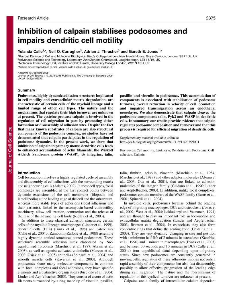
Results Calpain activity is required for disassembly of preformedpodosomes in migrating DCsUsing immunofluorescence and western blotting we foundthat DCs expressed both calpain 1 and calpain 2 and that theycan be found in a zone surrounding the actin core (Fig. 1). Inthe case of calpain 1, we also detected colocalisation withactin filaments at the edges of the actin core, mainly insmaller podosomes located towards the rear of the cluster(Fig. 1A, arrowheads). To test whether calpains play a role inthe formation and/or organisation of podosomes in migratingDCs we incubated DCs with the peptide inhibitors ALLM,ALLN or with a 27-amino-acid peptide containing a sequenceencoded by exon 1B of the highly specific endogenouscalpain inhibitor calpastatin, using as a negative control ascrambled peptide of the calpastatin sequence. The calpaininhibitors, but not inhibitors of cathepsins or the proteasome,induced a significant drop in calpain activity in DCs (Fig. 2A)
Journal of Cell Science 119 (11)
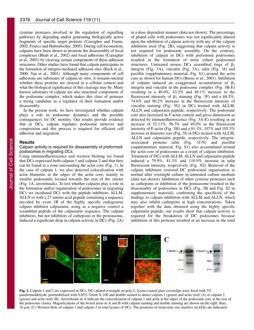
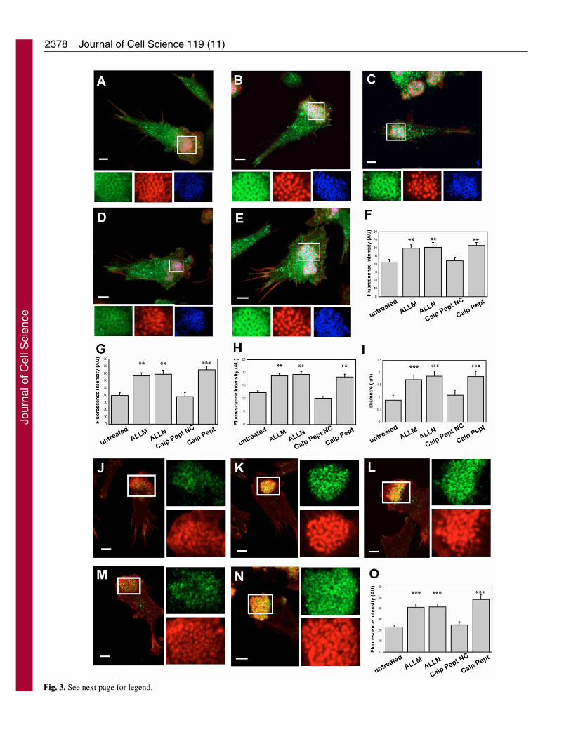
in a dose-dependent manner (data not shown). The percentageof plated cells with podosomes was not significantly alteredupon the inhibition of calpain activity with any of the calpaininhibitors used (Fig. 2B), suggesting that calpain activity isnot required for podosome assembly. On the contrary,inhibition of calpain in DCs with preformed podosomesresulted in the formation of more robust podosomalstructures. Untreated mouse DCs assembled rings of �2integrin (Fig. 3A), vinculin (Fig. 3A), talin (Fig. 3J) andpaxillin (supplementary material, Fig. S1) around the actincore as shown for human DCs (Burns et al., 2001). Inhibitionof calpain induced an exaggerated accumulation of �2integrin and vinculin in the podosome complex (Fig. 3B-E)resulting in a 40.0%, 43.2% and 48.1% increase in thefluorescent intensity of �2 staining (Fig. 3F) and a 68.5%,74.6% and 90.2% increase in the fluorescent intensity ofvinculin staining (Fig. 3G) in DCs treated with ALLM,ALLN and calpastatin peptide, respectively. The podosomecore also increased in F-actin content and gross dimension asdetected by immunofluorescence (Fig. 3A-E) resulting in anincrease of 52.11%, 56.3% and 49.0% in the fluorescentintensity of F-actin (Fig. 3H) and a 91.1%, 107% and 105.5%increase in diameter size (Fig. 3I) in DCs treated with ALLM,ALLN and calpastatin peptide, respectively. The integrin-associated proteins talin (Fig. 3J-N) and paxillin(supplementary material, Fig. S1) also accumulated aroundthe actin core of podosomes as a result of calpain inhibition.Treatment of DCs with ALLM, ALLN and calpastatin peptideinduced a 79.9%, 81.3% and 110.9% increase in talinfluorescent intensity, respectively (Fig. 3O). Removal of thecalpain inhibitors restored DC podosomal organisation tonormal after overnight culture in untreated culture medium(data not shown). Inhibition of other cysteine proteases suchas cathepsins or inhibition of the proteasome resulted in thedisassembly of podosomes in DCs (Fig. 2B and Fig. S2 insupplementary material), confirming the specificity of thefindings to calpain inhibition with ALLM and ALLN, whichmay also inhibit cathepsins at high concentrations. Takentogether with the data obtained using the highly specificcalpastatin peptide, our results show that calpain activity isrequired for the breakdown of DC podosomes becauseinhibition of this protease resulted in an increase in the total
Fig. 1. Calpain 1 and 2 are expressed in DCs. DCs plated overnight on poly-L-lysine-coated glass coverslips were fixed with 3%paraformaldehyde, permeabilised with 0.05% Triton X-100 and double stained to detect calpain 1 (green) and actin (red) (A) or calpain 2(green) and actin (red) (B). Arrowheads in A indicate the colocalisation of calpain 1 and actin at the edges of the podosome core at the rear ofthe podosome cluster. Magnifications of the boxed areas in A and B with calpain staining and double staining are shown on the right. Bars,10 �m. (C) Western blots of calpain 1 and calpain 2 in total lysates of DCs. The positions of molecular size markers (in kDa) are indicated.
Jour
nal o
f Cel
l Sci
ence
2377Calpain regulates podosome disassembly
F-actin within the actin core and the accumulation ofassociated podosomal proteins.
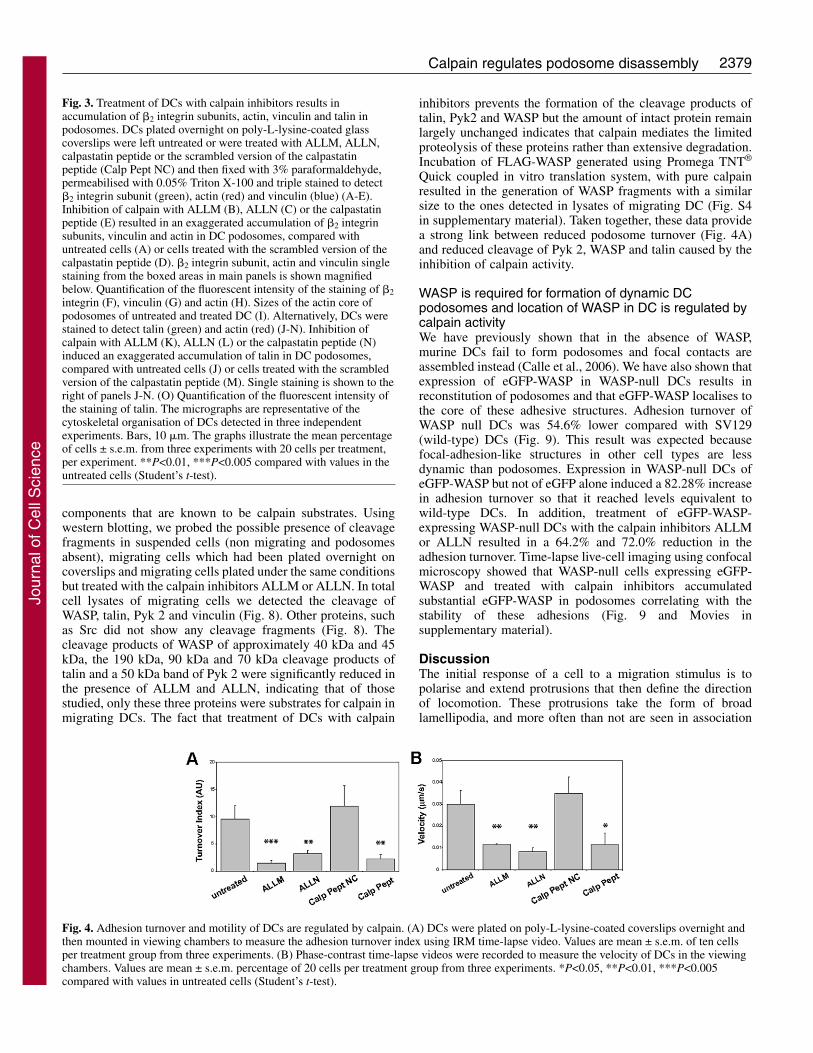
Calpain regulates adhesion turnover and motility of DCsPodosomes occupy the most prominent area of contact ofDCs to the substratum as shown by interference reflectionmicroscopy (IRM) and are clearly distinct from focalcomplexes and focal adhesions (Fig. S3 in supplementarymaterial). Using time-lapse IRM, we tested whether theformation of more robust podosomes by the inhibition ofcalpain resulted in an increase in the stability of theseadhesions. Treatment of DCs with the calpain inhibitorsALLM, ALLN or the calpastatin peptide reduced the turnoverindex of podosomes by 84.9%, 66.5% and 76.6%, respectively(Fig. 4A). This increased stability of DC adhesions correlatedwith a decrease in cell speed by 66.6%, 73.3% and 66.6% aftertreatment with ALLM, ALLN and the calpastatin peptide,respectively (Fig. 4B and Movies in supplementary material).These results show that calpain activity is essential for thedisassembly and turnover of podosomes and this process isessential for translocation of migrating DCs.
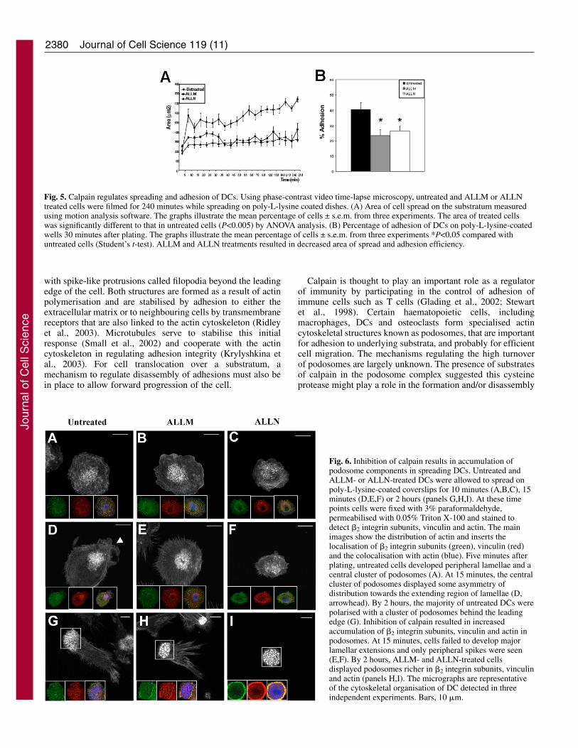
Calpain activity is essential for efficient adhesion inspreading DCsTo study the possible requirement of calpain activity for theassembly of new adhesions, especially podosomes, weanalysed the effect of calpain inhibition on freshly seeded DCs.As a result of pre-treatment of DCs with ALLM and ALLN,freshly seeded cells failed to spread to the same extent asuntreated cells (Fig. 5A) and the adhesion efficiency wasdecreased by 42.3% and 34.6%, respectively (Fig. 5B).
Using immunocytochemistry, we found that in the initial 10minutes of spreading, cells treated with ALLM and ALLNwere able to assemble centrally located podosomes and focalcomplexes similarly to untreated cells (Fig. 6A-C). However,with increasing time, podosomes grew in size and failed toreorganise in the cell body compared with untreated cells. Thusat 15 minutes, in untreated cells podosomes remained clusteredand displayed some asymmetry of distribution towards theextending lamellae (Fig. 6D, arrowhead). ALLM- and ALLN-treated cells failed to generate lamellar extensions but instead
displayed spikes around the whole periphery whereaspodosomes appeared centrally located and richer in actinfilaments (Fig. 6E,F). By 2 hours, the majority of untreatedDCs became elongated and polarised with a cluster ofpodosomes behind the leading edge (Fig. 6G). In ALLM-treated cells, podosomes were larger with more robust actincores and more defined �2 integrin and vinculin ringscompared to untreated cells (Fig. 6H). Similarly, ALLN treatedcells remained round with a central cluster of robustpodosomes rich in actin, �2 integrin and vinculin (Fig. 6I).
The accumulation of actin, �2 integrin subunits and vinculinin podosomes over time following inhibition of calpainsuggests that podosomal components are sequestered and notreleased to contribute to the formation of new adhesions thatwould support lamellar extensions.
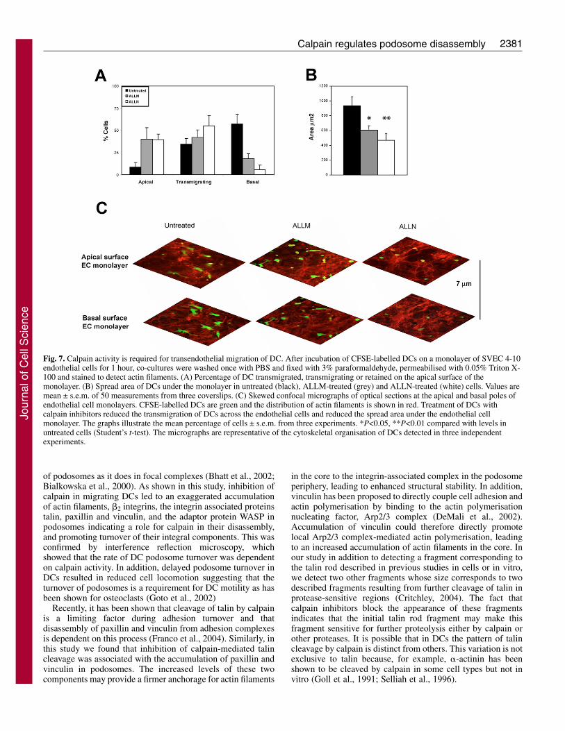
Calpain activity is required for endothelial transmigrationof DCsMigration across the endothelium is a requirement for DCs tosettle in peripheral tissues and, when pathogens are encountered,to leave the resident tissue and reach the lymph nodes for antigenpresentation (Imhof and Aurrand-Lions, 2004). Hence, wedecided to test whether the regulation of DC adhesion turnoverby calpain had any effect on the motility of DCs plated on themore relevant substratum provided by a monolayer ofendothelial cells. Treatment of DCs with calpain inhibitorsresulted in impaired transmigration of DCs across a monolayerof SVEC 4-10 endothelial cells. Calpain inhibition with bothALLM and ALLN decreased the percentage of transmigratedDCs across the monolayer by 68.0% and 90.2%, respectively(Fig. 7A). Consequently, there was a 4.6-fold increase in thepercentage of cells left on top of the monolayer with bothtreatments. In addition, the area of untreated DCs that had spreadunder the endothelial monolayer following transmigration wassignificantly higher than that of the few ALLM (1.5-fold) andALLN (1.9-fold) treated DCs (Fig. 7B,C).
The Wiskott Aldrich Syndrome protein (WASP), Pyk2and Talin are cleaved by calpain in migrating DCsIn order to study which podosomal proteins might be cleavedby calpain in migrating DCs we analysed defined podosome
Fig. 2. Calpain regulates podosome disassembly in DCs. (A) Calpain activity in the presence of inhibitors was detected. As expected, inhibitionof calpain activity was found using the calpain inhibitors ALLM (50 �M), ALLN (50 �M) or the calpastatin peptide (Calp Pept, 50 �M) butnot using cathepsin inhibitor 1 (Cathep. Inh,10 �M), proteosome inhibitor 1 (Proteo. Inh, 10 �M) or the scrambled version of the calpastatinpeptide (Calp Pept NC, 50 �M). (B) The percentage of DCs with podosomes was not affected by treatment with calpain inhibitors. Inhibitionof cathepsin B and proteosome resulted in DC podosome loss. Results are mean values ± s.e.m. from 50 cells with three coverslips examinedper experiment. *P<0.05 and ***P<0.005 compared with values in untreated cells (Student’s t-test).
Jour
nal o
f Cel
l Sci
ence
2378 Journal of Cell Science 119 (11)
Fig. 3. See next page for legend.
Jour
nal o
f Cel
l Sci
ence
2379Calpain regulates podosome disassembly
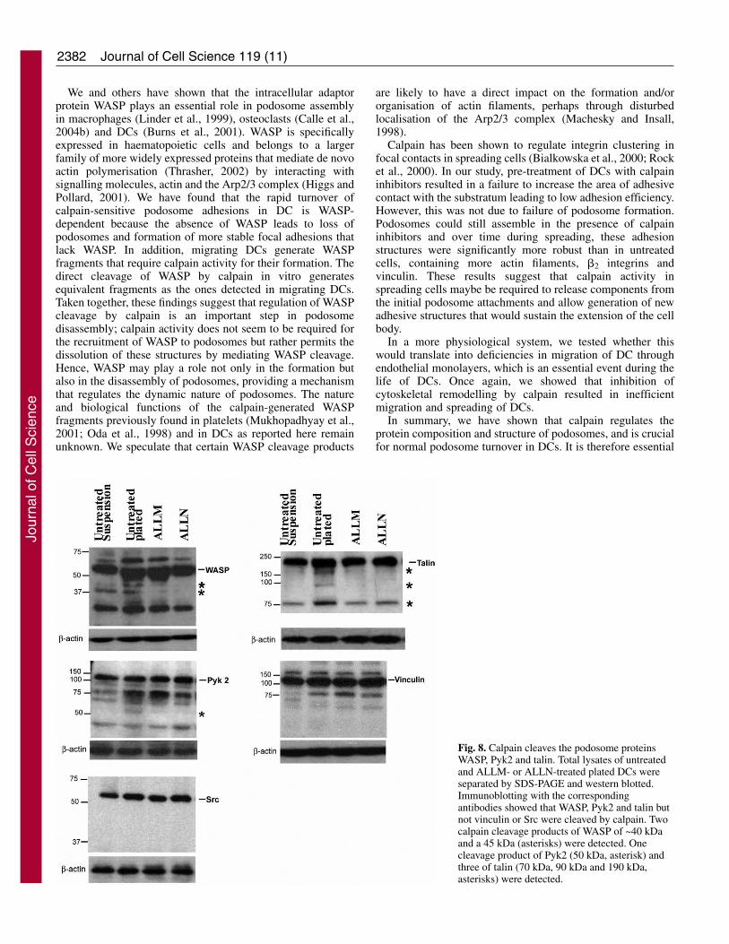
components that are known to be calpain substrates. Usingwestern blotting, we probed the possible presence of cleavagefragments in suspended cells (non migrating and podosomesabsent), migrating cells which had been plated overnight oncoverslips and migrating cells plated under the same conditionsbut treated with the calpain inhibitors ALLM or ALLN. In totalcell lysates of migrating cells we detected the cleavage ofWASP, talin, Pyk 2 and vinculin (Fig. 8). Other proteins, suchas Src did not show any cleavage fragments (Fig. 8). Thecleavage products of WASP of approximately 40 kDa and 45kDa, the 190 kDa, 90 kDa and 70 kDa cleavage products oftalin and a 50 kDa band of Pyk 2 were significantly reduced inthe presence of ALLM and ALLN, indicating that of thosestudied, only these three proteins were substrates for calpain inmigrating DCs. The fact that treatment of DCs with calpain
inhibitors prevents the formation of the cleavage products oftalin, Pyk2 and WASP but the amount of intact protein remainlargely unchanged indicates that calpain mediates the limitedproteolysis of these proteins rather than extensive degradation.Incubation of FLAG-WASP generated using Promega TNT®
Quick coupled in vitro translation system, with pure calpainresulted in the generation of WASP fragments with a similarsize to the ones detected in lysates of migrating DC (Fig. S4in supplementary material). Taken together, these data providea strong link between reduced podosome turnover (Fig. 4A)and reduced cleavage of Pyk 2, WASP and talin caused by theinhibition of calpain activity.
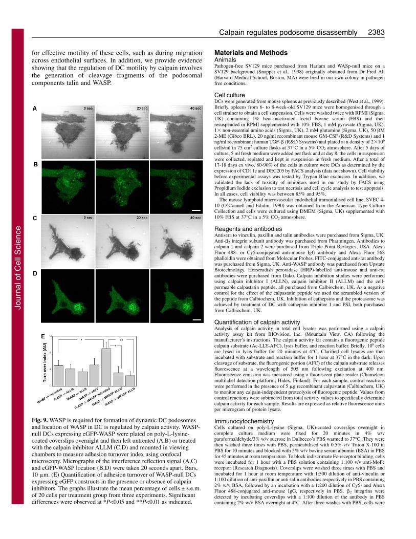
WASP is required for formation of dynamic DCpodosomes and location of WASP in DC is regulated bycalpain activityWe have previously shown that in the absence of WASP,murine DCs fail to form podosomes and focal contacts areassembled instead (Calle et al., 2006). We have also shown thatexpression of eGFP-WASP in WASP-null DCs results inreconstitution of podosomes and that eGFP-WASP localises tothe core of these adhesive structures. Adhesion turnover ofWASP null DCs was 54.6% lower compared with SV129(wild-type) DCs (Fig. 9). This result was expected becausefocal-adhesion-like structures in other cell types are lessdynamic than podosomes. Expression in WASP-null DCs ofeGFP-WASP but not of eGFP alone induced a 82.28% increasein adhesion turnover so that it reached levels equivalent towild-type DCs. In addition, treatment of eGFP-WASP-expressing WASP-null DCs with the calpain inhibitors ALLMor ALLN resulted in a 64.2% and 72.0% reduction in theadhesion turnover. Time-lapse live-cell imaging using confocalmicroscopy showed that WASP-null cells expressing eGFP-WASP and treated with calpain inhibitors accumulatedsubstantial eGFP-WASP in podosomes correlating with thestability of these adhesions (Fig. 9 and Movies insupplementary material).
DiscussionThe initial response of a cell to a migration stimulus is topolarise and extend protrusions that then define the directionof locomotion. These protrusions take the form of broadlamellipodia, and more often than not are seen in association
Fig. 3. Treatment of DCs with calpain inhibitors results inaccumulation of �2 integrin subunits, actin, vinculin and talin inpodosomes. DCs plated overnight on poly-L-lysine-coated glasscoverslips were left untreated or were treated with ALLM, ALLN,calpastatin peptide or the scrambled version of the calpastatinpeptide (Calp Pept NC) and then fixed with 3% paraformaldehyde,permeabilised with 0.05% Triton X-100 and triple stained to detect�2 integrin subunit (green), actin (red) and vinculin (blue) (A-E).Inhibition of calpain with ALLM (B), ALLN (C) or the calpastatinpeptide (E) resulted in an exaggerated accumulation of �2 integrinsubunits, vinculin and actin in DC podosomes, compared withuntreated cells (A) or cells treated with the scrambled version of thecalpastatin peptide (D). �2 integrin subunit, actin and vinculin singlestaining from the boxed areas in main panels is shown magnifiedbelow. Quantification of the fluorescent intensity of the staining of �2integrin (F), vinculin (G) and actin (H). Sizes of the actin core ofpodosomes of untreated and treated DC (I). Alternatively, DCs werestained to detect talin (green) and actin (red) (J-N). Inhibition ofcalpain with ALLM (K), ALLN (L) or the calpastatin peptide (N)induced an exaggerated accumulation of talin in DC podosomes,compared with untreated cells (J) or cells treated with the scrambledversion of the calpastatin peptide (M). Single staining is shown to theright of panels J-N. (O) Quantification of the fluorescent intensity ofthe staining of talin. The micrographs are representative of thecytoskeletal organisation of DCs detected in three independentexperiments. Bars, 10 �m. The graphs illustrate the mean percentageof cells ± s.e.m. from three experiments with 20 cells per treatment,per experiment. **P<0.01, ***P<0.005 compared with values in theuntreated cells (Student’s t-test).
Fig. 4. Adhesion turnover and motility of DCs are regulated by calpain. (A) DCs were plated on poly-L-lysine-coated coverslips overnight andthen mounted in viewing chambers to measure the adhesion turnover index using IRM time-lapse video. Values are mean ± s.e.m. of ten cellsper treatment group from three experiments. (B) Phase-contrast time-lapse videos were recorded to measure the velocity of DCs in the viewingchambers. Values are mean ± s.e.m. percentage of 20 cells per treatment group from three experiments. *P<0.05, **P<0.01, ***P<0.005compared with values in untreated cells (Student’s t-test).
Jour
nal o
f Cel
l Sci
ence
2380
with spike-like protrusions called filopodia beyond the leadingedge of the cell. Both structures are formed as a result of actinpolymerisation and are stabilised by adhesion to either theextracellular matrix or to neighbouring cells by transmembranereceptors that are also linked to the actin cytoskeleton (Ridleyet al., 2003). Microtubules serve to stabilise this initialresponse (Small et al., 2002) and cooperate with the actincytoskeleton in regulating adhesion integrity (Krylyshkina etal., 2003). For cell translocation over a substratum, amechanism to regulate disassembly of adhesions must also bein place to allow forward progression of the cell.
Calpain is thought to play an important role as a regulatorof immunity by participating in the control of adhesion ofimmune cells such as T cells (Glading et al., 2002; Stewartet al., 1998). Certain haematopoietic cells, includingmacrophages, DCs and osteoclasts form specialised actincytoskeletal structures known as podosomes, that are importantfor adhesion to underlying substrata, and probably for efficientcell migration. The mechanisms regulating the high turnoverof podosomes are largely unknown. The presence of substratesof calpain in the podosome complex suggested this cysteineprotease might play a role in the formation and/or disassembly
Journal of Cell Science 119 (11)
Fig. 5. Calpain regulates spreading and adhesion of DCs. Using phase-contrast video time-lapse microscopy, untreated and ALLM or ALLNtreated cells were filmed for 240 minutes while spreading on poly-L-lysine coated dishes. (A) Area of cell spread on the substratum measuredusing motion analysis software. The graphs illustrate the mean percentage of cells ± s.e.m. from three experiments. The area of treated cellswas significantly different to that in untreated cells (P<0.005) by ANOVA analysis. (B) Percentage of adhesion of DCs on poly-L-lysine-coatedwells 30 minutes after plating. The graphs illustrate the mean percentage of cells ± s.e.m. from three experiments *P<0.05 compared withuntreated cells (Student’s t-test). ALLM and ALLN treatments resulted in decreased area of spread and adhesion efficiency.
Fig. 6. Inhibition of calpain results in accumulation ofpodosome components in spreading DCs. Untreated andALLM- or ALLN-treated DCs were allowed to spread onpoly-L-lysine-coated coverslips for 10 minutes (A,B,C), 15minutes (D,E,F) or 2 hours (panels G,H,I). At these timepoints cells were fixed with 3% paraformaldehyde,permeabilised with 0.05% Triton X-100 and stained todetect �2 integrin subunits, vinculin and actin. The mainimages show the distribution of actin and inserts thelocalisation of �2 integrin subunits (green), vinculin (red)and the colocalisation with actin (blue). Five minutes afterplating, untreated cells developed peripheral lamellae and acentral cluster of podosomes (A). At 15 minutes, the centralcluster of podosomes displayed some asymmetry ofdistribution towards the extending region of lamellae (D,arrowhead). By 2 hours, the majority of untreated DCs werepolarised with a cluster of podosomes behind the leadingedge (G). Inhibition of calpain resulted in increasedaccumulation of �2 integrin subunits, vinculin and actin inpodosomes. At 15 minutes, cells failed to develop majorlamellar extensions and only peripheral spikes were seen(E,F). By 2 hours, ALLM- and ALLN-treated cellsdisplayed podosomes richer in �2 integrin subunits, vinculinand actin (panels H,I). The micrographs are representativeof the cytoskeletal organisation of DC detected in threeindependent experiments. Bars, 10 �m.
Jour
nal o
f Cel
l Sci
ence
2381Calpain regulates podosome disassembly
of podosomes as it does in focal complexes (Bhatt et al., 2002;Bialkowska et al., 2000). As shown in this study, inhibition ofcalpain in migrating DCs led to an exaggerated accumulationof actin filaments, �2 integrins, the integrin associated proteinstalin, paxillin and vinculin, and the adaptor protein WASP inpodosomes indicating a role for calpain in their disassembly,and promoting turnover of their integral components. This wasconfirmed by interference reflection microscopy, whichshowed that the rate of DC podosome turnover was dependenton calpain activity. In addition, delayed podosome turnover inDCs resulted in reduced cell locomotion suggesting that theturnover of podosomes is a requirement for DC motility as hasbeen shown for osteoclasts (Goto et al., 2002)
Recently, it has been shown that cleavage of talin by calpainis a limiting factor during adhesion turnover and thatdisassembly of paxillin and vinculin from adhesion complexesis dependent on this process (Franco et al., 2004). Similarly, inthis study we found that inhibition of calpain-mediated talincleavage was associated with the accumulation of paxillin andvinculin in podosomes. The increased levels of these twocomponents may provide a firmer anchorage for actin filaments
in the core to the integrin-associated complex in the podosomeperiphery, leading to enhanced structural stability. In addition,vinculin has been proposed to directly couple cell adhesion andactin polymerisation by binding to the actin polymerisationnucleating factor, Arp2/3 complex (DeMali et al., 2002).Accumulation of vinculin could therefore directly promotelocal Arp2/3 complex-mediated actin polymerisation, leadingto an increased accumulation of actin filaments in the core. Inour study in addition to detecting a fragment corresponding tothe talin rod described in previous studies in cells or in vitro,we detect two other fragments whose size corresponds to twodescribed fragments resulting from further cleavage of talin inprotease-sensitive regions (Critchley, 2004). The fact thatcalpain inhibitors block the appearance of these fragmentsindicates that the initial talin rod fragment may make thisfragment sensitive for further proteolysis either by calpain orother proteases. It is possible that in DCs the pattern of talincleavage by calpain is distinct from others. This variation is notexclusive to talin because, for example, �-actinin has beenshown to be cleaved by calpain in some cell types but not invitro (Goll et al., 1991; Selliah et al., 1996).
Fig. 7. Calpain activity is required for transendothelial migration of DC. After incubation of CFSE-labelled DCs on a monolayer of SVEC 4-10endothelial cells for 1 hour, co-cultures were washed once with PBS and fixed with 3% paraformaldehyde, permeabilised with 0.05% Triton X-100 and stained to detect actin filaments. (A) Percentage of DC transmigrated, transmigrating or retained on the apical surface of themonolayer. (B) Spread area of DCs under the monolayer in untreated (black), ALLM-treated (grey) and ALLN-treated (white) cells. Values aremean ± s.e.m. of 50 measurements from three coverslips. (C) Skewed confocal micrographs of optical sections at the apical and basal poles ofendothelial cell monolayers. CFSE-labelled DCs are green and the distribution of actin filaments is shown in red. Treatment of DCs withcalpain inhibitors reduced the transmigration of DCs across the endothelial cells and reduced the spread area under the endothelial cellmonolayer. The graphs illustrate the mean percentage of cells ± s.e.m. from three experiments. *P<0.05, **P<0.01 compared with levels inuntreated cells (Student’s t-test). The micrographs are representative of the cytoskeletal organisation of DCs detected in three independentexperiments.
Jour
nal o
f Cel
l Sci
ence
2382
We and others have shown that the intracellular adaptorprotein WASP plays an essential role in podosome assemblyin macrophages (Linder et al., 1999), osteoclasts (Calle et al.,2004b) and DCs (Burns et al., 2001). WASP is specificallyexpressed in haematopoietic cells and belongs to a largerfamily of more widely expressed proteins that mediate de novoactin polymerisation (Thrasher, 2002) by interacting withsignalling molecules, actin and the Arp2/3 complex (Higgs andPollard, 2001). We have found that the rapid turnover ofcalpain-sensitive podosome adhesions in DC is WASP-dependent because the absence of WASP leads to loss ofpodosomes and formation of more stable focal adhesions thatlack WASP. In addition, migrating DCs generate WASPfragments that require calpain activity for their formation. Thedirect cleavage of WASP by calpain in vitro generatesequivalent fragments as the ones detected in migrating DCs.Taken together, these findings suggest that regulation of WASPcleavage by calpain is an important step in podosomedisassembly; calpain activity does not seem to be required forthe recruitment of WASP to podosomes but rather permits thedissolution of these structures by mediating WASP cleavage.Hence, WASP may play a role not only in the formation butalso in the disassembly of podosomes, providing a mechanismthat regulates the dynamic nature of podosomes. The natureand biological functions of the calpain-generated WASPfragments previously found in platelets (Mukhopadhyay et al.,2001; Oda et al., 1998) and in DCs as reported here remainunknown. We speculate that certain WASP cleavage products
are likely to have a direct impact on the formation and/ororganisation of actin filaments, perhaps through disturbedlocalisation of the Arp2/3 complex (Machesky and Insall,1998).
Calpain has been shown to regulate integrin clustering infocal contacts in spreading cells (Bialkowska et al., 2000; Rocket al., 2000). In our study, pre-treatment of DCs with calpaininhibitors resulted in a failure to increase the area of adhesivecontact with the substratum leading to low adhesion efficiency.However, this was not due to failure of podosome formation.Podosomes could still assemble in the presence of calpaininhibitors and over time during spreading, these adhesionstructures were significantly more robust than in untreatedcells, containing more actin filaments, �2 integrins andvinculin. These results suggest that calpain activity inspreading cells maybe be required to release components fromthe initial podosome attachments and allow generation of newadhesive structures that would sustain the extension of the cellbody.
In a more physiological system, we tested whether thiswould translate into deficiencies in migration of DC throughendothelial monolayers, which is an essential event during thelife of DCs. Once again, we showed that inhibition ofcytoskeletal remodelling by calpain resulted in inefficientmigration and spreading of DCs.
In summary, we have shown that calpain regulates theprotein composition and structure of podosomes, and is crucialfor normal podosome turnover in DCs. It is therefore essential
Journal of Cell Science 119 (11)
Fig. 8. Calpain cleaves the podosome proteinsWASP, Pyk2 and talin. Total lysates of untreatedand ALLM- or ALLN-treated plated DCs wereseparated by SDS-PAGE and western blotted.Immunoblotting with the correspondingantibodies showed that WASP, Pyk2 and talin butnot vinculin or Src were cleaved by calpain. Twocalpain cleavage products of WASP of ~40 kDaand a 45 kDa (asterisks) were detected. Onecleavage product of Pyk2 (50 kDa, asterisk) andthree of talin (70 kDa, 90 kDa and 190 kDa,asterisks) were detected.
Jour
nal o
f Cel
l Sci
ence
2383Calpain regulates podosome disassembly
for effective motility of these cells, such as during migrationacross endothelial surfaces. In addition, we provide evidenceshowing that the regulation of DC motility by calpain involvesthe generation of cleavage fragments of the podosomalcomponents talin and WASP.
Materials and MethodsAnimalsPathogen-free SV129 mice purchased from Harlam and WASp-null mice on aSV129 background (Snapper et al., 1998) originally obtained from Dr Fred Alt(Harvard Medical School, Boston, MA) were bred in our own colony in pathogenfree conditions.
Cell cultureDCs were generated from mouse spleens as previously described (West et al., 1999).Briefly, spleens from 6- to 8-week-old SV129 mice were homogenised through acell strainer to obtain a cell suspension. Cells were washed twice with RPMI (Sigma,UK) containing 1% heat-inactivated foetal bovine serum (FBS) and thenresuspended in RPMI supplemented with 10% FBS, 1 mM pyruvate (Sigma, UK),1� non-essential amino acids (Sigma, UK), 2 mM glutamine (Sigma, UK), 50 �M2-ME (Gibco BRL), 20 ng/ml recombinant mouse GM-CSF (R&D Systems) and 1ng/ml recombinant human TGF-� (R&D Systems) and plated at a density of 2�106
cells/ml in 75 cm2 culture flasks at 37°C in a 5% CO2 atmosphere. After 5 days ofculture, 5 ml fresh medium were added per flask and at day 8, the cells in suspensionwere collected, replated and kept in suspension in fresh medium. After a total of17-18 days ex vivo, 80-90% of the cells in culture were DCs as determined by theexpression of CD11c and DEC205 by FACS analysis (data not shown). Cell viabilitybefore experimental assays was tested by Trypan Blue exclusion. In addition, wevalidated the lack of toxicity of inhibitors used in our study by FACS usingPropidium Iodide exclusion to test necrosis and cell cycle analysis to test apoptosis.In all cases, cell viability was between 85% and 95%.
The mouse lymphoid microvascular endothelial immortalised cell line, SVEC 4-10 (O’Connell and Edidin, 1990) was obtained from the American Type CultureCollection and cells were cultured using DMEM (Sigma, UK) supplemented with10% FBS at 37°C in a 5% CO2 atmosphere.
Reagents and antibodiesAntisera to vinculin, paxillin and talin antibodies were purchased from Sigma, UK.Anti-�2 integrin subunit antibody was purchased from Pharmingen. Antibodies tocalpain 1 and calpain 2 were purchased from Triple Point Biologics, USA. AlexaFluor 488- or Cy5-conjugated anti-mouse IgG antibody and Alexa Fluor 568phalloidin were obtained from Molecular Probes. FITC-conjugated anti-rat antibodywas purchased from Sigma, UK. Anti-WASP antibody was purchased from UpstateBiotechnology. Horseradish peroxidase (HRP)-labelled anti-mouse and anti-ratantibodies were purchased from Dako. Calpain inhibition studies were performedusing calpain inhibitor I (ALLN), calpain inhibitor II (ALLM) and the cell-permeable calpastatin peptide, all purchased from Calbiochem, UK. As a negativecontrol for the effect of the calpastatin peptide we used the scrambled version ofthe peptide from Calbiochem, UK. Inhibition of cathepsins and the proteasome wasachieved by treatment of DC with cathepsin inhibitor 1 and PSI, both purchasedfrom Calbiochem, UK.
Quantification of calpain activityAnalysis of calpain activity in total cell lysates was performed using a calpainactivity assay kit from BIOvision, Inc. (Mountain View, CA) following themanufacturer’s instructions. The calpain activity kit contains a fluorogenic peptidecalpain substrate (Ac-LLY-AFC), lysis buffer, and reaction buffer. Briefly, 106 cellsare lysed in lysis buffer for 20 minutes at 4°C. Clarified cell lysates are thenincubated with substrate and reaction buffer for 1 hour at 37°C in the dark. Uponcleavage of substrate, the fluorogenic portion (AFC) of the calpain substrate releasesfluorescence at a wavelength of 505 nm following excitation at 400 nm.Fluorescence emission was measured using a fluorescent plate reader (Chameleonmultilabel detection platform; Hidex, Finland). For each sample, control reactionswere performed in the presence of 5 �g recombinant calpastatin (Calbiochem, UK)to monitor any calpain-independent proteolysis of fluorogenic peptide. Values fromcontrol reactions were subtracted from total activity values to specifically determinecalpain activity for each sample. Results are expressed as relative fluorescence unitsper microgram of protein lysate.
ImmunocytochemistryCells cultured on poly-L-lysine (Sigma, UK)-coated coverslips overnight incomplete culture medium were fixed for 20 minutes in 4% w/vparaformaldehyde/3% w/v sucrose in Dulbecco’s PBS warmed to 37°C. They werethen washed three times with PBS, permeabilised with 0.5% v/v Triton X-100 inPBS for 10 minutes and blocked with 5% w/v bovine serum albumin (BSA) in PBSfor 45 minutes at room temperature. To block indiscrimate Fc-receptor binding, cellswere incubated for 1 hour with a PBS solution containing 1:100 v/v anti-MoFcreceptor (Research Diagnosis). Coverslips were washed three times with PBS andincubated for 1 hour at room temperature with 1:500 dilution of anti-vinculin or1:100 dilution of anti-paxillin or anti-talin antibodies respectively in PBS containing2% w/v BSA, followed by an incubation with a 1:200 dilution of Cy5- and AlexaFluor 488-conjugated anti-mouse IgG, respectively in PBS. �2 integrins weredetected by incubating coverslips with a 1:100 dilution of the antibody in PBScontaining 2% w/v BSA overnight at 4°C. After three washes with PBS, cells were
Fig. 9. WASP is required for formation of dynamic DC podosomesand location of WASP in DC is regulated by calpain activity. WASP-null DCs expressing eGFP-WASP were plated on poly-L-lysine-coated coverslips overnight and then left untreated (A,B) or treatedwith the calpain inhibitor ALLM (C,D) and mounted in viewingchambers to measure adhesion turnover index using confocalmicroscopy. Micrographs of the interference reflection signal (A,C)and eGFP-WASP location (B,D) were taken 20 seconds apart. Bars,10 �m. (E) Quantification of adhesion turnover of WASP-null DCsexpressing eGFP constructs in the presence or absence of calpaininhibitors. The graphs illustrate the mean percentage of cells ± s.e.m.of 20 cells per treatment group from three experiments. Significantdifferences were observed at *P<0.05 and **P<0.01 as indicated.
Jour
nal o
f Cel
l Sci
ence
2384
incubated in a dilution of 1:200 FITC-conjugated anti-rat antibody in PBS. Actinfilaments were detected by incubation with a solution of 0.1 �g/ml Alexa Fluor 568-conjugated phalloidin in PBS for 1 hour at 37°C. Coverslips were mounted ontoslides using Vectashield mounting medium (Vector Laboratories, UK) andvisualised using a Zeiss LSM 510 Meta confocal laser scanning head attached to aZeiss Axioplan 2 microscope. LSM 510 software was used to collect four sequentialimages from four separate optical sections in the z axis 0.2 �m apart. The samesoftware was used to obtain merged confocal images. Quantification of fluorescentintensity in confocal micrographs was performed using Kinetic Imaging Software,UK.
Analysis of adhesion turnoverWe used interference reflection microscopy (Curtis, 1964; Dunn and Jones, 2004)to visualise the adhesion-substratum interface of living cells. DCs in completeculture medium were plated on poly-L-lysine-coated glass coverslips and incubatedovernight at 37°C in a 5% CO2 atmosphere as previously described. Coverslips ofuntreated or treated DC with ALLM/ALLN (50 �M) or calpastatin peptide orscrambled calpastatin peptide (50 �M) were mounted onto viewing chambers inculture medium. Interference reflection micrographs were collected using a ZeissStandard 18 microscope fitted with an incident light fluorescence attachment.Exciter and barrier filters were removed from the LP420 reflector and replaced witha narrow band-pass filter to isolate the 546 nm line of the mercury arc source.Coverslips with attached cells were observed using a Zeiss 63� Neofluar Antiflexoil-immersion objective, NA 1.25. Images were collected digitally using in-housesoftware and were processed using Adobe Photoshop® version 7 to threshold theadhesion sites of the cells with the substratum.
The podosome turnover in DCs expressing eGFP constructs (see below) wasperformed by simultaneously visualising the GFP signal and adhesion-substratuminterface using a Zeiss LSM Meta confocal scanning head as described above, usingthe 488 nm line of an Argon laser and a 470-500 nm band-pass filter to detect theeGFP signal and a 505 nm long-pass filter to detect the interference reflection signal.To analyse the persistence of adhesion sites, ten images taken 30 seconds apart wereoverlapped using the ‘difference’ function in Adobe Photoshop. We thus obtaineda composite image with ten relevant grey levels. The areas of light-grey colour pixelsrepresent dynamic adhesions whereas areas of dark-grey and black pixels representincreasingly stable adhesions during the selected time course of measurement.Using the ‘histogram’ function of Adobe Photoshop, we could quantify thepercentage of pixels per image corresponding to each grey level, which allowed usto calculate a turnover index by dividing the percentage of pixels present in frames1 to 5 by the percentage of pixels present in frames 6 to 10 (Holt et al., personalcommunication). The Student’s t-test test was used to assess the statisticalsignificance of experimental results (*P<0.05).
Cell migration speedDCs were plated overnight on poly-L-lysine-coated glass coverslips as previouslydescribed and mounted onto viewing chambers in cell culture medium. Phase-contrast micrographs (magnification, 10� lens) taken 5 minutes apart for 4 hourswere collected using a Zeiss Axiovert 35 connected to a Hamamatsu digital cameraand recorded digitally using AQM software (Kinetic Imaging, Nottingham, UK).Cell migration was tracked using Kinetic Imaging motion analysis software andspeeds calculated from displacements of the cell nuclei.
Cell spreading assaySuspensions of DCs in complete culture medium in the presence or absence ofcalpain inhibitors were plated on poly-L-lysine-coated Petri dishes at 37°C in a 5%CO2 atmosphere. Phase-contrast micrographs (magnification, 10� lens) were takenevery 5 minutes for 4 hours and collected using the same set-up described above.Cell adhesion areas with respect to time were measured using motion analysissoftware as before.
Cell adhesion assayDCs were harvested, washed twice with RPMI and plated in RPMI onto poly-L-lysine-coated 96-well plates (50,000 cells/well) and incubated for 30 minutes at 37C in a 5% CO2 atmosphere. The assay was terminated by aspiration of the medium,followed by three washes with PBS to remove unbound or loosely bound cells.Substratum-bound cells were fixed with 4% w/v paraformaldehyde/3% w/v sucrosefor 20 minutes and then stained with a 0.1% solution of Crystal Violet for 2 hoursat room temperature. Excess crystal violet was washed off with distilled water andthe plate was air-dried. Cells were lysed with a 1% SDS solution and opticaldensity/well was measured at 580 nm in a Multiscan MCC/340 MkIIspectrophotometer. We found 95-100% DCs plated under the conditions describedwere securely bound onto the substratum after 4 hours, so we set this value as 100%adhesion of DCs for comparison with data collected for treated cell suspensions.
Cell transmigration assayA confluent monolayer of SVEC 4-10 cells was generated by plating 3�104 cellson endothelial cell attachment factor (TCS CellWork Ltd)-coated 13-mm-diametercoverslips in 24-well plates overnight. SVEC 4-10 cells were activated to induce
maximal expression of cell adhesion molecules by incubation with 250 U/ml TNF-� (R&D Systems) for 6 hours. DCs were fluorescently labelled by incubation inCFSE (Molecular Probes, UK) and 25�103 cells seeded per well in 0.5 ml RPMI.After 1 hour, co-cultures of DCs and SVEC were washed once with PBS at 37oCand fixed for 20 minutes in 4% w/v paraformaldehyde/3% w/v sucrose in PBS at37°C. Coverslips were stained with Alexa Fluor 568 phalloidin to detect F-actin andmounted onto slides. Three sequential confocal optical sections were taken at thetop, centre and bottom of the SVEC monolayer of randomly chosen fields. Wescored the percentage of DCs per coverslip found on either the surface of themonolayer, spanning the monolayer, or having fully crossed the monolayer for 25cells per coverslip and four coverslips per experiment.
Western blottingCalpain inhibitor treated or control DCs under the required experimental conditionswere lysed in RIPA lysis buffer containing 1% Triton X-100, 0.1% SDS, 150 mMNaCl, 50 mM Tris-HCl, 1 mM EDTA, 1 mM EGTA, 5 mM sodium molybdate, 20mM phenylphosphate with protease and phosphatase inhibitors [1 mMphenylmethylsulfonyl fluoride (PMSF), 10 �g/ml aprotinin, 20 �g/ml leupeptin, 20�g/ml pepstatin A, 50 mM NaF and 1 mM Sodium orthovanadate]. 20 �g total celllysate protein was loaded per lane in a 10% sodium dodecyl sulfate-polyacrylamide(SDS-PAGE) gel and subjected to electrophoresis. Proteins were blotted onto PVDFmembranes using a Bio-Rad Mini protein II transfer apparatus. Blots were blockedwith 5% dried milk solution diluted in TBS-T (10 mM Tris-HCl, pH 7.5, 100 mMNaCl, 0.1% Tween 20), incubated with indicated antibody and signal detectedwith horseradish peroxidase-conjugated secondary antibodies and enhancedchemiluminescence (ECL) detection system. Blots were stripped for 30 minuteswith 2% SDS and 0.7% �-mercaptoethanol for 1 hour at 50°C and reprobed for �-actin to confirm the total amount of protein loaded per lane.
Infections of DC using lentiviral vectorsLentiviral vector stocks were produced in 293T cells by co-transfecting the transfervector pHR�SINcPPT-SFFV-eGFP-(SEW) or SFFV-eGFP-WASp (SEWW), theenvelope plasmid pMD.G, and the packaging plasmid pCMVR8.91, as previouslydescribed (Zufferey et al., 1997). 1.5�107 cells were seeded onto 150 cm2 flasksand transfected with 10 �g DNA envelope, 30 �g DNA packaging and 40 �g DNAtransfer vector by precomplexing with 0.125 �M PEI (22 kDa) for 15 minutes atroom temperature in OptiMEM. After 4 hours at 37°C the medium was replacedwith fresh DMEM 10% FCS and virus were harvested 48 and 72 hours posttransfection. After filtering through a 0.45 �m-pore-size filter, the virus suspensionwas concentrated by centrifugation at 50,000 g for 2 hours at 4°C. The resultingpellet was resuspended in RPMI (Sigma, UK) and stored at –80°C until use. Thedesired number of DCs were plated in complete culture medium as described aboveusing phenol-free RPMI (Sigma, UK) and lentivirus-containing supernatant wasadded to the cells at an MOI of 100 and incubated for 24 hours. Medium wasreplaced with complete DC culture medium without phenol-free RPMI after 24hours and cells were cultured for a further 48 hours to allow maximal expressionof lentiviral vectors before being used in experiments.
This work was supported by grants from the Wellcome Trust (Y.C.,A.J.T., G.E.J.) and the Medical Research Council (G.E.J.).
ReferencesAbram, C. L., Seals, D. F., Pass, I., Salinsky, D., Maurer, L., Roth, T. M. and
Courtneidge, S. A. (2003). The adaptor protein fish associates with members of theADAMs family and localizes to podosomes of Src-transformed cells. J. Biol. Chem.278, 16844-16851.
Adams, J. C. (2002). Regulation of protrusive and contractile cell-matrix contacts. J. CellSci. 115, 257-265.
Bhatt, A., Kaverina, I., Otey, C. and Huttenlocher, A. (2002). Regulation of focalcomplex composition and disassembly by the calcium-dependent protease calpain. J.Cell Sci. 115, 3415-3425.
Bialkowska, K., Kulkarni, S., Du, X., Goll, D. E., Saido, T. C. and Fox, J. E. (2000).Evidence that beta3 integrin-induced Rac activation involves the calpain-dependentformation of integrin clusters that are distinct from the focal complexes and focaladhesions that form as Rac and RhoA become active. J. Cell Biol. 151, 685-696.
Binks, M., Jones, G. E., Brickell, P. M., Kinnon, C., Katz, D. R. and Thrasher, A. J.(1998). Intrinsic dendritic cell abnormalities in Wiskott-Aldrich syndrome. Eur. J.Immunol. 28, 3259-3267.
Brunton, V. G., MacPherson, I. R. and Frame, M. C. (2004). Cell adhesion receptors,tyrosine kinases and actin modulators: a complex three-way circuitry. Biochim.Biophys. Acta 1692, 121-144.
Buccione, R., Orth, J. D. and McNiven, M. A. (2004). Foot and mouth: podosomes,invadopodia and circular dorsal ruffles. Nat. Rev. Mol. Cell Biol. 5, 647-657.
Burns, S., Thrasher, A. J., Blundell, M. P., Machesky, L. and Jones, G. E. (2001).Configuration of human dendritic cell cytoskeleton by Rho GTPases, the WAS protein,and differentiation. Blood 98, 1142-1149.
Calle, Y., Chou, H. C., Thrasher, A. J. and Jones, G. E. (2004a). Wiskott-Aldrichsyndrome protein and the cytoskeletal dynamics of dendritic cells. J. Pathol. 204, 460-469.
Journal of Cell Science 119 (11)
Jour
nal o
f Cel
l Sci
ence
2385Calpain regulates podosome disassembly
Calle, Y., Jones, G. E., Jagger, C., Fuller, K., Blundell, M. P., Chow, J., Chambers,T. and Thrasher, A. J. (2004b). WASp deficiency in mice results in failure to formosteoclast sealing zones and defects in bone resorption. Blood 103, 3552-3561.
Calle, Y., Burns, S., Thrasher, A. and Jones, G. E. (2006). The leukocyte podosome.Eur. J. Cell Biol. 85, 151-157.
Carragher, N. O. and Frame, M. C. (2002). Calpain: a role in cell transformation andmigration. Int. J. Biochem. Cell Biol. 34, 1539-1543.
Carragher, N. O., Fincham, V. J., Riley, D. and Frame, M. C. (2001). Cleavage of focaladhesion kinase by different proteases during SRC-regulated transformation andapoptosis. Distinct roles for calpain and caspases. J. Biol. Chem. 276, 4270-4275.
Critchley, D. R. (2004). Cytoskeletal proteins talin and vinculin in integrin-mediatedadhesion. Biochem. Soc. Trans. 32, 831-836.
Curtis, A. S. (1964). The mechanism of adhesion of cells to glass. A study by interferencereflection microscopy. J. Cell Biol. 20, 199-215.
DeMali, K. A., Barlow, C. A. and Burridge, K. (2002). Recruitment of the Arp2/3complex to vinculin: coupling membrane protrusion to matrix adhesion. J. Cell Biol.159, 881-891.
Destaing, O., Saltel, F., Geminard, J. C., Jurdic, P. and Bard, F. (2003). Podosomesdisplay actin turnover and dynamic self-organization in osteoclasts expressing actin-green fluorescent protein. Mol. Biol. Cell 14, 407-416.
Dunn, G. A. and Jones, G. E. (2004). Cell motility under the microscope: vorsprungdurch technik. Nat. Rev. Mol. Cell Biol. 5, 667-672.
Evans, J. G., Correia, I., Krasavina, O., Watson, N. and Matsudaira, P. (2003).Macrophage podosomes assemble at the leading lamella by growth and fragmentation.J. Cell Biol. 161, 697-705.
Franco, S. J. and Huttenlocher, A. (2005). Regulating cell migration: calpains make thecut. J. Cell Sci. 118, 3829-3838.
Franco, S. J., Rodgers, M. A., Perrin, B. J., Han, J., Bennin, D. A., Critchley, D. R.and Huttenlocher, A. (2004). Calpain-mediated proteolysis of talin regulates adhesiondynamics. Nat. Cell Biol. 6, 977-983.
Gaidano, G., Bergui, L., Schena, M., Gaboli, M., Cremona, O., Marchisio, P. C. andCaligaris-Cappio, F. (1990). Integrin distribution and cytoskeleton organization innormal and malignant monocytes. Leukemia 4, 682-687.
Glading, A., Lauffenburger, D. A. and Wells, A. (2002). Cutting to the chase: calpainproteases in cell motility. Trends Cell Biol. 12, 46-54.
Goll, D. E., Dayton, W. R., Singh, I. and Robson, R. M. (1991). Studies of the alpha-actinin/actin interaction in the Z-disk by using calpain. J. Biol. Chem. 266, 8501-8510.
Goto, T., Maeda, H. and Tanaka, T. (2002). A selective inhibitor of matrixmetalloproteinases inhibits the migration of isolated osteoclasts by increasing the lifespan of podosomes. J. Bone Miner. Metab. 20, 98-105.
Higgs, H. N. and Pollard, T. D. (2001). Regulation of actin filament network formationthrough ARP2/3 complex: activation by a diverse array of proteins. Annu. Rev.Biochem. 70, 649-676.
Imhof, B. A. and Aurrand-Lions, M. (2004). Adhesion mechanisms regulating themigration of monocytes. Nat. Rev. Immunol. 4, 432-444.
Jones, G. E., Zicha, D., Dunn, G. A., Blundell, M. and Thrasher, A. (2002). Restorationof podosomes and chemotaxis in Wiskott-Aldrich syndrome macrophages followinginduced expression of WASp. Int. J. Biochem. Cell Biol. 34, 806-815.
Kanehisa, J., Yamanaka, T., Doi, S., Turksen, K., Heersche, J. N., Aubin, J. E. andTakeuchi, H. (1990). A band of F-actin containing podosomes is involved in boneresorption by osteoclasts. Bone 11, 287-293.
Kaverina, I., Stradal, T. E. and Gimona, M. (2003). Podosome formation in culturedA7r5 vascular smooth muscle cells requires Arp2/3-dependent de-novo actinpolymerization at discrete microdomains. J. Cell Sci. 116, 4915-4924.
Krylyshkina, O., Anderson, K. I., Kaverina, I., Upmann, I., Manstein, D. J., Small,J. V. and Toomre, D. K. (2003). Nanometer targeting of microtubules to focaladhesions. J. Cell Biol. 161, 853-859.
Lakkakorpi, P. T. and Vaananen, H. K. (1991). Kinetics of the osteoclast cytoskeletonduring the resorption cycle in vitro. J. Bone Miner. Res. 6, 817-826.
Linder, S. and Aepfelbacher, M. (2003). Podosomes: adhesion hot-spots of invasivecells. Trends Cell Biol. 13, 376-385.
Linder, S., Nelson, D., Weiss, M. and Aepfelbacher, M. (1999). Wiskott-Aldrichsyndrome protein regulates podosomes in primary human macrophages. Proc. Natl.Acad. Sci. USA 96, 9648-9653.
Machesky, L. M. and Insall, R. H. (1998). Scar1 and the related Wiskott-Aldrichsyndrome protein, WASP, regulate the actin cytoskeleton through the Arp2/3 complex.Curr. Biol. 8, 1347-1356.
Marchisio, P. C., Cirillo, D., Naldini, L., Primavera, M. V., Teti, A. and Zambonin-Zallone, A. (1984). Cell-substratum interaction of cultured avian osteoclasts ismediated by specific adhesion structures. J. Cell Biol. 99, 1696-1705.
Marchisio, P. C., Cirillo, D., Teti, A., Zambonin-Zallone, A. and Tarone, G. (1987).Rous sarcoma virus-transformed fibroblasts and cells of monocytic origin display apeculiar dot-like organization of cytoskeletal proteins involved in microfilament-membrane interactions. Exp. Cell Res. 169, 202-214.
Moreau, V., Tatin, F., Varon, C. and Genot, E. (2003). Actin can reorganize intopodosomes in aortic endothelial cells, a process controlled by Cdc42 and RhoA. Mol.Cell Biol. 23, 6809-6822.
Mukhopadhyay, S., Ramars, A. S., Ochs, H. D. and Dash, D. (2001). Bruton’s tyrosinekinase is a substrate of calpain in human platelets. FEBS Lett. 505, 37-41.
O’Connell, K. A. and Edidin, M. (1990). A mouse lymphoid endothelial cell lineimmortalized by simian virus 40 binds lymphocytes and retains functionalcharacteristics of normal endothelial cells. J. Immunol. 144, 521-525.
Oda, A., Ochs, H. D., Druker, B. J., Ozaki, K., Watanabe, C., Handa, M., Miyakawa,Y. and Ikeda, Y. (1998). Collagen induces tyrosine phosphorylation of Wiskott-Aldrich syndrome protein in human platelets. Blood 92, 1852-1858.
Oda, A., Ochs, H. D., Lasky, L. A., Spencer, S., Ozaki, K., Fujihara, M., Handa, M.,Ikebuchi, K. and Ikeda, H. (2001). CrkL is an adapter for Wiskott-Aldrich syndromeprotein and Syk. Blood 97, 2633-2639.
Osiak, A. E., Zenner, G. and Linder, S. (2005). Subconfluent endothelial cells formpodosomes downstream of cytokine and RhoGTPase signaling. Exp. Cell Res. 307,342-353.
Ridley, A. J., Schwartz, M. A., Burridge, K., Firtel, R. A., Ginsberg, M. H., Borisy,G., Parsons, J. T. and Horwitz, A. R. (2003). Cell migration: integrating signals fromfront to back. Science 302, 1704-1709.
Rock, M. T., Dix, A. R., Brooks, W. H. and Roszman, T. L. (2000). Beta1 integrin-mediated T cell adhesion and cell spreading are regulated by calpain. Exp. Cell Res.261, 260-270.
Selliah, N., Brooks, W. H. and Roszman, T. L. (1996). Proteolytic cleavage of alpha-actinin by calpain in T cells stimulated with anti-CD3 monoclonal antibody. J.Immunol. 156, 3215-3221.
Small, J. V., Geiger, B., Kaverina, I. and Bershadsky, A. (2002). How do microtubulesguide migrating cells? Nat. Rev. Mol. Cell Biol. 3, 957-964.
Snapper, S. B., Rosen, F. S., Mizoguchi, E., Cohen, P., Khan, W., Liu, C. H.,Hagemann, T. L., Kwan, S. P., Ferrini, R., Davidson, L. et al. (1998). Wiskott-Aldrich syndrome protein-deficient mice reveal a role for WASP in T but not B cellactivation. Immunity 9, 81-91.
Spinardi, L., Rietdorf, J., Nitsch, L., Bono, M., Tacchetti, C., Way, M. and Marchisio,P. C. (2004). A dynamic podosome-like structure of epithelial cells. Exp. Cell Res. 295,360-374.
Stewart, M. P., McDowall, A. and Hogg, N. (1998). LFA-1-mediated adhesion isregulated by cytoskeletal restraint and by a Ca2+-dependent protease, calpain. J. CellBiol. 140, 699-707.
Thrasher, A. J. (2002). WASp in immune-system organization and function. Nat. Rev.Immunol. 2, 635-646.
West, M. A., Antoniou, A. N., Prescott, A. R., Azuma, T., Kwiatkowski, D. J. andWatts, C. (1999). Membrane ruffling, macropinocytosis and antigen presentation inthe absence of gelsolin in murine dendritic cells. Eur. J. Immunol. 29, 3450-3455.
West, M. A., Wallin, R. P. A., Matthews, S. P., Svensson, H. G., Zaru, R., Ljunggren,H. G., Prescott, A. R. and Watts, C. (2004). Enhanced dendritic cell antigen capturevia toll-like receptor-induced actin remodeling. Science 305, 1153-1157.
Yan, B., Calderwood, D. A., Yaspan, B. and Ginsberg, M. H. (2001). Calpain cleavagepromotes talin binding to the beta 3 integrin cytoplasmic domain. J. Biol. Chem. 276,28164-28170.
Zambonin-Zallone, A., Teti, A., Carano, A. and Marchisio, P. C. (1988). Thedistribution of podosomes in osteoclasts cultured on bone laminae: effect of retinol. J.Bone Miner. Res. 3, 517-523.
Zufferey, R., Nagy, D., Mandel, R. J., Naldini, L. and Trono, D. (1997). Multiplyattenuated lentiviral vector achieves efficient gene delivery in vivo. Nat. Biotechnol.15, 871-875.
Jour
nal o
f Cel
l Sci
ence


![Controller Walk Through - 3rd Wingserver.3rd-wing.net/public/Manuels DCS/DCS World Input Controller... · Controller Walk Through. DCS [DCS World] ... After you have assigned your](https://img.pdfslide.us/doc/110x75/5ad5e44b7f8b9a6d708d6bd7/controller-walk-through-3rd-wingserver3rd-wingnetpublicmanuels-dcsdcs-world.jpg)