Embed Size (px)
Citation preview
IY29CH16-Hotamisligil ARI 4 February 2011 17:13
Inflammatory Mechanismsin ObesityMargaret F. Gregor and Gokhan S. HotamisligilDepartments of Genetics and Complex Diseases and Nutrition, Harvard School of PublicHealth, Boston, Massachusetts 02115; email: [email protected]
Annu. Rev. Immunol. 2011. 29:415–45
First published online as a Review in Advance onJanuary 3, 2011
The Annual Review of Immunology is online atimmunol.annualreviews.org
This article’s doi:10.1146/annurev-immunol-031210-101322
Copyright c© 2011 by Annual Reviews.All rights reserved
0732-0582/11/0423-0415$20.00
Keywords
adiposity, metaflammation, insulin resistance, inflammatory signaling,organelle stress, nutrient signals
Abstract
The modern rise in obesity and its strong association with insulin re-sistance and type 2 diabetes have elicited interest in the underlyingmechanisms of these pathologies. The discovery that obesity itself re-sults in an inflammatory state in metabolic tissues ushered in a researchfield that examines the inflammatory mechanisms in obesity. Here, wesummarize the unique features of this metabolic inflammatory state,termed metaflammation and defined as low-grade, chronic inflamma-tion orchestrated by metabolic cells in response to excess nutrients andenergy. We explore the effects of such inflammation in metabolic tissuesincluding adipose, liver, muscle, pancreas, and brain and its contribu-tion to insulin resistance and metabolic dysfunction. Another area inwhich many unknowns still exist is the origin or mechanism of initia-tion of inflammatory signaling in obesity. We discuss signals or triggersto the inflammatory response, including the possibility of endoplasmicreticulum stress as an important contributor to metaflammation. Fi-nally, we examine anti-inflammatory therapies for their potential in thetreatment of obesity-related insulin resistance and glucose intolerance.
415
Ann
u. R
ev. I
mm
unol
. 201
1.29
:415
-445
. Dow
nloa
ded
from
ww
w.a
nnua
lrev
iew
s.or
gby
Uni
vers
idad
e Fe
dera
l de
Juiz
de
Fora
on
08/2
2/11
. For
per
sona
l use
onl
y.
IY29CH16-Hotamisligil ARI 4 February 2011 17:13
MODERN THREAT OFMETABOLIC DISEASE:THE RISE AND CONSEQUENCESOF HUMAN OBESITY
In the past 50 years, the occurrence of humanobesity has risen tremendously across the globe.In the United States, obesity (defined as a bodymass index greater than 30) is now prevalentin more than 30% of the adult population (1).High-income countries are not the only onesaffected by obesity, as the condition is on analarming rise in the developing world as well(2). The World Health Organization reportsthat at least one billion adults are overweightand 300 million are obese, and these numbersare expected to rise in the future without inter-vention (3). Importantly, the obesity epidemicis now also affecting children, as the preva-lence of childhood obesity has tripled in thepast 30 years, leading to health problems in thissusceptible population (4).
Interestingly, carrying a large percentageof fat is not necessarily detrimental to ananimal’s health. Although obesity is rare inthe wild, there are naturally obese animals,such as Svalbard reindeer, seals, and polarbears. Moreover, in these cases the high degreeof adiposity does not preclude but actuallycontributes to their fitness, equipping them tosurvive in often harsh environments (5). Yetnaturally occurring obesity is in contrast tothe obesity found in modern humans, which isaccompanied by inflammation and often by dis-ease and disability. Although there is no clearand identifiable advantage to being obese inmodern humans, it is worth noting that humanobesity does not always result in disease, andtherefore, the threshold for tolerable fat differsamong individuals and may be determined byenvironmental and genetic variables.
The recent rise in human obesity is causedby increased energy intake and decreasedenergy expenditure that results in a massiveincrease in adipose tissue that is generallyharmful to health. Indeed, the rise in humanobesity is closely associated with an increasein diseases such as type 2 diabetes (T2D),
cardiovascular disease, hepatic steatosis, airwaydisease, neurodegeneration, biliary disease, andcertain cancers (6). These obesity-associatedmaladies are subsequently linked to reducedlife expectancy and premature death. In fact,most of the world’s population today lives incountries where individuals are more likely todie from the consequences of being overweightthan underweight (3). In light of these facts andthe massive burden of obesity-associated careon health-care systems, obesity and its relateddisease cluster are now leading global publichealth problems. Hence, understanding thebiological basis of obesity-related pathologiesand discovering medical therapies to restoremetabolic function is an urgent need forthe biomedical community. Therefore, thescientific question of paramount importanceis: What is the biology behind the transitionfrom metabolic fitness to illness?
HISTORICAL EVIDENCEFOR METABOLIC ANDIMMUNE INTERFACE
A turning point in the study of the biologyof T2D was the recognition and statement byH.P. Himsworth in 1936 that there were, infact, two different types of diabetics: those whowere insulin sensitive but simply lacked insulinand therefore presented with disease, and thosewho did not lack insulin but were insensitiveto its effects (7). This delineation of sensitiveversus insensitive diabetics paved the way forthe study of insulin resistance, but it was notuntil 1960 and the development of the insulinradioimmunoassay by Berson & Yalow (8) thatdirect measurement of insulin levels was pos-sible. This advance allowed for the discoverythat the insulin-resistant state exhibited hyper-insulinemia. Berson & Yalow themselves wenton to demonstrate that the obese diabetic hadincreased levels of insulin compared with leancontrols (9). The ability to measure insulinled to vigorous study of glucose homeostasisin diverse conditions. Importantly, the associa-tion between insulin resistance and immune re-sponses was recognized with the identification
416 Gregor · Hotamisligil
Ann
u. R
ev. I
mm
unol
. 201
1.29
:415
-445
. Dow
nloa
ded
from
ww
w.a
nnua
lrev
iew
s.or
gby
Uni
vers
idad
e Fe
dera
l de
Juiz
de
Fora
on
08/2
2/11
. For
per
sona
l use
onl
y.

IY29CH16-Hotamisligil ARI 4 February 2011 17:13
TNF-α: tumornecrosis factor-α
of insulin resistance in infectious states, begin-ning with studies in sepsis (10, 11). It is nowappreciated that many diseases with activeinflammatory responses display insulin resis-tance as a feature, such as hepatitis C, HIV,and rheumatoid arthritis (12–14). In addition,early clinical reports that connected the anti-inflammatory drug salicylate with improve-ments in insulin sensitivity [as summarizedelegantly by Shoelson and colleagues (15)] alsohinted at potential inflammatory underpin-nings to diabetes.
Meanwhile, developing almost in parallelwith the concept of insulin resistance wasthat of insulin antagonists. Scientists used theterm “insulin antagonist” to refer to substancesthat interfered with the action of insulin, andtheir studies focused on the characterization ofknown antagonists such as antibodies producedagainst insulin, growth hormone, and adrenalsteroid hormones; insulin-degrading enzymes;and the identification of unknown molecules(16, 17). In one case study, a diabetic patientwho acquired gangrene in the foot developedsevere insulin resistance. When this patient’sserum was used in mice, it provided protec-tion from insulin-induced hypoglycemia, sug-gesting that the serum contained an insulin an-tagonist (18). This interesting case again raisedthe association of infection and its resulting sol-uble products with insulin resistance and addedto it the idea of an insulin antagonist as a media-tor of this pathological response. The search atthat point did not include inflammatory medi-ators as possibilities for insulin antagonists butdid allow for the existence of unidentified an-tagonists. As Berson & Yalow (16) stated in aneditorial, “The possibility that one or anotherof the nonantibody antagonists is involved inthe causation of diabetes has been suggested onoccasion, but remains unproved.” Thirty-fiveyears later, a candidate inflammatory media-tor from obese adipose tissue, tumor necrosisfactor-α (TNF-α), emerged as one such antag-onist inhibiting signaling by the insulin recep-tor (as discussed below).
In the interim period, the evidence contin-ued to mount that T2D patients (those most
closely associated with obesity) may be undera unique inflammatory state. Epidemiologicalstudies showed a rise in acute-phase responseproteins in serum of T2D patients comparedwith controls (19, 20). During infections,patients exhibited a state of metabolic demise,including insulin resistance (21–23). However,a specific link between inflammatory andmetabolic responses was not yet forged. Thisconnection was later made with the discoverythat, compared with lean tissue, obese adiposetissue secretes inflammatory cytokines and thatthese inflammatory cytokines themselves caninhibit insulin signaling (24–26). These studiesfirst demonstrated the uniting of immuneand metabolic pathways and the detrimentaleffects this relationship can have on cellularand systemic metabolism. The definitive proofof such a connection between inflammatorymediators and insulin resistance in obesity andT2D came from genetic studies that interferedwith inflammatory mediators and demon-strated beneficial effects of this interferenceon insulin action, opening up a new field ofstudy in metabolic diseases (27). We now turnto a more in-depth description of this uniqueobesity-induced inflammation.
HALLMARKS OFOBESITY-INDUCEDINFLAMMATION
Investigators have appreciated for some timenow that the inflammatory state induced bymetabolic surplus is distinctive and outside theparadigm of classical inflammation as definedby the cardinal signs of redness, swelling, heat,and pain (reviewed in 6, 28). In addition, thisclassic response is uniformly associated with in-creased basal metabolic rate and represents thefocused and rapid response of the immune sys-tem to a site of injury or infection. Normally,such an insult is removed or neutralized, andthe inflammation is resolved. However, the in-flammatory response found in the obese state isof a different nature (6).
First, the inflammatory trigger in obe-sity is metabolic and caused by the excess
www.annualreviews.org • Inflammatory Mechanisms in Obesity 417
Ann
u. R
ev. I
mm
unol
. 201
1.29
:415
-445
. Dow
nloa
ded
from
ww
w.a
nnua
lrev
iew
s.or
gby
Uni
vers
idad
e Fe
dera
l de
Juiz
de
Fora
on
08/2
2/11
. For
per
sona
l use
onl
y.

IY29CH16-Hotamisligil ARI 4 February 2011 17:13
JNK: c-junN-terminal kinase
IKK: inhibitor of κ
kinase
PKR: proteinkinase R
TLR: Toll-likereceptor
HFD: high-fat diet
consumption of nutrients (for example, weightloss reverses the inflammation). Not only isthe trigger metabolic, but also the specializedmetabolic cells (such as adipocytes) are thecells that sustain the insult and whose responsebegins the inflammatory program, thus me-diating the interface between metabolic inputand inflammatory output. In other words,metabolic signals emerging from metaboliccells start the inflammatory responses anddamage metabolic homeostasis.
The first discovery of inflammation in obesetissues in the mouse revealed increased levels ofthe cytokine TNF-α in adipose tissue (and inadipocytes themselves) of obese mice comparedwith lean controls (25). This report was soonfollowed by a wealth of studies describing theinflammatory differences between obese andlean animals as well as humans. It is now ap-preciated that not only TNF-α but an array ofinflammatory cytokines are increased in obesetissues, including interleukin (IL)-6, IL-1β,CCL2, and others (15, 29). In addition, whilepredominant, adipose tissue is not the onlysite of such cytokine expression in obesity; wenow know that liver (30), pancreas (31), brain(32), and possibly muscle (33) all experience anincrease in inflammatory exposure in the obesestate. In certain cases, modest increases havebeen reported in systemic levels of cytokinesor acute-phase reactants in obese animals andhumans compared with lean controls (15, 34).Of note, the hallmark of such inflammatorycytokine expression in obese tissues is that itis significant but often modest or local whencompared with that of an infection, trauma, oracute immune response.
Investigations upstream of inflammatory cy-tokine expression identified the kinases c-junN-terminal kinase ( JNK), inhibitor of κ ki-nase (IKK), and more recently protein kinaseR (PKR) as major intracellular contributors tothe induction of inflammation in metabolic tis-sues (35, 36). Compared with lean controls,obese tissues such as adipose and liver displaymarkedly increased activation of these kinasesand their downstream signaling cascades. Ani-mal studies utilizing genetic deletion of these
kinases also point to their important role inmediating the inflammation found in obesity(30, 36, 37). In addition, the immune sensorknown as the inflammasome and the Toll-likereceptors (TLRs) of the innate immune sys-tem are also activated in obese tissues comparedwith lean controls (38–40). In sum, multiple sig-naling pathways in the metabolic cells may beactivated upon nutrient excess to stimulate aninflammatory response.
An additional feature of the inflammatorystate of obesity is increased infiltration of im-mune cells into the metabolic tissues. For ex-ample, the macrophage population is increasedin the adipose tissue of obese mice or mice feda high-fat diet (HFD) compared with lean micefed normal chow, and these cells also contributeto the increased tissue cytokine expression (41,42). Although the factors attracting and/or acti-vating immune cells in obese tissues are not yetfully understood, it has been reported that iso-lated bone marrow cells migrated toward cul-ture medium conditioned by adipose explantsfrom obese animals (43), displaying the strongconnection between obese tissues and immunecell infiltration.
Interestingly, the picture of immune cellshas been uncovered to reveal more complexitythan simple macrophage infiltration. In adiposetissue, for example, the state of activation of themacrophage population, be it proinflammatoryor anti-inflammatory (referred to as M1 orM2 activation, respectively), is important andinfluenced by metabolic factors. Obese mousetissues are reported to display an increasein the proinflammatory M1 population ofmacrophages, which in turn has been shownto negatively affect insulin sensitivity (44). Notonly macrophages, but also mast cells and nat-ural killer T (NKT) cells are known to increasein obese adipose tissue compared with leantissue and may contribute to the inflammatorymilieu and metabolic pathophysiology (45,46). In addition, recent publications reportedchanges in the adipose tissue T cell populationsin obesity (47–49). The ratio of CD8+ to CD4+
T cells increased as animals became obese,and the immunosuppressive T regulatory cells
418 Gregor · Hotamisligil
Ann
u. R
ev. I
mm
unol
. 201
1.29
:415
-445
. Dow
nloa
ded
from
ww
w.a
nnua
lrev
iew
s.or
gby
Uni
vers
idad
e Fe
dera
l de
Juiz
de
Fora
on
08/2
2/11
. For
per
sona
l use
onl
y.
IY29CH16-Hotamisligil ARI 4 February 2011 17:13
(Tregs) decreased, creating an environmentfavorable to immune activation. Although thepicture is far from clear, these data show thatmultiple types of immune cells are involvedin the response to the metabolic overload ofobesity, particularly in adipose tissue. Futurestudies will help to elucidate the orchestrationof these cell types during obesity, the metabolicsignals that engage these effectors, and theircontribution to pathological outcomes.
Another hallmark of the metabolic in-flammation found in obesity is its chronicity.The occurrences of inflammatory cytokineexpression and immune cell infiltration appearto happen gradually and to remain unresolvedover time. Again, this is in contrast to theacute inflammatory response normally ascribedto discussions of inflammation where rapidalert, response, and resolution occur at thesite of injury. The timing of obesity-inducedinflammation in mice needs more careful andexhaustive analysis, although a few studies havebegun to investigate the process by observingthe inflammatory state of adipose tissue atdifferent time points during the onset of obe-sity and insulin resistance. Xu and colleagues(41) showed that increases in expression ofmacrophage-related genes in adipose tissueoccurred early after HFD feeding (3 weeks),with a more dramatic increase at 16 and26 weeks on diet, the latter increase coincidingwith the onset of detectable systemic insulinresistance. Their conclusion was that as the fatmass increased, so did the inflammatory geneexpression, and the development of insulinresistance intensified this increase. A secondstudy reported a rise in macrophage-specificgene expression from 8 weeks of HFD thatincreased until the end of the time pointsstudied (weeks 16 and 20) (50). These authorscorrelated the macrophage infiltration with anincrease in adipocyte cell death and hypothe-sized that the macrophages may be responsiblefor the removal of the dead cells and remodel-ing of the adipose tissue. The issue of resolutionor tissue remodeling in obesity is an intriguingbut underexplored area. It would seem thatthe inflammatory state induced by metabolic
overload may not be dramatic enough to stimu-late a full resolution program, and therefore thelow-grade signals coming from the metabolictissues are maintained in a chronic state. Orit could be that a type of resolution unique tometabolic inflammation occurs that includesthe turnover of adipocytes or other unknownevents. Finally, the metabolic origin of thisinflammation may preclude the mountingof a resolution response. This deficiency ofresolution may be due to an obesity-induceddefect or lack of evolutionary selection todevelop such a response to metabolic signals.
There is also evidence that inflammatoryresponses may be acutely evoked by nutrients.For example, administration of lipids intomice for a few hours results in inflammatoryresponses such as activation of JNK in skeletalmuscle and liver tissues (51). Studies in humansalso indicate that nutrients may invoke a morerapid inflammatory response. Work by Aljadaand colleagues (52) showed that within hoursafter ingestion of a high-fat, high-carbohydratemeal, the circulating blood polymorphonuclearand mononuclear leukocytes exhibited signsof reactive oxygen species (ROS) and NF-κBactivation. The issue of the timing and pro-gression of the obese inflammatory responsestill requires careful time course studies, butone hypothesis is that the inflammatory signalsoccur at both early and later stages. Forexample, small signals that occur in response tonutrient overload (or even certain inflamma-tory nutrients) may build up over time to resultin more major changes such as immune cellactivation and infiltration. This question of thetiming of metabolic-induced inflammation isintriguing, as it now points to the possibility ofidentifying specific nutrient-induced immuneresponses. This is addressed further below.
Importantly, the inflammation discovered inobese tissues in the mouse has also been re-ported in humans. First, several studies describeinflammatory cytokine induction, increased ki-nase activity, and even macrophage infiltra-tion in the adipose tissue of obese humanswhen compared with their lean cohorts (25,53–55). Second, serum levels of inflammatory
www.annualreviews.org • Inflammatory Mechanisms in Obesity 419
Ann
u. R
ev. I
mm
unol
. 201
1.29
:415
-445
. Dow
nloa
ded
from
ww
w.a
nnua
lrev
iew
s.or
gby
Uni
vers
idad
e Fe
dera
l de
Juiz
de
Fora
on
08/2
2/11
. For
per
sona
l use
onl
y.
IY29CH16-Hotamisligil ARI 4 February 2011 17:13
IRS-1: insulinreceptor substrate 1
mediators such as C-reactive protein (CRP),plasminogen activator inhibitor-1 (PAI-1), andeven white blood cell count in humans are as-sociated with an increased risk for developmentof T2D (56–58). Third, recent large-scale ge-netic analyses have revealed significant systemiclinks between obesity and metabolic syndromephenotypes and inflammatory gene networks(59, 60).
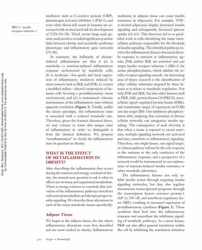
In summary, the hallmarks of obesity-induced inflammation are that it (a) ismetabolic—a nutrient-induced inflammatoryresponse orchestrated by metabolic cells;(b) is moderate—low-grade and local expres-sion of inflammatory mediators induced bystress sensors such as IKK and JNK; (c) createsa modified milieu—altered composition of im-mune cells favoring a proinflammatory tissueenvironment; and (d ) is maintained—chronicmaintenance of the inflammatory state withoutapparent resolution (Figure 1). Finally, unlikethe classic paradigm, this inflammatory stateis associated with a reduced metabolic rate.Therefore, given the features discussed above,we may venture to name this unique stateof inflammation in order to distinguish itfrom the classical definition. We propose“metaflammation” to clarify the inflammatorystate in question in obesity.
WHAT IS THE EFFECTOF METAFLAMMATION INOBESITY?
After describing the inflammation that occursduring the nutrient and energy overload of obe-sity, the natural next question to ask is what itseffects are on tissue and organismal metabolism.There is strong evidence to conclude that acti-vation of the inflammatory pathways interfereswith normal metabolism and disrupts proper in-sulin signaling. We describe these alterations ineach of the major metabolic tissues specifically.
Adipose Tissue
We begin in the adipose tissue, the site whereinflammatory alterations were first describedand are most studied in obesity. Inflammatory
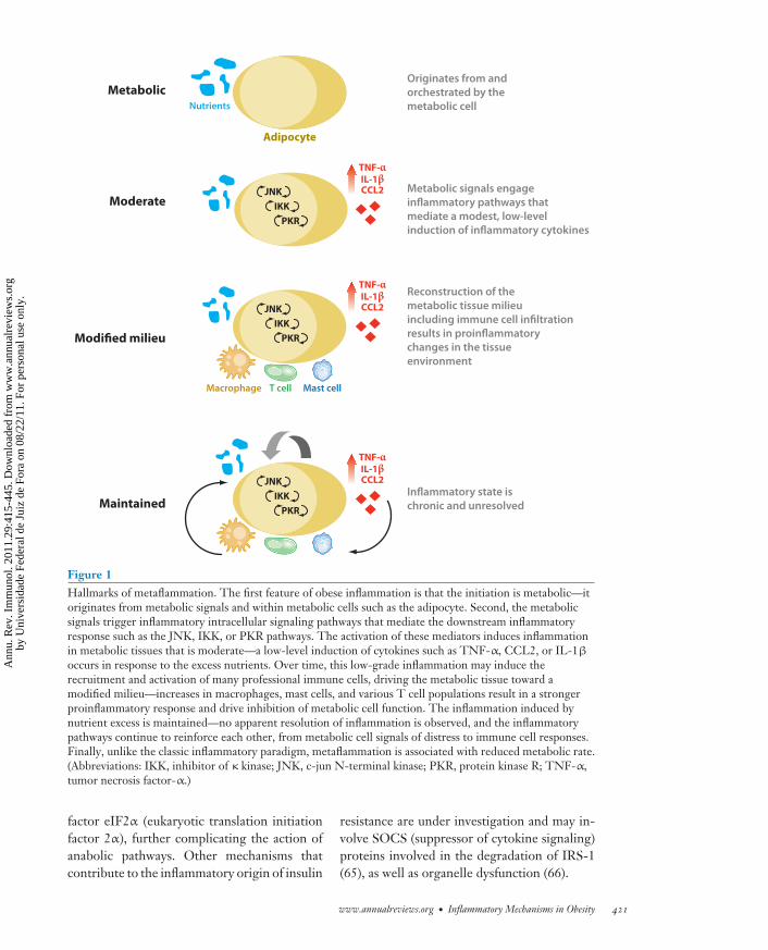
mediators in adipose tissue can cause insulinresistance in adipocytes. For example, TNF-α-treated adipocytes display decreased insulinsignaling and subsequently decreased glucoseuptake (61–63). This discovery led to or paral-leled work in cells identifying the major intra-cellular pathways responsible for the blockadeof insulin signaling. The identified pathways in-volve the inflammatory kinases discussed above.In response to nutrient or inflammatory sig-nals, JNK and/or IKK are activated and cantarget insulin receptor substrate 1 (IRS-1) forserine phosphorylation, which inhibits the in-sulin receptor signaling cascade. An interestingarea of future research is the identification ofother cellular substrates targeted by these ki-nases as it relates to metabolic regulation. Notonly JNK and IKK, but also other kinases suchas PKR, S6K, protein kinase Cθ (PKCθ), extra-cellular signal–regulated protein kinase (ERK),and mammalian target of rapamycin (mTOR)can also target IRS-1 for inhibitory phosphory-lation (64), implying that activation of diversecellular networks can antagonize insulin sig-naling. The consequence of such diversity isthat when a tissue is exposed to excess nutri-ents, multiple signaling networks are activatedand may contribute to inflammatory signaling.Therefore, one single kinase, one signal target,or a linear pathway will not be the sole responseto the nutrient or the only conductor of theinflammatory response, and a perspective of anetwork would be instrumental in our explana-tions of nutrient-induced insulin resistance orother metabolic adversities.
The inflammatory kinases not only in-hibit insulin action through targeting insulinsignaling molecules, but they also regulatedownstream transcriptional programs throughthe transcription factors activator protein-1(AP-1), NF-κB, and interferon regulatory fac-tor (IRF), resulting in increased expression ofproinflammatory cytokines (Figure 2). Thesecytokines then feed into the inflammatoryresponse and exacerbate the inhibitory signal-ing of metabolic pathways. As a stress kinase,PKR can also affect general translation withinthe cell by inhibiting the translation initiation
420 Gregor · Hotamisligil
Ann
u. R
ev. I
mm
unol
. 201
1.29
:415
-445
. Dow
nloa
ded
from
ww
w.a
nnua
lrev
iew
s.or
gby
Uni
vers
idad
e Fe
dera
l de
Juiz
de
Fora
on
08/2
2/11
. For
per
sona
l use
onl
y.
IY29CH16-Hotamisligil ARI 4 February 2011 17:13
TNF-αIL-1βCCL2
Metabolic
Moderate
Modified milieu
Maintained
Originates from and
orchestrated by the
metabolic cell
Adipocyte
Metabolic signals engage
inflammatory pathways that
mediate a modest, low-level
induction of inflammatory cytokines
Reconstruction of the
metabolic tissue milieu
including immune cell infiltration
results in proinflammatory
changes in the tissue
environment
Inflammatory state is
chronic and unresolved
Nutrients
Macrophage T cell Mast cell
JNK
IKK
PKR
TNF-αIL-1βCCL2JNK
IKK
PKR
TNF-αIL-1βCCL2JNK
IKK
PKR
Figure 1Hallmarks of metaflammation. The first feature of obese inflammation is that the initiation is metabolic—itoriginates from metabolic signals and within metabolic cells such as the adipocyte. Second, the metabolicsignals trigger inflammatory intracellular signaling pathways that mediate the downstream inflammatoryresponse such as the JNK, IKK, or PKR pathways. The activation of these mediators induces inflammationin metabolic tissues that is moderate—a low-level induction of cytokines such as TNF-α, CCL2, or IL-1β
occurs in response to the excess nutrients. Over time, this low-grade inflammation may induce therecruitment and activation of many professional immune cells, driving the metabolic tissue toward amodified milieu—increases in macrophages, mast cells, and various T cell populations result in a strongerproinflammatory response and drive inhibition of metabolic cell function. The inflammation induced bynutrient excess is maintained—no apparent resolution of inflammation is observed, and the inflammatorypathways continue to reinforce each other, from metabolic cell signals of distress to immune cell responses.Finally, unlike the classic inflammatory paradigm, metaflammation is associated with reduced metabolic rate.(Abbreviations: IKK, inhibitor of κ kinase; JNK, c-jun N-terminal kinase; PKR, protein kinase R; TNF-α,tumor necrosis factor-α.)
factor eIF2α (eukaryotic translation initiationfactor 2α), further complicating the action ofanabolic pathways. Other mechanisms thatcontribute to the inflammatory origin of insulin
resistance are under investigation and may in-volve SOCS (suppressor of cytokine signaling)proteins involved in the degradation of IRS-1(65), as well as organelle dysfunction (66).
www.annualreviews.org • Inflammatory Mechanisms in Obesity 421
Ann
u. R
ev. I
mm
unol
. 201
1.29
:415
-445
. Dow
nloa
ded
from
ww
w.a
nnua
lrev
iew
s.or
gby
Uni
vers
idad
e Fe
dera
l de
Juiz
de
Fora
on
08/2
2/11
. For
per
sona
l use
onl
y.
IY29CH16-Hotamisligil ARI 4 February 2011 17:13
Cytokine/TLR receptor
Nutrients
Metabolites InsulinreceptorOther
sensors
Translational control
Inflammatory response
Phosphotyrosine
Phosphoserine
Other mechanisms
Nutrienttransporters
IRS-1
PKR IKK JNK
IRF NF-κB AP-1
Generaltranslation
ER
ERInflammatorymediators
Nuclearreceptors
Mitochondria
Nutrientchaperones
(FABPs, RBP4, etc.)elF2α
elF2α
IRS-1
Insulin signaling
C Y T O S O L
N U C L E U S
Figure 2Inflammatory disruption of metabolic functions. Obese or high-fat diet conditions lead to the induction of inflammatory signalingpathways in metabolic cells through several paths. Nutrients or other metabolites may result in the activation of cytokine or Toll-likereceptor (TLR) pathways and have access to cellular targets directly or via chaperoning molecules. Three prominent kinasesdownstream of these receptors are JNK, IKK, and PKR, which play important roles in relaying stress signals throughout the cell andengaging metabolic responses. All three of these kinases can inhibit insulin signaling via serine phosphorylation of IRS-1. Thisphosphorylation leads to the ubiquitination and degradation of IRS-1, thus blocking insulin action downstream of receptor activation.In addition, PKR can negatively regulate the translation initiation factor eIF2α, leading to inhibition of general translation, andinfluence ER function. This property of PKR suggests that inflammatory signaling cascades, ER function, and insulin action may beregulated through metaflammation complexes or metaflammasomes containing these kinases. These three kinases can induce aninflammatory response through activation of the transcription factors AP-1, NF-κB, and IRF, which upregulate inflammatory mediatorgene expression. The increase in inflammatory cytokines can then lead to exacerbated receptor activation as the cytokine signalscombine with excess nutrients and establish a positive feedback loop of inflammation. (Abbreviations: AP-1, activator protein-1; eIF2α,eukaryotic translation initiation factor 2α; ER, endoplasmic reticulum; FABP, fatty acid–binding protein; IKK, inhibitor of κ kinase;IRF, interferon regulatory factor; IRS-1, insulin receptor substrate 1; JNK, c-jun N-terminal kinase; PKR, protein kinase R; RBP,retinol-binding protein.)
The functional link between inflammatorysignaling and insulin resistance has also beendemonstrated in vivo. Indeed, genetic loss-of-function mouse models for TNF-α, TNFR1/2,
JNK, TLR2, IKKε, and others have all demon-strated beneficial metabolic effects from lack oftheir respective inflammatory mediators whenthe animals are challenged with obesity or
422 Gregor · Hotamisligil
Ann
u. R
ev. I
mm
unol
. 201
1.29
:415
-445
. Dow
nloa
ded
from
ww
w.a
nnua
lrev
iew
s.or
gby
Uni
vers
idad
e Fe
dera
l de
Juiz
de
Fora
on
08/2
2/11
. For
per
sona
l use
onl
y.

IY29CH16-Hotamisligil ARI 4 February 2011 17:13
HFDs (27, 37, 67–69). For example, TNF-α−/− mice undergoing diet-induced obesity dis-play decreased blood glucose and insulin lev-els compared with control mice, along withimproved glucose and insulin sensitivity (27).Importantly, these models have also shown apreservation of local insulin signaling and sen-sitivity in adipose tissue when the inflammatorymediators are absent, indicating that protectionof insulin signaling in the specialized metabolictissues leads to systemic improvements.
The effects of inflammation in adipose tis-sue are not limited to insulin signaling alone.For instance, treatment of adipocytes in vitrowith inflammatory cytokines such as TNF-αcan induce lipolysis, itself a feature of obeseadipose tissue pathology (70). In addition, in-flammatory signaling in the adipocyte can alsodownregulate the activity of the nuclear recep-tor peroxisome proliferator-activated receptorγ (PPARγ), which is essential to adipogene-sis and to maintenance of adipocyte gene ex-pression and function (71). In support of thisfinding, TNF-α treatment blocks the differ-entiation of preadipocytes in vitro (72). In-deed, without PPARγ the adipocyte’s abilityto make and store lipids and to maintain in-sulin sensitivity is compromised (71). One im-portant consequence of inflammation-inducedPPARγ downregulation is reduced adiponectinexpression, a cytokine specific to adipocytes thatis also involved in regulating systemic insulinsensitivity (73). Relief of inflammation restoresthe adipocyte’s capacity for lipogenesis andadiponectin expression (74). Therefore, collec-tively we observe that the effects of inflamma-tion on adipose tissue are multifaceted but seemto have one goal in common: shutting downnormal adipocyte processes in favor of stressresponses. Without the proper functioning andendocrine action of adipose tissue, the body’snutrient deposition is disrupted, and systemicglucose homeostasis is thrown into imbalance.
Immune Cell Contribution
The work described above focuses on iso-lated adipocytes or germ-line genetic models
involving inflammatory mediators wheredeletion occurs in every tissue. Given thatimmune cell infiltration is one of the hallmarksof obese inflammation, many researchers havefocused on immune cell studies, primarilyof macrophages, to determine the effects ofthe immune cells on adipose tissue function.Deletion of macrophage-specific genes ofvarious inflammatory signaling componentshas revealed the contribution of macrophagefunctions in obesity-induced insulin resistance.For example, deletion of the chemoattractantreceptor CCR2 in HFD-fed mice resulted indecreased macrophage infiltration into adiposetissue, a decrease in adipose inflammatorygene expression, and also improved insulinsensitivity compared with control animals (75).Conversely, overexpression of the chemoat-tractant CCL2 specifically in adipose tissuecauses increased macrophage infiltration andresults in systemic insulin resistance andhepatosteatosis (76, 77). Again, in these studiesthe animals were carrying germ-line deletions;therefore, macrophage-specific effects were notdistinguishable. To answer the question aboutmacrophage-specific effects, several studieshave utilized macrophage-specific deletion orbone marrow transplant models to pinpointmacrophage/myeloid contribution in the obeseinflammatory phenotype. Deletion of IKKβ inthe myeloid lineage protected mice on HFDsfrom glucose intolerance (78). Additionalstudies investigating Cbl-associated protein(Cap), CCR2, fatty acid–binding protein 4(FABP4/ap2), and TLR4 loss of functionin macrophages also revealed decreases inobesity-induced inflammation and insulinresistance of varying degrees (43, 79–81).Interestingly, myeloid-specific deletion of JNKwas not associated with a beneficial metabolicphenotype, supporting the conclusion thatactivity of inflammatory pathways in cells suchas adipocytes is sufficient for obesity-inducedinsulin resistance and metabolic deterioration(82, 83). The only studies reporting advan-tageous molecules in the macrophages weredone with PPARγ and PPARδ. Loss of PPARγ
or PPARδ in macrophages actually worsens
www.annualreviews.org • Inflammatory Mechanisms in Obesity 423
Ann
u. R
ev. I
mm
unol
. 201
1.29
:415
-445
. Dow
nloa
ded
from
ww
w.a
nnua
lrev
iew
s.or
gby
Uni
vers
idad
e Fe
dera
l de
Juiz
de
Fora
on
08/2
2/11
. For
per
sona
l use
onl
y.
IY29CH16-Hotamisligil ARI 4 February 2011 17:13
the metabolic phenotype of the mouse, andthis has been attributed to a failure of theM2 alternate activation of the macrophagesand a subsequent switch to a proinflam-matory state (84, 85). Therefore, a generalconclusion drawn from these studies is thatinterference with the proinflammatory actionof the macrophage is beneficial for the adiposetissue environment and organismal glucosehomeostasis.
What about removing entire cell types?Taking these investigations one step further,many groups have used techniques that depleteentire cell types in the immune population. Forinstance, one study depleted mice of CD11c+
immune cells, which include macrophages,dendritic cells, and neutrophils, among otherimmune cells, and showed that insulin sen-sitivity was improved in these obese animals(86). Studies involving T cell populationsalso demonstrated that depletion of CD8+
T cells or transfer/enhancement of CD4+ Tcells or Tregs improved the insulin sensitivityof diet-induced obese mice (47–49). Finally,studies in mast cells and NKT cells alsoshowed that depletion of these cell types leadsto decreased inflammation in the adipose tissueand improvements in glucose homeostasis(45, 46). Again, the data seem to point to theinflammatory actions of the immune systemas important contributors to the disruptionof metabolic function, and when these areremoved it is beneficial for the anabolic insulinpathways. This benefit can occur whether youare manipulating the system to act in an anti-inflammatory manner (for example, adoptivetransfer of Tregs) or inhibiting its proinflam-matory actions (i.e., through gene deletionof chemoattractants). In sum, in the adiposetissue we see the outcropping of inflammationleading to inhibition of insulin signaling andother adipocyte anabolic functions throughcomplex interactions between adipocytes anda series of immune effectors. Taken together,large amounts of data point to a causal role ofadipose inflammation in the manifestation ofobesity-induced insulin resistance.
Liver
The liver represents another major metabolicorgan in its ability to control not only gluconeo-genesis and glycogen storage, but also massiveamounts of lipogenesis and cholesterol synthe-sis and secretion. In contrast to adipose tissue,the liver does not experience an infiltration ofmacrophages during the onset of obesity but in-stead undergoes an activation of inflammationwithin cells of the liver, including the residentmacrophage-like Kupffer cells (87). In animalmodels of obesity, inflammatory cytokine ex-pression is increased in the liver compared withlean controls (30). Indeed, obesity is associatedwith fatty liver (or hepatosteatosis) that oftenleads to the more advanced inflammatory stateof steatohepatitis. What effects does this in-flammation have on liver metabolic functions?
First, as described in adipose tissue, inflam-matory mediators have the ability to inhibitinsulin signaling, and these same inhibitorypathways have been shown to be active inthe obese liver (88). Specifically, activation ofthe NF-κB pathway appears to be critical ininflammation-induced insulin resistance. Micewith liver-specific activation of IKKβ displaydecreased glucose tolerance and insulin sensi-tivity and decreased insulin signaling in the liveritself (30). Conversely, a loss of IKKβ in hepa-tocytes decreased the HFD induction of inflam-matory cytokines in the liver and rendered themice more insulin sensitive (78). This reduc-tion in insulin sensitivity and signaling affectsother aspects of liver metabolism, most notablygluconeogenesis. Normally, gluconeogenesisis suppressed by insulin signaling, but in obeseconditions this regulation is lost, and hepaticglucose production subsequently contributesto hyperglycemia. Again, upon inhibition ofinflammatory signaling, gluconeogenesis isproperly suppressed by insulin (78). JNK kinaseis also activated in the liver during obesity,and loss-of-function models have revealeddecreased inflammatory markers and increasedinsulin sensitivity in the liver of HFD-fedJNK1−/− mice (37). Interestingly, unlike wholeliver inhibition of JNK, hepatocyte-specific
424 Gregor · Hotamisligil
Ann
u. R
ev. I
mm
unol
. 201
1.29
:415
-445
. Dow
nloa
ded
from
ww
w.a
nnua
lrev
iew
s.or
gby
Uni
vers
idad
e Fe
dera
l de
Juiz
de
Fora
on
08/2
2/11
. For
per
sona
l use
onl
y.
IY29CH16-Hotamisligil ARI 4 February 2011 17:13
deletion of the JNK1 isoform resulted in glu-cose intolerance and increased glucose and lipidproduction in the liver of lean mice (89). In thissetting, neither the impact of the remainingJNK isoforms ( JNK2 or 3) nor the effects ofobesity have been addressed. A possible expla-nation for the discrepancy of liver JNK actionis that JNK activation may operate on multiplecell types in the liver (such as hepatocytes,Kupffer cells, and others) and that this collectiveaction is detrimental for metabolism. This hy-pothesis is supported by the inhibitor studies us-ing small molecules, dominant-negative JNK,and RNAi-mediated blockade of JNK activity,all of which affected multiple cell types in theliver and resulted in increased insulin sensitivityand improved systemic metabolism (90–93).
Lipogenic effects of inflammation are animportant but understudied area. Early studieshave shown that in vivo administration ofTNF-α or IL-6 can induce hepatic lipogenesisand increase hepatic triglyceride production(94, 95). This augmented production leads toan increase in very-low-density lipoprotein(VLDL) (and particularly apoB100) secretionfrom the liver and an overall increase in serumtriglyceride levels. Although it seems thatinflammatory cytokines can induce lipogenesispathways in the liver, mechanistically thisprocess is not well understood and necessitatesfurther study. Another effect of inflammationin the obese liver is the activation of a secretoryresponse involving inflammatory mediatorsand acute-phase reactants. For example, serumlevels of cytokines such as CRP, PAI-1, serumamyloid A, and IL-6 produced by the liver areincreased in obese animals and humans com-pared with lean controls (15, 34). Some of thesecytokines have been shown to have adversemetabolic effects on peripheral organs. Onestudy found that liver IKK activation leads to anincrease in systemic IL-6 that negatively influ-ences muscle insulin sensitivity (30). Therefore,the secretory profile of the liver in obesity maybe a strong contributor to the malfunction ofperipheral tissues under such nutrient excess.
If the inflammation in the liver esca-lates, cell death may occur, resulting in the
recruitment of immune cells and the patholog-ical state known as steatohepatitis. The stresskinase JNK has been shown to be required forsaturated free fatty acid–induced apoptosis inhepatocytes, providing a possible link betweenthe excess lipids of obesity and the resultinginflammation-induced cell death (96). Thus,the liver remains an important site wheremetabolic and immune cell signals converge tolimit the tissue’s response to nutrients.
Muscle
The muscle is a key site of glucose uptake andenergy consumption in the body and in thiscapacity is an important contributor to glucosehomeostasis. Consensus has not been reachedregarding the data on muscle inflammationin obesity, and many of the following pointsmay be debated. Thus far, obesity does notappear necessarily or uniformly to induce in-flammation in muscle tissue, but inflammatorymediators from other sites such as liver and adi-pose can influence muscle metabolism (30, 97).For example, morphologically, macrophageinfiltration is not observed in muscle fibers perse in obese animals, but the adjacent adiposetissue displays increased infiltration comparedwith lean tissues (42). Also, unlike adiposeor liver tissues, muscle does not express orrelease significant amounts TNF-α or IL-6in T2D patients compared with controls andtherefore is not likely to be a source for the risein inflammatory mediators (98, 99). However,data contrary to this also exist, as local increasesin TNF-α expression in muscle tissue of obesehumans reportedly correlate strongly withimpaired insulin sensitivity (33). Mechanisticstudies in mice activating the IKK/NF-κBpathway in muscle reveal a muscle wastingphenotype but no induction of inflammatorycytokines (100). Inhibition of the NF-κBpathway through muscle-specific repression ofNF-κB or deletion of IKK2 also revealed noeffect on muscle insulin sensitivity or systemicglucose homeostasis, leading to the conclusionthat at least this major inflammatory pathwaydoes not appear to regulate muscle metabolic
www.annualreviews.org • Inflammatory Mechanisms in Obesity 425
Ann
u. R
ev. I
mm
unol
. 201
1.29
:415
-445
. Dow
nloa
ded
from
ww
w.a
nnua
lrev
iew
s.or
gby
Uni
vers
idad
e Fe
dera
l de
Juiz
de
Fora
on
08/2
2/11
. For
per
sona
l use
onl
y.

IY29CH16-Hotamisligil ARI 4 February 2011 17:13
dysfunction during obesity (100, 101). Muscle-specific deletion of JNK1 also presents withcomplex results, displaying increased insulinsensitivity in the muscle but impaired liver andadipose tissue metabolism and, as a result, nosystemic benefit (102).
Although the muscle cell does not seemto be an origin of inflammatory signals inobesity or to regulate metabolism throughinflammatory kinase action, the influence ofperipheral inflammation on muscle functionis well established. For example, inflammatorycytokines are able to induce insulin resistancein muscle cells in culture (103, 104) and in vivoby infusion into humans, resulting in decreasedglucose uptake and glycogen synthesis (105). Ithas been proposed that TNF-α, IL-6, CCL2,and/or retinol-binding protein-4 are inflamma-tory mediators from sources such as adipose andliver tissue that can act on the muscle (30, 97,103). Indeed, the TNFR−/− mouse is resistantto the decrease in muscle glucose uptake andinsulin resistance in response to TNF-α admin-istration (106), and treatment with antibodiesto TNF-α can increase muscle insulin sensitiv-ity (107). In addition, liver-specific activation ofIKK in mice results in increases in systemic IL-6levels. The muscle displays increased IL-6 sig-naling and decreased insulin sensitivity and glu-cose uptake. When antibodies to IL-6 are ad-ministered, muscle IL-6 signaling is decreasedand glucose homeostasis improves (30). Therole of IL-6 in muscle insulin sensitivity is con-troversial and complex, however. Several stud-ies report no effect or insulin-sensitizing effectsof IL-6, as in the context of exercise-inducedIL-6 release from muscle (reviewed in 108).
Given that muscle is a tissue of paramountimportance for glucose uptake and glyco-gen synthesis, further studies are necessaryto elucidate the connections between musclemetabolism and inflammatory pathways duringobesity.
Brain
Although not usually classified as a metabolicorgan per se, the brain is the site of central
regulation of appetite control and energyexpenditure. The brain, and specifically the hy-pothalamus, responds to metabolic endocrinesignals, including nutrients themselves, insulinreleased from the pancreas, and leptin secretedfrom the adipose tissue. Insulin and leptin bothhave important appetite-suppressant effectsmediated through hypothalamic signaling.Interestingly, in obesity the hypothalamusitself exhibits both insulin and leptin resis-tance, leading to a loss of this control ofappetite and feeding behavior, exacerbating thealready excessive body weight gain (reviewedin 109). In addition to influencing appetite,the central nervous system can also influenceperipheral organ response to nutrients, as seenin hypothalamic control of hepatic glucoseproduction (110). Intriguingly, more recentstudies have revealed that inflammatory path-ways in the hypothalamus are also activatedduring obesity. De Souza et al. (32) reported anincrease in expression of TNF-α, IL-1β, andIL-6, among other immune-related molecules,in HFD-fed rats compared with lean controls.In addition, HFD-fed rats display evidence ofincreased apoptosis in the hypothalamus (111).What are the effects of such inflammationin the brain and subsequently on metabolichomeostasis? Again, the few studies availableon this subject point to a relationship not onlywith insulin resistance but also with leptinresistance and the resulting feeding behaviorand energy expenditure. For example, inHFD-fed mice, brain-specific activation ofthe inflammatory kinase IKKβ resulted in anincrease in food intake and body weight alongwith significant hypothalamic insulin andleptin resistance (112). Conversely, inhibitionof IKKβ signaling in the brain restored insulinand leptin sensitivity and protected micefrom HFD-induced weight gain, resultingin overall improved glucose homeostasis.Further evidence to support the idea that braininflammation may play a causal role in obesity-induced insulin resistance comes from studiesdone upstream of IKK signaling, namely inthe TLR pathway. The TLR pathway of theinnate immune system is involved in sensing
426 Gregor · Hotamisligil
Ann
u. R
ev. I
mm
unol
. 201
1.29
:415
-445
. Dow
nloa
ded
from
ww
w.a
nnua
lrev
iew
s.or
gby
Uni
vers
idad
e Fe
dera
l de
Juiz
de
Fora
on
08/2
2/11
. For
per
sona
l use
onl
y.

IY29CH16-Hotamisligil ARI 4 February 2011 17:13
pathogens and eliciting a proper immuneresponse. It is thought that under HFD con-ditions, saturated fatty acids themselves mayactivate TLR signaling. Infusion of a TLR4antibody via intracerebroventricular canulationprotected rats from the inflammatory responseelicited by saturated fatty acids and concomi-tantly decreased food intake and body weightgain (113). In addition, brain-specific deletionof MyD88, a downstream adaptor moleculefor TLR signaling, enhanced leptin signalingin HFD-fed mice compared with controllittermates (114). This recovery of leptinsignaling was accompanied by a decrease infood intake and weight gain and improvementsin glucose homeostasis. The role of the JNKkinase in central regulation of metabolismis substantial, yet complex. Studies utilizingbrain-specific deletion of JNK1 under HFDconditions demonstrated protection againstinsulin resistance, hyperinsulinemia, andglucose intolerance in the mice lacking JNK1compared with controls (115, 116), implicatingthe brain as a key site of JNK action. However,mice that have undergone JNK1 brain deletionare smaller than their wild-type counterpartsand exhibit increased thyroid hormone sig-naling and thermogenesis. Therefore, furtherstudy is needed to distinguish the effects ongeneral body growth from those on energymetabolism and adiposity. Nonetheless, theadvances described above reveal that inflamma-tory pathways in the brain during obesity maybe major players in the imbalance of systemicmetabolic homeostasis by contributing toinhibition of insulin and leptin signaling andthereby impeding regulation of food intake,body mass, and systemic metabolism.
Pancreas
The pancreas is at the heart of glucose home-ostasis, given its role as the source of insulinand glucagon production. Failure of this organto produce sufficient insulin in response to ris-ing systemic glucose levels is at the center ofdiabetic disease. Although the immune originof type 1 diabetes has long been appreciated,
given the autoimmune mechanism that destroysthe insulin-producing β cells, evidence is nowaccumulating to implicate inflammation in thedysfunction of the T2D pancreas. During thecourse of obesity, peripheral insulin resistancerequires ever increasing amounts of insulin toremove glucose from the circulation. This in-creased production results in stress on the β
cell, eventually leading to its hyperproliferationand surrender to apoptosis.
Indeed, recent data have shown that duringthe time course of a HFD, inflammatorycytokine expression in the pancreas increasesand macrophage infiltration occurs, in parallelwith the onset of glucose intolerance (31).Inflammatory activity in the pancreas has longbeen known to disturb insulin production andβ cell survival, two of the main mediators beingIL-1β and IFN-γ. IL-1β activates the NF-κBpathway in pancreatic islets, and the effectsof this activation have been studied in thepancreas using a nondegradable inhibitor ofNF-κB signaling known as IκBα. Overexpres-sion of this inhibitor in human islets in vitroprotects cells from IL-1β-induced nitric oxide(NO) production and apoptosis (117). A trans-genic mouse expressing this same inhibitorspecifically in β cells also resulted in decreasedNO production and apoptosis in response toinflammatory cytokines and significant in vivoprotection from streptozotocin (STZ)-induceddiabetes, a disease model characterized byselective β cell loss (118). JNK signaling hasalso been implicated in pancreatic protectionfrom apoptosis: JNK1−/− mice are resistant toSTZ-induced diabetes (119), and expression ofa dominant-negative JNK in transplanted isletselevated insulin levels and decreased bloodglucose in STZ-treated mice (120). In culturedcells, JNK inhibition protects from loss ofinsulin gene expression and secretion inducedby oxidative or cytokine stress (120, 121). Thus,these two kinases at the core of inflammatorysignaling can affect major β cell functions suchas insulin production and survival.
However, what is really needed to un-derstand these processes are studies in thecontext of obesity. The work described above
www.annualreviews.org • Inflammatory Mechanisms in Obesity 427
Ann
u. R
ev. I
mm
unol
. 201
1.29
:415
-445
. Dow
nloa
ded
from
ww
w.a
nnua
lrev
iew
s.or
gby
Uni
vers
idad
e Fe
dera
l de
Juiz
de
Fora
on
08/2
2/11
. For
per
sona
l use
onl
y.
IY29CH16-Hotamisligil ARI 4 February 2011 17:13
investigates inflammatory molecules in isolatedβ cells or in pancreas-destroying models ofdiabetes. Obesity differs from these models inits distinct features of lipid overload, peripheralinsulin resistance, and slowly progressing timecourse of pathology. Studies in pancreas-specific genetic models performed duringthe development of obesity and T2D will beuseful to investigate the role of inflammatorymediators on pancreatic β cell function. For ex-ample, it has been hypothesized that low-levelinflammation in the pancreas (namely IL-1β
expression) may promote β cell proliferationand that only exacerbated inflammation resultsin apoptosis (122). Given that one of the char-acteristics of obesity-induced inflammation isa low level of cytokine expression, this maydrive islet hyperproliferation and be neededfor adaptation to peripheral insulin resistance.However, this adaptive hyperinsulinemia itselfcan also be a driver of metabolic complications.As a result, it is favorable to inhibit inflamma-tion as conditions worsen to preserve β cellsurvival and function for the organism.
Inflammatory Mechanismsin the Gastrointestinal Tract
One exciting new area of discovery is the in-fluence of gut microbiota upon obesity andmetabolism. The interactions between mi-crobes of the intestine and host responses canaffect weight gain, insulin sensitivity, and theinflammatory state, not only of the gut but ofperipheral organs as well.
A first critical observation in this area camewith the discovery that populations of gutmicrobiota are significantly different betweenlean and obese animals (123). Specifically, at thedivision level genetically obese (ob/ob) micedisplay decreased levels of Bacteroidetes withan increase in Firmicutes bacteria comparedwith lean controls. A similar population changewas also shown to be present in obese versuslean humans (124), and changing the diet topromote weight loss was able to change themicrobiota profile toward a lean phenotype.Intriguingly, this altered flora has a potential
activity of its own, as transfer of obese gutmicrobiota to lean mice caused them togain more weight when compared withmice receiving wild-type microbiota (125).The mechanism behind this difference isthought to be increased energy harvest in theobese intestine due to its specific microbiotamakeup, although further study is neededto fully understand the process behind thisphenomenon.
The picture becomes more complex whenthe inflammatory status of the intestine isconsidered. The idea that inflammation ofthe gut could play a role in determining bodyweight was supported by the fact that transferof microbiota from normal or conventionalizedmice into germ-free mouse donors caused asignificant weight gain accompanied by in-creased insulin resistance (126). In addition,germ-free mice were protected from bodyweight gain, insulin resistance, and glucoseintolerance induced by HFD compared withconventionalized mice (127). More specifically,studies investigating the bacterial cell wallcomponent lipopolysaccharide (LPS), whichactivates the host’s innate immune systemthrough TLR4, reveal a relationship betweenHFD and increased levels of LPS exposure(128). In addition, a study in rats reported thatthose animals with a propensity for obesitydisplayed altered gut microbiota and increasedTLR4 activation (129). Treatment of obesemice with antibiotics decreased levels of LPSand TNF-α expression in the intestine and ledto a decrease in body weight and serum insulinand improvements in glucose tolerance (130,131). Finally, one intriguing study providesevidence that host inflammatory mediators canactually influence intestinal microbiota andwhole body metabolism. Vijay-Kumar et al.(132) report that Tlr5−/− mice develop obesityalong with features of metabolic syndromeincluding glucose intolerance, fatty liver,insulin resistance, and islet hyperplasia. Ofnote, transfer of gut microbiota from Tlr5−/−
cecum to wild-type recipients recapitulatedsome aspects of the metabolic syndromephenotype in the wild-type animals. The
428 Gregor · Hotamisligil
Ann
u. R
ev. I
mm
unol
. 201
1.29
:415
-445
. Dow
nloa
ded
from
ww
w.a
nnua
lrev
iew
s.or
gby
Uni
vers
idad
e Fe
dera
l de
Juiz
de
Fora
on
08/2
2/11
. For
per
sona
l use
onl
y.

IY29CH16-Hotamisligil ARI 4 February 2011 17:13
metabolic phenotype in these animals is mild,however, and not likely to account for theextent of pathology observed in the obese,type 2 diabetic state. The transferred Tlr5−/−
microbiota also resulted in increased TNF-αand IL-1β in the colon of recipient animals,pointing to enhanced inflammation as a pos-sible factor in the regulation of the metabolicphenotype. In sum, these observations supportthe concept that bacterial and host interactionswithin the gastrointestinal tract may influencesystemic metabolic homeostasis and point toinflammatory molecules as potential mediatorsof these effects. This is an important area toexplore, as our understanding of the communi-cation of the various microbes to the complexarray of host tissues and cell types is limited;research into this area will uncover theseinteractions and their influence on whole bodymetabolism.
HOW DOES METABOLICINFLAMMATION OCCUR?
We now come to the question of how theinflammatory response occurs in obesity inthe first place. Starting signals for a tradi-tional inflammatory response are usually de-scribed in terms of pathogens—a molecule froma microbe, parasite, foreign body, or injuredtissue—that will engage a cell signaling path-way (either in the host cell or sentinel immunecell) to initiate an immune response. With over-feeding or HFDs, however, the starting signalis unclear and remains a critical area of futureresearch. Although the initiating factor is notfully known, it is thought that the insult-to-signaling pathway originates within the spe-cialized metabolic cell, e.g., in the adipocyte,hepatocyte, or myocyte. As even nonimmunecells possess their own defensive mechanisms,these metabolic cells are fully capable of engag-ing inflammatory signaling and communicatingin response to danger signals. For example, theadipocyte can execute many of the downstreaminflammatory signaling events such as inflam-masome and TLR activation, JNK and NF-κBsignaling, and production of inflammatory cy-
tokines in response to metabolic stress signalsthat are normally viewed as the territory of pro-fessional immune cells.
One theory to explain the origin of the in-flammatory response in obesity is that nutrientsthemselves are naturally inflammatory. That isto say, there is a normal physiological responsefrom metabolic cells encountering nutrientsthat results in a low level of inflammation. Theidea is that food or nutrients are not self andtherefore elicit a slight immune response asthey are encountered and metabolized. Indeed,digestion of food and its metabolic traffickingis a daily yet intense experience as cells en-counter the flood of energy and nutrients ina short period of time (i.e., lunch), and such astimulus may be anticipated to induce a briefepisode of stress signaling in the target cells.If this is the case, then there may be mech-anisms to mitigate this physiological nutrientresponse in metabolic cells and tissues. Onestudy supporting this hypothesis demonstratedthat the six-transmembrane protein STAMP2may play just such a role in metabolic tis-sues encountering nutrients (133). STAMP2expression was increased in adipose tissue dur-ing feeding and displayed anti-inflammatoryproperties in response to nutrient stimulationin adipocytes. When cultured adipocytes lackSTAMP2, they produce an uncontrolled in-flammatory output when exposed to nutrients.Indeed, Stamp2−/− mice exhibited increasedadipose tissue inflammation when fed a reg-ular diet and systemic metabolic dysfunctionsuch as insulin resistance and glucose intoler-ance. These results suggest that STAMP2 mayfunction as an immune suppressor in metabolicand immune cells as they respond to nutri-ents. Under obese conditions, the upregula-tion of STAMP2 expression during feeding islost, correlating with increased inflammatoryconditions. While the STAMP2 molecule pos-sesses a metalloreductase/oxidoreductase activ-ity, the exact substrates to which it responds orthe molecular mechanisms by which it preparesthe cells for incoming nutrients and prevents in-flammatory responses remain important areasof future research.
www.annualreviews.org • Inflammatory Mechanisms in Obesity 429
Ann
u. R
ev. I
mm
unol
. 201
1.29
:415
-445
. Dow
nloa
ded
from
ww
w.a
nnua
lrev
iew
s.or
gby
Uni
vers
idad
e Fe
dera
l de
Juiz
de
Fora
on
08/2
2/11
. For
per
sona
l use
onl
y.

IY29CH16-Hotamisligil ARI 4 February 2011 17:13
A second hypothesis is that feeding natu-rally couples the nutrients with inflammatorymolecules. For example, there is increasedpermeability of the intestine after feeding,probably to ensure maximal nutrient uptake.Interestingly, studies in mice and humansrevealed that serum LPS was increased afterfeeding, suggesting the hypothesis that in-testinal permeability releases inflammatorymolecules into the circulation, coupling nutri-ent entry with inflammatory signals (128, 134).Therefore, the metabolic cells would adapt tocoping with both. In lean animals postfeedingor during fasting, levels of nutrients and LPSare low, indicating the system has effectivelyhandled the nutrient load. However, in obeseanimals the intestine is constantly more per-meable than in lean animals, and serum levelsof LPS are higher, indicating that the systemno longer functions efficiently and there may
Fasting/feeding cycles over time
Degree ofinflammatory
response
Obeseinflammatory
response
Leaninflammatory
response
Recruit-ment of
professionalimmune cells
Figure 3Pulsatile inflammatory response during feeding: normal versus obese reactionsover time. Fasting/feeding cycles induce low-level inflammatory responses inmetabolic cells of lean, healthy animals that are easily resolved. During thehigh-fat diet or excess feeding of obesity, responses to food become moreintense and frequent, and resolution of the inflammatory response becomes lessefficient, raising the baseline of inflammation in metabolic tissues. Once thelevel of inflammatory response reaches a certain threshold in the metaboliccells, professional immune cells such as macrophages, mast cells, and T cells arerecruited and activated. Their participation in the inflammatory response altersthe tissue environment toward a proinflammatory milieu and exacerbates theinflammation even further.
be a constant source of inflammatory signalpresent. Higher LPS levels in obesity could alsobe explained in that increased nutrient/LPSexposures heighten the baseline inflammatoryresponse of metabolic tissues.
Hence, a general hypothesis may bedrawn from the two examples given above,as follows: During the feeding/fasting cycle,a pulsatile inflammatory response occurs inthe metabolic cells (Figure 3). Under normalconditions, the low-level inflammation peakswith feeding and resolves after the nutrientsare metabolized. With overfeeding or obeseconditions, however, a constant stimulus fromnutrient intake results in a more consistentlyactive inflammatory response. These low-levelsignals accumulate over time, amplify witheach nutrient exposure, begin to impair normalmetabolic pathways, and may reach a levelwhere the professional immune cells are alertedand called to duty. Once the immune cellsare activated and participate in the responseto inflammation, the severity of the responseincreases, and disruption of metabolic cellfunction becomes more complete.
Another theory to explain the origin of theinflammation found in obesity is that the nutri-ents themselves are not naturally inflammatorybut in excess can engage the classical pathogen-sensing or immune-response pathways. This isalmost a mistaken identity theory—the systemis tricked into thinking that these abundant nu-trients are pathological. This mistaken iden-tity may be due to a dose-dependent loss ofspecificity that exploits the ability of pathogensensors to recognize similar structural enti-ties, such as lipids. We focus on two ex-amples from the literature that may supportthis hypothesis. The first is in the study ofTLRs, the pathogen-sensing receptors of theinnate immune system. Again, as the specializedmetabolic cells possess their own defense sys-tem, they express various TLRs. Adipocytes, forexample, express functional TLR4 and TLR2.Studies in mice have revealed that adiposeTLR4 can be activated by infusion of saturatedfatty acids and that this activation contributesto the resulting insulin resistance (39). Also,
430 Gregor · Hotamisligil
Ann
u. R
ev. I
mm
unol
. 201
1.29
:415
-445
. Dow
nloa
ded
from
ww
w.a
nnua
lrev
iew
s.or
gby
Uni
vers
idad
e Fe
dera
l de
Juiz
de
Fora
on
08/2
2/11
. For
per
sona
l use
onl
y.

IY29CH16-Hotamisligil ARI 4 February 2011 17:13
increasing the level of fatty acids in the systemvia HFD feeding causes an increase in TLR4expression, and genetic loss of TLR4 in theseconditions may ameliorate insulin resistance(81, 135).
A second example is the pathogen-sensingkinase PKR, known to sense double-strandedRNA, which is indicative of viral invasion ofthe cell, and to initiate an inflammatory re-sponse (136). Recently, our group has shownthat PKR is activated (in the absence of virus)during obesity or lipid infusion in mice (36). In-terestingly, PKR itself can orchestrate the ac-tivity of JNK and downregulate insulin signal-ing in metabolic cells, all from signals receivedvia a HFD. Thus, the model derived fromthese examples is that nutrient overload inducespathogen sensors (Figure 4). The ability ofPKR to interact with JNK and IKK as well asinsulin signaling components and translationalcontrol through eIF2α also leads us to postulatethat such complexes may represent metaflam-masomes and may in fact be the recognitionmechanisms for metabolic signals that triggerinflammation. Under regular conditions, the
level of nutrients pouring into the system is lowenough that only normal metabolic pathwaysare engaged and fidelity is ensured. However,upon overfeeding, the organism is exposed tohigh nutrient levels, which begin to directly ac-tivate pathogen-sensing molecules or pathwayswithin the cells. This spillover of signals mayeven reach a third level as immune cells areactivated and coordinate a multilayer responsewithin the tissue, effectively blocking nutrient-induced anabolic activity.
Signals to Inflammatory Pathways
Evidence for the direct engagement of nu-trients with pathogen or immune sensors isstill lacking, but this remains one of the mostfascinating areas of obesity research. An im-portant future area in this respect is the sys-tematic characterization of nutrients for theirability to directly engage innate immune re-sponses through pathogen sensors or othermechanisms. Such platforms could give rise togroundbreaking insights regarding the healtheffects and underlying mechanistic actions of
Healthy nutrient intake Excess nutrient intake
Drain
Metabolicpathways
Pathogen-sensing
pathways
Immune-responsepathways
Prolonged excess nutrient intake
pat ays
Nutrientinput
Figure 4Overload of nutrient signals spills from metabolic pathways to immune-response pathways. Nutrient input under normal or healthyconditions should engage metabolic pathways within the cells, leaving immune-response pathways inactive. With increased nutrientintake, the levels of nutrients flooding the system may rise enough that the overflow stimulates pathogen-sensing pathways. Becausethese pathways recognize biological molecules (such as specific fatty acids), nutrient moieties in excess may also be able to activate suchsensors. Once the immune sensors are activated, they may be antagonistic to the metabolic pathways, in effect blocking the drain ofnutrient metabolism. If the nutrient excess persists to an extreme state, immune-response pathways of the professional immune cellsmay also be activated. The involvement of these pathways will intensify the inhibition of metabolic pathways and contribute to thebacklog of nutrients in the system.
www.annualreviews.org • Inflammatory Mechanisms in Obesity 431
Ann
u. R
ev. I
mm
unol
. 201
1.29
:415
-445
. Dow
nloa
ded
from
ww
w.a
nnua
lrev
iew
s.or
gby
Uni
vers
idad
e Fe
dera
l de
Juiz
de
Fora
on
08/2
2/11
. For
per
sona
l use
onl
y.
IY29CH16-Hotamisligil ARI 4 February 2011 17:13
UPR: unfoldedprotein response
nutrients. For example, is there a nutrient thatengages the TLR system? If so, at what concen-tration does the interaction take place? As men-tioned above, the biological targets and actionsof the overwhelming majority of nutrients,exogenous or endogenous, remain unknown.The concept of a nutrient-induced immune re-sponse provides an opportunity to approach asmall but tractable portion of this vast question.
To date, studies in cultured cells have identi-fied several candidates for inducers of metabolicinflammation. As implicated above, free fattyacids and especially saturated free fatty acidscan activate inflammatory signaling in variouscell types (137, 138). In addition, high glu-cose, hypoxia, and cell damage and death haveall been proposed as initiators of inflammationduring obesity (139, 140), often in combina-tion. For example, one scenario may be that asthe obese adipocyte expands, it reaches a me-chanical limit to its storage capacity. The tissuemay be less vascularized due to expansion, andthe cell’s stress response and perhaps even deathresponse occur, releasing cytokines and excessfatty acids. These in turn are sensed by inflam-matory kinases (such as JNK, PKR or IKK), theinflammasome, or TLRs, and an inflammatorycascade begins. There may also be specializedsignaling complexes, metaflammasomes, thatare organized by sensing molecules such as PKRand that coordinate the responses based on thenature and level of nutrient exposure. SuchPKR complexes could also be integrating or-ganelle function with metabolic and inflamma-tory outcomes (see below), and exploring theseintriguing possibilities requires further work.
Organelle Stress
A key contributor to metabolic deteriora-tion and the inflammatory response that de-serves discussion is organelle dysfunction of themetabolic cells. Before we arrive at a single cy-tokine or free fatty acid inducer, we should con-sider that, in response to nutrient excess, thecell experiences functional stress that then gen-erates an inflammatory response. For instance,we know that, compared with lean tissues, obese
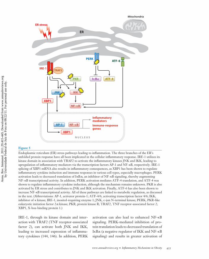
liver and adipose display increased levels of en-doplasmic reticulum (ER) stress (141). As theER is the primary site of protein folding inthe cell, ER stress is measured by the activa-tion of the unfolded protein response (UPR),which is driven by three main transmembranesensors that reside on the ER, namely PERK(PKR-like eukaryotic initiation factor 2α ki-nase), IRE-1 (inositol-requiring enzyme 1), andATF-6 (activating transcription factor 6) (re-viewed in 142). Briefly, PERK, which possessesa kinase domain, inhibits general protein trans-lation through phosphorylation of eIF2α, lead-ing to alternative translation of ATF-4 and itsdownstream targets. IRE-1, a transmembraneprotein that has kinase and endoribonucleaseactivities, cleaves the mRNA of the transcrip-tion factor XBP1 (X-box-binding protein 1),resulting in translation of an activated formof XBP1 responsible for upregulation of manychaperone genes. Finally, ATF-6 is a transcrip-tion factor that resides on the ER membraneand is cleaved in response to ER stress, pro-ducing an active factor that translocates to thenucleus and upregulates transcription of chap-erone genes. The goal of activating the threearms of the UPR is to restore ER homeostasisby halting protein synthesis, increasing degra-dation of proteins from the ER, and increas-ing the level of chaperone proteins to assist inprotein folding. If proper ER function cannotbe achieved or if the stress continues, the UPRmay also initiate apoptotic pathways. For in-depth discussion of this response and its effectson metabolism, we direct the reader to a recentreview (66).
Interestingly, there are many connectionsbetween the UPR and inflammatory signalingpathways (Figure 5). First, chemical agentsthat target the UPR such as tunicamycin andthapsigargin lead to the induction of inflam-matory kinases and the production of an arrayof inflammatory genes including Il6, CXCL8,CCL2, or Tnfa (143, 144). ER stress can also bea source of the production of oxidative stressor apoptosis, processes that themselves canlead to inflammation (145). Investigations intothe mechanism behind this effect revealed that
432 Gregor · Hotamisligil
Ann
u. R
ev. I
mm
unol
. 201
1.29
:415
-445
. Dow
nloa
ded
from
ww
w.a
nnua
lrev
iew
s.or
gby
Uni
vers
idad
e Fe
dera
l de
Juiz
de
Fora
on
08/2
2/11
. For
per
sona
l use
onl
y.
IY29CH16-Hotamisligil ARI 4 February 2011 17:13
IRE-1
PERK
TRAF2
ATF-6
Inflammatorymediators
Immune-responsegenes
PKR IκBα
ER
Mitochondria
N U C L E U S
ER stress
NF-κB
IKK
AP-1
JNK
ATF-4
XBP1
XBP1
NF-κB
Figure 5Endoplasmic reticulum (ER) stress pathways leading to inflammation. The three branches of the ER’sunfolded protein response have all been implicated in the cellular inflammatory response. IRE-1 utilizes itskinase domain in association with TRAF2 to activate the inflammatory kinases JNK and IKK, leading toupregulation of inflammatory mediators via the transcription factors AP-1 and NF-κB, respectively. IRE-1splicing of XBP1 mRNA also results in inflammatory consequences, as XBP1 has been shown to regulateinflammatory cytokine induction and immune responses in various cell types, especially macrophages. PERKactivation leads to decreased translation of IκBα, an inhibitor of NF-κB signaling, thereby augmentingNF-κB transcriptional activity. In addition, PERK activation mediates ATF-4 translation, and ATF-4 wasshown to regulate inflammatory cytokine induction, although the mechanism remains unknown. PKR is alsoactivated by ER stress and contributes to JNK and IKK activation. Finally, ATF-6 has also been shown toincrease NF-κB transcriptional activity. All of these pathways are linked to metabolic regulation, as discussedin the text. (Abbreviations: AP-1, activator protein-1; ATF-4/6, activating transcription factor 4/6; IKK,inhibitor of κ kinase; IRE-1, inositol-requiring enzyme 1; JNK, c-jun N-terminal kinase; PERK, PKR-likeeukaryotic initiation factor 2α kinase; PKR, protein kinase R; TRAF2, TNF receptor-associated factor 2;XBP1, X-box-binding protein 1.)
IRE-1, through its kinase domain and inter-action with TRAF2 (TNF receptor-associatedfactor 2), can activate both JNK and IKK,leading to increased expression of inflamma-tory cytokines (144, 146). In addition, PERK
activation can also lead to enhanced NF-κBsignaling. PERK-mediated inhibition of pro-tein translation leads to decreased translation ofIκBα (a negative regulator of IKK and NF-κBsignaling) and results in greater activation of
www.annualreviews.org • Inflammatory Mechanisms in Obesity 433
Ann
u. R
ev. I
mm
unol
. 201
1.29
:415
-445
. Dow
nloa
ded
from
ww
w.a
nnua
lrev
iew
s.or
gby
Uni
vers
idad
e Fe
dera
l de
Juiz
de
Fora
on
08/2
2/11
. For
per
sona
l use
onl
y.
IY29CH16-Hotamisligil ARI 4 February 2011 17:13
NF-κB and the expression of its proinflamma-tory target genes Tnfa and Il6 (147–149). ERstress can also activate PKR, itself an eIF2α
kinase (36). Interestingly, PKR is required forfull JNK activation in response to ER stressand also augments NF-κB signaling during in-flammation, suggesting a central role for PKRin integrating organelle stress to inflammatoryresponses (36, 150). The UPR transcriptionfactor ATF-6 has also been shown to increaseNF-κB signaling (151), whereas ATF-4 andXBP1 have been implicated in the inductionof inflammatory cytokines during UPR acti-vation in various cell types (143, 152, 153).XBP1 in particular is necessary for a properhost response to pathogens in the inductionof inflammatory cytokines, as evidenced bystudies in Caenorhabditis elegans and in murinemacrophages in vivo (153–155), suggesting aconserved mechanism whereby the ER and itsUPR are intimately connected with host-celldefense. Finally, many of the above listedtriggers (saturated free fatty acids, hypoxia,high glucose) that can induce inflammatorysignaling also induce ER stress (145).
How do we bring these relationships to bearin the context of obesity? Given that obeseanimals exhibit an increased UPR activationcompared with lean controls, the various armsof the UPR may be sources of the inflam-matory response in the specialized metaboliccells. Of note, a mouse model bearing genetichaploinsufficiency of XBP1 exhibited an ag-gravated UPR response compared with wild-type mice, and intriguingly, these mice alsodisplayed increased body weight, insulin resis-tance, and glucose intolerance (141). We sug-gest that organelle dysfunction in response tonutrient overload causes a unique inflammatoryresponse in the metabolic cells that then leadsto inhibition of insulin signaling, beginning thepathological process of insulin resistance andhomeostatic dysfunction. This is a hypothesis,and as yet no definitive study exists that con-nects the UPR to inflammation during the on-set of obesity and its complications. Althoughactivation of inflammatory kinases such as JNKand IKKβ are clearly linked to ER stress, fu-
ture study in this area will reveal the impor-tance of the UPR as a mechanism of metabolicinflammation.
ARE ANTI-INFLAMMATORYTHERAPIES IMPORTANTTO OBESITY TREATMENT?
As described above, inflammation causesadverse metabolic consequences in every tissueinvestigated thus far. This striking findingnaturally leads to the question of therapeuticintervention by modulation of inflammatorycascades. Will inhibition of obesity-inducedinflammation lead to beneficial effects onorganismal metabolism? If so, what are theoptimal strategies to design such agents?Studies from the literature targeting actualinflammatory molecules in metabolic diseaseare few in number. In one study, blockingTNF-α in a small group of obese T2D patientsdecreased plasma levels of inflammatory mark-ers but did not alter systemic insulin sensitivity(156). However, treatment of insulin-resistantrheumatoid arthritic patients with TNF-αantibody did improve insulin resistance (157),indicating that additional and more detailedstudies with these agents in a larger number ofsubjects are required to form a conclusion. Onepromising very recent study also reported thattreatment of obese T2D patients with etaner-cept, a TNF-α antagonist, resulted in reducedblood glucose and increased high molecularweight adiponectin levels (158). Another im-portant study using recombinant IL-1 receptorantagonist also yielded positive metabolic re-sults such as improved glycemia and increasedβ cell secretion of insulin (159). These studiesare important as direct proof-of-principle thataddressing the inflammatory component ofobesity results in improved insulin sensitivityin humans. However, whether blocking theaction of a single cytokine is a feasible andsufficiently potent intervention in obesity andT2D remains a subject of ongoing debate.
Targets to reduce obesity-associated in-flammation may also include the kinases orpathogen-sensing pathways described above,
434 Gregor · Hotamisligil
Ann
u. R
ev. I
mm
unol
. 201
1.29
:415
-445
. Dow
nloa
ded
from
ww
w.a
nnua
lrev
iew
s.or
gby
Uni
vers
idad
e Fe
dera
l de
Juiz
de
Fora
on
08/2
2/11
. For
per
sona
l use
onl
y.

IY29CH16-Hotamisligil ARI 4 February 2011 17:13
and it is likely that such interventions mayprove to be more effective. In obese mice,administration of either synthetic or pep-tide JNK inhibitors, interfering RNAs, ordominant-negative approaches led to sig-nificant metabolic improvements, includingincreased glucose tolerance and recovery of in-sulin sensitivity (90, 92, 93, 160). The effi-cacy of the JNK-targeted molecules in humansremains to be evaluated. However, it appearsthat preventing activation of these upstreamkinases (which influence a program of media-tors) would be far more effective than interfer-ing with individual classic inflammatory medi-ators known at this time. Support for such anidea also comes from the extensive studies per-formed with the anti-inflammatory drug fam-ily of salicylates, hypothesized to decrease in-flammation through inhibition of IKK activity.Multiple independent studies have supportedthe finding that treatment of T2D patients withsalsalate or aspirin lowers systemic inflamma-tion, improves glycemia, lowers blood lipid lev-els, and increases circulating adiponectin (161–163). Although the precise target of salsalatesremains a subject of discussion, these agentsclearly prevent inflammation in a broad man-ner and diminish the pathological activation ofmultiple signaling networks that are critical inmetabolic deterioration.
Cell-based immunotherapy also presentsitself as an option, achieved through manip-ulation of the immune cells themselves. Asdiscussed above, elimination or inhibition ofvarious cell types such as CD11c+ immunecells, various populations of T cells, and evenmast cells in obese mouse models have yieldedbeneficial metabolic effects (45, 47–49, 86).This area is just beginning to develop and mayhold promise in obesity-related disease. Again,in this context it will be important to understandthe metabolic signals that give rise to activationof these immune effectors and prevent theiraction specifically, rather than interfering withthe entire immunological cell population.
Finally, agents that target the original sig-nals to inflammation in obesity are also un-der investigation. For example, ameliorating
ER stress in the specialized metabolic cell maycut off the source of inflammatory signals andrestore insulin sensitivity. Indeed, obese micetreated with two different chemical chaperonesexhibited reduced ER stress in metabolic tis-sues and substantial increases in glucose toler-ance and insulin sensitivity, accompanied by adecrease in JNK activity (164). One of thesemolecules has also been tested in obese hu-mans and in a small clinical trial demon-strated promising insulin-sensitizing activity(165). Thus, amelioration of ER stress carriesstrong potential for therapeutic intervention.
Restoring natural or endogenous anti-inflammatory molecules to metabolic cells mayalso be beneficial. The best example of this isPPARγ activation in adipose tissue. PPARγ is atranscription factor that controls adipogenesisand adipocyte-specific functions. The group ofcompounds known as thiazolidienes (TZDs) isa potent agonist of PPARγ already in clinicaluse as insulin sensitizers (although their useis controversial). TZDs not only activatePPARγ and restore lipogenic function toadipocytes, but also possess anti-inflammatoryproperties (74, 166, 167). In addition, onePPARγ target, adiponectin, is an adipokinethat acts as an endogenous anti-inflammatorymolecule. Adiponectin can upregulate IL-10expression in macrophages and leukocytes,resulting in potent anti-inflammatory activity(168, 169). TZDs as a therapeutic agentincrease adiponectin expression and possessanti-inflammatory activities. Whether this isthe primary mechanism of improving insulinsensitivity is not yet known. Adiponectinitself as a treatment for T2D has not beeninvestigated in humans, although in micedelivery of recombinant protein reduces theproduction of inflammatory cytokines such asTNF-α, lowers blood glucose levels, relievesfatty liver, and may reduce atherosclerosis(170–173). In obese humans, one recent studyshowed that blocking TNF-α increased thelevel of biologically active, high molecularweight, adiponectin levels (158).
A new area of promising research intherapeutic approaches against obesity
www.annualreviews.org • Inflammatory Mechanisms in Obesity 435
Ann
u. R
ev. I
mm
unol
. 201
1.29
:415
-445
. Dow
nloa
ded
from
ww
w.a
nnua
lrev
iew
s.or
gby
Uni
vers
idad
e Fe
dera
l de
Juiz
de
Fora
on
08/2
2/11
. For
per
sona
l use
onl
y.

IY29CH16-Hotamisligil ARI 4 February 2011 17:13
and metabolic disease is the use of anti-inflammatory nutrients provided throughdiet. The most studied example may be theomega-3 polyunsaturated fatty acids (n-3PUFAs). This fatty acid in particular possessesanti-inflammatory properties via its metabo-lites EPA and DHA and has already beenshown to ameliorate inflammatory diseasessuch as cardiovascular disease, atherosclerosis,and inflammatory bowel disease (174). Mousestudies supplementing HFDs with n-3 PUFAsreveal decreased expression of inflammatorycytokines and macrophage infiltration inadipose tissue and increased insulin sensitivityin liver and muscle (175, 176). In humans,there are few data investigating the effectsof n-3 PUFAs in obesity and T2D. In astudy of T2D women, n-3 PUFA intake wasassociated with decreased risk of developingcardiovascular disease (177), and a second studyreported decreased triglyceride levels in T2Dpatients supplemented with n-3 PUFAs (178).Additional studies in this area will be importantto assess the benefits or lack thereof of n-3PUFAs on obese pathologies. Interestingly,our lab recently identified a lipokine fromadipose tissue, C16:1n7-palmitoleate, thatpossesses potent insulin-sensitizing properties,including stimulation of glucose removal bymuscle and inhibition of lipogenic pathwaysin the liver (179). This lipokine can alsoneutralize some of the detrimental and in-flammatory effects of saturated fatty acids andmay contribute to regulation of metabolicallytriggered inflammatory responses (179). Aspalmitoleate is a naturally occurring lipid,dietary supplement during metabolic disease isan attractive therapeutic option to be explored.In fact, recent studies have found significantassociation between palmitoleate levels and
metabolic disease in humans, providing furthersupport for such possibilities (180–181). Thus,the difference between inflammatory and anti-inflammatory nutrients remains a fascinatingarea of future work, including the identificationof other nutrients with such anti-inflammatoryproperties and the effects these may have ondysfunctional tissues.
Discussion of anti-inflammatory therapyraises the fundamental question of whether weshould inhibit inflammation in the first place.Inflammation is a host response to danger, usu-ally in the form of a pathogen or injury, whichis necessary for wound repair and survival. Re-moving this signal altogether before the threatis resolved may leave the system vulnerable tocontinuing damage or subsequent exposures.We assume that in obesity the inflammation iscaused by nutrient signals, and therefore inhibi-tion of inflammatory mediators will not removethe troublesome excess nutrients or the accom-panying organelle dysfunction that is relatedto the metabolic signals. However, if inflam-mation is regulating how the cells react to theexcess nutrients and therefore exacerbating in-sulin resistance, inhibition may still be helpful.Ultimately, the most effective strategies shouldaddress the root causes as well the inflammatoryconsequences of chronic metabolic disease. Thestrategies should focus on the right mechanis-tic bases or targets without compromising theimmune response in general.
In conclusion, we propose that in obesity im-mune and metabolic pathways unite to affectcell function. Therefore, anti-inflammatorytherapies are valuable in that they compromisethe engagement of these two pathways and mayeffectually take out one of the eyes of the mon-ster that is driving the insulin resistance of obe-sity and its related pathologies.
SUMMARY POINTS
1. Obesity-induced inflammation may be best described as metaflammation: a chronic,low-grade inflammatory response initiated by excess nutrients in metabolic cells. Theinflammatory signaling conducted by the metabolic cell eventually causes activation ofspecialized immune cells and leads to an unresolved inflammatory response within thetissue.
436 Gregor · Hotamisligil
Ann
u. R
ev. I
mm
unol
. 201
1.29
:415
-445
. Dow
nloa
ded
from
ww
w.a
nnua
lrev
iew
s.or
gby
Uni
vers
idad
e Fe
dera
l de
Juiz
de
Fora
on
08/2
2/11
. For
per
sona
l use
onl
y.
IY29CH16-Hotamisligil ARI 4 February 2011 17:13
2. Metaflammation in general has inhibitory effects on insulin action through the inflam-matory kinases JNK, IKK, and PKR in metabolic tissues and disrupts nutrient and energymetabolism through these or other mechanisms.
3. Obesity may induce metaflammation when nutrient excess engages pathogen sensingeither through physiological nutrient-induced stress taken to extremes or through adose-dependent loss of specificity that activates immune-response pathways.
4. Therapeutic interventions to inhibit inflammatory pathways in obesity are showing ben-eficial effects on insulin sensitivity in mouse models and human trials.
FUTURE ISSUES
1. Is there such a thing as inflammatory versus noninflammatory nutrients? How can specificnutrients that trigger metaflammation be identified?
2. Is there a metaflammasome(s) where multiple endogenous and exogenous metabolicsignals are received and conveyed into inflammatory signaling complexes and networks?
3. Can metabolic health be preserved or recovered despite inflammation by alternativeinterventions or is it mandatory to dampen metaflammation?
4. Are there any beneficial effects from the inflammation associated with obesity?
DISCLOSURE STATEMENT
G.S.H. is on the scientific advisory board, is a shareholder in, and receives research supportfrom Syndexa Pharmaceuticals. M.F.G. is not aware of any affiliations, memberships, funding, orfinancial holdings that might be perceived as affecting the objectivity of this review.
ACKNOWLEDGMENTS
We thank members of the Hotamisligil laboratory for helpful discussion and comments.
LITERATURE CITED
1. Flegal KM, Carroll MD, Ogden CL, Curtin LR. 2010. Prevalence and trends in obesity among USadults, 1999–2008. JAMA 303:235–41
2. Hossain P, Kawar B, El Nahas M. 2007. Obesity and diabetes in the developing world—a growingchallenge. N. Engl. J. Med. 356:213–15
3. WHO. 2010. WHO Fact Files: Ten facts on obesity. Geneva: WHO. http://www.who.int/features/factfiles/obesity/en/index.html
4. Ogden CL, Carroll MD, Curtin LR, Lamb MM, Flegal KM. 2010. Prevalence of high body mass indexin US children and adolescents, 2007–2008. JAMA 303:242–49
5. Pond C. 1998. The Fats of Life. Cambridge, UK: Cambridge Univ. Press6. Hotamisligil GS. 2006. Inflammation and metabolic disorders. Nature 444:860–677. Himsworth HP. 1936. Diabetes mellitus: its differentiation into insulin-sensitive and insulin-insensitive
types. Lancet i:127–308. Yalow RS, Berson SA. 1960. Immunoassay of endogenous plasma insulin in man. J. Clin. Investig.
39:1157–75
www.annualreviews.org • Inflammatory Mechanisms in Obesity 437
Ann
u. R
ev. I
mm
unol
. 201
1.29
:415
-445
. Dow
nloa
ded
from
ww
w.a
nnua
lrev
iew
s.or
gby
Uni
vers
idad
e Fe
dera
l de
Juiz
de
Fora
on
08/2
2/11
. For
per
sona
l use
onl
y.
IY29CH16-Hotamisligil ARI 4 February 2011 17:13
9. Yalow RS, Glick SM, Roth J, Berson SA. 1965. Plasma insulin and growth hormone levels in obesity anddiabetes. Ann. N. Y. Acad. Sci. 131:357–73
10. Clowes GH Jr, Martin H, Walji S, Hirsch E, Gazitua R, Goodfellow R. 1978. Blood insulin responsesto blood glucose levels in high output sepsis and septic shock. Am. J. Surg. 135:577–83
11. Wichterman KA, Chaudry IH, Baue AE. 1979. Studies of peripheral glucose uptake during sepsis.Arch. Surg. 114:740–45
12. Bahtiyar G, Shin JJ, Aytaman A, Sowers JR, McFarlane SI. 2004. Association of diabetes and hepatitisC infection: epidemiologic evidence and pathophysiologic insights. Curr. Diabetes Rep. 4:194–98
13. Pao V, Lee GA, Grunfeld C. 2008. HIV therapy, metabolic syndrome, and cardiovascular risk.Curr. Atheroscler. Rep. 10:61–70
14. Sidiropoulos PI, Karvounaris SA, Boumpas DT. 2008. Metabolic syndrome in rheumatic diseases: epi-demiology, pathophysiology, and clinical implications. Arthritis Res. Ther. 10:207
15. Shoelson S, Lee J, Goldfine A. 2006. Inflammation and insulin resistance. J. Clin. Investig. 116:1793–80116. Berson SA, Yalow RS. 1958. Insulin antagonists, insulin antibodies and insulin resistance. Am. J. Med.
25:155–5917. Vallance-Owen J. 1960. Insulin antagonists. Br. Med. Bull. 16:214–1818. Field JB. 1962. Insulin resistance in diabetes. Annu. Rev. Med. 13:249–6019. Fearnley GR, Vincent CT, Chakrabarti R. 1959. Reduction of blood fibrinolytic activity in diabetes
mellitus by insulin. Lancet 2:106720. Ogston D, McAndrew GM. 1964. Fibrinolysis in obesity. Lancet 2:1205–721. Baker EH, Wood DM, Brennan AL, Clark N, Baines DL, Philips BJ. 2006. Hyperglycaemia and pul-
monary infection. Proc. Nutr. Soc. 65:227–3522. Van Cromphaut SJ, Vanhorebeek I, Van Den Berghe G. 2008. Glucose metabolism and insulin resistance
in sepsis. Curr. Pharm. Des. 14:1887–9923. Bloom JD. 1969. Glucose intolerance in pulmonary tuberculosis. Am. Rev. Respir. Dis. 100:38–4124. Hotamisligil GS, Shargill NS, Spiegelman BM. 1993. Adipose expression of tumor necrosis factor-α:
direct role in obesity-linked insulin resistance. Science 259:87–9125. Hotamisligil GS, Arner P, Caro JF, Atkinson RL, Spiegelman BM. 1995. Increased adipose tissue ex-
pression of tumor necrosis factor-α in human obesity and insulin resistance. J. Clin. Investig. 95:2409–1526. Hotamisligil GS, Peraldi P, Budavari A, Ellis R, White MF, Spiegelman BM. 1996. IRS-1-mediated
inhibition of insulin receptor tyrosine kinase activity in TNF-α- and obesity-induced insulin resistance.Science 271:665–68
27. Uysal KT, Wiesbrock SM, Marino MW, Hotamisligil GS. 1997. Protection from obesity-induced insulinresistance in mice lacking TNF-α function. Nature 389:610–14
28. Medzhitov R. 2008. Origin and physiological roles of inflammation. Nature 454:428–3529. Berg AH, Scherer PE. 2005. Adipose tissue, inflammation, and cardiovascular disease. Circ. Res. 96:939–
4930. Cai D, Yuan M, Frantz DF, Melendez PA, Hansen L, et al. 2005. Local and systemic insulin resistance
resulting from hepatic activation of IKK-β and NF-κB. Nat. Med. 11:183–9031. Ehses JA, Perren A, Eppler E, Ribaux P, Pospisilik JA, et al. 2007. Increased number of islet-associated
macrophages in type 2 diabetes. Diabetes 56:2356–7032. De Souza CT, Araujo EP, Bordin S, Ashimine R, Zollner RL, et al. 2005. Consumption of a fat-rich diet
activates a proinflammatory response and induces insulin resistance in the hypothalamus. Endocrinology146:4192–99
33. Saghizadeh M, Ong JM, Garvey WT, Henry RR, Kern PA. 1996. The expression of TNF α by humanmuscle. Relationship to insulin resistance. J. Clin. Investig. 97:1111–16
34. Pickup JC, Crook MA. 1998. Is type II diabetes mellitus a disease of the innate immune system?Diabetologia 41:1241–48
35. Solinas G, Karin M. 2010. JNK1 and IKKβ: molecular links between obesity and metabolic dysfunction.FASEB J. 24:2596–611
36. Nakamura T, Furuhashi M, Li P, Cao H, Tuncman G, et al. 2010. Double-stranded RNA-dependentprotein kinase links pathogen sensing with stress and metabolic homeostasis. Cell 140:338–48
438 Gregor · Hotamisligil
Ann
u. R
ev. I
mm
unol
. 201
1.29
:415
-445
. Dow
nloa
ded
from
ww
w.a
nnua
lrev
iew
s.or
gby
Uni
vers
idad
e Fe
dera
l de
Juiz
de
Fora
on
08/2
2/11
. For
per
sona
l use
onl
y.

IY29CH16-Hotamisligil ARI 4 February 2011 17:13
37. Hirosumi J, Tuncman G, Chang L, Gorgun CZ, Uysal KT, et al. 2002. A central role for JNK in obesityand insulin resistance. Nature 420:333–36
38. Schroder K, Zhou R, Tschopp J. 2010. The NLRP3 inflammasome: a sensor for metabolic danger?Science 327:296–300
39. Shi H, Kokoeva MV, Inouye K, Tzameli I, Yin H, Flier JS. 2006. TLR4 links innate immunity and fattyacid-induced insulin resistance. J. Clin. Investig. 116:3015–25
40. Song MJ, Kim KH, Yoon JM, Kim JB. 2006. Activation of Toll-like receptor 4 is associated with insulinresistance in adipocytes. Biochem. Biophys. Res. Commun. 346:739–45
41. Xu H, Barnes GT, Yang Q, Tan G, Yang D, et al. 2003. Chronic inflammation in fat plays a crucial rolein the development of obesity-related insulin resistance. J. Clin. Investig. 112:1821–30
42. Weisberg SP, McCann D, Desai M, Rosenbaum M, Leibel RL, Ferrante AW Jr. 2003. Obesity isassociated with macrophage accumulation in adipose tissue. J. Clin. Investig. 112:1796–808
43. Ito A, Suganami T, Yamauchi A, Degawa-Yamauchi M, Tanaka M, et al. 2008. Role of CC chemokinereceptor 2 in bone marrow cells in the recruitment of macrophages into obese adipose tissue. J. Biol.Chem. 283:35715–23
44. Olefsky JM, Glass CK. 2010. Macrophages, inflammation, and insulin resistance. Annu. Rev. Physiol.72:219–46
45. Liu J, Divoux A, Sun J, Zhang J, Clement K, et al. 2009. Genetic deficiency and pharmacologicalstabilization of mast cells reduce diet-induced obesity and diabetes in mice. Nat. Med. 15:940–45
46. Ohmura K, Ishimori N, Ohmura Y, Tokuhara S, Nozawa A, et al. 2010. Natural killer T cells are involvedin adipose tissues inflammation and glucose intolerance in diet-induced obese mice. Arterioscler. Thromb.Vasc. Biol. 30:193–99
47. Feuerer M, Herrero L, Cipolletta D, Naaz A, Wong J, et al. 2009. Lean, but not obese, fat is enrichedfor a unique population of regulatory T cells that affect metabolic parameters. Nat. Med. 15:930–39
48. Nishimura S, Manabe I, Nagasaki M, Eto K, Yamashita H, et al. 2009. CD8+ effector T cells contributeto macrophage recruitment and adipose tissue inflammation in obesity. Nat. Med. 15:914–20
49. Winer S, Chan Y, Paltser G, Truong D, Tsui H, et al. 2009. Normalization of obesity-associated insulinresistance through immunotherapy. Nat. Med. 15:921–29
50. Strissel KJ, Stancheva Z, Miyoshi H, Perfield JW 2nd, DeFuria J, et al. 2007. Adipocyte death, adiposetissue remodeling, and obesity complications. Diabetes 56:2910–18
51. Watt MJ, Hevener A, Lancaster GI, Febbraio MA. 2006. Ciliary neurotrophic factor prevents acutelipid-induced insulin resistance by attenuating ceramide accumulation and phosphorylation of c-JunN-terminal kinase in peripheral tissues. Endocrinology 147:2077–85
52. Aljada A, Mohanty P, Ghanim H, Abdo T, Tripathy D, et al. 2004. Increase in intranuclear nuclear factorκB and decrease in inhibitor κB in mononuclear cells after a mixed meal: evidence for a proinflammatoryeffect. Am. J. Clin. Nutr. 79:682–90
53. Harman-Boehm I, Bluher M, Redel H, Sion-Vardy N, Ovadia S, et al. 2007. Macrophage infiltrationinto omental versus subcutaneous fat across different populations: effect of regional adiposity and thecomorbidities of obesity. J. Clin. Endocrinol. Metab. 92:2240–47
54. Fried SK, Bunkin DA, Greenberg AS. 1998. Omental and subcutaneous adipose tissues of obese sub-jects release interleukin-6: depot difference and regulation by glucocorticoid. J. Clin. Endocrinol. Metab.83:847–50
55. Bashan N, Dorfman K, Tarnovscki T, Harman-Boehm I, Liberty IF, et al. 2007. Mitogen-activated pro-tein kinases, inhibitory-κB kinase, and insulin signaling in human omental versus subcutaneous adiposetissue in obesity. Endocrinology 148:2955–62
56. Pradhan AD, Manson JE, Rifai N, Buring JE, Ridker PM. 2001. C-reactive protein, interleukin 6, andrisk of developing type 2 diabetes mellitus. JAMA 286:327–34
57. Vozarova B, Weyer C, Lindsay RS, Pratley RE, Bogardus C, Tataranni PA. 2002. High white bloodcell count is associated with a worsening of insulin sensitivity and predicts the development of type 2diabetes. Diabetes 51:455–61
58. Festa A, D’Agostino R Jr, Tracy RP, Haffner SM. 2002. Elevated levels of acute-phase proteins andplasminogen activator inhibitor-1 predict the development of type 2 diabetes: the insulin resistanceatherosclerosis study. Diabetes 51:1131–37
www.annualreviews.org • Inflammatory Mechanisms in Obesity 439
Ann
u. R
ev. I
mm
unol
. 201
1.29
:415
-445
. Dow
nloa
ded
from
ww
w.a
nnua
lrev
iew
s.or
gby
Uni
vers
idad
e Fe
dera
l de
Juiz
de
Fora
on
08/2
2/11
. For
per
sona
l use
onl
y.

IY29CH16-Hotamisligil ARI 4 February 2011 17:13
59. Emilsson V, Thorleifsson G, Zhang B, Leonardson AS, Zink F, et al. 2008. Genetics of gene expressionand its effect on disease. Nature 452:423–28
60. Chen Y, Zhu J, Lum PY, Yang X, Pinto S, et al. 2008. Variations in DNA elucidate molecular networksthat cause disease. Nature 452:429–35
61. Hotamisligil GS, Murray DL, Choy LN, Spiegelman BM. 1994. Tumor necrosis factor α inhibitssignaling from the insulin receptor. Proc. Natl. Acad. Sci. USA 91:4854–58
62. Engelman JA, Berg AH, Lewis RY, Lisanti MP, Scherer PE. 2000. Tumor necrosis factor α-mediatedinsulin resistance, but not dedifferentiation, is abrogated by MEK1/2 inhibitors in 3T3-L1 adipocytes.Mol. Endocrinol. 14:1557–69
63. Stephens JM, Lee J, Pilch PF. 1997. Tumor necrosis factor-α-induced insulin resistance in 3T3-L1adipocytes is accompanied by a loss of insulin receptor substrate-1 and GLUT4 expression without aloss of insulin receptor-mediated signal transduction. J. Biol. Chem. 272:971–76
64. Boura-Halfon S, Zick Y. 2009. Phosphorylation of IRS proteins, insulin action, and insulin resistance.Am. J. Physiol. Endocrinol. Metab. 296:E581–91
65. Lebrun P, Van Obberghen E. 2008. SOCS proteins causing trouble in insulin action. Acta Physiol.192:29–36
66. Hotamisligil GS. 2010. Endoplasmic reticulum stress and the inflammatory basis of metabolic disease.Cell 140:900–17
67. Himes RW, Smith CW. 2010. Tlr2 is critical for diet-induced metabolic syndrome in a murine model.FASEB J. 24:731–39
68. Chiang SH, Bazuine M, Lumeng CN, Geletka LM, Mowers J, et al. 2009. The protein kinase IKKε
regulates energy balance in obese mice. Cell 138:961–7569. Uysal KT, Wiesbrock SM, Hotamisligil GS. 1998. Functional analysis of tumor necrosis factor (TNF)
receptors in TNF-α-mediated insulin resistance in genetic obesity. Endocrinology 139:4832–3870. Feingold KR, Doerrler W, Dinarello CA, Fiers W, Grunfeld C. 1992. Stimulation of lipolysis in cul-
tured fat cells by tumor necrosis factor, interleukin-1, and the interferons is blocked by inhibition ofprostaglandin synthesis. Endocrinology 130:10–16
71. Guilherme A, Virbasius JV, Puri V, Czech MP. 2008. Adipocyte dysfunctions linking obesity to insulinresistance and type 2 diabetes. Nat. Rev. Mol. Cell Biol. 9:367–77
72. Pape ME, Kim KH. 1988. Effect of tumor necrosis factor on acetyl-coenzyme A carboxylase geneexpression and preadipocyte differentiation. Mol. Endocrinol. 2:395–403
73. Trujillo ME, Scherer PE. 2005. Adiponectin—journey from an adipocyte secretory protein to biomarkerof the metabolic syndrome. J. Intern. Med. 257:167–75
74. Ruan H, Pownall HJ, Lodish HF. 2003. Troglitazone antagonizes tumor necrosis factor-α-inducedreprogramming of adipocyte gene expression by inhibiting the transcriptional regulatory functions ofNF-κB. J. Biol. Chem. 278:28181–92
75. Weisberg SP, Hunter D, Huber R, Lemieux J, Slaymaker S, et al. 2006. CCR2 modulates inflammatoryand metabolic effects of high-fat feeding. J. Clin. Investig. 116:115–24
76. Kamei N, Tobe K, Suzuki R, Ohsugi M, Watanabe T, et al. 2006. Overexpression of monocyte chemoat-tractant protein-1 in adipose tissues causes macrophage recruitment and insulin resistance. J. Biol. Chem.281:26602–14
77. Kanda H, Tateya S, Tamori Y, Kotani K, Hiasa K, et al. 2006. MCP-1 contributes to macrophage infiltra-tion into adipose tissue, insulin resistance, and hepatic steatosis in obesity. J. Clin. Investig. 116:1494–505
78. Arkan MC, Hevener AL, Greten FR, Maeda S, Li ZW, et al. 2005. IKK-β links inflammation to obesity-induced insulin resistance. Nat. Med. 11:191–98
79. Furuhashi M, Fucho R, Gorgun CZ, Tuncman G, Cao H, Hotamisligil GS. 2008. Adipocyte/macrophagefatty acid-binding proteins contribute to metabolic deterioration through actions in both macrophagesand adipocytes in mice. J. Clin. Investig. 118:2640–50
80. Lesniewski LA, Hosch SE, Neels JG, de Luca C, Pashmforoush M, et al. 2007. Bone marrow-specificCap gene deletion protects against high-fat diet-induced insulin resistance. Nat. Med. 13:455–62
81. Saberi M, Woods NB, de Luca C, Schenk S, Lu JC, et al. 2009. Hematopoietic cell-specific deletion ofToll-like receptor 4 ameliorates hepatic and adipose tissue insulin resistance in high-fat-fed mice. CellMetab. 10:419–29
440 Gregor · Hotamisligil
Ann
u. R
ev. I
mm
unol
. 201
1.29
:415
-445
. Dow
nloa
ded
from
ww
w.a
nnua
lrev
iew
s.or
gby
Uni
vers
idad
e Fe
dera
l de
Juiz
de
Fora
on
08/2
2/11
. For
per
sona
l use
onl
y.
IY29CH16-Hotamisligil ARI 4 February 2011 17:13
82. Sabio G, Das M, Mora A, Zhang Z, Jun JY, et al. 2008. A stress signaling pathway in adipose tissueregulates hepatic insulin resistance. Science 322:1539–43
83. Vallerie SN, Furuhashi M, Fucho R, Hotamisligil GS. 2008. A predominant role for parenchymal c-Junamino terminal kinase ( JNK) in the regulation of systemic insulin sensitivity. PLoS ONE 3:e3151
84. Odegaard JI, Ricardo-Gonzalez RR, Goforth MH, Morel CR, Subramanian V, et al. 2007. Macrophage-specific PPARγ controls alternative activation and improves insulin resistance. Nature 447:1116–20
85. Kang K, Reilly SM, Karabacak V, Gangl MR, Fitzgerald K, et al. 2008. Adipocyte-derived Th2 cytokinesand myeloid PPARδ regulate macrophage polarization and insulin sensitivity. Cell Metab. 7:485–95
86. Patsouris D, Li PP, Thapar D, Chapman J, Olefsky JM, Neels JG. 2008. Ablation of CD11c-positivecells normalizes insulin sensitivity in obese insulin resistant animals. Cell Metab. 8:301–9
87. Baffy G. 2009. Kupffer cells in non-alcoholic fatty liver disease: the emerging view. J. Hepatol. 51:212–2388. Feinstein R, Kanety H, Papa MZ, Lunenfeld B, Karasik A. 1993. Tumor necrosis factor-α suppresses
insulin-induced tyrosine phosphorylation of insulin receptor and its substrates. J. Biol. Chem. 268:26055–58
89. Sabio G, Cavanagh-Kyros J, Ko HJ, Jung DY, Gray S, et al. 2009. Prevention of steatosis by hepaticJNK1. Cell Metab. 10:491–98
90. Kaneto H NY, Miyatsuka T, Kawamori D, Matsuoka TA, Matsuhisa M, et al. 2004. Possible noveltherapy for diabetes with cell-permeable JNK-inhibitory peptide. Nat. Med. 10:1128–32
91. Liu G, Rondinone CM. 2005. JNK: bridging the insulin signaling and inflammatory pathway. Curr.Opin. Investig. Drugs 6:979–87
92. Nakatani Y, Kaneto H, Kawamori D, Hatazaki M, Miyatsuka T, et al. 2004. Modulation of the JNKpathway in liver affects insulin resistance status. J. Biol. Chem. 279:45803–9
93. Wilcox DM, Yang R, Morgan SJ, Nguyen PT, Voorbach MJ, et al. 2006. Delivery of RNAi reagents inmurine models of obesity and diabetes. J. RNAi Gene Silenc. 3:225–36
94. Feingold KR, Grunfeld C. 1987. Tumor necrosis factor-α stimulates hepatic lipogenesis in the rat invivo. J. Clin. Investig. 80:184–90
95. Grunfeld C, Adi S, Soued M, Moser A, Fiers W, Feingold KR. 1990. Search for mediators of the lipogeniceffects of tumor necrosis factor: potential role for interleukin 6. Cancer Res. 50:4233–38
96. Malhi H, Bronk SF, Werneburg NW, Gores GJ. 2006. Free fatty acids induce JNK-dependent hepato-cyte lipoapoptosis. J. Biol. Chem. 281:12093–101
97. Yang Q, Graham TE, Mody N, Preitner F, Peroni OD, et al. 2005. Serum retinol binding protein 4contributes to insulin resistance in obesity and type 2 diabetes. Nature 436:356–62
98. Carey AL, Bruce CR, Sacchetti M, Anderson MJ, Olsen DB, et al. 2004. Interleukin-6 and tumor necrosisfactor-α are not increased in patients with type 2 diabetes: evidence that plasma interleukin-6 is relatedto fat mass and not insulin responsiveness. Diabetologia 47:1029–37
99. Febbraio MA, Steensberg A, Starkie RL, McConell GK, Kingwell BA. 2003. Skeletal muscle interleukin-6 and tumor necrosis factor-α release in healthy subjects and patients with type 2 diabetes at rest andduring exercise. Metabolism 52:939–44
100. Cai D, Frantz JD, Tawa NE Jr, Melendez PA, Oh BC, et al. 2004. IKKβ/NF-κB activation causes severemuscle wasting in mice. Cell 119:285–98
101. Rohl M, Pasparakis M, Baudler S, Baumgartl J, Gautam D, et al. 2004. Conditional disruption of IκBkinase 2 fails to prevent obesity-induced insulin resistance. J. Clin. Investig. 113:474–81
102. Sabio G, Kennedy NJ, Cavanagh-Kyros J, Jung DY, Ko HJ, et al. 2010. Role of muscle c-Jun NH2-terminal kinase 1 in obesity-induced insulin resistance. Mol. Cell. Biol. 30:106–15
103. Sell H, Dietze-Schroeder D, Kaiser U, Eckel J. 2006. Monocyte chemotactic protein-1 is a potentialplayer in the negative cross-talk between adipose tissue and skeletal muscle. Endocrinology 147:2458–67
104. del Aguila LF, Claffey KP, Kirwan JP. 1999. TNF-α impairs insulin signaling and insulin stimulationof glucose uptake in C2C12 muscle cells. Am. J. Physiol. 276:E849–55
105. Plomgaard P, Bouzakri K, Krogh-Madsen R, Mittendorfer B, Zierath JR, Pedersen BK. 2005. Tumornecrosis factor-α induces skeletal muscle insulin resistance in healthy human subjects via inhibition ofAkt substrate 160 phosphorylation. Diabetes 54:2939–45
www.annualreviews.org • Inflammatory Mechanisms in Obesity 441
Ann
u. R
ev. I
mm
unol
. 201
1.29
:415
-445
. Dow
nloa
ded
from
ww
w.a
nnua
lrev
iew
s.or
gby
Uni
vers
idad
e Fe
dera
l de
Juiz
de
Fora
on
08/2
2/11
. For
per
sona
l use
onl
y.

IY29CH16-Hotamisligil ARI 4 February 2011 17:13
106. Steinberg GR, Michell BJ, van Denderen BJ, Watt MJ, Carey AL, et al. 2006. Tumor necrosis factorα-induced skeletal muscle insulin resistance involves suppression of AMP-kinase signaling. Cell Metab.4:465–74
107. Borst SE, Bagby GJ. 2002. Neutralization of tumor necrosis factor reverses age-induced impairment ofinsulin responsiveness in skeletal muscle of Sprague-Dawley rats. Metabolism 51:1061–64
108. Carey AL, Febbraio MA. 2004. Interleukin-6 and insulin sensitivity: friend or foe? Diabetologia 47:1135–42
109. Obici S, Rossetti L. 2003. Minireview: nutrient sensing and the regulation of insulin action and energybalance. Endocrinology 144:5172–78
110. Obici S, Zhang BB, Karkanias G, Rossetti L. 2002. Hypothalamic insulin signaling is required forinhibition of glucose production. Nat. Med. 8:1376–82
111. Moraes JC, Coope A, Morari J, Cintra DE, Roman EA, et al. 2009. High-fat diet induces apoptosis ofhypothalamic neurons. PLoS ONE 4:e5045
112. Zhang X, Zhang G, Zhang H, Karin M, Bai H, Cai D. 2008. Hypothalamic IKKβ/NF-κB and ER stresslink overnutrition to energy imbalance and obesity. Cell 135:61–73
113. Milanski M, Degasperi G, Coope A, Morari J, Denis R, et al. 2009. Saturated fatty acids producean inflammatory response predominantly through the activation of TLR4 signaling in hypothalamus:implications for the pathogenesis of obesity. J. Neurosci. 29:359–70
114. Kleinridders A, Schenten D, Konner AC, Belgardt BF, Mauer J, et al. 2009. MyD88 signaling in theCNS is required for development of fatty acid-induced leptin resistance and diet-induced obesity. CellMetab. 10:249–59
115. Sabio G, Cavanagh-Kyros J, Barrett T, Jung DY, Ko HJ, et al. 2010. Role of the hypothalamic-pituitary-thyroid axis in metabolic regulation by JNK1. Genes Dev. 24:256–64
116. Belgardt BF, Mauer J, Wunderlich FT, Ernst MB, Pal M, et al. 2010. Hypothalamic and pituitary c-JunN-terminal kinase 1 signaling coordinately regulates glucose metabolism. Proc. Natl. Acad. Sci. USA107:6028–33
117. Giannoukakis N, Rudert WA, Trucco M, Robbins PD. 2000. Protection of human islets from the effectsof interleukin-1β by adenoviral gene transfer of an IκB repressor. J. Biol. Chem. 275:36509–13
118. Eldor R, Yeffet A, Baum K, Doviner V, Amar D, et al. 2006. Conditional and specific NF-κB blockadeprotects pancreatic β cells from diabetogenic agents. Proc. Natl. Acad. Sci. USA 103:5072–77
119. Fukuda K, Tesch GH, Nikolic-Paterson DJ. 2008. c-Jun amino terminal kinase 1 deficient mice areprotected from streptozotocin-induced islet injury. Biochem. Biophys. Res. Commun. 366:710–16
120. Kaneto H, Xu G, Fujii N, Kim S, Bonner-Weir S, Weir GC. 2002. Involvement of c-Jun N-terminalkinase in oxidative stress-mediated suppression of insulin gene expression. J. Biol. Chem. 277:30010–18
121. Maedler K, Schulthess FT, Bielman C, Berney T, Bonny C, et al. 2008. Glucose and leptin induceapoptosis in human β-cells and impair glucose-stimulated insulin secretion through activation of c-JunN-terminal kinases. FASEB J. 22:1905–13
122. Donath MY, Schumann DM, Faulenbach M, Ellingsgaard H, Perren A, Ehses JA. 2008. Islet inflamma-tion in type 2 diabetes: from metabolic stress to therapy. Diabetes Care 31(Suppl. 2):S161–64
123. Ley RE, Backhed F, Turnbaugh P, Lozupone CA, Knight RD, Gordon JI. 2005. Obesity alters gutmicrobial ecology. Proc. Natl. Acad. Sci. USA 102:11070–75
124. Ley RE, Turnbaugh PJ, Klein S, Gordon JI. 2006. Microbial ecology: human gut microbes associatedwith obesity. Nature 444:1022–23
125. Turnbaugh PJ, Ley RE, Mahowald MA, Magrini V, Mardis ER, Gordon JI. 2006. An obesity-associatedgut microbiome with increased capacity for energy harvest. Nature 444:1027–31
126. Backhed F, Ding H, Wang T, Hooper LV, Koh GY, et al. 2004. The gut microbiota as an environmentalfactor that regulates fat storage. Proc. Natl. Acad. Sci. USA 101:15718–23
127. Backhed F, Manchester JK, Semenkovich CF, Gordon JI. 2007. Mechanisms underlying the resistanceto diet-induced obesity in germ-free mice. Proc. Natl. Acad. Sci. USA 104:979–84
128. Cani PD, Amar J, Iglesias MA, Poggi M, Knauf C, et al. 2007. Metabolic endotoxemia initiates obesityand insulin resistance. Diabetes 56:1761–72
442 Gregor · Hotamisligil
Ann
u. R
ev. I
mm
unol
. 201
1.29
:415
-445
. Dow
nloa
ded
from
ww
w.a
nnua
lrev
iew
s.or
gby
Uni
vers
idad
e Fe
dera
l de
Juiz
de
Fora
on
08/2
2/11
. For
per
sona
l use
onl
y.
IY29CH16-Hotamisligil ARI 4 February 2011 17:13
129. de La Serre CB, Ellis CL, Lee J, Hartman AL, Rutledge JC, Raybould HE. 2010. Propensity to high-fat diet-induced obesity in rats is associated with changes in the gut microbiota and gut inflammation.Am. J. Physiol. Gastrointest. Liver Physiol. 299:G440–48
130. Cani PD, Bibiloni R, Knauf C, Waget A, Neyrinck AM, et al. 2008. Changes in gut microbiota controlmetabolic endotoxemia-induced inflammation in high-fat diet-induced obesity and diabetes in mice.Diabetes 57:1470–81
131. Membrez M, Blancher F, Jaquet M, Bibiloni R, Cani PD, et al. 2008. Gut microbiota modulation withnorfloxacin and ampicillin enhances glucose tolerance in mice. FASEB J. 22:2416–26
132. Vijay-Kumar M, Aitken JD, Carvalho FA, Cullender TC, Mwangi S, et al. 2010. Metabolic syndromeand altered gut microbiota in mice lacking Toll-like receptor 5. Science 328:228–31
133. Wellen KE, Fucho R, Gregor MF, Furuhashi M, Morgan C, et al. 2007. Coordinated regulation ofnutrient and inflammatory responses by STAMP2 is essential for metabolic homeostasis. Cell 129:537–48
134. Erridge C, Attina T, Spickett CM, Webb DJ. 2007. A high-fat meal induces low-grade endotoxemia:evidence of a novel mechanism of postprandial inflammation. Am. J. Clin. Nutr. 86:1286–92
135. Tsukumo DM, Carvalho-Filho MA, Carvalheira JB, Prada PO, Hirabara SM, et al. 2007. Loss-of-function mutation in Toll-like receptor 4 prevents diet-induced obesity and insulin resistance. Diabetes56:1986–98
136. Garcia MA, Gil J, Ventoso I, Guerra S, Domingo E, et al. 2006. Impact of protein kinase PKR in cellbiology: from antiviral to antiproliferative action. Microbiol. Mol. Biol. Rev. 70:1032–60
137. Boden G, Shulman GI. 2002. Free fatty acids in obesity and type 2 diabetes: defining their role in thedevelopment of insulin resistance and β-cell dysfunction. Eur. J. Clin. Investig. 32(Suppl. 3):14–23
138. Iyer A, Fairlie DP, Prins JB, Hammock BD, Brown L. 2010. Inflammatory lipid mediators in adipocytefunction and obesity. Nat. Rev. Endocrinol. 6:71–82
139. Wellen KE, Hotamisligil GS. 2005. Inflammation, stress, and diabetes. J. Clin. Investig. 115:1111–19140. Ye J. 2009. Emerging role of adipose tissue hypoxia in obesity and insulin resistance. Int. J. Obes. 33:54–66141. Ozcan U, Cao Q, Yilmaz E, Lee AH, Iwakoshi NN, et al. 2004. Endoplasmic reticulum stress links
obesity, insulin action, and type 2 diabetes. Science 306:457–61142. Ron D, Walter P. 2007. Signal integration in the endoplasmic reticulum unfolded protein response. Nat.
Rev. Mol. Cell Biol. 8:519–29143. Gargalovic PS, Gharavi NM, Clark MJ, Pagnon J, Yang WP, et al. 2006. The unfolded protein response
is an important regulator of inflammatory genes in endothelial cells. Arterioscler. Thromb. Vasc. Biol.26:2490–96
144. Hu P, Han Z, Couvillon AD, Kaufman RJ, Exton JH. 2006. Autocrine tumor necrosis factor α linksendoplasmic reticulum stress to the membrane death receptor pathway through IRE1α-mediated NF-κBactivation and down-regulation of TRAF2 expression. Mol. Cell. Biol. 26:3071–84
145. Gregor MG, Hotamisligil GS. 2007. Adipocyte stress: the endoplasmic reticulum and metabolic disease.J. Lipid Res. 48:1905–14
146. Urano F, Wang X, Bertolotti A, Zhang Y, Chung P, et al. 2000. Coupling of stress in the ER to activationof JNK protein kinases by transmembrane protein kinase IRE1. Science 287:664–66
147. Deng J, Lu PD, Zhang Y, Scheuner D, Kaufman RJ, et al. 2004. Translational repression mediatesactivation of nuclear factor κB by phosphorylated translation initiation factor 2. Mol. Cell. Biol. 24:10161–68
148. Jiang HY, Wek SA, McGrath BC, Scheuner D, Kaufman RJ, et al. 2003. Phosphorylation of the αsubunitof eukaryotic initiation factor 2 is required for activation of NF-κB in response to diverse cellular stresses.Mol. Cell. Biol. 23:5651–63
149. Wu S, Tan M, Hu Y, Wang JL, Scheuner D, Kaufman RJ. 2004. Ultraviolet light activates NFκBthrough translational inhibition of IκBα synthesis. J. Biol. Chem. 279:34898–902
150. Bonnet MC, Weil R, Dam E, Hovanessian AG, Meurs EF. 2000. PKR stimulates NF-κB irrespectiveof its kinase function by interacting with the IκB kinase complex. Mol. Cell. Biol. 20:4532–42
151. Yamazaki H, Hiramatsu N, Hayakawa K, Tagawa Y, Okamura M, et al. 2009. Activation of the Akt-NF-κB pathway by subtilase cytotoxin through the ATF6 branch of the unfolded protein response.J. Immunol. 183:1480–87
www.annualreviews.org • Inflammatory Mechanisms in Obesity 443
Ann
u. R
ev. I
mm
unol
. 201
1.29
:415
-445
. Dow
nloa
ded
from
ww
w.a
nnua
lrev
iew
s.or
gby
Uni
vers
idad
e Fe
dera
l de
Juiz
de
Fora
on
08/2
2/11
. For
per
sona
l use
onl
y.
IY29CH16-Hotamisligil ARI 4 February 2011 17:13
152. Smith JA, Turner MJ, DeLay ML, Klenk EI, Sowders DP, Colbert RA. 2008. Endoplasmic reticulumstress and the unfolded protein response are linked to synergistic IFN-β induction via X-box bindingprotein 1. Eur. J. Immunol. 38:1194–203
153. Martinon F, Chen X, Lee AH, Glimcher LH. 2010. TLR activation of the transcription factor XBP1regulates innate immune responses in macrophages. Nat. Immunol. 11:411–18
154. Richardson CE, Kooistra T, Kim DH. 2010. An essential role for XBP-1 in host protection againstimmune activation in C. elegans. Nature 463:1092–95
155. Bischof LJ, Kao CY, Los FC, Gonzalez MR, Shen Z, et al. 2008. Activation of the unfolded proteinresponse is required for defenses against bacterial pore-forming toxin in vivo. PLoS Pathog. 4:e1000176
156. Dominguez H, Storgaard H, Rask-Madsen C, Steffen Hermann T, Ihlemann N, et al. 2005. Metabolicand vascular effects of tumor necrosis factor-α blockade with etanercept in obese patients with type 2diabetes. J. Vasc. Res. 42:517–25
157. Gonzalez-Gay MA, De Matias JM, Gonzalez-Juanatey C, Garcia-Porrua C, Sanchez-Andrade A, et al.2006. Anti-tumor necrosis factor-α blockade improves insulin resistance in patients with rheumatoidarthritis. Clin. Exp. Rheumatol. 24:83–86
158. Stanley TL, Zanni MV, Johnsen S, Rasheed S, Makimura H, et al. 2011. TNF-α antagonism withEtanercept decreases glucose and increases the proportion of high molecular weight adiponectin inobese subjects with features of the metabolic syndrome. J. Clin. Endocrinol. Metab. 96:E146–50
159. Larsen CM, Faulenbach M, Vaag A, Volund A, Ehses JA, et al. 2007. Interleukin-1-receptor antagonistin type 2 diabetes mellitus. N. Engl. J. Med. 356:1517–26
160. Siddiqui MA, Reddy PA. Small molecule JNK (c-Jun N-terminal kinase) inhibitors. J. Med. Chem53:3005–12
161. Hundal RS, Petersen KF, Mayerson AB, Randhawa PS, Inzucchi S, et al. 2002. Mechanism by whichhigh-dose aspirin improves glucose metabolism in type 2 diabetes. J. Clin. Investig. 109:1321–26
162. Fleischman A, Shoelson SE, Bernier R, Goldfine AB. 2008. Salsalate improves glycemia and inflammatoryparameters in obese young adults. Diabetes Care 31:289–94
163. Goldfine AB, Fonseca V, Jablonski KA, Pyle L, Staten MA, Shoelson SE. 2010. The effects of salsalateon glycemic control in patients with type 2 diabetes: a randomized trial. Ann. Intern. Med. 152:346–57
164. Ozcan U, Yilmaz E, Ozcan L, Furuhashi M, Vaillancourt E, et al. 2006. Chemical chaperones reduceER stress and restore glucose homeostasis in a mouse model of type 2 diabetes. Science 313:1137–40
165. Kars M, Yang L, Gregor MF, Mohammed BS, Pietka TA, et al. 2010. Tauroursodeoxycholic acid mayimprove liver and muscle but not adipose tissue insulin sensitivity in obese men and women. Diabetes59:1899–905
166. Kolak M, Yki-Jarvinen H, Kannisto K, Tiikkainen M, Hamsten A, et al. 2007. Effects of chronic rosigli-tazone therapy on gene expression in human adipose tissue in vivo in patients with type 2 diabetes.J. Clin. Endocrinol. Metab. 92:720–24
167. Peraldi P, Xu M, Spiegelman BM. 1997. Thiazolidinediones block tumor necrosis factor-α-inducedinhibition of insulin signaling. J. Clin. Investig. 100:1863–69
168. Kumada M, Kihara S, Ouchi N, Kobayashi H, Okamoto Y, et al. 2004. Adiponectin specifically in-creased tissue inhibitor of metalloproteinase-1 through interleukin-10 expression in human macrophages.Circulation 109:2046–49
169. Wolf AM, Wolf D, Rumpold H, Enrich B, Tilg H. 2004. Adiponectin induces the anti-inflammatorycytokines IL-10 and IL-1RA in human leukocytes. Biochem. Biophys. Res. Commun. 323:630–35
170. Xu A, Wang Y, Keshaw H, Xu LY, Lam KS, Cooper GJ. 2003. The fat-derived hormone adiponectinalleviates alcoholic and nonalcoholic fatty liver diseases in mice. J. Clin. Investig. 112:91–100
171. Okamoto Y, Kihara S, Ouchi N, Nishida M, Arita Y, et al. 2002. Adiponectin reduces atherosclerosis inapolipoprotein E-deficient mice. Circulation 106:2767–70
172. Yamauchi T, Kamon J, Waki H, Imai Y, Shimozawa N, et al. 2003. Globular adiponectin protectedob/ob mice from diabetes and ApoE-deficient mice from atherosclerosis. J. Biol. Chem. 278:2461–68
173. Yamauchi T, Kamon J, Waki H, Terauchi Y, Kubota N, et al. 2001. The fat-derived hormone adiponectinreverses insulin resistance associated with both lipoatrophy and obesity. Nat. Med. 7:941–46
174. Wall R, Ross RP, Fitzgerald GF, Stanton C. 2010. Fatty acids from fish: the anti-inflammatory potentialof long-chain omega-3 fatty acids. Nutr. Rev. 68:280–89
444 Gregor · Hotamisligil
Ann
u. R
ev. I
mm
unol
. 201
1.29
:415
-445
. Dow
nloa
ded
from
ww
w.a
nnua
lrev
iew
s.or
gby
Uni
vers
idad
e Fe
dera
l de
Juiz
de
Fora
on
08/2
2/11
. For
per
sona
l use
onl
y.
IY29CH16-Hotamisligil ARI 4 February 2011 17:13
175. Oh DY, Talukdar S, Bae EJ, Imamura T, Morinaga H, et al. 2010. GPR120 is an omega-3 fatty acidreceptor mediating potent anti-inflammatory and insulin-sensitizing effects. Cell 142:687–98
176. Todoric J, Loffler M, Huber J, Bilban M, Reimers M, et al. 2006. Adipose tissue inflammation inducedby high-fat diet in obese diabetic mice is prevented by n-3 polyunsaturated fatty acids. Diabetologia49:2109–19
177. Hu FB, Cho E, Rexrode KM, Albert CM, Manson JE. 2003. Fish and long-chain omega-3 fatty acidintake and risk of coronary heart disease and total mortality in diabetic women. Circulation 107:1852–57
178. Kesavulu MM, Kameswararao B, Apparao C, Kumar EG, Harinarayan CV. 2002. Effect of omega-3fatty acids on lipid peroxidation and antioxidant enzyme status in type 2 diabetic patients. Diabetes Metab.28:20–26
179. Cao H, Gerhold K, Mayers JR, Wiest MM, Watkins SM, Hotamisligil GS. 2008. Identification of alipokine, a lipid hormone linking adipose tissue to systemic metabolism. Cell 134:933–44
180. Mozaffarian D, Cao H, King IB, Lemaitre RN, Song X, et al. 2010. Circulating palmitoleic acid and riskof metabolic abnormalities and new-onset diabetes. Am. J. Clin. Nutr. 92:1350–58
181. Mozaffarian D, Cao H, King IB, Lemaitre RN, Song X, et al. 2010. Trans-palmitoleic acid, metabolicrisk factors, and new-onset diabetes in US adults. Ann. Intern. Med. 153:790–99
www.annualreviews.org • Inflammatory Mechanisms in Obesity 445
Ann
u. R
ev. I
mm
unol
. 201
1.29
:415
-445
. Dow
nloa
ded
from
ww
w.a
nnua
lrev
iew
s.or
gby
Uni
vers
idad
e Fe
dera
l de
Juiz
de
Fora
on
08/2
2/11
. For
per
sona
l use
onl
y.

IY29-Frontmatter ARI 4 February 2011 21:56
Annual Review ofImmunology
Volume 29, 2011Contents
Innate Antifungal Immunity: The Key Role of PhagocytesGordon D. Brown � � � � � � � � � � � � � � � � � � � � � � � � � � � � � � � � � � � � � � � � � � � � � � � � � � � � � � � � � � � � � � � � � � � � � � � � � � � � � � 1
Stromal Cell–Immune Cell InteractionsRamon Roozendaal and Reina E. Mebius � � � � � � � � � � � � � � � � � � � � � � � � � � � � � � � � � � � � � � � � � � � � � � � � � � �23
Nonredundant Roles of Basophils in ImmunityHajime Karasuyama, Kaori Mukai, Kazushige Obata, Yusuke Tsujimura,
and Takeshi Wada � � � � � � � � � � � � � � � � � � � � � � � � � � � � � � � � � � � � � � � � � � � � � � � � � � � � � � � � � � � � � � � � � � � � � � � � �45
Regulation and Functions of IL-10 Family of Cytokinesin Inflammation and DiseaseWenjun Ouyang, Sascha Rutz, Natasha K. Crellin, Patricia A. Valdez,
and Sarah G. Hymowitz � � � � � � � � � � � � � � � � � � � � � � � � � � � � � � � � � � � � � � � � � � � � � � � � � � � � � � � � � � � � � � � � � � �71
Prevention and Treatment of Papillomavirus-Related CancersThrough ImmunizationIan H. Frazer, Graham R. Leggatt, and Stephen R. Mattarollo � � � � � � � � � � � � � � � � � � � � � � � � 111
HMGB1 Is a Therapeutic Target for Sterile Inflammation and InfectionUlf Andersson and Kevin J. Tracey � � � � � � � � � � � � � � � � � � � � � � � � � � � � � � � � � � � � � � � � � � � � � � � � � � � � � � � � 139
Plasmacytoid Dendritic Cells: Recent Progress and Open QuestionsBoris Reizis, Anna Bunin, Hiyaa S. Ghosh, Kanako L. Lewis, and Vanja Sisirak � � � � � 163
Nucleic Acid Recognition by the Innate Immune SystemRoman Barbalat, Sarah E. Ewald, Maria L. Mouchess, and Gregory M. Barton � � � � � � 185
Trafficking of B Cell Antigen in Lymph NodesSantiago F. Gonzalez, Søren E. Degn, Lisa A. Pitcher, Matthew Woodruff,
Balthasar A. Heesters, and Michael C. Carroll � � � � � � � � � � � � � � � � � � � � � � � � � � � � � � � � � � � � � � � � � 215
Natural Innate and Adaptive Immunity to CancerMatthew D. Vesely, Michael H. Kershaw, Robert D. Schreiber,
and Mark J. Smyth � � � � � � � � � � � � � � � � � � � � � � � � � � � � � � � � � � � � � � � � � � � � � � � � � � � � � � � � � � � � � � � � � � � � � � 235
Immunoglobulin Responses at the Mucosal InterfaceAndrea Cerutti, Kang Chen, and Alejo Chorny � � � � � � � � � � � � � � � � � � � � � � � � � � � � � � � � � � � � � � � � � � � 273
HLA/KIR Restraint of HIV: Surviving the FittestArman A. Bashirova, Rasmi Thomas, and Mary Carrington � � � � � � � � � � � � � � � � � � � � � � � � � � � 295
v
Ann
u. R
ev. I
mm
unol
. 201
1.29
:415
-445
. Dow
nloa
ded
from
ww
w.a
nnua
lrev
iew
s.or
gby
Uni
vers
idad
e Fe
dera
l de
Juiz
de
Fora
on
08/2
2/11
. For
per
sona
l use
onl
y.

IY29-Frontmatter ARI 4 February 2011 21:56
Mechanisms that Promote and Suppress Chromosomal Translocationsin LymphocytesMonica Gostissa, Frederick W. Alt, and Roberto Chiarle � � � � � � � � � � � � � � � � � � � � � � � � � � � � � � � � 319
Pathogenesis and Host Control of Gammaherpesviruses: Lessons fromthe MouseErik Barton, Pratyusha Mandal, and Samuel H. Speck � � � � � � � � � � � � � � � � � � � � � � � � � � � � � � � � � 351
Genetic Defects in Severe Congenital Neutropenia: Emerging Insights intoLife and Death of Human Neutrophil GranulocytesChristoph Klein � � � � � � � � � � � � � � � � � � � � � � � � � � � � � � � � � � � � � � � � � � � � � � � � � � � � � � � � � � � � � � � � � � � � � � � � � � � � � 399
Inflammatory Mechanisms in ObesityMargaret F. Gregor and Gokhan S. Hotamisligil � � � � � � � � � � � � � � � � � � � � � � � � � � � � � � � � � � � � � � � � 415
Human TLRs and IL-1Rs in Host Defense: Natural Insightsfrom Evolutionary, Epidemiological, and Clinical GeneticsJean-Laurent Casanova, Laurent Abel, and Lluis Quintana-Murci � � � � � � � � � � � � � � � � � � � � 447
Integration of Genetic and Immunological Insights into a Model of CeliacDisease PathogenesisValerie Abadie, Ludvig M. Sollid, Luis B. Barreiro, and Bana Jabri � � � � � � � � � � � � � � � � � � � 493
Systems Biology in Immunology: A Computational Modeling PerspectiveRonald N. Germain, Martin Meier-Schellersheim, Aleksandra Nita-Lazar,
and Iain D.C. Fraser � � � � � � � � � � � � � � � � � � � � � � � � � � � � � � � � � � � � � � � � � � � � � � � � � � � � � � � � � � � � � � � � � � � � � 527
Immune Response to Dengue Virus and Prospects for a VaccineBrian R. Murphy and Stephen S. Whitehead � � � � � � � � � � � � � � � � � � � � � � � � � � � � � � � � � � � � � � � � � � � � � 587
Follicular Helper CD4 T Cells (TFH)Shane Crotty � � � � � � � � � � � � � � � � � � � � � � � � � � � � � � � � � � � � � � � � � � � � � � � � � � � � � � � � � � � � � � � � � � � � � � � � � � � � � � � � 621
SLAM Family Receptors and SAP Adaptors in ImmunityJennifer L. Cannons, Stuart G. Tangye, and Pamela L. Schwartzberg � � � � � � � � � � � � � � � � � 665
The Inflammasome NLRs in Immunity, Inflammation,and Associated DiseasesBeckley K. Davis, Haitao Wen, and Jenny P.-Y. Ting � � � � � � � � � � � � � � � � � � � � � � � � � � � � � � � � � � � 707
Indexes
Cumulative Index of Contributing Authors, Volumes 19–29 � � � � � � � � � � � � � � � � � � � � � � � � � � � 737
Cumulative Index of Chapter Titles, Volumes 19–29 � � � � � � � � � � � � � � � � � � � � � � � � � � � � � � � � � � � 744
Errata
An online log of corrections to Annual Review of Immunology articles may be found athttp://immunol.annualreviews.org/errata.shtml
vi Contents
Ann
u. R
ev. I
mm
unol
. 201
1.29
:415
-445
. Dow
nloa
ded
from
ww
w.a
nnua
lrev
iew
s.or
gby
Uni
vers
idad
e Fe
dera
l de
Juiz
de
Fora
on
08/2
2/11
. For
per
sona
l use
onl
y.