Embed Size (px)
Citation preview
Anti-Inflammatory Mechanisms of Apolipoprotein A-IMimetic Peptide in Acute Respiratory Distress SyndromeSecondary to SepsisOleg F. Sharifov1, Xin Xu1, Amit Gaggar1,2, William E. Grizzle3, Vinod K. Mishra1, Jaideep Honavar1,
Silvio H. Litovsky3, Mayakonda N. Palgunachari1, C. Roger White1, G. M. Anantharamaiah4,
Himanshu Gupta1,2*
1Department of Medicine, University of Alabama at Birmingham, Birmingham, Alabama, United States of America, 2Medicine Service, United States Department of
Veterans Affairs Medical Center, Birmingham, Alabama, United States of America, 3Department of Pathology, University of Alabama at Birmingham, Birmingham,
Alabama, United States of America, 4Department of Medicine, Department of Biochemistry and Molecular Genetics, University of Alabama at Birmingham, Birmingham,
Alabama, United States of America
Abstract
Acute respiratory distress syndrome (ARDS) due to sepsis has a high mortality rate with limited treatment options. Highdensity lipoprotein (HDL) exerts innate protective effects in systemic inflammation. However, its role in ARDS has not beenwell studied. Peptides such as L-4F mimic the secondary structural features and functions of apolipoprotein (apo)A-I, themajor protein component of HDL. We set out to measure changes in HDL in sepsis-mediated ARDS patients, and to studythe potential of L-4F to prevent sepsis-mediated ARDS in a rodent model of lipopolysaccharide (LPS)-mediated acute lunginjury, and a combination of primary human leukocytes and human ARDS serum. We also analyzed serum from non-lungdisease intubated patients (controls) and sepsis-mediated ARDS patients. Compared to controls, ARDS demonstratesincreased serum endotoxin and IL-6 levels, and decreased HDL, apoA-I and activity of anti-oxidant HDL-associatedparaoxanase-1. L-4F inhibits the activation of isolated human leukocytes and neutrophils by ARDS serum and LPS in vitro.Further, L-4F decreased endotoxin activity and preserved anti-oxidant properties of HDL both in vitro and in vivo. In a ratmodel of severe endotoxemia, L-4F significantly decreased mortality and reduces lung and liver injury, even whenadministered 1 hour post LPS. Our study suggests the protective role of the apoA-I mimetic peptide L-4F in ARDS and gram-negative endotoxemia and warrant further clinical evaluation. The main protective mechanisms of L-4F are due to directinhibition of endotoxin activity and preservation of HDL anti-oxidant activity.
Citation: Sharifov OF, Xu X, Gaggar A, Grizzle WE, Mishra VK, et al. (2013) Anti-Inflammatory Mechanisms of Apolipoprotein A-I Mimetic Peptide in AcuteRespiratory Distress Syndrome Secondary to Sepsis. PLoS ONE 8(5): e64486. doi:10.1371/journal.pone.0064486
Editor: Jorge I F Salluh, D’or Institute of Research and Education, Brazil
Received January 13, 2013; Accepted April 14, 2013; Published May 14, 2013
Copyright: � 2013 Sharifov et al. This is an open-access article distributed under the terms of the Creative Commons Attribution License, which permitsunrestricted use, distribution, and reproduction in any medium, provided the original author and source are credited.
Funding: This work was supported by NIH grants NHLBI K08HL085282 (H.G.), R01 HL102371 (A.G.), 5R01GM 082952 (C.R.W.), and NHLBI HL 34343 (G.M.A.). Thefunders had no role in study design, data collection and analysis, decision to publish, or preparation of the manuscript.
Competing Interests: Dr. Amit Gaggar, the co-author of this manuscript, is a PLOS ONE Editorial Board member. This does not alter the authors’ adherence toall the PLOS ONE policies on sharing data and materials. Dr. G. M. Anantharamaiah, the co-investigator and co-author of this manuscript, is inventor of the peptideL-4F. Dr. G. M. Anantharamaiah is a principal in Bruin Pharma. He has several patents on apoA-I mimetic peptides. However, since he is involved in basic researchusing these peptides as tools to understand HDL structure and function, this does not alter the authors’ adherence to all the PLOS ONE policies on sharing dataand materials.
* E-mail: [email protected]
Introduction
Sepsis is frequently associated with acute respiratory distress
syndrome (ARDS), which is a leading cause of morbidity and
mortality in critically ill patients [1]. Lipopolysaccharide (LPS), an
endotoxic component of the outer membrane of gram-negative
bacteria, mediates many of the toxic effects associated with sepsis,
including inflammation and oxidative stress [2,3]. LPS activates
Toll-like receptors present on leukocytes and endothelium leading
to stimulation of NF-kB and mitogen-activated protein kinase
pathways that cause synthesis and liberation of various pro-
inflammatory mediators [4–6]. TNF-a and IL-6 are important
mediators of the LPS-activated inflammatory cascade leading to
liver injury and acute lung injury (ALI) [7–9]. A high plasma IL-6
level serves as a poor prognostic indicator for sepsis-related
ARDS/ALI [10–13].
Plasma lipoproteins play an important role in LPS neutraliza-
tion by binding to circulating LPS and transporting it to the liver
where it gets metabolized and excreted in the bile [14–16]. High
density lipoprotein (HDL) possesses the highest binding capacity
for LPS compared to other lipoproteins [15,17]. Septic patients
have decreased plasma HDL levels [18]. In sepsis, HDL undergoes
remodeling and is converted to an acute-phase lipoprotein with
pro-inflammatory and pro-oxidant properties [18–20]. Plasma
levels of anti-inflammatory apolipoprotein A-I (apoA-I), the major
protein constituent of HDL, and anti-oxidant enzyme paraox-
onase 1 (PON1), are also reduced by sepsis [18,20–22]. Decreased
activity of PON1 in septic patients is associated with oxidative
stress and inflammation progress [20–22]. Raising plasma HDL is
associated with reduction in LPS-induced inflammation in humans
[23] and improved survival in experimental animal models [24].
Reconstituted HDL attenuates ALI in endotoxemic rats [25].
PLOS ONE | www.plosone.org 1 May 2013 | Volume 8 | Issue 5 | e64486
Administration of human apoA-I also inhibits ALI and improves
survival in endotoxemic mice [26]. Although raising plasma HDL
represents an important goal for sepsis treatment [20], obtaining
therapeutic quantities of HDL/apoA-I is impractical. Moreover,
the mechanisms for the protective effects of apoA-I in ARDS are
not well understood.
To develop an alternative approach for HDL therapy, we have
studied apoA-I mimetic peptides [27,28]. The 4F peptide,
composed of 18 amino acids, mimics the helical repeating
domains of apoA-I and has strong anti-atherogenic and anti-
inflammatory properties [27–30]. We previously reported that 4F
reduces cardiovascular dysfunction and improves survival in
rodent models of sepsis [31,32]. It is hypothesized that 4F may
exert similar protective effects against tissue injury induced by
gram-negative sepsis in humans, including sepsis-mediated com-
plication such as ARDS/ALI. To support this hypothesis and
study mechanisms, we utilized a combination of primary human
leukocytes, a rodent model of LPS-mediated ALI and human
ARDS serum. Some of the results of these studies have been
previously reported in the form of an abstract [33].
Materials and Methods
Study approvalAll human studies were approved by the University of Alabama
at Birmingham (UAB) IRB. All subjects or their representatives
(when applicable) provided written informed consent. All proce-
dures involving animals were conducted in accordance with the
guidelines of the Care and Use of Laboratory Animals and the
National Institutes of Health and approved by the IACUC of the
UAB.
Peptides and LPSL-4F, composed of the L-amino acid sequence Ac-DWFKA-
FYDKVAEKFKEAF-NH2 was synthesized by the solid phase
peptide synthesis method [34]. The scrambled peptide (Sc-4F, Ac-
DWFAKDYFKKAFVEEFAK-NH2) was synthesized by rear-
ranging the amino acid sequence of L-4F. Sc-4F is unable to
form an amphipathic helix and was used as a control for L-4F.
Peptide purity was ascertained by mass spectral analysis and
analytical high-performance liquid chromatography. LPS (Esche-
richia coli 026:B6, Sigma-Aldrich) and Bodipy-LPS (Escherichia
coli 055:B5, Molecular Probes) was obtained commercially.
In vitro experimentsPatient Populations and Blood Collection. ARDS patients
secondary to sepsis (n = 6) and non-lung disease patients (n = 6),
who were also intubated and mechanically ventilated, were
recruited from the UAB hospital. Patient’s age varied from 33 to
69 years old (52.863.2 y.o., Table S1). They were either African-
American (50%) or Caucasian (50%) and included both male
(58%) and female (42%). All subjects carried the diagnosis of
ARDS based on accepted diagnostic criteria [35]. Blood samples
were collected and centrifuged at 2500 rpm for 15 min, and sera
were stored at 280uC for later use. Blood samples from healthy
volunteers were collected by venipuncture into heparinized
VacutainerTM tubes (Becton Dickinson) or VACUETTEH serum
tubes (Greiner Bio-One).
In vitro experiments with isolated
neutrophils. Neutrophils were isolated from peripheral blood
of healthy volunteers as previously described [36] by separation on
a Ficoll-Histopaque gradient (Sigma-Aldrich, St. Louis, MO). The
final suspension of neutrophils contained more than 96% of viable
cells as evaluated by trypan blue exclusion. The pre-warmed
neutrophils suspensions (37uC, 3.56106 cells/ml) were incubated
in serum from either healthy donors or ARDS patients with or
without L-4F (40 mg/ml) or Sc-4F for 1 h at 37uC in 5% CO2.
Following incubation, neutrophils were isolated by gentle centri-
fugation and stained for measuring CD11b expression, and cell-
free supernatant was analyzed for levels of MPO and endotoxin
and activity of HDL-associated PON1.
In vitro measurement of oxidative stress using lucigenin-
amplified chemiluminescence. Fresh human leukocytes were
collected from the blood of healthy volunteers by aspiration of a
buffy coat. Any remaining red blood cells in buffy coat were lysed;
leukocytes were then centrifuged and twice washed in PBS.
Leukocytes (1–26106 cell/ml) were incubated in serum from
either healthy donor or ARDS patients in the absence and
presence of 40 mg/ml L-4F. Lucigenin (10 mM) was added 2 min
before adding L-4F and onset of measurements. Lucigenin-
amplified chemiluminescence was measured for 3 h as a marker
of superoxide formation [37]. Area under curves (AUC) of relative
photon emission (RPE) over time were calculated and compared.
In vitro experiments with Bodipy-LPS. To investigate
interaction of LPS with blood cells in the presence and absence of
L-4F, fluorescent Bodipy-LPS (E. Coli 055:B5, Molecular Probes)
with or without L-4F was incubated in the whole blood for 30 min.
White blood cells were isolated from the blood by aspiration of a
buffy coat as described above and then analyzed on Becton
Dickinson FACSCalibur flow cytometer for Bodipy signal (Ex488/
Em530). White blood cells were gated according to physical
characteristics (FSC/SSC) with assistance of an experienced
scientist from flow-cytometry core facility. We measured Bodipy
mean fluorescent intensity (MFI) in each major leukocyte subtypes
(granulocytes, monocytes and lymphocytes). Changes in Bodipy-
LPS and L-4F-induced MFI, relative to background MFI in
control cells (no Bodipy-LPS) were analyzed. Biological activity of
Bodipy-LPS was determined by incubating in the whole human
blood and following measurements of cytokine production.
In vitro experiments with human blood. To 1 ml of blood
from a healthy volunteer either: 1) saline; 2) L-4F 40 mg/ml; 3) Sc-
4F 40 mg/ml; 4) LPS 1 mg/ml (E. coli 026:B6, Sigma-Aldrich); 5)
LPS and L-4F; or 6) LPS and Sc-4F was added and incubated for
12 h at 37uC in 5%CO2. The blood was then centrifuged at 4uC
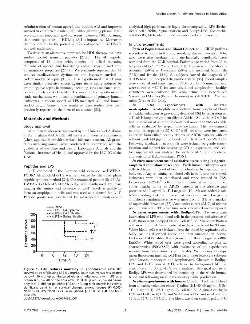
Figure 1. L-4F reduces mortality in endotoxemic rats. Ratsurvival at 24 h following LPS (30 mg/kg, i.p., n = 26) versus rats treatedby L-4F (10 mg/kg) administered either simultaneously with LPS as apremix (i.p., n = 20) or one hour after LPS (L-4F given i.v., n = 26). Salineonly (n = 10) did not get either LPS or L-4F. Log-rank analysis indicates asignificant trend in rat survival changes among groups (P,0.005)*P,0.05 vs. LPS, {P,0.05 vs. Saline (sham), `P,0.05 vs. L-4F one hourpost LPS.doi:10.1371/journal.pone.0064486.g001
Apolipoprotein A-I Mimetic Peptide in Septic ARDS
PLOS ONE | www.plosone.org 2 May 2013 | Volume 8 | Issue 5 | e64486
Apolipoprotein A-I Mimetic Peptide in Septic ARDS
PLOS ONE | www.plosone.org 3 May 2013 | Volume 8 | Issue 5 | e64486
for 20 minutes at 300 g. Collected plasma aliquots were frozen (for
cytokine and endotoxin analysis) or kept at 4uC.Measurement of molecular interaction of L-4F with LPS
using Circular Dichroism spectroscopy. Circular dichroism
(CD) spectroscopy of L-4F 100 mg/ml with and without LPS
1000 mg/ml was performed in PBS. The CD spectra were
recorded using JASCO J-815 CD spectrometer (JASCO model
PTC-423S/15). The CD spectra were measured from 250 to
195 nm every 1 nm with 4 s averaging per point and a 2 nm
bandwidth. A 0.1 cm path length cell was used for obtaining the
spectra. The CD spectra were signal averaged by adding four
scans, baseline corrected, and smoothed. All the CD spectra were
recorded at 37uC. The mean residue ellipticity, [H]MRE (deg cm2
dmol21), was calculated using the following equation: [H]MRE =
(MRW 6 H)/(10cl), where, MRW is the mean residue weight
(molecular weight of the peptide divided by the number of amino
acids in the peptide), H is the observed ellipticity in degrees, c is
the concentration of the peptide in grams per milliliter, and l is the
path length of the cell in centimeters. MRE was measured at
222 nm as previously published [34,38].
Flow cytometry for cd11b detection. Following incubation
with patient serum (see above), neutrophils were washed with PBS
and incubated with BSA-based FACS buffer for 15 min at room
temperature. Neutrophils were again washed with PBS and were
incubated in BSA-based FACS buffer with mouse anti-human
cd11b (activation epitope) allophycocyanin (APC) (eBioscience) for
30 min at 4uC. After washing with PBS, the cells were fixed in 2%
paraformaldehyde. Samples were controlled for by corresponding
negative controls (including unstained samples and mouse IgG1 K
Isotype Control APC, eBioscience). CD11b expression (based on
mean fluorescent intensity) was measured by flow cytometry using
FACS Calibur (Becton Dickinson). Results were normalized to
control as a percent change.
In vivo rat experimentsExperimental design. Adult male Sprague-Dawley rats,
weighing 32268 g (Charles Rivers, Wilmington, MA), were used
throughout the study. Prior to experiments, the rats were
acclimatized for one week in a 12 h light/dark cycle with free
access to food and water. Nonfasted rats underwent either
injection of LPS (Escherichia coli 026:B6, Sigma-Aldrich,
30 mg/kg, i.p.) or Saline (1 ml). One hour after LPS injection,
rats were randomized to receive either L-4F (10 mg/kg, i.v.) or
saline (1 ml). After 6 h, rats were euthanized and either lungs or
bronchoalveolar lavage fluid (BALF) and liver were collected. In a
separate experiment, L-4F was injected either concurrently or one
hour after LPS administration. Cumulative survival was evaluated
at 24 h.
Tissue collection protocol. After ketamine/xylazine anes-
thesia, abdominal cavity and chest were opened and heparinized
blood sample was collected from the right ventricle, followed by
exsanguination via the abdominal aorta. The right liver lobe was
cut and frozen in liquid nitrogen, and then lungs or bronchola-
violar lavage fluid (BALF) was collected. In the lung group, the
cannula was inserted into the lung artery via the right ventricle,
and the lung vascular system was slowly perfused with 10 ml of
cold PBS, followed by perfusion with 10 ml of cold 4% buffered
formalin. The lungs were dissected and placed in 4% buffered
formalin for 18 h. In the BALF group, the cannula was fixed in the
trachea, and the lungs were lavaged gently, via this cannula, to
avoid tissue rupture. Lavage fluid (5 mL) was slowly injected into
the lung using a syringe and then sucked out again. The operation
was repeated 3 times with fresh cold PBS. Total BALF of 15 ml
was collected and centrifuged at 3006 g for 10 min at 4uC. Thesupernatants were kept frozen at 280uC until assayed, and the cell
pellet was resuspended in sterile PBS for cell counting.
Histological analysis. Paraffin-embedded lung and frozen
liver tissues were sectioned at 5 mm and stained with hematoxylin
and eosin for morphological analysis. Sections of lungs (5–8 rats/
group) and liver (4 rats/group) were analyzed using standard
histological techniques and the level of tissue inflammation/
damage was subjectively assessed by blinded experienced pathol-
ogists. Lung microscopy was performed using Zeiss Axio Imager,
M2 microscope equipped with a Zeiss Axio-Cam MRC5 camera.
Color images were acquired with Zeiss 33 software (v.4.8) using
X100 is Cplan-NeoFLUAR 10X/0.3 and X400 is a Plan-
APOCHROMAT 40X/0.95 objectives. Liver microscopy was
performed using an Olympus BX51 microscope equipped with a
Retiga 1300 camera (Q imaging). Color (12-bit) images were
acquired with Bioquant 2012 software using UPlanApo objective
(206, numerical aperture.70). In lung and liver sections, neutro-
phil counts were performed based on the segmented morphology
in high power fields (640) in 30 measurements from 10 different
randomly selected alveolar or portal areas, respectively.
Plasma analysisMeasurements of endotoxin activity. Endotoxin activity in
rat and human samples and in saline mixture of LPS with L-4F or
Sc-4F was measured by limulus amoebocyte lysate (LAL) assay
using kinetic-colormetric test (Endochrome-K, Charles River) in
accordance with manufacturer protocol.
Measurement of plasma cytokines. The concentration of
TNF-a and IL-6 in the human plasma/serum and the concen-
tration of TNF-a, IL-6, and IL-10 in rat plasma was quantified by
using commercially obtained ELISA kits (BD Biosciences) specific
for humans and for rats, respectively.
Measurement of Myeloperoxidase. The CalbiochemHInnoZymeTM Myeloperoxidase Activity Kit (EMD Millipore)
was used to quantify specific active human myeloperoxidase
(MPO). The specific MPO Fluorometric Detection Kit (Enzo Life
Science, Plymouth Meeting, PA. USA) was used to quantify total
MPO in rat plasma. Measurements were performed in accordance
with manufacturer’s protocols.
HDL isolation. Human serum or plasma (50 mL) or rat
plasma (50 mL) was combined with the magnesium/dextran
sulfate reagent (5 mL, Pointe Scientific, Inc., MI, USA). The
mixture was vortexed and incubated for 10 min at room
temperature letting the reagent to precipitate the LDL and VLDL
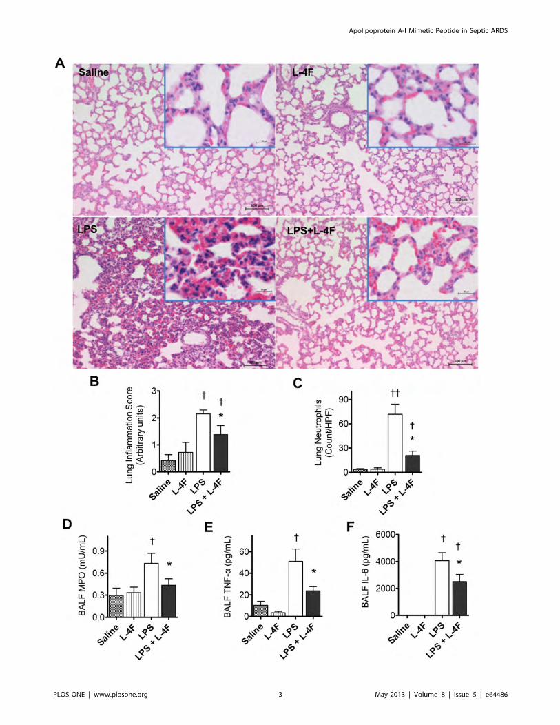
Figure 2. L-4F inhibits lung tissue inflammation in endotoxemic rats. A: Lung H&E stained sections (magnification 6100, the insert is amagnification6630). In saline treated rats, the alveolar walls appear normal and do not contain extensive numbers of polymorphonuclear leukocytes(PMNs). In L-4F treated rats, lung tissue is similar to control, but some PMNs are identified in alveolar capillaries In LPS treated rats, the alveolar wallsare thickened and contain congested capillaries with numerous PMNs. Some of these cells have infiltrated outside the capillaries. In rats treated withL-4F, the lung is less affected than the lungs of rats receiving LPS and is similar to the lungs which received L-4F alone. Scale bars, 100 mm (in inserts,20 mm). B: Blinded analysis of lung inflammation score in 5–8 rat/group. C: Neutrophil counts in lung parenchyma. The vast majority of theneutrophils were found in the septae, and fewer neutrophils were in the alveolar spaces. Neutrophil counts were performed based on the segmentedmorphology in high power fields (640) in 30 measurements from 10 different randomly selected portal areas (n = 5/group). D, E, F: BALF MPO, TNF-a, and IL-6 respectively (n = 4 for controls, n = 8 for LPS groups). *P,0.05 vs. LPS, {P,0.05 and {{P,0.01 vs. Saline and L-4F.doi:10.1371/journal.pone.0064486.g002
Apolipoprotein A-I Mimetic Peptide in Septic ARDS
PLOS ONE | www.plosone.org 4 May 2013 | Volume 8 | Issue 5 | e64486
Apolipoprotein A-I Mimetic Peptide in Septic ARDS
PLOS ONE | www.plosone.org 5 May 2013 | Volume 8 | Issue 5 | e64486
fractions, leaving the HDL fraction in solution. After centrifuga-
tion, HDL fraction was collected. In some human in vitro
experiments, HDL fractions were collected by size exclusion
chromatography using a Bio-Logic Fast Protein Liquid Chroma-
tography system (Bio Rad) as previously described [31,39].
Measurement of HDL-associated paraoxonase-1
activity. Paraoxonase-1 (PON1) activity was determined by
adding HDL or HDL fractions separated by FPLC to the buffer
containing paraoxon (Sigma-Aldrich), and measuring the rate of
release of 4-nitrophenol at 405 nm as previously published
[30,31]. Briefly, 2-mL of serum or HDL fractions were added to
200 mL buffer (100 mmol/L Tris containing 2 mmol/L CaCl2,
pH 8.0) containing paraoxon (1 mmol/L O, O-diethyl-O-p-
nitrophenylphosphate), and the rate of release of 4-nitrophenol
was determined spectrophotometrically. The assay was performed
in a 96-well plate, and readings were taken every 2 minutes at
405 nm. The quantity of 4-nitrophenol formed was calculated
from the molar extinction coefficient of 17 100 mol/L21cm21.
One unit of PON activity was defined as 1 nmol of 4-nitrophenol
formed per minute.
Measurement of serum and HDL cholesterol in
humans. Total serum/plasma and HDL cholesterol was
measured using commercial kit (Thermo Fisher Scientific Inc.).
Liver function test panel in rats. Plasma levels of alanine
transaminase (ALT), aspartate transaminase (AST), alkaline
phosphatase (ALP), total and direct bilirubin were measured by
Animallab (Birmingham, AL) using ACE clinical chemistry system
(Alfa Wassermann Inc.). Plasma triglycerides and total cholesterol
were measured using corresponding commercial kits (Thermo
Scientific).
Measurement of relative change of reactive oxygen
species level in rat plasma. Relative change of reactive
oxygen species level in plasma was measured using a 29,79-
dichlorofluorescin (DCF, Sigma-Aldrich) assay. Rat plasma (5 mL)was diluted in 200 mL of PBS containing 50 mM DCFH and
incubated in 96-well black plates for 1 h at 37uC in dark.
Fluorescence was then measured with excitation and emission
wavelengths of 485 nm and 530 nm, respectively, and then
normalized by total plasma cholesterol.
Measurement of apoA-I level in human serum. Absolute
values of apoA-I levels in patient’s serum were measured using
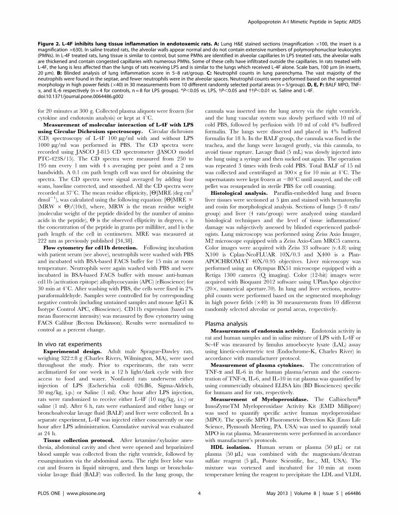
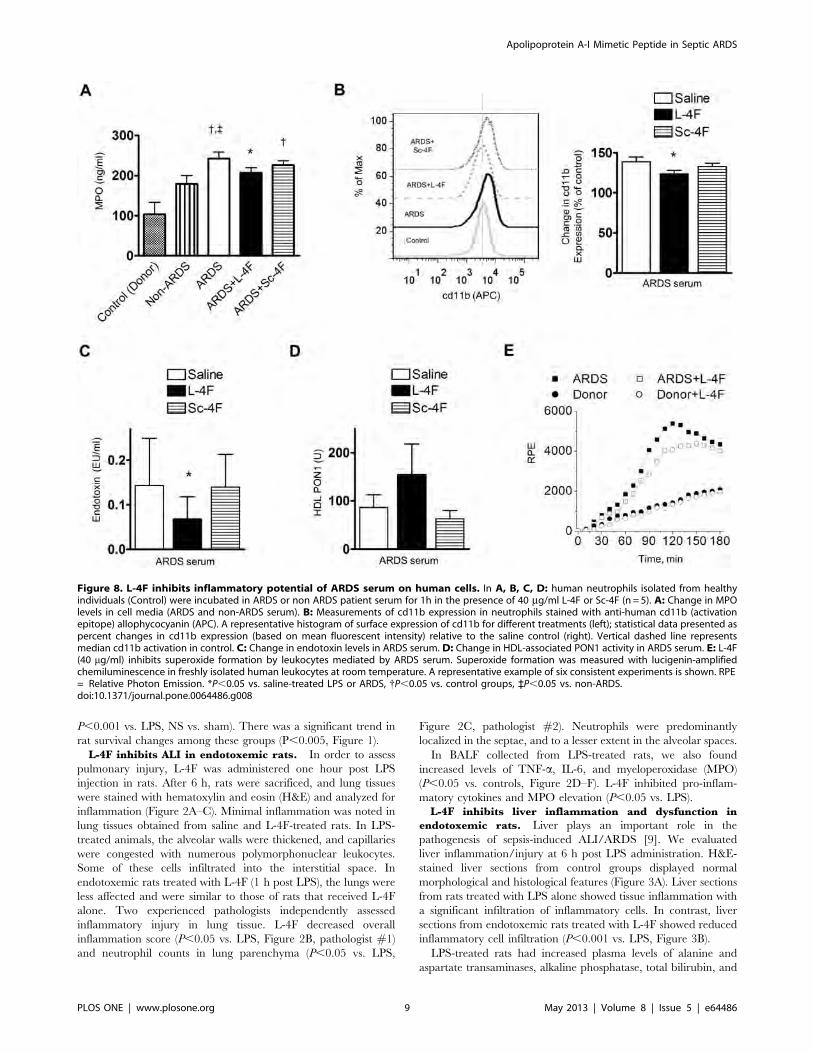
Figure 3. L-4F inhibits liver inflammation in endotoxemic rats. A: Liver sections (620), stained with H&E. Liver sections from saline and L-4F-treated animals showed normal morphological and histological features without signs of inflammation or tissue damage. Liver sections from ratstreated with LPS demonstrated significant infiltration of inflammatory cells, whereas liver sections from rats treated with LPS and L-4F had reducedinfiltration of inflammatory cells. Scale bars, 50 mm. B: Blinded analysis of neutrophil infiltration in liver tissue. Neutrophil counts were performedbased on the segmented morphology in high power fields (640) in 30 measurements from 10 different randomly selected portal areas (n = 4/group).C, D, E, F, G: Plasma ALT, AST, ALP, Total Bilirubin, triglycerides, respectively (n = 8–12/group). *P,0.05 vs. LPS, {P,0.05 vs. Saline and L-4F.doi:10.1371/journal.pone.0064486.g003
Figure 4. L-4F reduces systemic inflammation in endotoxemic rats. Plasma measurements taken at 6 h following LPS injection: A: Endotoxinlevels; B: IL-6 concentration; C: MPO activity; D: Relative levels of reactive oxygen species as measured by DCF assay (normalized to plasmacholesterol); E: Western analysis of relative levels of apoA-I (based on plasma SDS-PAGE gel electrophoresis followed by immunobloting for apoA-I);F: HDL-cholesterol (HDL-C) levels; G: HDL-associated PON1 activity. *P,0.05 vs. LPS, {P,0.05 vs. Saline (n = 8 for controls, n = 12–14 for LPS groups).doi:10.1371/journal.pone.0064486.g004
Apolipoprotein A-I Mimetic Peptide in Septic ARDS
PLOS ONE | www.plosone.org 6 May 2013 | Volume 8 | Issue 5 | e64486
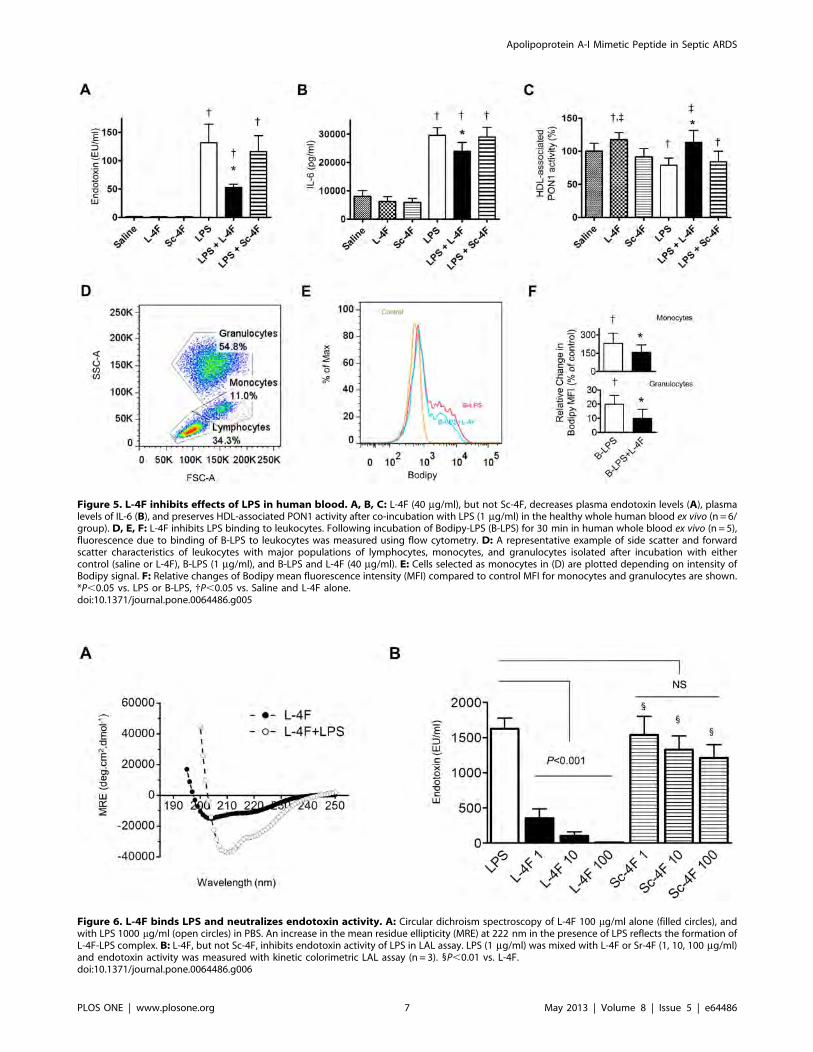
Figure 5. L-4F inhibits effects of LPS in human blood. A, B, C: L-4F (40 mg/ml), but not Sc-4F, decreases plasma endotoxin levels (A), plasmalevels of IL-6 (B), and preserves HDL-associated PON1 activity after co-incubation with LPS (1 mg/ml) in the healthy whole human blood ex vivo (n = 6/group). D, E, F: L-4F inhibits LPS binding to leukocytes. Following incubation of Bodipy-LPS (B-LPS) for 30 min in human whole blood ex vivo (n = 5),fluorescence due to binding of B-LPS to leukocytes was measured using flow cytometry. D: A representative example of side scatter and forwardscatter characteristics of leukocytes with major populations of lymphocytes, monocytes, and granulocytes isolated after incubation with eithercontrol (saline or L-4F), B-LPS (1 mg/ml), and B-LPS and L-4F (40 mg/ml). E: Cells selected as monocytes in (D) are plotted depending on intensity ofBodipy signal. F: Relative changes of Bodipy mean fluorescence intensity (MFI) compared to control MFI for monocytes and granulocytes are shown.*P,0.05 vs. LPS or B-LPS, {P,0.05 vs. Saline and L-4F alone.doi:10.1371/journal.pone.0064486.g005
Figure 6. L-4F binds LPS and neutralizes endotoxin activity. A: Circular dichroism spectroscopy of L-4F 100 mg/ml alone (filled circles), andwith LPS 1000 mg/ml (open circles) in PBS. An increase in the mean residue ellipticity (MRE) at 222 nm in the presence of LPS reflects the formation ofL-4F-LPS complex. B: L-4F, but not Sc-4F, inhibits endotoxin activity of LPS in LAL assay. LPS (1 mg/ml) was mixed with L-4F or Sr-4F (1, 10, 100 mg/ml)and endotoxin activity was measured with kinetic colorimetric LAL assay (n = 3). 1P,0.01 vs. L-4F.doi:10.1371/journal.pone.0064486.g006
Apolipoprotein A-I Mimetic Peptide in Septic ARDS
PLOS ONE | www.plosone.org 7 May 2013 | Volume 8 | Issue 5 | e64486
ELISA kit for human apoA-I (Mabthech USA) in accordance with
manufacturer’s protocol. These absolute apoA-I values were
compared with relative apoA-I values measured based on
immunobloting analysis similar to that described below, except
the patient’s serum and goat anti-human apoA-I antibody
(Brookwood Biomedical, Birmingham, AL) were used. We found
a very strong correlation (r = 0.95, P,0.0001, Pearson) between
relative apoA-I levels measured based on WB data and absolute
values measured by ELISA.
SDS Polyacrylamide Gel Electrophoreisis (SDS-PAGE);
Immunobloting Analysis. Rat plasma aliquot (0.35 ml) was
separated by 4–20% Tris-HEPES-SDS Polyacrylamide gel
(Thermo) at 100 V and transferred to a nitrocellulose membrane
(100 V, 2 h, 4uC). Following transfer, the membrane was blocked
with TBS/3% gelatin for 1 hour and then washed three times
(10 min each) with TBST (10 mM Tris pH 7.40, 150 mM NaCl,
0.1% Tween 20). The membrane was then probed with 0.1 mg/ml
polyclonal HRP-labeled rabbit anti-mouse (cross reactive) apoA-I
antibody (Brookwood Biomedical, Birmingham, AL) for 1 hour at
room temperature, and then washed with TBST. The membrane
was then incubated with 0.1 mg/ml strepavidin alkaline phospha-
tase (Bio Rad) with TBST/1%gelatin for 1h, followed by an
additional wash with TBST first, and then with TBS. Color was
developed with alkaline phosphatase kit (Bio Rad). Reaction was
stopped by washing the membrane in water. After membrane was
air dried, it was optically scanned and apoA-I bands were analyzed
with Image-J Software (NIH, Bethesda, MD).
Statistical AnalysisAll results, unless otherwise specified, are reported as the mean
6 SEM. Statistical analysis was performed using GraphPad Prism
V.4.0.1 (GraphPad Software Inc.). Where appropriate, differences
between groups were assessed by paired Wilcoxon signed rank test
or unpaired Mann-Whitney test (for two experimental groups) or
one-way ANOVA with post-hoc Bonferroni’s Multiple Compar-
ison test (for three or more groups). Pearson correlation was used
for analysis of relationship between HDL-C, apoA-I, and PON1
activity in patient serum. Survival data was assessed by log-rank
analysis (Kaplan-Meier survival method). Differences in survival at
24 h between groups in rat experiments were analyzed with
Fisher’s exact test. A value of P,0.05 was considered to be
statistically significant.
Results
Effects of L-4F in endotoxemic ratsL-4F improves rat survival. Over a 24 h observation
period, there was no mortality in the group of sham rats
(Figure 1). In contrast, administration of 30 mg/kg LPS resulted
in rat mortality, which occurred between the 12 and 24 h time
points in all groups (Figure 1). Among endotoxemic rats, minimal
survival rate at 24 h was observed in saline-treated rats (38%,
P,0.001 vs. sham). L-4F, administered one hour post LPS
injection, significantly improved rat survival to 65% (P,0.05 vs.
LPS, P,0.05 vs. sham). When administered concurrently with
LPS, L-4F virtually prevented rat mortality (90% survival rate,
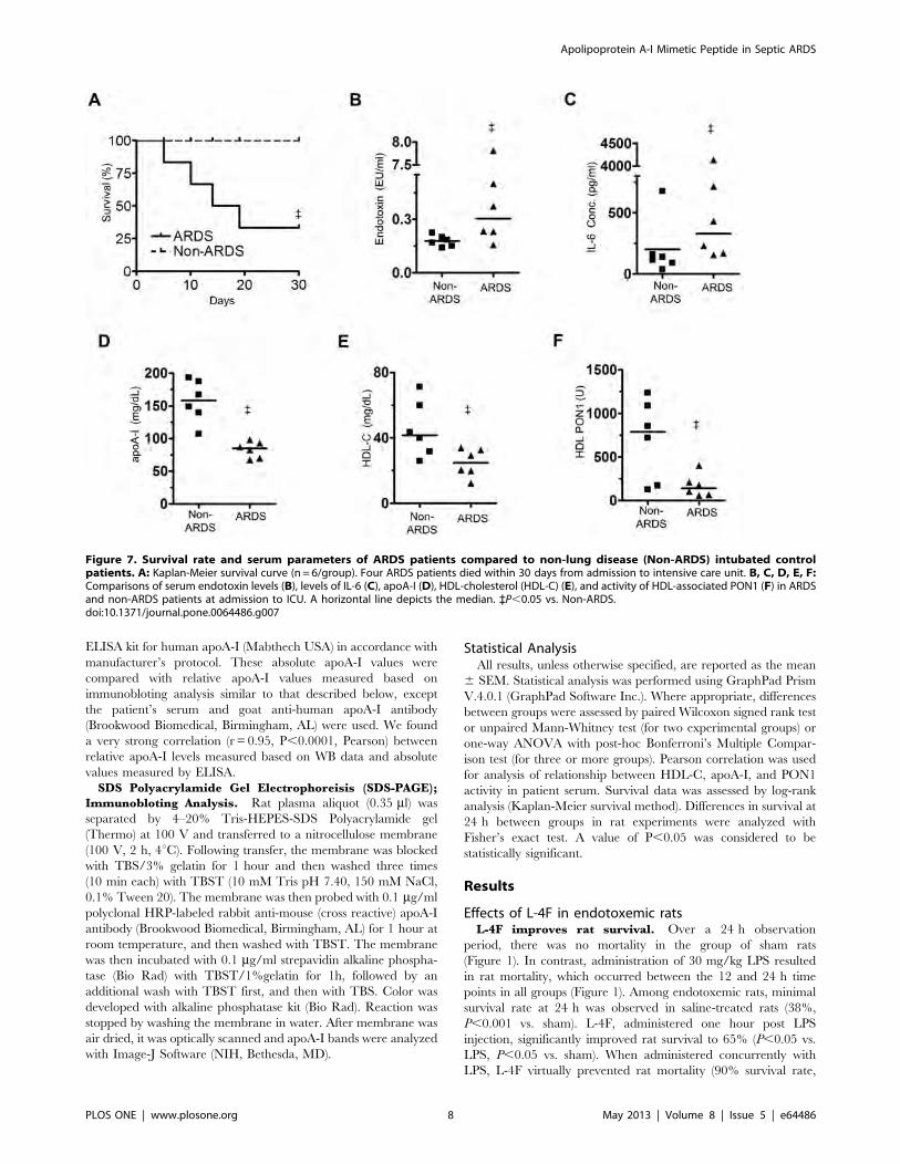
Figure 7. Survival rate and serum parameters of ARDS patients compared to non-lung disease (Non-ARDS) intubated controlpatients. A: Kaplan-Meier survival curve (n = 6/group). Four ARDS patients died within 30 days from admission to intensive care unit. B, C, D, E, F:Comparisons of serum endotoxin levels (B), levels of IL-6 (C), apoA-I (D), HDL-cholesterol (HDL-C) (E), and activity of HDL-associated PON1 (F) in ARDSand non-ARDS patients at admission to ICU. A horizontal line depicts the median. `P,0.05 vs. Non-ARDS.doi:10.1371/journal.pone.0064486.g007
Apolipoprotein A-I Mimetic Peptide in Septic ARDS
PLOS ONE | www.plosone.org 8 May 2013 | Volume 8 | Issue 5 | e64486
P,0.001 vs. LPS, NS vs. sham). There was a significant trend in
rat survival changes among these groups (P,0.005, Figure 1).
L-4F inhibits ALI in endotoxemic rats. In order to assess
pulmonary injury, L-4F was administered one hour post LPS
injection in rats. After 6 h, rats were sacrificed, and lung tissues
were stained with hematoxylin and eosin (H&E) and analyzed for
inflammation (Figure 2A–C). Minimal inflammation was noted in
lung tissues obtained from saline and L-4F-treated rats. In LPS-
treated animals, the alveolar walls were thickened, and capillaries
were congested with numerous polymorphonuclear leukocytes.
Some of these cells infiltrated into the interstitial space. In
endotoxemic rats treated with L-4F (1 h post LPS), the lungs were
less affected and were similar to those of rats that received L-4F
alone. Two experienced pathologists independently assessed
inflammatory injury in lung tissue. L-4F decreased overall
inflammation score (P,0.05 vs. LPS, Figure 2B, pathologist #1)
and neutrophil counts in lung parenchyma (P,0.05 vs. LPS,
Figure 2C, pathologist #2). Neutrophils were predominantly
localized in the septae, and to a lesser extent in the alveolar spaces.
In BALF collected from LPS-treated rats, we also found
increased levels of TNF-a, IL-6, and myeloperoxidase (MPO)
(P,0.05 vs. controls, Figure 2D–F). L-4F inhibited pro-inflam-
matory cytokines and MPO elevation (P,0.05 vs. LPS).
L-4F inhibits liver inflammation and dysfunction in
endotoxemic rats. Liver plays an important role in the
pathogenesis of sepsis-induced ALI/ARDS [9]. We evaluated
liver inflammation/injury at 6 h post LPS administration. H&E-
stained liver sections from control groups displayed normal
morphological and histological features (Figure 3A). Liver sections
from rats treated with LPS alone showed tissue inflammation with
a significant infiltration of inflammatory cells. In contrast, liver
sections from endotoxemic rats treated with L-4F showed reduced
inflammatory cell infiltration (P,0.001 vs. LPS, Figure 3B).
LPS-treated rats had increased plasma levels of alanine and
aspartate transaminases, alkaline phosphatase, total bilirubin, and
Figure 8. L-4F inhibits inflammatory potential of ARDS serum on human cells. In A, B, C, D: human neutrophils isolated from healthyindividuals (Control) were incubated in ARDS or non ARDS patient serum for 1h in the presence of 40 mg/ml L-4F or Sc-4F (n = 5). A: Change in MPOlevels in cell media (ARDS and non-ARDS serum). B: Measurements of cd11b expression in neutrophils stained with anti-human cd11b (activationepitope) allophycocyanin (APC). A representative histogram of surface expression of cd11b for different treatments (left); statistical data presented aspercent changes in cd11b expression (based on mean fluorescent intensity) relative to the saline control (right). Vertical dashed line representsmedian cd11b activation in control. C: Change in endotoxin levels in ARDS serum. D: Change in HDL-associated PON1 activity in ARDS serum. E: L-4F(40 mg/ml) inhibits superoxide formation by leukocytes mediated by ARDS serum. Superoxide formation was measured with lucigenin-amplifiedchemiluminescence in freshly isolated human leukocytes at room temperature. A representative example of six consistent experiments is shown. RPE= Relative Photon Emission. *P,0.05 vs. saline-treated LPS or ARDS, {P,0.05 vs. control groups, `P,0.05 vs. non-ARDS.doi:10.1371/journal.pone.0064486.g008
Apolipoprotein A-I Mimetic Peptide in Septic ARDS
PLOS ONE | www.plosone.org 9 May 2013 | Volume 8 | Issue 5 | e64486
triglycerides (P,0.05 vs. saline, Figure 3C–G). L-4F decreased
plasma transaminases and triglycerides levels in endotoxemic rats
(P,0.05 vs. LPS).
L-4F inhibits systemic inflammation in endotoxemic
rats. Six hours post LPS administration, plasma endotoxin
level was significantly elevated (P,0.05 vs. saline, Figure 4A).
Similarly, plasma levels of IL-6, MPO and reactive oxygen species
as measured by DCF assay was increased, whereas plasma levels of
apoA-I decreased (P,0.05 vs. saline, Figure 4B–E). Although
there was reduction in HDL cholesterol in LPS group, it was not
statistically significant (Figure 4F). However, there was significant
reduction in HDL-associated PON1 activity (P,0.05 vs. saline,
Figure 4G). L-4F decreased plasma endotoxin level in LPS-treated
rats and reversed the effect of LPS on IL-6, apoA-I, and PON1
activity (P,0.05 vs. LPS).
Effects of L-4F ex vivo in the presence of LPSL-4F inhibits LPS-mediated effects in human blood. We
measured activity of L-4F against endotoxin-mediated inflamma-
tory effects in whole human blood. LPS (1 mg/ml) was incubated
in human blood for 12 h and was associated with high level of
endotoxin activity in plasma (Figure 5A). LPS caused a strong
inflammatory response, which was associated with high plasma IL-
6 levels (Figure 5B) and decreased activity of HDL-associated
PON1 (Figure 5C). L-4F, but not control peptide Sc-4F,
significantly decreased endotoxin activity, inhibited plasma IL-6
elevation and preserved activity of HDL-associated PON1
(P,0.05 vs. LPS for all, Figure 5A–C).
L-4F inhibits LPS binding to leukocytes. Since LPS
mediates many of its inflammatory effects via binding to
leukocytes, we measured effects of L-4F on LPS binding/
internalization by leukocytes in human blood using fluorescent
Bodipy-LPS (1 mg/ml) and flow cytometry. Bodipy-LPS demon-
strate biologic effects similar to LPS, which is inhibited by L-4F
(data not shown). Leukocytes isolated from human blood were
gated based on side scatter and forward scatter characteristics
(Figure 5D). Monocytes, incubated with Bodipy-LPS, revealed a
two-fold increase in Bodipy fluorescence compared to controls
(P,0.05 vs. control cells, Figure 5E–F). A similar phenomenon
was observed in granulocytes (Figure 5F). L-4F inhibited Bodipy-
LPS-mediated fluorescence in each cell population (P,0.05 vs.
Bodipy-LPS, Figure 5F).
L-4F binds LPS and decreases endotoxin activity. A
potential physical interaction between LPS and L-4F was
suggested in our previous study as a mechanism to explain the
peptide-mediated inhibition of LPS action [30]. To characterize
the potential physical interaction purely between LPS and L-4F,
we performed circular dichroism spectroscopy of L-4F, alone and
in the presence of LPS, in PBS (Figure 6A). An increase in the
mean residue ellipticity (MRE) at 222 nm in the presence of LPS
(approximately by 150%) reflects higher helical content of L-4F
due to the formation of LPS-L-4F complex.
To test whether LPS, bound to L-4F, decreases endotoxin
activity, we incubated LPS (1 mg/ml) with L-4F (1, 10, and
100 mg/ml) in saline and assessed endotoxin activity using the
limulus amebocyte lysate assay. L-4F reduced endotoxin activity in
a dose-dependent manner (P,0.001 vs. LPS, Figure 6B). At the
same concentrations, Sc-4F did not have significant effect on
endotoxin activity.
Effect of L-4F on inflammatory potential of ARDS patientserum ex vivo
ARDS patient physiological characteristics and
survival. Compared to non-lung disease intubated (non-ARDS)
patients, ARDS patients had lower indices of respiratory function
and higher acute physiology and chronic health evaluation scores
(APACHE II) as shown in Table S1. Transaminase levels were
higher in ARDS likely due to ongoing systemic inflammation
related to sepsis (Table S1). Four ARDS patients (67%) died by
19 days after admission to ICU, whereas no non-ARDS patients
died at least within 30 days (Figure 7A).
Septic inflammation markers in ARDS patient
serum. On the day of intubation, ARDS patient serum had
elevated levels of endotoxin activity and IL-6 compared to non-
ARDS patients (1.55161.253 EU/ml vs. 0.17860.013 EU/ml,
and 973.16638.6 pg/ml vs. 203.1696.17 pg/ml, respectively,
P,0.05 for both, Figure 7B–C). ARDS patients also had lower
levels of total serum cholesterol and PON1 activity than non-
ARDS patients (8167 mg/dL vs. 159617 mg/dL, P,0.005, and
259676 U vs. 10336278 U, P,0.05, respectively). Serum apoA-I
levels were lower in ARDS compared to non-ARDS patients
(P,0.05, Figure 7D). HDL cholesterol (HDL-C) and HDL-
associated PON1 activity (2263 mg/dL vs. 4166 mg/dL,
P,0.05, Figure 7E, and 170662 U vs. 7006183 U, P,0.05,
Figure 7F, respectively) were also reduced.
A strong association between PON1 activity and levels of HDL-
C and apoA-I has been shown in healthy humans [40]. In ARDS
patients, we observed that PON1 activity (standardized to HDL-C
level) was almost two-fold lower than in non-ARDS patients
(7.962.3 vs. 14.563.6, NS). In non-ARDS patients, there was a
strong correlation between levels of HDL-C, HDL-associated
PON1 activity and apoA-I (r = 0.89, P=0.016, and r = 0.88,
P=0.021, respectively, two tailed Pearson). No such relationship
was found in ARDS. This suggests that ARDS is not only
associated with reduced HDL, but also with pathologically
remodeled HDL.
L-4F inhibits neutrophil activation induced by serum
from ARDS patients. Neutrophils are major mediators of
ARDS/ALI. Freshly isolated human neutrophils were incubated
with non-ARDS and with ARDS patient serum, and the efficacy
of L-4F (40 mg/ml) in reducing the inflammatory potential of
ARDS serum was tested. Neutrophil activity was assessed by
measuring the MPO level in cell media. Only ARDS serum
induced a significant increase in MPO levels (P,0.05 vs. control
and non-ARDS), which was inhibited by L-4F but not by Sc-4F
(Figure 8A). We also measured changes in the expression of cd11b
on neutrophil membranes, a marker of neutrophil activation using
flow cytometry (Figure 8B). ARDS serum increased cd11b
expression by 39% compared to non-ARDS controls (Figure 8B).
L-4F added to ARDS serum reduced cd11b expression by
approximately 40% (P,0.05, vs. ARDS), whereas Sc-4F had
minimal effect. These effects of L-4F were accompanied by a
reduction in endotoxin activity in ARDS serum (P,0.05 vs. saline-
treated ARDS, Figure 8C).
L-4F inhibits superoxide formation by leukocytes induced
by ARDS patient serum. Oxidative stress is important in the
pathogenesis of ARDS/ALI. Freshly isolated human leukocytes
were incubated in ARDS patient serum, and superoxide formation
as a marker of oxidative stress was measured using lucigenin-
amplified chemiluminescence. Leukocytes incubated in ARDS
serum caused stronger chemiluminescence than leukocytes incu-
bated in healthy serum (Figure 8E). L-4F in ARDS samples
inhibited the rate as well as peak chemiluminescence emission. L-
4F produced similar effects in all ARDS samples resulting in
decreased superoxide formation (P,0.05 vs. ARDS, n= 6).
Apolipoprotein A-I Mimetic Peptide in Septic ARDS
PLOS ONE | www.plosone.org 10 May 2013 | Volume 8 | Issue 5 | e64486
Discussion
Treatment options for ARDS patients are limited and generally
rely on supportive therapies to maintain adequate oxygenation
and hemodynamic status. Despite the recognition that HDL and
apoA-I modulate immune cell function, their role in reducing
complications associated with ARDS is not clear. Herein, we
demonstrate that the 18 amino acid residue apoA-I mimetic
peptide L-4F, which bears a similar class A amphipathic structure
as that found in helical repeats of apoA-I, interacts with LPS and
prevents activation of leukocytes or neutrophils by LPS and septic
ARDS serum. In a clinically relevant model, L-4F inhibits lung
and liver injury and improves mortality in rodents by inhibiting
endotoxin activity and by improving the functional properties of
HDL. Our study therefore provides the rationale for the clinical
testing of the L-4F peptide in sepsis-associated ARDS.
Several mechanisms are involved in beneficial effects of L-4F in
our study. First, L-4F directly binds to and neutralizes LPS.
Formation of L-4F-LPS complexes has been suggested by us
previously [30]. Here we demonstrate that L-4F can directly
interact with LPS. We also demonstrate that L-4F directly
decreases the endotoxin activity of LPS. LPS mediates many of
its inflammatory effects via binding to leukocytes and activating
Toll-like receptors [2,3]. Further, we demonstrate that L-4F
decreases LPS binding to leukocytes. This effect is likely mediated
by high affinity of L-4F to LPS. L-4F competes with lipopolysac-
charide-binding protein for LPS binding as shown by us previously
[30].
Another mechanism of L-4F is via its effects on HDL. In serum
of sepsis-mediated ARDS patients, we found high levels of
endotoxin and low levels of apoA-I and HDL. Increasing plasma
apoA-I or HDL has been shown to reduce endotoxemia-associated
complications [24,26], and so did L-4F in our present study and in
previous experiments [31]. HDL neutralizes LPS by binding to
LPS and mobilizing LPS to the liver for its degradation and
excretion [14,23,24]. It has been suggested that L-4F facilitates
LPS binding to HDL in endotoxemic rats [31]. In addition, HDL
via apoA-I down-regulates CD14 and CD11b expression in
monocytes and neutrophils [23,41]. L-4F has been recently shown
to produce similar effects in human monocyte-derived macro-
phages [42] and down-regulates cell-surface TLRs in these cells
[43].
We also found that the structural and functional integrity of
HDL in ARDS was compromised. In our rodent model of ARDS,
plasma apoA-I was severely reduced. LPS induces acute inflam-
mation/injury in rat liver which is the major site for apoA-I
biosynthesis [44]. Reduction of apoA-I synthesis in the liver likely
occurs through LPS-mediated NF-kB activation [45]. L-4F
treatment significantly reduces hepatic inflammation. Further,
apoA-I is a selective target for MPO-catalyzed oxidation, which
leads to pro-inflammatory HDL [46]. LPS causes an increase in
MPO activity which is prevented by L-4F. L-4F and apoA-I share
structural and functional properties, it has been recently shown
that 4F can serve as a reactive substrate for MPO-derived
hypochlorous acid [47].
ARDS is associated with increased oxidant stress. HDL is a
major antioxidant due to presence of apoA-I and PON1 as its
constituents [20,48]. PON1 protects lipoproteins and plasma
membranes against oxidation by hydrolyzing lipid peroxides
[49,50], and reduces oxidative cytotoxicity [20]. PON1 levels
and activity is reduced in acute inflammatory state, which
decreases overall HDL antioxidant activity [20] and leads to
inability of acute-phase HDL to protect against lipid oxidation
[18,20]. Our results indicate that L-4F preserves HDL-associated
PON-1 activity and therefore facilitates PON1-mediated reduction
in lipid hydroperoxides. Other investigators including a murine
model of asthma have previously demonstrated the ability of L-4F
to promote anti-inflammatory effects of HDL, including increases
in PON1 [51–53].
In the current study, we found significant mortality reduction in
L-4F-treated rats even when it is administered 1 hour post LPS.
This is associated with significant reduction in hepatic and
pulmonary inflammation. Similar to our results, another recent
study also found improvement in mortality and ALI after L-4F
treatment in endotoxemic rats [54]. This was associated with
stimulation of pulmonary sphingosine-1-phosphate receptor 1,
activation of Akt and down regulation of NF-kB pathway [54].
Our data though suggest that major protective mechanisms of L-
4F in LPS-mediated inflammation and ALI in this [54] and our
models include inhibition of endotoxin activity by direct interac-
tion with the bacterial lipopolysaccharide along with a preserva-
tion of HDL function.
Our study has a number of limitations. The rodent model of
LPS-mediated endotoxemia reproduces only partially the patho-
physiological mechanisms of septic inflammation and organ injury
(including ALI/ARDS) induced by gram-negative bacteria in
humans [55]. Nevertheless, LPS-related model is extensively used
and could be considered as fast and reliable approach for the
‘‘proof-of-concept’’ or new drug screening. Another limitation is
related to short period of follow-up in mortality study. The death
of L-4F-treated rats could hypothetically be delayed. In our
preliminary experiment (n = 6/group) with 10-days follow-up, no
rat died after 24 h post LPS in saline-treated and L-4F-treated
groups. This is consistent with the results of a recent 72-hours
follow-up study performed in relatively similar conditions [54]. In
the present study, we used only single injection of L-4F. Since L-4F
is non-toxic and rapidly metabolized in the organism, repeated L-
4F administrations in septic animals would potentially produce
even better anti-inflammatory effects and this option could be
tested in future research. Another study limitation is related to lack
of Sc-4F treatment group in our in vivo experiments. However, a
number of laboratories reported the failure of scrambled form of
4F to inhibit effects of LPS in in vivo experiment [31] as well as to
reproduce anti-inflammatory and anti-oxidant effects of 4F in
apoE null mice [56] and in murine model of asthma [53].
Despite an improved understanding of ARDS over the past
30 years, the impact on modulating disease-related outcomes has
been relatively modest [57]. To date, the only therapy which has
shown impact on ARDS-related mortality has been low-tidal
volume ventilation [58]. Current 60-day mortality rates for ARDS
patients are approximately 35%, although those individuals with
sepsis as an underlying cause demonstrate increased mortality
rates (45–60%) [59]. Similarly, our cohort of sepsis-related ARDS
patients demonstrated increased mortality despite an appropriate
ventilator strategy. For these individuals, the need for a specific
disease-modifying therapeutic compound is critical, and L-4F
peptide may be one such potential agent. Acceptable safety profile
for paranteral and oral forms of 4F has been recently shown in
selected patient groups with high-risk cardiovascular disease
[60,61]. Using L-4F in ARDS would likely provide beneficial
effects on both inflammatory and oxidative pathogenic pathways
of ARDS. Our data showing improvement in ALI indices and
survival in endotoxemic rats along with the successful ex vivo LPS
neutralization in ARDS patient serum by L-4F peptide strongly
suggest the therapeutic potential of this peptide in early sepsis-
related ARDS.
Apolipoprotein A-I Mimetic Peptide in Septic ARDS
PLOS ONE | www.plosone.org 11 May 2013 | Volume 8 | Issue 5 | e64486
Supporting Information
Table S1 Patients characteristics on admission to ICU.(TIF)
Acknowledgments
The authors would like to thank Dr. Victor J. Thannickal for helpful
discussion of the manuscript and Dr. David W. Garber for assistance with
FPLC system.
GMA is a Principal in Bruin Pharma, a start-up Biotech company.
Author Contributions
Conceived and designed the experiments: OFS AG CRW GMA HG.
Performed the experiments: OFS XX JH MNP. Analyzed the data: OFS
XX WEG SHL VKM GMA HG. Contributed reagents/materials/
analysis tools: MNP GMA. Wrote the paper: OFS AG VKM CRW GMA
HG.
References
1. Rubenfeld GD, Caldwell E, Peabody E, Weaver J, Martin DP, et al. (2005)
Incidence and outcomes of acute lung injury. N Engl J Med 353: 1685–1693.
2. Cohen J (2002) The immunopathogenesis of sepsis. Nature 420: 885–891.
3. Munford RS (2006) Severe sepsis and septic shock: the role of gram-negative
bacteremia. Annu Rev Pathol 1: 467–496.
4. Bannerman DD, Goldblum SE (2003) Mechanisms of bacterial lipopolysaccha-ride-induced endothelial apoptosis. American journal of physiology Lung
cellular and molecular physiology 284: L899–914.
5. Baumgarten G, Knuefermann P, Schuhmacher G, Verveolgyi V, von RappardJ, et al. (2006) Toll-like receptor 4, nitric oxide, and myocardial depression in
endotoxemia. Shock (Augusta, Ga) 25: 43–49.
6. Guha M, Mackman N (2001) LPS induction of gene expression in humanmonocytes. Cellular signalling 13: 85–94.
7. Calkins CM, Heimbach JK, Bensard DD, Song Y, Raeburn CD, et al. (2001)
TNF receptor I mediates chemokine production and neutrophil accumulation inthe lung following systemic lipopolysaccharide. J Surg Res 101: 232–237.
8. Nowak M, Gaines GC, Rosenberg J, Minter R, Bahjat FR, et al. (2000) LPS-
induced liver injury in D-galactosamine-sensitized mice requires secreted TNF-alpha and the TNF-p55 receptor. Am J Physiol Regul Integr Comp Physiol 278:
R1202–1209.
9. Siore AM, Parker RE, Stecenko AA, Cuppels C, McKean M, et al. (2005)Endotoxin-induced acute lung injury requires interaction with the liver.
Am J Physiol Lung Cell Mol Physiol 289: L769–776.
10. Meduri GU, Headley S, Kohler G, Stentz F, Tolley E, et al. (1995) Persistent
elevation of inflammatory cytokines predicts a poor outcome in ARDS. Plasma
IL-1 beta and IL-6 levels are consistent and efficient predictors of outcome overtime. Chest 107: 1062–1073.
11. Damas P, Ledoux D, Nys M, Vrindts Y, De Groote D, et al. (1992) Cytokine
serum level during severe sepsis in human IL-6 as a marker of severity. Ann Surg215: 356–362.
12. Bouros D, Alexandrakis MG, Antoniou KM, Agouridakis P, Pneumatikos I, et
al. (2004) The clinical significance of serum and bronchoalveolar lavageinflammatory cytokines in patients at risk for Acute Respiratory Distress
Syndrome. BMC Pulm Med 4: 6.
13. Remick DG, Bolgos GR, Siddiqui J, Shin J, Nemzek JA (2002) Six at six:interleukin-6 measured 6 h after the initiation of sepsis predicts mortality over 3
days. Shock 17: 463–467.
14. Read TE, Harris HW, Grunfeld C, Feingold KR, Calhoun MC, et al. (1993)
Chylomicrons enhance endotoxin excretion in bile. Infect Immun 61: 3496–
3502.
15. Kitchens RL, Thompson PA, Munford RS, O’Keefe GE (2003) Acute
inflammation and infection maintain circulating phospholipid levels and
enhance lipopolysaccharide binding to plasma lipoproteins. J Lipid Res 44:2339–2348.
16. Feingold KR, Funk JL, Moser AH, Shigenaga JK, Rapp JH, et al. (1995) Role
for circulating lipoproteins in protection from endotoxin toxicity. Infect Immun63: 2041–2046.
17. Levels JH, Abraham PR, van Barreveld EP, Meijers JC, van Deventer SJ (2001)
Distribution and kinetics of lipoprotein-bound lipoteichoic acid. Infect Immun71: 3280–3284.
18. van Leeuwen HJ, Heezius EC, Dallinga GM, van Strijp JA, Verhoef J, et al.(2003) Lipoprotein metabolism in patients with severe sepsis. Crit Care Med 31:
1359–1366.
19. Van Lenten BJ, Hama SY, de Beer FC, Stafforini DM, McIntyre TM, et al.(1995) Anti-inflammatory HDL becomes pro-inflammatory during the acute
phase response. Loss of protective effect of HDL against LDL oxidation in aortic
wall cell cocultures. J Clin Invest 96: 2758–2767.
20. Wu A, Hinds CJ, Thiemermann C (2004) High-density lipoproteins in sepsis and
septic shock: metabolism, actions, and therapeutic applications. Shock 21: 210–
221.
21. Novak F, Vavrova L, Kodydkova J, Novak F, Sr., Hynkova M, et al. (2010)
Decreased paraoxonase activity in critically ill patients with sepsis. Clin Exp Med
10: 21–25.
22. Draganov D, Teiber J, Watson C, Bisgaier C, Nemzek J, et al. (2010) PON1 and
oxidative stress in human sepsis and an animal model of sepsis. Adv Exp MedBiol 660: 89–97.
23. Pajkrt D, Doran JE, Koster F, Lerch PG, Arnet B, et al. (1996)
Antiinflammatory effects of reconstituted high-density lipoprotein during humanendotoxemia. J Exp Med 184: 1601–1608.
24. Levine DM, Parker TS, Donnelly TM, Walsh A, Rubin AL (1993) In vivo
protection against endotoxin by plasma high density lipoprotein. Proc Natl AcadSci U S A 90: 12040–12044.
25. McDonald MC, Dhadly P, Cockerill GW, Cuzzocrea S, Mota-Filipe H, et al.
(2003) Reconstituted high-density lipoprotein attenuates organ injury andadhesion molecule expression in a rodent model of endotoxic shock. Shock 20:
551–557.
26. Yan YJ, Li Y, Lou B, Wu MP (2006) Beneficial effects of ApoA-I on LPS-induced acute lung injury and endotoxemia in mice. Life Sci 79: 210–215.
27. Garber DW, Datta G, Chaddha M, Palgunachari MN, Hama SY, et al. (2001) A
new synthetic class A amphipathic peptide analogue protects mice from diet-induced atherosclerosis. J Lipid Res 42: 545–552.
28. Navab M, Anantharamaiah GM, Reddy ST, Van Lenten BJ, Datta G, et al.
(2004) Human apolipoprotein A-I and A-I mimetic peptides: potential foratherosclerosis reversal. Curr Opin Lipidol 15: 645–649.
29. Van Lenten BJ, Wagner AC, Jung CL, Ruchala P, Waring AJ, et al. (2008) Anti-
inflammatory apoA-I-mimetic peptides bind oxidized lipids with much higher
affinity than human apoA-I. J Lipid Res 49: 2302–2311.
30. Gupta H, Dai L, Datta G, Garber DW, Grenett H, et al. (2005) Inhibition of
lipopolysaccharide-induced inflammatory responses by an apolipoprotein AI
mimetic peptide. Circ Res 97: 236–243.
31. Dai L, Datta G, Zhang Z, Gupta H, Patel R, et al. (2010) The apolipoprotein A-
I mimetic peptide 4F prevents defects in vascular function in endotoxemic rats.
J Lipid Res 51: 2695–2705.
32. Zhang Z, Datta G, Zhang Y, Miller AP, Mochon P, et al. (2009) Apolipoprotein
A-I mimetic peptide treatment inhibits inflammatory responses and improves
survival in septic rats. Am J Physiol Heart Circ Physiol 297: H866–873.
33. Sharifov OF, Xu X, Gaggar A, Grizzle WE, Palgunachari MN, et al. (2012)
ApoA-I Mimetic Peptide, L-4F Inhibits Endotoxin-Induced Acute Lung Injury
And Systemic Inflammation by Inhibiting Neutrophil Activation. Am J RespirCrit Care Med 185: A2213.
34. Datta G, Chaddha M, Hama S, Navab M, Fogelman AM, et al. (2001) Effects of
increasing hydrophobicity on the physical-chemical and biological properties ofa class A amphipathic helical peptide. Journal of lipid research 42: 1096–1104.
35. Fowler AA, Hamman RF, Zerbe GO, Benson KN, Hyers TM (1985) Adult
respiratory distress syndrome. Prognosis after onset. Am Rev Respir Dis 132:472–478.
36. Xu X, Jackson PL, Tanner S, Hardison MT, Abdul Roda M, et al. (2011) A self-
propagating matrix metalloprotease-9 (MMP-9) dependent cycle of chronicneutrophilic inflammation. PLoS One 6: e15781.
37. Iovine NM, Elsbach P, Weiss J (1997) An opsonic function of the neutrophil
bactericidal/permeability-increasing protein depends on both its N- and C-terminal domains. Proc Natl Acad Sci U S A 94: 10973–10978.
38. Mishra VK, Palgunachari MN, Datta G, Phillips MC, Lund-Katz S, et al. (1998)
Studies of synthetic peptides of human apolipoprotein A-I containing tandemamphipathic alpha-helixes. Biochemistry 37: 10313–10324.
39. Garber DW, Kulkarni KR, Anantharamaiah GM (2000) A sensitive and
convenient method for lipoprotein profile analysis of individual mouse plasmasamples. J Lipid Res 41: 1020–1026.
40. Blatter Garin MC, Moren X, James RW (2006) Paraoxonase-1 and serum
concentrations of HDL-cholesterol and apoA-I. J Lipid Res 47: 515–520.
41. Murphy AJ, Woollard KJ, Suhartoyo A, Stirzaker RA, Shaw J, et al. (2011)Neutrophil activation is attenuated by high-density lipoprotein and apolipopro-
tein A-I in in vitro and in vivo models of inflammation. Arterioscler ThrombVasc Biol 31: 1333–1341.
42. Smythies LE, White CR, Maheshwari A, Palgunachari MN, Anantharamaiah
GM, et al. (2010) Apolipoprotein A-I mimetic 4F alters the function of humanmonocyte-derived macrophages. Am J Physiol Cell Physiol 298: C1538–1548.
43. White CR, Smythies LE, Crossman DK, Palgunachari MN, Anantharamaiah
GM, et al. (2012) Regulation of Pattern Recognition Receptors by theApolipoprotein A-I Mimetic Peptide 4F. Arterioscler Thromb Vasc Biol 32:
2631–2639.
44. Haddad IA, Ordovas JM, Fitzpatrick T, Karathanasis SK (1986) Linkage,evolution, and expression of the rat apolipoprotein A-I, C-III, and A-IV genes.
J Biol Chem 261: 13268–13277.
45. Morishima A, Ohkubo N, Maeda N, Miki T, Mitsuda N (2003) NFkappaBregulates plasma apolipoprotein A-I and high density lipoprotein cholesterol
through inhibition of peroxisome proliferator-activated receptor alpha. J Biol
Chem 278: 38188–38193.
Apolipoprotein A-I Mimetic Peptide in Septic ARDS
PLOS ONE | www.plosone.org 12 May 2013 | Volume 8 | Issue 5 | e64486
46. Undurti A, Huang Y, Lupica JA, Smith JD, DiDonato JA, et al. (2009)
Modification of high density lipoprotein by myeloperoxidase generates a pro-
inflammatory particle. J Biol Chem 284: 30825–30835.
47. White CR, Datta G, Buck AK, Chaddha M, Reddy G, et al. (2012) Preservation
of biological function despite oxidative modification of the apolipoprotein A-I
mimetic peptide 4F. J Lipid Res 53: 1576–1587.
48. Bashtovyy D, Jones MK, Anantharamaiah GM, Segrest JP (2011) Sequence
conservation of apolipoprotein A-I affords novel insights into HDL structure-
function. J Lipid Res 52: 435–450.
49. Aviram M, Rosenblat M, Bisgaier CL, Newton RS, Primo-Parmo SL, et al.
(1998) Paraoxonase inhibits high-density lipoprotein oxidation and preserves its
functions. A possible peroxidative role for paraoxonase. J Clin Invest 101: 1581–
1590.
50. Ferretti G, Bacchetti T, Masciangelo S, Bicchiega V (2010) HDL-paraoxonase
and membrane lipid peroxidation: a comparison between healthy and obese
subjects. Obesity (Silver Spring) 18: 1079–1084.
51. Imaizumi S, Grijalva V, Navab M, Van Lenten BJ, Wagner AC, et al. (2010) L-
4F differentially alters plasma levels of oxidized fatty acids resulting in more anti-
inflammatory HDL in mice. Drug Metab Lett 4: 139–148.
52. Vakili L, Hama S, Kim JB, Tien D, Safarpoor S, et al. (2010) The Effect of HDL
Mimetic Peptide 4F on PON1. Adv Exp Med Biol 660: 167–172.
53. Nandedkar SD, Weihrauch D, Xu H, Shi Y, Feroah T, et al. (2011) D-4F, an
apoA-1 mimetic, decreases airway hyperresponsiveness, inflammation, and
oxidative stress in a murine model of asthma. J Lipid Res 52: 499–508.
54. Kwon WY, Suh GJ, Kim KS, Kwak YH, Kim K (2012) 4F, apolipoprotein AI
mimetic peptide, attenuates acute lung injury and improves survival inendotoxemic rats. J Trauma Acute Care Surg 72: 1576–1583.
55. Floer M, Gotte M, Wild MK, Heidemann J, Gassar ES, et al. (2010) Enoxaparin
improves the course of dextran sodium sulfate-induced colitis in syndecan-1-deficient mice. Am J Pathol 176: 146–157.
56. Navab M, Anantharamaiah GM, Reddy ST, Hama S, Hough G, et al. (2004)Oral D-4F causes formation of pre-beta high-density lipoprotein and improves
high-density lipoprotein-mediated cholesterol efflux and reverse cholesterol
transport from macrophages in apolipoprotein E-null mice. Circulation 109:3215–3220.
57. Ware LB, Matthay MA (2000) The acute respiratory distress syndrome.N Engl J Med 342: 1334–1349.
58. ARDS-Network (2000) Ventilation with lower tidal volumes as compared withtraditional tidal volumes for acute lung injury and the acute respiratory distress
syndrome. The Acute Respiratory Distress Syndrome Network. N Engl J Med
342: 1301–1308.59. Gaggar A, Olman MA (2006) Biologic markers of mortality in acute lung injury.
Clin Chim Acta 372: 24–32.60. Bloedon LT, Dunbar R, Duffy D, Pinell-Salles P, Norris R, et al. (2008) Safety,
pharmacokinetics, and pharmacodynamics of oral apoA-I mimetic peptide D-4F
in high-risk cardiovascular patients. J Lipid Res 49: 1344–1352.61. Watson CE, Weissbach N, Kjems L, Ayalasomayajula S, Zhang Y, et al. (2011)
Treatment of patients with cardiovascular disease with L-4F, an apo-A1mimetic, did not improve select biomarkers of HDL function. J Lipid Res 52:
361–373.
Apolipoprotein A-I Mimetic Peptide in Septic ARDS
PLOS ONE | www.plosone.org 13 May 2013 | Volume 8 | Issue 5 | e64486