Embed Size (px)
Citation preview
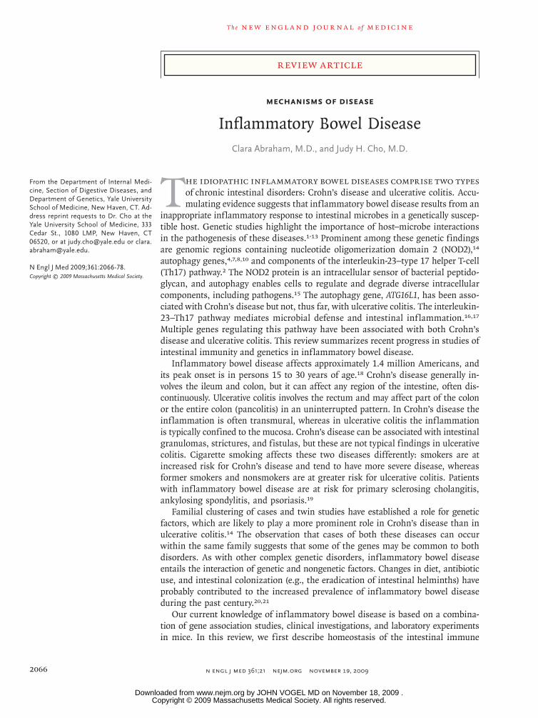
review article
T h e n e w e ngl a nd j o u r na l o f m e dic i n e
n engl j med 361;21 nejm.org november 19, 20092066
Mechanisms of Disease
Inflammatory Bowel DiseaseClara Abraham, M.D., and Judy H. Cho, M.D.
From the Department of Internal Medi-cine, Section of Digestive Diseases, and Department of Genetics, Yale University School of Medicine, New Haven, CT. Ad-dress reprint requests to Dr. Cho at the Yale University School of Medicine, 333 Cedar St., 1080 LMP, New Haven, CT 06520, or at [email protected] or [email protected].
N Engl J Med 2009;361:2066-78.Copyright © 2009 Massachusetts Medical Society.
The idiopathic inflammatory bowel diseases comprise two types of chronic intestinal disorders: Crohn’s disease and ulcerative colitis. Accu-mulating evidence suggests that inflammatory bowel disease results from an
inappropriate inflammatory response to intestinal microbes in a genetically suscep-tible host. Genetic studies highlight the importance of host–microbe interactions in the pathogenesis of these diseases.1-13 Prominent among these genetic findings are genomic regions containing nucleotide oligomerization domain 2 (NOD2),14 autophagy genes,4,7,8,10 and components of the interleukin-23–type 17 helper T-cell (Th17) pathway.2 The NOD2 protein is an intracellular sensor of bacterial peptido-glycan, and autophagy enables cells to regulate and degrade diverse intracellular components, including pathogens.15 The autophagy gene, ATG16L1, has been asso-ciated with Crohn’s disease but not, thus far, with ulcerative colitis. The interleukin-23–Th17 pathway mediates microbial defense and intestinal inflammation.16,17 Multiple genes regulating this pathway have been associated with both Crohn’s disease and ulcerative colitis. This review summarizes recent progress in studies of intestinal immunity and genetics in inflammatory bowel disease.
Inflammatory bowel disease affects approximately 1.4 million Americans, and its peak onset is in persons 15 to 30 years of age.18 Crohn’s disease generally in-volves the ileum and colon, but it can affect any region of the intestine, often dis-continuously. Ulcerative colitis involves the rectum and may affect part of the colon or the entire colon (pancolitis) in an uninterrupted pattern. In Crohn’s disease the inflammation is often transmural, whereas in ulcerative colitis the inflammation is typically confined to the mucosa. Crohn’s disease can be associated with intestinal granulomas, strictures, and fistulas, but these are not typical findings in ulcerative colitis. Cigarette smoking affects these two diseases differently: smokers are at increased risk for Crohn’s disease and tend to have more severe disease, whereas former smokers and nonsmokers are at greater risk for ulcerative colitis. Patients with inflammatory bowel disease are at risk for primary sclerosing cholangitis, ankylosing spondylitis, and psoriasis.19
Familial clustering of cases and twin studies have established a role for genetic factors, which are likely to play a more prominent role in Crohn’s disease than in ulcerative colitis.14 The observation that cases of both these diseases can occur within the same family suggests that some of the genes may be common to both disorders. As with other complex genetic disorders, inflammatory bowel disease entails the interaction of genetic and nongenetic factors. Changes in diet, antibiotic use, and intestinal colonization (e.g., the eradication of intestinal helminths) have probably contributed to the increased prevalence of inflammatory bowel disease during the past century.20,21
Our current knowledge of inflammatory bowel disease is based on a combina-tion of gene association studies, clinical investigations, and laboratory experiments in mice. In this review, we first describe homeostasis of the intestinal immune
Copyright © 2009 Massachusetts Medical Society. All rights reserved. Downloaded from www.nejm.org by JOHN VOGEL MD on November 18, 2009 .
Mechanisms of Disease
n engl j med 361;21 nejm.org november 19, 2009 2067
system in health and then focus on advances in our understanding of how genetic alterations in this system contribute to the development of inflammatory bowel disease.
The In tes tina l Immune S ys tem
The Intestinal Microbiome and Inflammatory Bowel Disease
The intestinal microbiome consists of the micro-organisms that inhabit the gut. The intraluminal microbiota affects the development of the intes-tinal immune system, supplies key nutrients, and modulates energy metabolism.22 The intestinal microbiota is acquired at birth but changes rapidly during the first year of life. In adults, each per-son’s unique population of fecal microbiota is fairly stable over time, but fluctuations occur in response to environmental and developmental factors and in disease.21,23,24
Host–microbiome interactions can be mutual-ly beneficial or can be deleterious, inciting intes-tinal inflammation. Observations in patients with inflammatory bowel disease and in animal mod-els point to the role of bacteria in such inflam-mation. For example, antibiotics are effective in some patients with inflammatory bowel disease, and most mouse models of colitis require intes-tinal bacteria for inflammation to occur.25 Bac-teria that can adhere to and invade the intestinal mucosa may be particularly important, as in the case of Escherichia coli.26 Although a number of specific pathogens have been incriminated in the development of inflammatory bowel disease, none have been confirmed as causal; rather, microbial antigens that are normally present in the intes-tinal lumen seem to drive inflammation in the gut. As compared with control subjects, patients with Crohn’s disease and those with ulcerative colitis have depletion and reduced diversity of members of the mucosa-associated phyla Firmi-cutes and Bacteroidetes.21,27 Whether these altera-tions contribute to the disease or merely reflect secondary changes caused by the inflammation is not known.
The Intestinal Epithelium
The intestinal epithelium at the interface between the intestinal microbiome and the lymphoid tissue associated with the gastrointestinal system plays a critical role in shaping the mucosal immune response. Intestinal epithelial cells are a physical
barrier against excessive entry of bacteria and other antigens from the intestinal lumen into the circulation. An intact mucosal barrier depends on intercellular junctions, which help to seal the space between adjacent epithelial cells (the para-cellular space), and tight junctions, which are the key elements of the seal.28 In inflammatory bowel disease, the paracellular space has increased per-meability, and the regulation of tight junctions is defective.28 These abnormalities may be due to a primary defect in barrier function or may be an outcome of inflammation.28-32
Additional defenses against bacterial invasion consist of specialized epithelial cells, including goblet cells and Paneth cells. Goblet cells regulate the production of mucus and factors that con-tribute to epithelial repair and regulation of in-flammation.33,34 Paneth cells secrete antimicro-bial peptides such as α-defen sins. Intestinal mucus overlies the epithelium, thereby limiting contact between bacteria and epithelial cells. Epithelial regeneration and repair serve to control and ul-timately resolve the inflammatory response to in-jury. In inflammatory bowel disease, however, the inflammatory response often results in continued epithelial injury, which causes erosions, ulcer-ations, and a decrease in the production of de-fensin.35,36 The result is increased exposure to intestinal microbiota and amplification of the in-flammatory response.
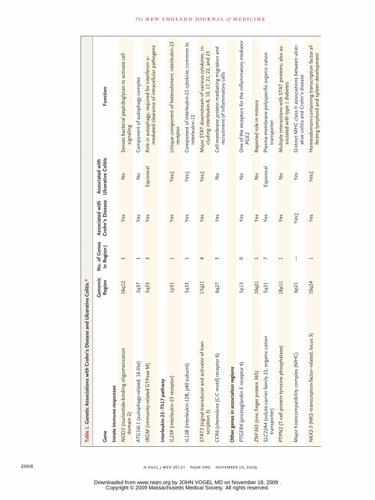
In mouse models of inflammatory bowel dis-ease, several types of epithelial dysfunction can cause intestinal inflammation. These include de-fects in epithelial-cell development or prolifera-tion, barrier function, cell-matrix adhesion, endo-plasmic reticulum stress, and epithelial restitution after injury.28,37,38 Prostaglandin E receptor 4 (EP4) contributes to mucosal repair and barrier func-tion; in mice that are deficient in EP4, the colitis that develops in response to chemical injury is more severe than that in wild-type animals.39 Polymorphisms in proximity to the gene encod-ing EP4 (PTGER4) were recently implicated in Crohn’s disease in humans6 (Table 1). In mice in which the gene encoding MUC2, a major mucin component, has been deleted, intestinal inflam-mation develops41; moreover, a variant in a ge-nomic region that includes the gene encoding MUC19 has been associated with Crohn’s dis-ease.2 Endoplasmic reticulum stress (a cellular response to various environmental changes) is increased in inflamed intestinal epithelial cells,42
Copyright © 2009 Massachusetts Medical Society. All rights reserved. Downloaded from www.nejm.org by JOHN VOGEL MD on November 18, 2009 .
T h e n e w e ngl a nd j o u r na l o f m e dic i n e
n engl j med 361;21 nejm.org november 19, 20092068
Tabl
e 1.
Gen
etic
Ass
ocia
tions
with
Cro
hn’s
Dis
ease
and
Ulc
erat
ive
Col
itis.
*
Gen
eG
enom
ic
Reg
ion
No.
of G
enes
in
Reg
ion†
Ass
ocia
ted
with
C
rohn
’s D
isea
seA
ssoc
iate
d w
ith
Ulc
erat
ive
Col
itis
Func
tion
Inna
te im
mun
e re
spon
ses
NO
D2
(nuc
leot
ide-
bind
ing
olig
omer
izat
ion
do
mai
n 2)
16q1
21
Yes
No
Sens
es b
acte
rial
pep
tidog
lyca
n to
act
ivat
e ce
ll si
gnal
ing
ATG
16L1
(au
toph
agy-
rela
ted,
16-
like)
2q37
1Ye
sN
oC
ompo
nent
of a
utop
hagy
com
plex
IRG
M (
imm
unity
-rel
ated
GTP
ase
M)
5q33
3Ye
sEq
uivo
cal
Rol
e in
aut
opha
gy; r
equi
red
for
inte
rfer
on-γ
–m
edia
ted
clea
ranc
e of
intr
acel
lula
r pa
thog
ens
Inte
rleu
kin-
23–T
h17
path
way
IL23
R (
inte
rleu
kin-
23 r
ecep
tor)
1p31
1Ye
sYe
s‡U
niqu
e co
mpo
nent
of h
eter
odim
eric
inte
rleu
kin-
23
rece
ptor
IL12
B (
inte
rleu
kin-
12B
, p40
sub
unit)
5q33
1Ye
sYe
s‡C
ompo
nent
of i
nter
leuk
in-2
3 cy
toki
ne; c
omm
on to
in
terl
euki
n-12
STA
T3 (
sign
al tr
ansd
ucer
and
act
ivat
or o
f tra
n-sc
ript
ion
3)17
q21
4Ye
sYe
s‡M
ajor
STA
T do
wns
trea
m o
f var
ious
cyt
okin
es, i
n-cl
udin
g in
terl
euki
n-6,
10,
17,
21,
22,
and
23
CC
R6
(che
mok
ine
[C-C
mot
if ] r
ecep
tor
6)6q
273
Yes
No
Cel
l-mem
bran
e pr
otei
n m
edia
ting
mig
ratio
n an
d re
crui
tmen
t of i
nfla
mm
ator
y ce
lls
Oth
er g
enes
in a
ssoc
iatio
n re
gion
s
PTG
ER4
(pro
stag
land
in E
rec
epto
r 4)
5p13
0Ye
sN
oO
ne o
f the
rec
epto
rs fo
r th
e in
flam
mat
ory
med
iato
r PG
E2
ZN
F365
(zi
nc fi
nger
pro
tein
365
)10
q21
1Ye
sN
oR
epor
ted
role
in m
itosi
s
SLC
22A
4 (s
olut
e-ca
rrie
r fa
mily
22,
org
anic
-cat
ion
tran
spor
ter)
5q31
7Ye
sEq
uivo
cal
Plas
ma
mem
bran
e po
lysp
ecifi
c or
gani
c ca
tion
tran
spor
ter
PTPN
2 (T
-cel
l pro
tein
tyro
sine
pho
spha
tase
)18
p11
1Ye
sN
oM
ultip
le in
tera
ctio
ns w
ith S
TAT
prot
eins
; als
o as
-so
ciat
ed w
ith ty
pe 1
dia
bete
s
Maj
or h
isto
com
patib
ility
com
plex
(M
HC
) 6p
21—
Yes‡
Yes
Dis
tinct
MH
C c
lass
II a
ssoc
iatio
ns b
etw
een
ulce
r-at
ive
colit
is a
nd C
rohn
’s d
isea
se
NKX
2-3
(NK2
–tra
nscr
iptio
n-fa
ctor
–rel
ated
, loc
us 3
)10
q24
1Ye
sYe
s‡H
omeo
dom
ain-
cont
aini
ng tr
ansc
ript
ion
fact
or a
f-fe
ctin
g ly
mph
oid
and
sple
en d
evel
opm
ent
Copyright © 2009 Massachusetts Medical Society. All rights reserved. Downloaded from www.nejm.org by JOHN VOGEL MD on November 18, 2009 .
Mechanisms of Disease
n engl j med 361;21 nejm.org november 19, 2009 2069
and deletion from murine intestinal epithelium of the XBP1 gene, which encodes X-box binding protein 1, a key component of the endoplasmic reticulum stress response, results in inflamma-tion in the small intestine.38
The Inflammatory Response in Inflammatory Bowel Disease
The intestinal lamina propria contains a complex population of immune cells that balance the re-quirement for immune tolerance of luminal mi-crobiota with the need to defend against patho-gens, the excessive entry of luminal microbiota, or both (Fig. 1 and 2A). The hallmark of active inflammatory bowel disease is a pronounced in-filtration into the lamina propria of innate im-mune cells (neutrophils, macrophages, dendritic cells, and natural killer T cells) and adaptive im-mune cells (B cells and T cells). Increased num-bers and activation of these cells in the intestinal mucosa elevate local levels of tumor necrosis fac-tor α (TNF-α), interleukin-1β, interferon-γ, and cytokines of the interleukin-23–Th17 pathway (Fig. 2B).
The initial immune response to intestinal microbiota is tightly regulated, and this regula-tion determines whether immune tolerance or a defensive inflammatory response ensues. Distur-bance of the balance of these responses can lead to inflammatory bowel disease: in mouse models, perturbation of the proteins essential to immune function can incite intestinal inflammation.25 In experimental colitis, some intestinal lymphocytes respond to microbial antigens, but the extent to which specific intestinal microbial antigens drive intestinal lymphocytes in inflammatory bowel disease is unknown.
Innate Immune Recognition
The innate arm of the immune system provides an initial, rapid response to microbes. Cells of the innate system display receptors that recognize general microbial patterns (pattern-recognition receptors), in contrast to antigen-specific recog-nition by receptors of the adaptive immune sys-tem. The intestinal epithelial layer expresses vari-ous types of innate immune receptors (Fig. 1) that mediate defenses against luminal microbiota but also condition epithelial and antigen-pre-senting cells for inducing the tolerance mecha-nisms that maintain immune homeostasis in the intestine.43-46 The expression of plasma-mem-M
ST1
(mac
roph
age
stim
ulat
ing
1)3p
2135
Yes
Yes‡
Invo
lved
in m
acro
phag
e ch
emot
axis
and
act
ivat
ion
follo
win
g pr
oinf
lam
mat
ory
sign
als
PLA
2G2E
(se
cret
ory
phos
phol
ipas
e A
2)1p
360§
N
oYe
sR
elea
ses
arac
hido
nic
acid
from
mem
bran
e ph
os-
phol
ipid
s
IL10
(in
terl
euki
n-10
)1q
321¶
Eq
uivo
cal
Yes
Imm
unos
uppr
essi
ve c
ytok
ine
with
a c
entr
al r
ole
in
regu
latin
g in
test
inal
infla
mm
atio
n
IFN
G (
inte
rfer
on-γ
)12
q15
2§
No
Yes
Cri
tical
cyt
okin
e in
inna
te a
nd a
dapt
ive
imm
unity
ag
ains
t int
race
llula
r pa
thog
ens
* Ea
ch g
enom
ic r
egio
n lis
ted
in t
he t
able
has
bee
n re
port
ed t
o be
hig
hly
asso
ciat
ed w
ith e
ither
Cro
hn’s
dis
ease
or
ulce
rativ
e co
litis
(P<
10−1
1 ). B
ecau
se e
pide
mio
logi
c st
udie
s pr
edic
t th
at
som
e ge
netic
loci
will
dem
onst
rate
sim
ilar
asso
ciat
ion
tren
ds in
Cro
hn’s
dis
ease
and
ulc
erat
ive
colit
is, l
oci s
tron
gly
asso
ciat
ed in
one
dis
ease
hav
e be
en t
este
d fo
r as
soci
atio
n in
the
co
nver
se p
heno
type
. Bec
ause
in t
his
situ
atio
n th
e m
ultip
le t
estin
g bu
rden
is r
educ
ed, l
ess
stri
ngen
t th
resh
olds
for
asso
ciat
ion
in t
he c
onve
rse
phen
otyp
e pr
ovid
e ev
iden
ce fo
r di
seas
e as
soci
atio
n.†
The
num
ber
of g
enes
in t
he a
ssoc
iate
d re
gion
was
obt
aine
d fr
om B
arre
tt e
t al
.2 unl
ess
othe
rwis
e sp
ecifi
ed. I
n so
me
case
s, t
he a
ssoc
iatio
n si
gnal
iden
tifie
d in
gen
omew
ide
asso
ciat
ion
stud
ies
is c
onfin
ed t
o a
geno
mic
reg
ion
cont
aini
ng o
nly
one
gene
, str
ongl
y im
plic
atin
g th
at g
ene
in t
he p
atho
gene
sis
(e.g
., ch
rom
osom
es 1
6q12
, 2q3
7, 1
p31,
and
5p3
3). I
n ot
her
case
s,
the
asso
ciat
ion
sign
al e
ncom
pass
es m
ultip
le g
enes
and
the
cau
sal g
ene
or g
enes
are
unk
now
n, m
ost
nota
bly
the
chro
mos
ome
3p21
ass
ocia
tion
sign
al e
ncom
pass
ing
35 e
xpre
ssed
tr
ansc
ript
s. A
t th
e ot
her
extr
eme,
the
ass
ocia
tion
can
be c
onfin
ed t
o a
regi
on c
onta
inin
g no
gen
es (
e.g.
, chr
omos
ome
5p13
). H
owev
er, i
n th
e ca
se o
f the
chr
omos
ome
5p13
ass
ocia
tion
sign
al, P
TGER
4 is
the
clo
sest
gen
e to
the
ass
ocia
tion
sign
al a
nd is
a c
ompe
lling
func
tiona
l can
dida
te g
ene;
pol
ymor
phis
ms
in t
his
regi
on t
hat
are
asso
ciat
ed w
ith C
rohn
’s d
isea
se r
egu-
late
PTG
ER4
mes
seng
er R
NA
leve
ls.6
‡ T
here
is le
ss s
igni
fican
t ev
iden
ce fo
r an
ass
ocia
tion
in t
he c
onve
rse
phen
otyp
e.§
The
num
ber
of g
enes
in t
he a
ssoc
iate
d re
gion
was
obt
aine
d fr
om S
ilver
berg
et
al.40
¶
The
num
ber
of g
enes
in t
he a
ssoc
iate
d re
gion
was
obt
aine
d fr
om F
rank
e et
al.11
Copyright © 2009 Massachusetts Medical Society. All rights reserved. Downloaded from www.nejm.org by JOHN VOGEL MD on November 18, 2009 .
T h e n e w e ngl a nd j o u r na l o f m e dic i n e
n engl j med 361;21 nejm.org november 19, 20092070
Copyright © 2009 Massachusetts Medical Society. All rights reserved. Downloaded from www.nejm.org by JOHN VOGEL MD on November 18, 2009 .
Mechanisms of Disease
n engl j med 361;21 nejm.org november 19, 2009 2071
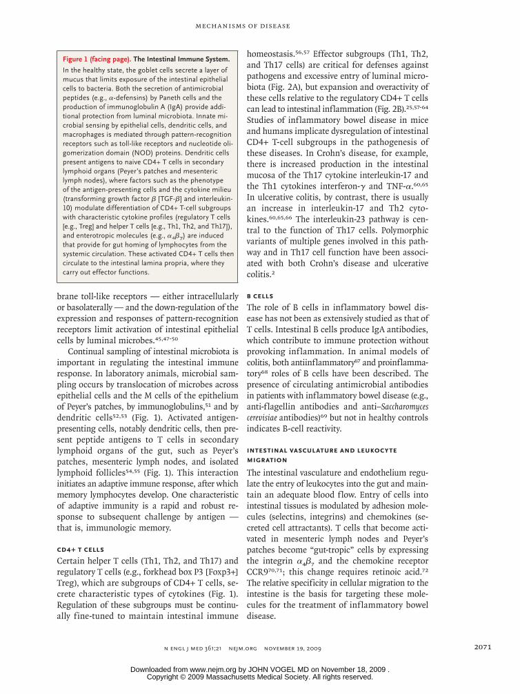
brane toll-like receptors — either intracellularly or basolaterally — and the down-regulation of the expression and responses of pattern-recognition receptors limit activation of intestinal epithelial cells by luminal microbes.45,47-50
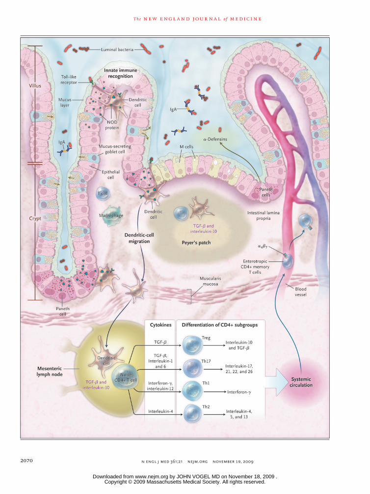
Continual sampling of intestinal microbiota is important in regulating the intestinal immune response. In laboratory animals, microbial sam-pling occurs by translocation of microbes across epithelial cells and the M cells of the epithelium of Peyer’s patches, by immunoglobulins,51 and by dendritic cells52,53 (Fig. 1). Activated antigen-presenting cells, notably dendritic cells, then pre-sent peptide antigens to T cells in secondary lymphoid organs of the gut, such as Peyer’s patches, mesenteric lymph nodes, and isolated lymphoid follicles54,55 (Fig. 1). This interaction initiates an adaptive immune response, after which memory lymphocytes develop. One characteristic of adaptive immunity is a rapid and robust re-sponse to subsequent challenge by antigen — that is, immunologic memory.
CD4+ T Cells
Certain helper T cells (Th1, Th2, and Th17) and regulatory T cells (e.g., forkhead box P3 [Foxp3+]Treg), which are subgroups of CD4+ T cells, se-crete characteristic types of cytokines (Fig. 1). Regulation of these subgroups must be continu-ally fine-tuned to maintain intestinal immune
homeostasis.56,57 Effector subgroups (Th1, Th2, and Th17 cells) are critical for defenses against pathogens and excessive entry of luminal micro-biota (Fig. 2A), but expansion and overactivity of these cells relative to the regulatory CD4+ T cells can lead to intestinal inflammation (Fig. 2B).25,57-64 Studies of inflammatory bowel disease in mice and humans implicate dysregulation of intestinal CD4+ T-cell subgroups in the pathogenesis of these diseases. In Crohn’s disease, for example, there is increased production in the intestinal mucosa of the Th17 cytokine interleukin-17 and the Th1 cytokines interferon-γ and TNF-α.60,65 In ulcerative colitis, by contrast, there is usually an increase in interleukin-17 and Th2 cyto-kines.60,65,66 The interleukin-23 pathway is cen-tral to the function of Th17 cells. Polymorphic variants of multiple genes involved in this path-way and in Th17 cell function have been associ-ated with both Crohn’s disease and ulcerative colitis.2
B Cells
The role of B cells in inflammatory bowel dis-ease has not been as extensively studied as that of T cells. Intestinal B cells produce IgA antibodies, which contribute to immune protection without provoking inflammation. In animal models of colitis, both antiinflammatory67 and proinflamma-tory68 roles of B cells have been described. The presence of circulating antimicrobial antibodies in patients with inflammatory bowel disease (e.g., anti-flagellin antibodies and anti–Saccharomyces cerevisiae antibodies)69 but not in healthy controls indicates B-cell reactivity.
Intestinal Vasculature and Leukocyte Migration
The intestinal vasculature and endothelium regu-late the entry of leukocytes into the gut and main-tain an adequate blood flow. Entry of cells into intestinal tissues is modulated by adhesion mole-cules (selectins, integrins) and chemokines (se-creted cell attractants). T cells that become acti-vated in mesenteric lymph nodes and Peyer’s patches become “gut-tropic” cells by expressing the integrin α4β7 and the chemokine receptor CCR970,71; this change requires retinoic acid.72 The relative specificity in cellular migration to the intestine is the basis for targeting these mole-cules for the treatment of inflammatory bowel disease.
Figure 1 (facing page). The Intestinal Immune System.
In the healthy state, the goblet cells secrete a layer of mucus that limits exposure of the intestinal epithelial cells to bacteria. Both the secretion of antimicrobial peptides (e.g., α-defensins) by Paneth cells and the production of immunoglobulin A (IgA) provide addi-tional protection from luminal microbiota. Innate mi-crobial sensing by epithelial cells, dendritic cells, and macrophages is mediated through pattern-recognition receptors such as toll-like receptors and nucleotide oli-gomerization domain (NOD) proteins. Dendritic cells present antigens to naive CD4+ T cells in secondary lymphoid organs (Peyer’s patches and mesenteric lymph nodes), where factors such as the phenotype of the antigen-presenting cells and the cytokine milieu (transforming growth factor β [TGF-β] and interleukin-10) modulate differentiation of CD4+ T-cell subgroups with characteristic cytokine profiles (regulatory T cells [e.g., Treg] and helper T cells [e.g., Th1, Th2, and Th17]), and enterotropic molecules (e.g., α4β7) are induced that provide for gut homing of lymphocytes from the systemic circulation. These activated CD4+ T cells then circulate to the intestinal lamina propria, where they carry out effector functions.
Copyright © 2009 Massachusetts Medical Society. All rights reserved. Downloaded from www.nejm.org by JOHN VOGEL MD on November 18, 2009 .
T h e n e w e ngl a nd j o u r na l o f m e dic i n e
n engl j med 361;21 nejm.org november 19, 20092072
Copyright © 2009 Massachusetts Medical Society. All rights reserved. Downloaded from www.nejm.org by JOHN VOGEL MD on November 18, 2009 .
Mechanisms of Disease
n engl j med 361;21 nejm.org november 19, 2009 2073
An accumulation of leukocytes in intestinal tissues is characteristic of inflammatory bowel disease. Leukocyte adherence and recruitment are increased in the microvessels in chronic dis-ease,73 mediated in part by up-regulation of ad-hesion molecules on vascular endothelial cells by TNF-α and interleukin-1. Moreover, increased levels of tissue-specific and inflammatory chemo-kines enhance leukocyte migration.74 Abnormali-ties in microvascular function probably contribute to inflammation, ischemia, and impaired mu-cosal healing.73 Ischemia causes local tissue hy-poxia, which in turn regulates factors that contrib-ute to both intestinal injury and protection.75,76
Innate-Immune-R esponse Genes a nd Crohn’s Dise a se
NOD2 and Crohn’s Disease
The importance of responses to intestinal bacte-ria in inflammatory bowel disease is highlighted by the association between Crohn’s disease and the NOD2 gene77 (Table 1), which encodes an in-tracellular sensor of peptidoglycan, a component in bacterial cell walls.78,79 The association includes three NOD2 polymorphisms that change the ami-no acids in NOD2, each impairing responses to peptidoglycan. These three polymorphisms oc-cur with increased frequency in persons of Euro-pean ancestry but are not present in Asian patients and are significantly less frequent in African Americans with Crohn’s disease.14 Approximate-ly 30% of patients of European ancestry have at least one of the three polymorphisms. NOD2 car-riers are more likely than noncarriers to have ileal involvement and complications related to fibro-stenosis and to require intestinal resection.80 Heterozygosity for a polymorphism confers an increased risk of Crohn’s disease (by a factor of 1.75 to 4), whereas homozygosity confers a much greater risk (by a factor of 11 to 27)81; these are the highest relative risks observed for any of the genes associated with this disease. NOD2 poly-morphisms alone, however, are not sufficient to cause Crohn’s disease, which is indicative of the complexities of a multifactorial disorder.
Epithelial cells, Paneth cells, macrophages, dendritic cells, and endothelial cells all express NOD2.77 The activation of the NOD2 protein by bacterial peptidoglycan activates the nuclear fac-tor κB (NF-κB) and mitogen-activated protein
Figure 2 (facing page). The Intestinal Immune System in Health and Disease.
In healthy persons (Panel A), the lamina propria normal-ly contains a diverse array of immune cells and secreted cytokines. These include antiinflammatory mediators (transforming growth factor β [TGF-β] and interleukin-10) that down-regulate immune responses, as well as proinflammatory mediators from both innate and adap-tive immune cells that limit excessive entry of intestinal microbiota and defend against pathogens. Noninflam-matory defenses, such as phagocytosis by macrophages, probably assist in defending against bacteria entering the lamina propria while minimizing tissue injury. A homeo-static balance is maintained between regulatory T cells (e.g., Treg) and effector T cells (Th1, Th2, and Th17). In persons with intestinal inflammation (Panel B), sev-eral events contribute to increased bacterial exposure, including disruption of the mucus layer, dysregulation of epithelial tight junctions, increased intestinal permea-bility, and increased bacterial adherence to epithelial cells. In inflammatory bowel disease, innate cells pro-duce increased levels of tumor necrosis factor α (TNF-α), interleukin-1β, interleukin-6, interleukin-12, interleukin-23, and chemokines. There is marked expansion of the lamina propria, with increased numbers of CD4+ T cells, especially proinflammatory T-cell subgroups, which also secrete increased levels of cytokines and chemokines. Increased production of chemokines results in recruit-ment of additional leukocytes, resulting in a cycle of inflammation. At present, therapeutic approaches to inflammatory bowel disease (labels in red) focus on in-hibiting proinflammatory cytokines, inhibiting the en-try of cells into intestinal tissues (dashed arrow), and in-hibiting T-cell activation and proliferation. Additional investigational biologic therapies include blockade of costimulatory signals that enhance interactions between innate cells and adaptive cells, administration of epithe-lial growth factors, and enhancement of tolerance through a variety of mechanisms. CD4+ Th17 cells (in-set) express surface molecules such as the interleu-kin-23 receptor (a component of the interleukin-23–re-ceptor complex, which consists of the interleukin-23 receptor and the interleukin-12 receptor B1) and CCR6. Interleukin-23 (comprising subunits p19 and p40) is secreted by antigen-presenting cells, and engagement of interleukin-23 with the interleukin-23–receptor com-plex results in activation of the Janus-associated kinase (JAK2) signal transducers and activators of transcrip-tion (STAT3), thereby regulating transcriptional activation. Interleukin-23 contributes to Th17-cell proliferation, sur-vival, or both, and its actions are enhanced by tumor ne-crosis factor (ligand) superfamily, member 15 (TNFS15). Of the top 30 genetic associations with Crohn’s disease, at least six genes can be implicated in Th17 cells and in-terleukin-23 signaling. A number of these genes are not unique to interleukin-23–Th17 signaling. Genes in the in-terleukin-23–Th17 pathway that have been associated with Crohn’s disease are designated by red stars, and those with ulcerative colitis by blue stars.
Copyright © 2009 Massachusetts Medical Society. All rights reserved. Downloaded from www.nejm.org by JOHN VOGEL MD on November 18, 2009 .
T h e n e w e ngl a nd j o u r na l o f m e dic i n e
n engl j med 361;21 nejm.org november 19, 20092074
(MAP) kinase signaling pathways, which leads to the production of cytokines (e.g., TNF and inter-leukin-1β) and antimicrobial peptides.77,82 De-creased secretion of proinflammatory cytokines and decreased activation of NF-κB on acute stimu-lation of NOD2 with bacterial peptidoglycan components have been detected in NOD2 carri-ers.77 Intestinal inflammation does not develop in NOD2-deficient mice, as is the case with most human NOD2 risk-allele carriers.82 Normally, se-cretion of proinflammatory cytokines by intes-tinal antigen-presenting cells is minimal,55,83 yet bacterial killing occurs, implying that the intes-tinal immune system can defend against luminal microbiota while minimizing tissue injury.83 In contrast, the gut in inflammatory bowel disease contains an increased number of antigen-pre-senting cells that secrete proinflammatory cyto-kines.84 Various factors within the intestinal en-vironment contribute to the down-regulation of proinflammatory cytokines by intestinal antigen–presenting cells. These include inhibitory cyto-kines (transforming growth factor β [TGF-β] and interleukin-10) and chronic stimulation through pattern-recognition receptors, such as chronic peptidoglycan stimulation through NOD2.85,86 The NOD2-mediated mechanisms that down-regulate proinflammatory cytokines during chronic NOD2 stimulation are defective in carriers of genetic variants that impair NOD2 function.85 How loss-of-function NOD2 polymorphisms increase suscep-tibility to Crohn’s disease is unknown, but most likely this outcome reflects the myriad functions of NOD2 and the unique features of the intesti-nal environment.
Autophagy Genes and Crohn’s Disease
Associations with Crohn’s disease have been established for ATG16L1 and immunity-related GTPase M protein (IRGM)4,7,8,10 (Table 1), two genes involved in autophagy. Autophagy is a mechanism for clearing intracellular components, including organelles, apoptotic bodies, and microbes.15 In mice with low expression of ATG16L1, the mor-phologic features and gene expression of Paneth cells are abnormal.87 ATG16L1 carriers with Crohn’s disease also have abnormal Paneth-cell morphol-ogy.87 In mice, ATG16L1 appears to regulate se-cretion of interleukin-1β and inhibit intestinal inflammation.88
A da p ti v e Immuni t y a nd Infl a mm at or y Bow el Dise a se
Alterations in T-Cell Tolerance
The inhibitory cytokines interleukin-10 and TGF-β in Peyer’s patches, mesenteric lymph nodes, and lamina propria are involved in T-cell tolerance in the intestine.25 Regulatory T cells can differentiate in Peyer’s patches and mesenteric lymph nodes through the actions of TGF-β and retinoic acid.89 Defects in the development and function of regulatory T cells, or alterations in the ability to respond to them, can result in intes-tinal inflammation in mice.56,90 The autophagy pathway contributes to T-cell tolerance at multi-ple levels, which suggests that polymorphisms of autophagy genes associated with Crohn’s disease could increase a patient’s susceptibility to intes-tinal inflammation through defects in T-cell tol-erance.15,91 Moreover, there is a genetic association between the inhibitory cytokine interleukin-10 and ulcerative colitis.11 This association corresponds with animal models showing that interleukin-10 participates in down-regulating intestinal inflam-mation; for example, colitis and intestinal dys-plasia develop spontaneously in interleukin-10–deficient mice.25 The central importance and sufficiency of defective interleukin-10 signaling in the mediation of intestinal inflammation are further highlighted by the finding that uncom-mon, recessive loss-of-function mutations in ei-ther the IL10RA or the IL10RB component of the interleukin-10 receptor result in Crohn’s dis-ease.92
Th17 Cells and Interleukin-23 Signaling in Inflammatory Bowel Disease
Interleukin-23 signaling is mediated through the engagement of heterodimeric interleukin-23 (com-prising p19 and p40 subunits) with its heterodi-meric receptor (comprising interleukin-23R and interleukin-12RB1). The engagement activates the JAK-STAT (Janus-associated kinase–signal trans-ducers and activators of transcription) signaling pathway, which regulates the transcription of several genes. The importance of interleukin-23 signaling in mediating inflammation has been shown in animal models.17,59,61,64,65,93 Moreover, reports of highly significant genetic associations between IL23R and inflammatory bowel disease,3
Copyright © 2009 Massachusetts Medical Society. All rights reserved. Downloaded from www.nejm.org by JOHN VOGEL MD on November 18, 2009 .
Mechanisms of Disease
n engl j med 361;21 nejm.org november 19, 2009 2075
psoriasis,94 and ankylosing spondylitis95 indicate that inflammatory bowel disease shares genetic associations with certain other autoimmune dis-eases. Interleukin-23, secreted by macrophages and dendritic cells, may contribute to TR17 pro-liferation, survival, or both.96 Interleukin-23 also contributes to intestinal inflammation through Th17-independent pathways.93 Levels of interleu-kin-23 and Th17 cytokines are elevated in the co-lonic mucosa in both Crohn’s disease and ulcer-ative colitis.58,60,63
Multiple independent associations within the IL23R gene region have been found in inflamma-tory bowel disease, most notably for an Arg381Gln variant of the gene: carriers of this uncommon glutamine allele are less likely to have inflam-matory bowel disease, by a factor of 2 to 3, than persons who carry only the common arginine allele.3 The effect of the IL23R polymorphisms on interleukin-23R function has not been defined. In addition to IL23R, associations with Crohn’s disease have been observed in genomic regions encompassing multiple genes involved in inter-leukin-23–Th17 signaling2 (Fig. 2 and Table 1).
Compa r ati v e Gene A sso ci ation S t udies in Crohn’s Dise a se
a nd Ul cer ati v e Coli tis
It is estimated that known genetic associations account for only about 20% of the genetic vari-ance underlying susceptibility to inflammatory bowel disease; the remaining genetic factors have not been identified. Consistent with predictions inferred from epidemiologic studies, genome wide association studies have identified loci associat-ed solely with Crohn’s disease or ulcerative colitis and other loci associated with both disorders (Table 1). For example, multiple genes along the interleukin-23 signaling pathway — notably IL23R, JAK2, STAT3, and p40 — have been associ-ated with both these diseases.2,11,40,97 Genomic associations with Crohn’s disease alone include NOD2 and the autophagy gene, ATG16L1,2 but a number of well-powered studies have not shown associations between these genes and ulcerative colitis. Given the broad functional contributions of autophagy, a more general role in either Crohn’s disease or ulcerative colitis may eventu-ally be established. Significant associations be-
tween ulcerative colitis and a region on chromo-some 12q15 encompassing the interferon-γ and interleukin-26 genes have been observed, but no such associations with Crohn’s disease have thus far been reported.40 In the initial genomewide association studies in ulcerative colitis, a highly significant association was observed for a com-mon polymorphism in a region on chromosome 1q32 containing the IL10 gene, with only a very modest association observed in Crohn’s disease.11 However, the intriguing finding that complete, loss-of-function mutations in the interleukin-10 receptor92 result in a Crohn’s disease–predomi-nant phenotype highlights the complex pheno-typic overlap between these two diseases.
Associations within the major histocompati-bility complex class II region near HLA-DRA (alpha chain) are the most significant observed in genomewide association studies of ulcerative colitis.11,40,97 Distinct HLA-DRB1 (beta-chain) al-leles have been associated with both diseases.97
Ther a peu tic Implic ations
Treatment of inflammatory bowel disease includes lifestyle alterations (e.g., smoking cessation for patients with Crohn’s disease), medical manage-ment, and surgical interventions. A seminal ad-vance was the introduction of treatment with an anti–TNF-α monoclonal antibody, which is par-ticularly effective in Crohn’s disease. The efficacy of this therapy alone probably reflects the pleio-tropic effects of TNF; however, the therapy is often limited by a loss of efficacy, underscoring the need for novel therapies.
Anti-p40 monoclonal antibodies have been reported to be effective in psoriasis98 and Crohn’s disease.99,100 The p40 cytokine subunit is common to both interleukin-23 and interleukin-12, and monoclonal antibodies against p40 inhibit both pathways. Selective blockade of interleukin-23 can be achieved by targeting the p19 subunit, and this approach has been reported to be effective in many,59,64 although not all,101 animal models of inflammatory bowel disease. Selective inhibition of interleukin-23 may, however, deregulate other, cross-regulated pathways and T-cell subgroups, with unintended consequences.100 Moreover, some Th17 cytokines may also have protective features; for example, interleukin-22 ameliorates disease in
Copyright © 2009 Massachusetts Medical Society. All rights reserved. Downloaded from www.nejm.org by JOHN VOGEL MD on November 18, 2009 .
T h e n e w e ngl a nd j o u r na l o f m e dic i n e
n engl j med 361;21 nejm.org november 19, 20092076
an animal model of colitis.102 A major question that remains to be resolved is whether selective interleukin-23 blockade will be more or less ef-fective than combined interleukin-12–interleu-kin-23 blockade in the treatment of inflamma-tory bowel disease.
Other treatments under investigation include the infusion of interleukin-10–producing T cells and the administration of interleukin-10–pro-ducing bacteria.103,104 Certain bacterial compo-nents, commensal bacteria, and “probiotic” bacte-ria more generally are also being investigated.48,105 The increased levels of tissue-specific and inflam-matory chemokines that enhance intestinal leuko-cyte migration74 are the basis for targeting these molecules in inflammatory bowel disease.104,106 With the development of new, potent antiin-flammatory agents, one must consider balancing therapeutic benefit with side effects resulting
from an increased risk of infection or reactivation of infections (JC virus–induced multifocal leuko-encephalopathy in the case of natalizumab107 and tuberculosis in the case of anti-TNF-α mono-clonal antibody108). Future progress in disease monitoring and therapy will depend on the de-velopment of a more refined and integrated under-standing of the mechanisms mediating intesti-nal immune homeostasis.
Supported by grants from the National Institutes of Health (DK P30-34989, to Drs. Abraham and Cho; R01DK077905, to Dr. Abraham; and R01DK072373, U01 DK62429, U01 DK062422, and UL1 RR024139, to Dr. Cho), the Burroughs Wellcome Medi-cal Foundation (to Dr. Cho), the Bohmfalk Funds for Medical Research (to Dr. Cho), and the Crohn’s and Colitis Foundation of America (to Drs. Abraham and Cho).
Dr. Cho reports being listed as a coinventor on a patent for NOD2 polymorphisms owned by Prometheus Labs and receiving lecture fees from Millennium Pharmaceuticals. No other poten-tial conflict of interest relevant to this article was reported.
We thank Graeme Bell, Eric Elton, Fred Gorelick, and Cathy Nagler for reviewing an earlier version of the manuscript.
References
Wellcome Trust Case Control Consor-1. tium. Genome-wide association study of 14,000 cases of seven common diseases and 3,000 shared controls. Nature 2007; 447:661-78.
Barrett JC, Hansoul S, Nicolae DL, et al. 2. Genome-wide association defines more than 30 distinct susceptibility loci for Crohn’s disease. Nat Genet 2008;40:955-62.
Duerr RH, Taylor KD, Brant SR, et al. 3. A genome-wide association study identi-fies IL23r as an inflammatory bowel dis-ease gene. Science 2006;314:1461-3.
Hampe J, Franke A, Rosenstiel P, et al. 4. A genome-wide association scan of non-synonymous SNPs identifies a susceptibil-ity variant for Crohn disease in ATG16L1. Nat Genet 2007;39:207-11.
Kugathasan S, Baldassano RN, Brad-5. field JP, et al. Loci on 20q13 and 21q22 are associated with pediatric-onset inflam-matory bowel disease. Nat Genet 2008; 40:1211-5.
Libioulle C, Louis E, Hansoul S, et al. 6. Novel Crohn disease locus identified by genome-wide association maps to a gene desert on 5p13.1 and modulates expres-sion of PTGER4. PLoS Genet 2007;3(4): e58.
Parkes M, Barrett JC, Prescott NJ, et al. 7. Sequence variants in the autophagy gene IRGM and multiple other replicating loci contribute to Crohn’s disease susceptibil-ity. Nat Genet 2007;39:830-2.
Rioux JD, Xavier RJ, Taylor KD, et al. 8. Genome-wide association study identifies new susceptibility loci for Crohn disease and implicates autophagy in disease patho-genesis. Nat Genet 2007;39:596-604.
Yamazaki K, McGovern D, Ragoussis 9. J, et al. Single nucleotide polymorphisms in TNFSF15 confer susceptibility to Crohn’s disease. Hum Mol Genet 2005; 14:3499-506.
McCarroll SA, Huett A, Kuballa P, et al. 10. Deletion polymorphism upstream of IRGM associated with altered IRGM ex-pression and Crohn’s disease. Nat Genet 2008;40:1107-12.
Franke A, Balschun T, Karlsen TH, et 11. al. Sequence variants in IL10, ARPC2 and multiple other loci contribute to ulcer-ative colitis susceptibility. Nat Genet 2008;40:1319-23.
Franke A, Balschun T, Karlsen TH, et 12. al. Replication of signals from recent studies of Crohn’s disease identifies pre-viously unknown disease loci for ulcer-ative colitis. Nat Genet 2008;40:713-5.
Fisher SA, Tremelling M, Anderson 13. CA, et al. Genetic determinants of ulcer-ative colitis include the ECM1 locus and five loci implicated in Crohn’s disease. Nat Genet 2008;40:710-2.
Cho JH, Weaver CT. The genetics of 14. inflammatory bowel disease. Gastroen-terology 2007;133:1327-39.
Levine B, Deretic V. Unveiling the 15. roles of autophagy in innate and adaptive immunity. Nat Rev Immunol 2007;7:767-77.
Mangan PR, Harrington LE, O’Quinn 16. DB, et al. Transforming growth factor-beta induces development of the T(H)17 lineage. Nature 2006;441:231-4.
McGeachy MJ, Cua DJ. The link be-17. tween IL-23 and Th17 cell-mediated im-mune pathologies. Semin Immunol 2007; 19:372-6.
Loftus EV Jr, Sandborn WJ. Epidemiol-18. ogy of inflammatory bowel disease. Gas-troenterol Clin North Am 2002;31:1-20.
Bernstein CN, Wajda A, Blanchard JF. 19. The clustering of other chronic inflam-matory diseases in inflammatory bowel disease: a population-based study. Gastro-enterology 2005;129:827-36.
Weinstock JV. Helminths and mucosal 20. immune modulation. Ann N Y Acad Sci 2006;1072:356-64.
Eckburg PB, Relman DA. The role of 21. microbes in Crohn’s disease. Clin Infect Dis 2007;44:256-62.
Bäckhed F, Ley RE, Sonnenburg JL, 22. Peterson DA, Gordon JI. Host-bacterial mutualism in the human intestine. Sci-ence 2005;307:1915-20.
Turnbaugh PJ, Hamady M, Yatsunenko 23. T, et al. A core gut microbiome in obese and lean twins. Nature 2009;457:480-4.
Wu L, Estrada O, Zaborina O, et al. 24. Recognition of host immune activation by Pseudomonas aeruginosa. Science 2005; 309:774-7.
Elson CO, Cong Y, McCracken VJ, 25. Dimmitt RA, Lorenz RG, Weaver CT. Ex-perimental models of inflammatory bowel disease reveal innate, adaptive, and regu-latory mechanisms of host dialogue with the microbiota. Immunol Rev 2005;206: 260-76.
Barnich N, Carvalho FA, Glasser AL, 26. et al. CEACAM6 acts as a receptor for ad-herent-invasive E. coli, supporting ileal mucosa colonization in Crohn disease. J Clin Invest 2007;117:1566-74.
Frank DN, St Amand AL, Feldman RA, 27. Boedeker EC, Harpaz N, Pace NR. Molec-ular-phylogenetic characterization of mi-
Copyright © 2009 Massachusetts Medical Society. All rights reserved. Downloaded from www.nejm.org by JOHN VOGEL MD on November 18, 2009 .
Mechanisms of Disease
n engl j med 361;21 nejm.org november 19, 2009 2077
crobial community imbalances in human inflammatory bowel diseases. Proc Natl Acad Sci U S A 2007;104:13780-5.
Turner JR. Molecular basis of epithe-28. lial barrier regulation: from basic mecha-nisms to clinical application. Am J Pathol 2006;169:1901-9.
Wang F, Schwarz BT, Graham WV, et 29. al. IFN-gamma-induced TNFR2 expres-sion is required for TNF-dependent intes-tinal epithelial barrier dysfunction. Gas-troenterology 2006;131:1153-63.
Musch MW, Clarke LL, Mamah D, et al. 30. T cell activation causes diarrhea by increas-ing intestinal permeability and inhibiting epithelial Na+/K+-ATPase. J Clin Invest 2002;110:1739-47.
Bruewer M, Luegering A, Kucharzik T, 31. et al. Proinflammatory cytokines disrupt epithelial barrier function by apoptosis-independent mechanisms. J Immunol 2003;171:6164-72.
Dahan S, Roda G, Pinn D, et al. Epi-32. thelial: lamina propria lymphocyte inter-actions promote epithelial cell differenti-ation. Gastroenterology 2008;134:192-203.
Taupin D, Podolsky DK. Trefoil fac-33. tors: initiators of mucosal healing. Nat Rev Mol Cell Biol 2003;4:721-32. [Erratum, Nat Rev Mol Cell Biol 2003;4:819.]
McVay LD, Keilbaugh SA, Wong TM, 34. et al. Absence of bacterially induced RELMbeta reduces injury in the dextran sodium sulfate model of colitis. J Clin In-vest 2006;116:2914-23.
Wehkamp J, Salzman NH, Porter E, 35. et al. Reduced Paneth cell alpha-defensins in ileal Crohn’s disease. Proc Natl Acad Sci U S A 2005;102:18129-34.
Simms LA, Doecke JD, Walsh MD, 36. Huang N, Fowler EV, Radford-Smith GL. Reduced alpha-defensin expression is asso-ciated with inflammation and not NOD2 mutation status in ileal Crohn’s disease. Gut 2008;57:903-10.
Habtezion A, Toivola DM, Butcher EC, 37. Omary MB. Keratin-8-deficient mice de-velop chronic spontaneous Th2 colitis amenable to antibiotic treatment. J Cell Sci 2005;118:1971-80.
Kaser A, Lee AH, Franke A, et al. XBP1 38. links ER stress to intestinal inflamma-tion and confers genetic risk for human inf lammatory bowel disease. Cell 2008; 134:743-56.
Kabashima K, Saji T, Murata T, et al. 39. The prostaglandin receptor EP4 suppresses colitis, mucosal damage and CD4 cell ac-tivation in the gut. J Clin Invest 2002;109: 883-93.
Silverberg MS, Cho JH, Rioux JD, et al. 40. Ulcerative colitis-risk loci on chromo-somes 1p36 and 12q15 found by genome-wide association study. Nat Genet 2009; 41:216-20.
Van der Sluis M, De Koning BA, De 41. Bruijn AC, et al. Muc2-deficient mice spontaneously develop colitis, indicating
that MUC2 is critical for colonic protec-tion. Gastroenterology 2006;131:117-29.
Shkoda A, Ruiz PA, Daniel H, et al. 42. Interleukin-10 blocked endoplasmic retic-ulum stress in intestinal epithelial cells: impact on chronic inflammation. Gastro-enterology 2007;132:190-207.
Rakoff-Nahoum S, Paglino J, Eslami-43. Varzaneh F, Edberg S, Medzhitov R. Rec-ognition of commensal microflora by toll-like receptors is required for intestinal homeostasis. Cell 2004;118:229-41.
Vijay-Kumar M, Sanders CJ, Taylor RT, 44. et al. Deletion of TLR5 results in sponta-neous colitis in mice. J Clin Invest 2007; 117:3909-21.
Lee J, Mo JH, Katakura K, et al. Main-45. tenance of colonic homeostasis by distinc-tive apical TLR9 signalling in intestinal epithelial cells. Nat Cell Biol 2006;8:1327-36.
Bashir ME, Louie S, Shi HN, Nagler-46. Anderson C. Toll-like receptor 4 signaling by intestinal microbes influences suscep-tibility to food allergy. J Immunol 2004; 172:6978-87.
Gewirtz AT, Navas TA, Lyons S, 47. Godowski PJ, Madara JL. Cutting edge: bacterial f lagellin activates basolaterally expressed TLR5 to induce epithelial pro-inflammatory gene expression. J Immunol 2001;167:1882-5.
Neish AS, Gewirtz AT, Zeng H, et al. 48. Prokaryotic regulation of epithelial re-sponses by inhibition of IkappaB-alpha ubiquitination. Science 2000;289:1560-3.
Melmed G, Thomas LS, Lee N, et al. 49. Human intestinal epithelial cells are broadly unresponsive to Toll-like receptor 2-dependent bacterial ligands: implica-tions for host-microbial interactions in the gut. J Immunol 2003;170:1406-15.
Otte JM, Cario E, Podolsky DK. Mech-50. anisms of cross hyporesponsiveness to Toll-like receptor bacterial ligands in in-testinal epithelial cells. Gastroenterology 2004;126:1054-70.
Yoshida M, Claypool SM, Wagner JS, 51. et al. Human neonatal Fc receptor medi-ates transport of IgG into luminal secre-tions for delivery of antigens to mucosal dendritic cells. Immunity 2004;20:769-83.
Rescigno M, Urbano M, Valzasina B, 52. et al. Dendritic cells express tight junc-tion proteins and penetrate gut epithelial monolayers to sample bacteria. Nat Immu-nol 2001;2:361-7.
Niess JH, Brand S, Gu X, et al. CX3-53. CR1-mediated dendritic cell access to the intestinal lumen and bacterial clearance. Science 2005;307:254-8.
Lorenz RG, Newberry RD. Isolated 54. lymphoid follicles can function as sites for induction of mucosal immune responses. Ann N Y Acad Sci 2004;1029:44-57.
Johansson C, Kelsall BL. Phenotype 55. and function of intestinal dendritic cells. Semin Immunol 2005;17:284-94.
Makita S, Kanai T, Oshima S, et al. 56. CD4+CD25bright T cells in human intes-tinal lamina propria as regulatory cells. J Immunol 2004;173:3119-30.
Izcue A, Coombes JL, Powrie F. Regu-57. latory T cells suppress systemic and mu-cosal immune activation to control intes-tinal inflammation. Immunol Rev 2006; 212:256-71.
Annunziato F, Cosmi L, Santarlasci V, 58. et al. Phenotypic and functional features of human Th17 cells. J Exp Med 2007; 204:1849-61.
Elson CO, Cong Y, Weaver CT, et al. 59. Monoclonal anti-interleukin 23 reverses active colitis in a T cell-mediated model in mice. Gastroenterology 2007;132:2359-70.
Fujino S, Andoh A, Bamba S, et al. In-60. creased expression of interleukin 17 in inflammatory bowel disease. Gut 2003;52: 65-70.
Hue S, Ahern P, Buonocore S, et al. 61. Interleukin-23 drives innate and T cell-mediated intestinal inflammation. J Exp Med 2006;203:2473-83.
Nguyen DD, Maillard MH, Cotta-de-62. Almeida V, et al. Lymphocyte-dependent and Th2 cytokine-associated colitis in mice deficient in Wiskott-Aldrich syndrome protein. Gastroenterology 2007;133:1188-97.
Saruta M, Yu QT, Avanesyan A, Flesh-63. ner PR, Targan SR, Papadakis KA. Pheno-type and effector function of CC chemo-kine receptor 9-expressing lymphocytes in small intestinal Crohn’s disease. J Im-munol 2007;178:3293-300.
Yen D, Cheung J, Scheerens H, et al. 64. IL-23 is essential for T cell-mediated coli-tis and promotes inflammation via IL-17 and IL-6. J Clin Invest 2006;116:1310-6.
Kobayashi T, Okamoto S, Hisamatsu 65. T, et al. IL-23 differentially regulates the Th1/Th17 balance in ulcerative colitis and Crohn’s disease. Gut 2008;57:1682-9.
Fuss IJ, Heller F, Boirivant M, et al. 66. Nonclassical CD1d-restricted NK T cells that produce IL-13 characterize an atypi-cal Th2 response in ulcerative colitis. J Clin Invest 2004;113:1490-7.
Velázquez P, Wei B, Braun J. Surveil-67. lance B lymphocytes and mucosal immu-noregulation. Springer Semin Immuno-pathol 2005;26:453-62.
Olson TS, Bamias G, Naganuma M, 68. et al. Expanded B cell population blocks regulatory T cells and exacerbates ileitis in a murine model of Crohn disease. J Clin Invest 2004;114:389-98.
Lodes MJ, Cong Y, Elson CO, et al. 69. Bacterial f lagellin is a dominant antigen in Crohn disease. J Clin Invest 2004;113: 1296-306.
Mora JR, Bono MR, Manjunath N, et al. 70. Selective imprinting of gut-homing T cells by Peyer’s patch dendritic cells. Nature 2003;424:88-93.
Jaensson E, Uronen-Hansson H, Pabst 71.
Copyright © 2009 Massachusetts Medical Society. All rights reserved. Downloaded from www.nejm.org by JOHN VOGEL MD on November 18, 2009 .
n engl j med 361;21 nejm.org november 19, 20092078
Mechanisms of Disease
O, et al. Small intestinal CD103+ dendrit-ic cells display unique functional proper-ties that are conserved between mice and humans. J Exp Med 2008;205:2139-49.
Iwata M, Hirakiyama A, Eshima Y, 72. Kagechika H, Kato C, Song SY. Retinoic acid imprints gut-homing specificity on T cells. Immunity 2004;21:527-38.
Hatoum OA, Heidemann J, Binion DG. 73. The intestinal microvasculature as a ther-apeutic target in inflammatory bowel dis-ease. Ann N Y Acad Sci 2006;1072:78-97.
Mora JR, von Andrian UH. T-cell hom-74. ing specificity and plasticity: new concepts and future challenges. Trends Immunol 2006;27:235-43.
Hegazi RA, Rao KN, Mayle A, Sepul-75. veda AR, Otterbein LE, Plevy SE. Carbon monoxide ameliorates chronic murine colitis through a heme oxygenase 1-depen-dent pathway. J Exp Med 2005;202:1703-13.
Robinson A, Keely S, Karhausen J, 76. Gerich ME, Furuta GT, Colgan SP. Muco-sal protection by hypoxia-inducible factor prolyl hydroxylase inhibition. Gastroen-terology 2008;134:145-55.
Abraham C, Cho JH. Functional con-77. sequences of NOD2 (CARD15) mutations. Inflamm Bowel Dis 2006;12:641-50.
Inohara N, Ogura Y, Fontalba A, et al. 78. Host recognition of bacterial muramyl di-peptide mediated through NOD2: impli-cations for Crohn’s disease. J Biol Chem 2003;278:5509-12.
Girardin SE, Boneca IG, Viala J, et al. 79. Nod2 is a general sensor of peptidoglycan through muramyl dipeptide (MDP) detec-tion. J Biol Chem 2003;278:8869-72.
Lesage S, Zouali H, Cézard JP, et al. 80. CARD15/NOD2 mutational analysis and genotype-phenotype correlation in 612 patients with inflammatory bowel disease. Am J Hum Genet 2002;70:845-57.
Economou M, Trikalinos TA, Loizou 81. KT, Tsianos EV, Ioannidis JP. Differential effects of NOD2 variants on Crohn’s dis-ease risk and phenotype in diverse popu-lations: a metaanalysis. Am J Gastroen-terol 2004;99:2393-404.
Kobayashi KS, Chamaillard M, Ogura Y, 82. et al. Nod2-dependent regulation of in-nate and adaptive immunity in the intesti-nal tract. Science 2005;307:731-4.
Smythies LE, Sellers M, Clements RH, 83. et al. Human intestinal macrophages dis-play profound inf lammatory anergy de-
spite avid phagocytic and bacteriocidal activity. J Clin Invest 2005;115:66-75.
Kamada N, Hisamatsu T, Okamoto S, 84. et al. Unique CD14 intestinal macrophages contribute to the pathogenesis of Crohn disease via IL-23/IFN-gamma axis. J Clin Invest 2008;118:2269-80.
Hedl M, Li J, Cho JH, Abraham C. 85. Chronic stimulation of Nod2 mediates tolerance to bacterial products. Proc Natl Acad Sci U S A 2007;104:19440-5.
Watanabe T, Asano N, Murray PJ, et al. 86. Muramyl dipeptide activation of nucleo-tide-binding oligomerization domain 2 pro tects mice from experimental colitis. J Clin Invest 2008;118:545-59.
Cadwell K, Liu JY, Brown SL, et al. 87. A key role for autophagy and the autophagy gene Atg16L1 in mouse and human intes-tinal Paneth cells. Nature 2008;456:259-63.
Saitoh T, Fujita N, Jang MH, et al. 88. Loss of the autophagy protein Atg16L1 enhances endotoxin-induced IL-1beta pro-duction. Nature 2008;456:264-8.
Mucida D, Park Y, Kim G, et al. Recip-89. rocal TH17 and regulatory T cell differen-tiation mediated by retinoic acid. Science 2007;317:256-60.
Monteleone G, Del Vecchio Blanco G, 90. Monteleone I, et al. Post-transcriptional regulation of Smad7 in the gut of patients with inflammatory bowel disease. Gastro-enterology 2005;129:1420-9.
Nedjic J, Aichinger M, Emmerich J, 91. Mizushima N, Klein L. Autophagy in thy-mic epithelium shapes the T-cell reper-toire and is essential for tolerance. Nature 2008;455:396-400.
Glocker E-O, Kotlarz D, Boztug K, et 92. al. Inflammatory bowel disease and mu-tations affecting the interleukin-10 recep-tor. N Engl J Med 2009;361:2033-45.
Izcue A, Hue S, Buonocore S, et al. 93. Interleukin-23 restrains regulatory T cell activity to drive T cell-dependent colitis. Immunity 2008;28:559-70.
Cargill M, Schrodi SJ, Chang M, et al. 94. A large-scale genetic association study confirms IL12B and leads to the identifi-cation of IL23R as psoriasis-risk genes. Am J Hum Genet 2007;80:273-90.
Burton PR, Clayton DG, Cardon LR, 95. et al. Association scan of 14,500 nonsyn-onymous SNPs in four diseases identi-fies auto immunity variants. Nat Genet 2007;39:1329-37.
McGeachy MJ, Cua DJ. Th17 cell dif-96. ferentiation: the long and winding road. Immunity 2008;28:445-53.
Fernando MM, Stevens CR, Walsh EC, 97. et al. Defining the role of the MHC in auto-immunity: a review and pooled analysis. PLoS Genet 2008;4(4):e1000024.
Krueger GG, Langley RG, Leonardi C, 98. et al. A human interleukin-12/23 mono-clonal antibody for the treatment of pso-riasis. N Engl J Med 2007;356:580-92.
Mannon PJ, Fuss IJ, Mayer L, et al. 99. Anti–interleukin-12 antibody for active Crohn’s disease. N Engl J Med 2004;351: 2069-79. [Erratum, N Engl J Med 2005;352: 1276.]
Sandborn WJ, Feagan BG, Fedorak 100. RN, et al. A randomized trial of usteki-numab, a human interleukin-12/23 mono-clonal antibody, in patients with moderate-to-severe Crohn’s disease. Gastroenterology 2008;135:1130-41.
Becker C, Dornhoff H, Neufert C, 101. et al. Cutting edge: IL-23 cross-regulates IL-12 production in T cell-dependent ex-perimental colitis. J Immunol 2006;177: 2760-4.
Sugimoto K, Ogawa A, Mizoguchi E, 102. et al. IL-22 ameliorates intestinal inflam-mation in a mouse model of ulcerative colitis. J Clin Invest 2008;118:534-44.
Van Montfrans C, Hooijberg E, Rodri-103. guez Pena MS, et al. Generation of regula-tory gut-homing human T lymphocytes using ex vivo interleukin 10 gene transfer. Gastroenterology 2002;123:1877-88.
Baumgart DC, Sandborn WJ. Inflam-104. matory bowel disease: clinical aspects and established and evolving therapies. Lancet 2007;369:1641-57.
Mazmanian SK, Round JL, Kasper 105. DL. A microbial symbiosis factor prevents intestinal inflammatory disease. Nature 2008;453:620-5.
Ghosh S, Goldin E, Gordon FH, et al. 106. Natalizumab for active Crohn’s disease. N Engl J Med 2003;348:24-32.
Van Assche G, Van Ranst M, Sciot R, 107. et al. Progressive multifocal leukoenceph-alopathy after natalizumab therapy for Crohn’s disease. N Engl J Med 2005;353: 362-8.
Keane J, Gershon S, Wise RP, et al. 108. Tuberculosis associated with infliximab, a tumor necrosis factor α–neutralizing agent. N Engl J Med 2001;345:1098-104.Copyright © 2009 Massachusetts Medical Society.
collections of articles on the journal’s web site
The Journal’s Web site (NEJM.org) sorts published articles into more than 50 distinct clinical collections, which can be used as convenient
entry points to clinical content. In each collection, articles are cited in reverse chronologic order, with the most recent first.
Copyright © 2009 Massachusetts Medical Society. All rights reserved. Downloaded from www.nejm.org by JOHN VOGEL MD on November 18, 2009 .