Embed Size (px)
Citation preview
ORIGINAL RESEARCHpublished: 02 February 2016
doi: 10.3389/fphar.2016.00007
Edited by:Banasri Hazra,
Jadavpur University, India
Reviewed by:Subhalakshmi Ghosh,
Jadavpur University, IndiaParimal C. Sen,
Bose Institute, India
*Correspondence:Chun W. Mai
Specialty section:This article was submitted to
Ethnopharmacology,a section of the journal
Frontiers in Pharmacology
Received: 19 November 2015Accepted: 11 January 2016
Published: 02 February 2016
Citation:Mai CW, Yap KSI, Kho MT, Ismail NH,
Yusoff K, Shaari K, Chin SYand Lim ESH (2016) MechanismsUnderlying the Anti-Inflammatory
Effects of Clinacanthus nutans LindauExtracts: Inhibition of Cytokine
Production and Toll-Like Receptor-4Activation. Front. Pharmacol. 7:7.
doi: 10.3389/fphar.2016.00007
Mechanisms Underlying theAnti-Inflammatory Effects ofClinacanthus nutans LindauExtracts: Inhibition of CytokineProduction and Toll-Like Receptor-4ActivationChun W. Mai1*, Kok S. I. Yap2, Mee T. Kho3, Nor H. Ismail4, Khatijah Yusoff5,6,Khozirah Shaari5, Swee Y. Chin1 and Erin S. H. Lim7
1 Department of Pharmaceutical Chemistry, School of Pharmacy, International Medical University, Bukit Jalil, Malaysia,2 Department of Life Sciences, School of Pharmacy, International Medical University, Bukit Jalil, Malaysia, 3 School ofPostgraduate Studies and Research, International Medical University, Bukit Jalil, Malaysia, 4 Atta-ur-Rahman Institute forNatural Products Discovery, Universiti Teknologi MARA, Shah Alam, Malaysia, 5 Institute of Bioscience, Universiti PutraMalaysia, Serdang, Malaysia, 6 Department of Microbiology, Faculty of Biotechnology and Biomolecular Sciences, UniversitiPutra Malaysia, Serdang, Malaysia, 7 Perdana University-Royal College of Surgeons Ireland, Perdana University, Serdang,Malaysia
Clinacanthus nutans has had a long history of use in folk medicine in Malaysia andSoutheast Asia; mostly in the relief of inflammatory conditions. In this study, weinvestigated the effects of different extracts of C. nutans upon lipopolysaccharide (LPS)induced inflammation in order to identify its mechanism of action. Extracts of leavesand stem bark of C. nutans were prepared using polar and non-polar solvents toproduce four extracts, namely polar leaf extract (LP), non-polar leaf extract (LN), polarstem extract (SP), and non-polar stem extracts (SN). The extracts were standardized bydetermining its total phenolic and total flavonoid contents. Its anti-inflammatory effectswere assessed on LPS induced nitrite release in RAW264.7 macrophages and Toll-likereceptor (TLR-4) activation in TLR-4 transfected human embryonic kidney cells (HEK-BlueTM-hTLR4 cells). The levels of inflammatory cytokines (TNF-α, IFN-γ, IL-1β, IL-6,IL-12p40, and IL-17) in treated RAW264.7 macrophages were quantified to verify itsanti-inflammatory effects. Western blotting was used to investigate the effect of the mostpotent extract (LP) on TLR-4 related inflammatory proteins (p65, p38, ERK, JNK, IRF3)in RAW264.7 macrophages. All four extracts produced a significant, concentration-dependent reduction in LPS-stimulated nitric oxide, LPS-induced TLR-4 activationin HEK-BlueTM-hTLR4 cells and LPS-stimulated cytokines production in RAW264.7macrophages. The most potent extract, LP, also inhibited all LPS-induced TLR-4inflammatory proteins. These results provide a basis for understanding the mechanismsunderlying the previously demonstrated anti-inflammatory activity of C. nutans extracts.
Keywords: anti-inflammatory agents, Clinacanthus nutans, Toll-like receptor 4, macrophages, total flavonoidcontent
Frontiers in Pharmacology | www.frontiersin.org 1 February 2016 | Volume 7 | Article 7
Mai et al. Anti-Inflammatory Effects of Clinacanthus nutans
INTRODUCTION
Clinacanthus nutans (‘Belalai Gajah’ in Malay, or ‘You DunCao’ in Mandarin) has long been used in Thailand to improvebladder function and has also been used traditionally inMalaysia as a folk medicine for kidney and bladder disease(Low et al., 2011). C. nutans Lindau (Acanthaceae) leaves andbranches have been known to relief nettle rash, dysentery,fever, burns, scalds, insect stings, and oral inflammatorysymptoms (Cheeptham and Towers, 2002). Traditionally,C. nutans have been consumed as crude extract of the leavesor stem bark. The leaf extracts of C. nutans has been reportedto possess antioxidant (Khoo et al., 2015), analgesic andanti-inflammatory activities against varicella zoster virus(Thawaranantha et al., 1992) as well as inhibitory activity againstscorpion venom-induced fibroblast lysis (Uawonggul et al., 2006).Kittisiripornkul (1984) reported the anti-inflammatory activityof a n-butanol-soluble fraction from the leaves (Kittisiripornkul,1984) and a methanol extract of the whole plant reducedcarrageenan-induced paw oedema and ethyl phenylpropiolate-induced ear oedema in rats (Wanikiat et al., 2008). Theextract also concentration-dependently inhibited humanneutrophil chemokinesis, as well as N-formyl-methionyl-leucyl-phenylalanine-induced chemotaxis, superoxide aniongeneration, and myeloperoxidase and elastase release (Wanikiatet al., 2008). However, the exact cellular mechanisms underlyingthe anti-inflammatory actions of C. nutans extracts remainunknown.
In a recent study (Khoo et al., 2015), it has been highlightedthat both polar and non-polar compounds in C. nutans couldplay important roles through its medicinal properties; thereforein our study, we have included both the polar and non-polar fractions of C. nutans leaves and stem bark. In orderto examine the anti-inflammatory effects of C. nutans, weinvestigated its effects on human embryonic kidney (HEK)cells stably transfected with human TLR (HEK-BlueTM-4) andin murine macrophages (RAW264.7) challenged with LPS, theGram-negative bacterial cell wall component, a well-establishedactivator of TLR-4 (Mai et al., 2013). TLR-4 is the first lineof host defense against acute and chronic inflammation and isone of the key pro-inflammatory signaling receptors (Park et al.,2009). Activation of TLR-4 by LPS enhances the productionof NO and inflammatory cytokines, through activating nuclearfactor κB (NF κB) and IRF3. Inhibition of TLR-4 activationmay produce potent anti-inflammatory effects since TLR-4 is the
Abbreviations:BSA, Bovine serum albumin; DMSO, Dimethyl sulfoxide; ERK,Extracellular signal-regulated kinase; HEK-BlueTM-hTLR4, TLR-4 transfectedhuman embryonic kidney cells; IC50NO, Concentration inhibits 50% of LPSinduced NO production; IC50TLR4, Concentration inhibits 50% of LPS inducedTLR-4 activity; IFN-γ, Interferon-gamma; IL, Interleukin; IRF3, Interferonregulatory factor 3; JNK, c-Jun N-terminal kinase 1/2; LN, Non-polar leaf extract;LP, Polar leaf extract; LPS, Lipopolysaccharide; mg GAE/g dm, Gallic acidequivalent (mg) per dried material (g); mg QE/g dm, Quercetin equivalent (mg)per dried material (g); MTT, Methyl thiazolyl tetrazolium; NF κB, Nuclear factorκB; NO, Nitric oxide; SDS-PAGE, Sodium dodecyl sulfate polyacrylamide gelelectrophoresis; SEAP, Secreted embryonic alkaline phosphatase; SP, Polar stemextract; SN, Non-polar stem extract; TFC, Total flavonoid content; TLR-4, Toll-likereceptor-4; TNF-α, Tumor necrosis factor-alpha; TPC, Total phenolic content.
upstream receptor that activates both NF κB and IRF3 signaling,the hallmarks of inflammation (Hatziieremia et al., 2006; Maiet al., 2013).
MATERIALS AND METHODS
ReagentsAll materials, unless specified, were purchased from Sigma–Aldrich (St Louis, MO,USA). Ultrapure LPS from Escherichia coliwas purchased from InvivoGen (San Diego, CA, USA) and wasreconstituted using endotoxin-free water (InvivoGen, San Diego,CA, USA).
Preparation of Plant ExtractsClinacanthus nutans was collected from an orchard in Temerloh,Pahang Malaysia, and its identity was kindly verified by Dr.Richard Chung at the Forest Research Institute of Malaysia. Theplant was also deposited in the Malaysian Agricultural Researchand Development Institute herbarium with the specimennumbers MDI 12807 and MDI 12808. The plant was separatedinto leaves and stem bark, which were dried and powdered. Thepowdered leaves were extracted with polar solvents (methanoland dichloromethane) or non-polar solvents (hexane and diethylether) through immersion in the solvent for three days at roomtemperature. A total of four extracts were prepared, namelypolar leave extracts (LP), non-polar leave extracts (LN), polarstem extracts (SP), and non-polar stem extracts (SN). Theextracts were then gravity filtered and the solvents removedunder vacuum using a rotary evaporator at 60◦C. The driedextracts were subjected to further experiments. In order tostandardize the extracts, we determined the total phenoliccontent (TPC) and total flavonoid content (TFC) of extractsusing standardized assays as per described in previous studies(Mai et al., 2009a,b). TPC of the extracts were expressed as gallicacid equivalents in milligram per gram of dried material (mgGAE/g dm) while TFC of the extracts were expressed as quercetinequivalent in milligram per gram of dried material (mg QE/gdm).
Cell Lines and Cell CultureMacrophages RAW264.7 were obtained from the AmericanType Culture Collection (ATCC, TIB-71TM) and maintainedin Dulbecco’s Modification of Eagle’s Medium (DMEM)supplemented with 10% inactivated foetal bovine serum (FBS)and 1% penicillin–streptomycin. All the cells were culturedand maintained at 37◦C in a 5% carbon dioxide incubator.HEK-BlueTM-4 cells, obtained from InvivoGen (San Diego, CA,USA), are HEK that are stably transfected with human Toll-likereceptor-4 (hTLR-4), myeloid differentiation factor-2/clusterof differentiation-14 and SEAP reporter gene. The cells werecultured in complete DMEM which contained 4.5 g/L glucoseand L-glutamine, 10% heat inactivate FBS, 1% penicillin-streptomycin, NormocinTM (InvivoGen, San Diego, CA, USA)and HEK-BlueTM Selection Medium (InvivoGen, San Diego,CA, USA). All cells were harvested within 20 passages using
Frontiers in Pharmacology | www.frontiersin.org 2 February 2016 | Volume 7 | Article 7
Mai et al. Anti-Inflammatory Effects of Clinacanthus nutans
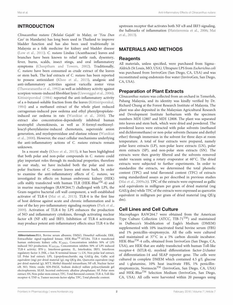
FIGURE 1 | Cytotoxicity of Clinacanthus nutans extracts on cells. Dose response curve of LP, LN, SP, and SN on cell viability of human embryonic kidneycells, HEK-BlueTMhTLR-4 (A) and murine macrophages, RAW264.7 (B). All values were not significant difference when compared to 0.1% DMSO (one-way ANOVApost hoc Dunnett’s t-test). Each value represents means ± SD from three independent experiments. (C) No significant morphological changes inHEK-BlueTMhTLR-4 and RAW264.7 cells after 72 hours treatment with 100 μg/mL of extracts or 0.1% DMSO.
the cell scraper, without addition of trypsin for further analysisaccording to the manufacturer’s instruction.
Cytotoxicity AssayThe MTT cell viability assay was used to access the cytotoxicityof extracts on HEK-BlueTM-hTLR4 cells and RAW264.7 cells, asdescribed previously with modification (Mai et al., 2009a,b, 2013,
2014; Tan et al., 2013). Briefly, all extracts (LP, SP, LN, or SN) werereconstituted using DMSO to 100 mg/mL and further dilutedto required concentrations (1–100 μg/mL) using ultra-purifiedsterile water prior to the assays. Cells were treated with variousconcentrations of extracts or 0.1% DMSO (negative control) for72 h before the reaction was terminated with the MTT reagent.The absorbance was recorded at a test wavelength of 570 nm and
Frontiers in Pharmacology | www.frontiersin.org 3 February 2016 | Volume 7 | Article 7
Mai et al. Anti-Inflammatory Effects of Clinacanthus nutans
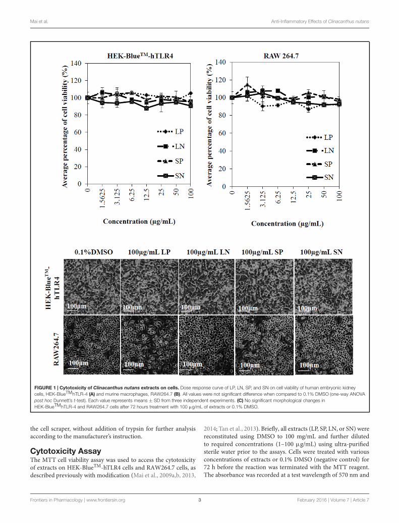
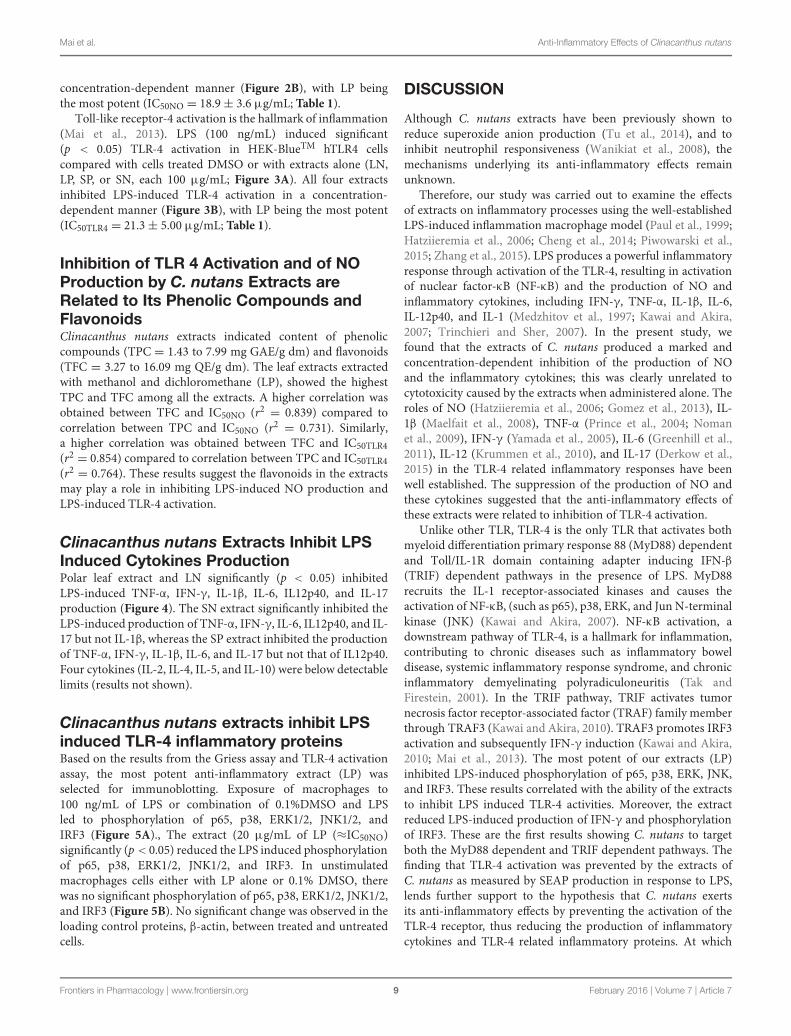
FIGURE 2 | Effect of C. nutans extracts on NO production in RAW264.7 cells. (A) As compared to cells treated with 0.1% DMSO, cells treated with100 μg/mL of LP, LN, SP, SN failed to induce significant NO production, while cells treated with 100 ng/mL of LPS induced significant NO production (∗p < 0.05,one-way ANOVA post hoc Dunnett’s t-test). (B) Cells were treated with various concentrations of extracts (1.5625–100 μg/mL) or 0.1% DMSO for 1 h followed by18 h of LPS induction (100 ng/mL). Statistical significant difference from LPS induction are indicated as ∗p < 0.05 by one-way ANOVA post hoc Dunnet’s t-test.Each value represents means ± SD from three independent experiments.
a reference wavelength of 630 nm using the Tecan Infinite F200plate reader (Männedorf, Switzerland). The mean absorbance forthe negative control (0.1% DMSO) was normalized as 100%.
Griess AssayIn the presence of LPS,NO is generated by inducible NO synthasein macrophages, as a hallmark of inflammation (Gross andWolin, 1995; Matsuno et al., 1998). The production of NO canbe quantified by measuring the level of nitrite production, the
stable metabolite of NO as described in the Griess assay (Kimet al., 1995; Paul et al., 1997; Tsai et al., 1999; Shweash et al., 2011).RAW264.7 macrophages were plated at 2 × 10⁵cells/mL in a 12-well plate and treated with or without extracts (LP, SP, LN, or SN)for 1 h followed by stimulation with or without LPS (100 ng/mL)for 18 h. Supernatants (50 μL) were removed and mixed withequal amounts of Griess reagents (Kim et al., 1995; Paul et al.,1997; Tsai et al., 1999; Shweash et al., 2011). The solutions werethen left for 10 min at room temperature before measurement
Frontiers in Pharmacology | www.frontiersin.org 4 February 2016 | Volume 7 | Article 7
Mai et al. Anti-Inflammatory Effects of Clinacanthus nutans
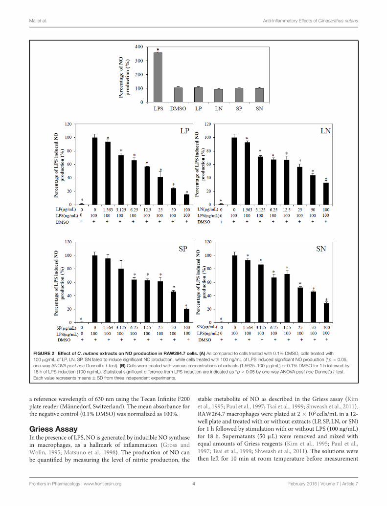
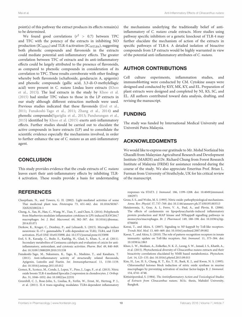
TABLE 1 | Correlation between TPC and TFC of extracts and its IC50 values in Griess (IC50NO) and TLR-4 activation assay (IC50TLR4).
Extracts TPC(mg GAE/ g dm)
TFC(mg QE/ g dm)
IC50NO
(μg/mL)Correlationbetween TPCand IC50NO
Correlationbetween TFCand IC50NO
IC50TLR4
(μg/mL)Correlationbetween TPCand IC50TLR4
Correlationbetween TFCand IC50TLR4
LP 7.99 ± 0.6 16.09 ± 4.2 18.9 ± 3.6 r2 = 0.731 r2 = 0.839 21.3 ± 5.0 r2 = 0.764 r2 = 0.854
LN 3.26 ± 0.9 4.97 ± 1.3 37.1 ± 7.2 29.4 ± 9.0
SP 2.47 ± 0.4 3.75 ± 0.7 43.1 ± 4.7 27.2 ± 1.0
SN 1.43 ± 0.1 3.27 ± 1.1 33.8 ± 2.5 27.5 ± 6.3
Each value represents means ± standard deviation from three independent experiments.
on a microplate reader at 540 nm. The IC50NO, indicating theconcentration at which extracts inhibited 50% of LPS inducedNOproduction was determined.
TLR-4 Activation AssayHEK-BlueTM hTLR4 cells were plated at 1 × 10⁵ cells/mL andextracts were added simultaneously. DMSO (0.1%) was addedas a control solvent. In the presence of a TLR-4 agonist, suchas LPS, the TLR-4 is expected to be activated. Activated TLR-4 induces NF-κB and activator protein-1 (AP-1) activation,under the control of NF-κB/AP-1 promoter, following which,the promoter will then induce secreted embryonic alkalinephosphatase (SEAP) production. The levels of SEAP productionwere determined using the HEK-BlueTM Detection Medium(InvivoGen, San Diego, CA, USA), a detection medium whichchanges color into purple or blue in the presence of SEAP after24 h incubation. An extract will be accepted as a TLR-4 activatorif the percentage of TLR-4 activation is more than 100% ascompared cells treated with solvent control, 0.1% DMSO in theabsence of LPS. Conversely, an extract will be concluded as aTLR-4 inhibitor if the extract can inhibit LPS induced TLR-4 activation. The SEAP levels were quantified using the TecanInfinite F200 plate reader (Männedorf, Switzerland) at 630nm.The increase in the level of SEAP is directly proportionate toincreased NF-κB activation, resulting in higher degree of TLR-4activation. Finally, the IC50TLR4, indicating the concentration inwhich extracts inhibited 50% of LPS induced TLR-4 activity wasdetermined.
Cytokine AssayMacrophages RAW264.7 cells were plated and either non-treated or treated with extracts (LP, SP, LN, SN) for 1 hfollowed by exposure to LPS for 18 h. Ten cytokines, namelythe interleukin IL-1β, IL-2, IL-4, IL-5, IL-6, IL-10, IL-12p40,IL-17, IFN-γ, and tumor necrosis factor-alpha (TNF-α), wereassayed in the supernatants using the MILLIPLEX R© MAPMouseCytokine/Chemokine Magnetic Bead panel (Millipore, Germany)according to the manufacturer’s instructions. For the platewashing steps, a handheld magnet attached to a plate holder wasused and the assay was performed on the Luminex 200 multiplexanalyser (Austin, USA) using the Luminex Software xPONENTR©
3.1 (Austin, USA) for data acquisition. The Median FluorescentIntensity data using a 5-parameter logistic or spline curve-fittingmethod was used for calculating cytokine concentrations in
samples. The results were normalised with cells treated with100 ng/mL of LPS.
ImmunoblottingProtein lysates from the macrophages treated with 0.1% DMSO,LPS (100 ng/mL); the most potent extract (LP 20 μg/mL)with or without LPS (100 ng/mL) were extracted in an ice-cold lysis buffer (1%-NP-40, 1 mM dithiothreitol and proteaseinhibitors cocktail in phosphate buffer saline, PBS). Proteins(50 μg) were separated by 7.5% SDS-PAGE, and transferred ontopolyvinylidene fluoride (PVDF) membranes. The membraneswere blocked for non-specific binding for 1 h in 5% BSA dilutedwith PBST (PBS and Tween-20). The blots were incubatedovernight with respective 50 ng/mL primary antibodies, such asphosphorylated p65 (p-p65), p65, phosphorylated p38 mitogenactivated protein kinase (p-p38), p38, phosphorylated ERKs 1/2(p-ERK 1/2), ERK1/2, phosphorylated c-Jun N-terminal kinase1/2 (p-JNK 1/2), JNK1/2, phosphorylated interferon regulatoryfactor 3 (p-IRF3), IRF3, and β-actin. The blots were washed withPBST before incubated with respective horseradish peroxidise-conjugated secondary antibody. All antibodies used in this studywere obtained from Cell Signalling Technology, USA. The blotswere subjected to enhanced luminol-based chemiluminescentreagents.
Statistical AnalysisAll data were reported as mean ± standard deviation froma minimum of three independent experiments. Statisticalsignificance was analysed using one-way analysis of variance(ANOVA) and post testing using Dunnett’s test through SPSS(version 18.0) for Windows. A p-value of less than 0.05 (p < 0.05)was considered significantly different compared to negativecontrol, treatment with 0.1% DMSO.
RESULTS
Clinacanthus nutans Extracts are NotCytotoxic to HEK-BlueTM-hTLR4 or RAW264.7 CellsCells (HEK-BlueTM-hTLR4 cells and macrophages) weretreated with various concentrations (1.5625–100 μg/mL)of LP, LN, SP, or SN for 72 h. The viability of HEK cells(HEK-BlueTM-hTLR4) and murine macrophages (RAW264.7) were not significantly reduced by any of the extracts
Frontiers in Pharmacology | www.frontiersin.org 5 February 2016 | Volume 7 | Article 7
Mai et al. Anti-Inflammatory Effects of Clinacanthus nutans
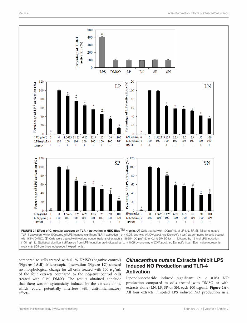
FIGURE 3 | Effect of C. nutans extracts on TLR-4 activation in HEK-BlueTM-4 cells. (A) Cells treated with 100μg/mL of LP, LN, SP, SN failed to induceTLR-4 activation, while 100ng/mL of LPS induced significant TLR-4 activation (∗p < 0.05, one-way ANOVA post hoc Dunnett’s t-test) as compared to cells treatedwith 0.1% DMSO. (B) Cells were treated with various concentrations of extracts (1.5625–100 μg/mL) or 0.1% DMSO for 1 h followed by 18 h of LPS induction(100 ng/mL). Statistical significant difference from LPS induction are indicated as ∗p < 0.05 by one-way ANOVA post hoc Dunnet’s t-test. Each value representsmeans ± SD from three independent experiments.
compared to cells treated with 0.1% DMSO (negative control)(Figures 1A,B). Microscopic observation (Figure 1C) showedno morphological change for all cells treated with 100 μg/mLof the four extracts compared to the negative control cellstreated with 0.1% DMSO. The results obtained concludethat there was no cytotoxicity induced by the extracts alone,which could potentially interfere with anti-inflammatoryeffects.
Clinacanthus nutans Extracts Inhibit LPSInduced NO Production and TLR-4ActivationLipopolysaccharide induced significant (p < 0.05) NOproduction compared to cells treated with DMSO or withextracts alone (LN, LP, SP, or SN, each 100 μg/mL; Figure 2A).All four extracts inhibited LPS induced NO production in a
Frontiers in Pharmacology | www.frontiersin.org 6 February 2016 | Volume 7 | Article 7
Mai et al. Anti-Inflammatory Effects of Clinacanthus nutans
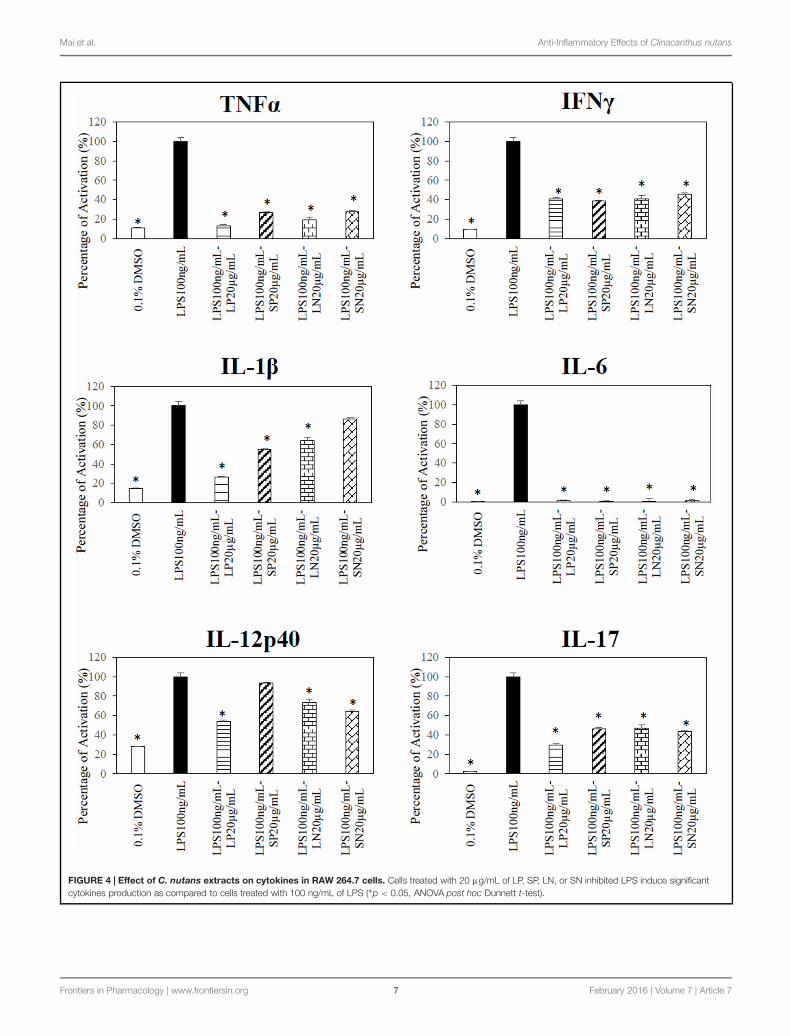
FIGURE 4 | Effect of C. nutans extracts on cytokines in RAW 264.7 cells. Cells treated with 20 μg/mL of LP, SP, LN, or SN inhibited LPS induce significantcytokines production as compared to cells treated with 100 ng/mL of LPS (∗p < 0.05, ANOVA post hoc Dunnett t-test).
Frontiers in Pharmacology | www.frontiersin.org 7 February 2016 | Volume 7 | Article 7
Mai et al. Anti-Inflammatory Effects of Clinacanthus nutans
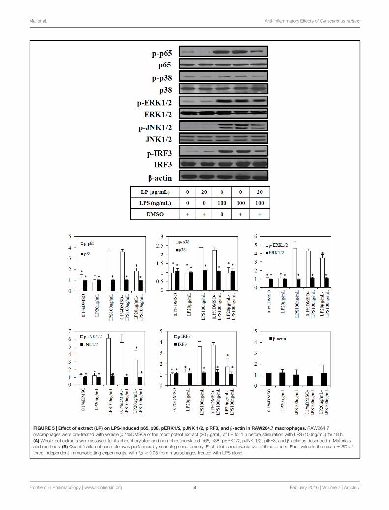
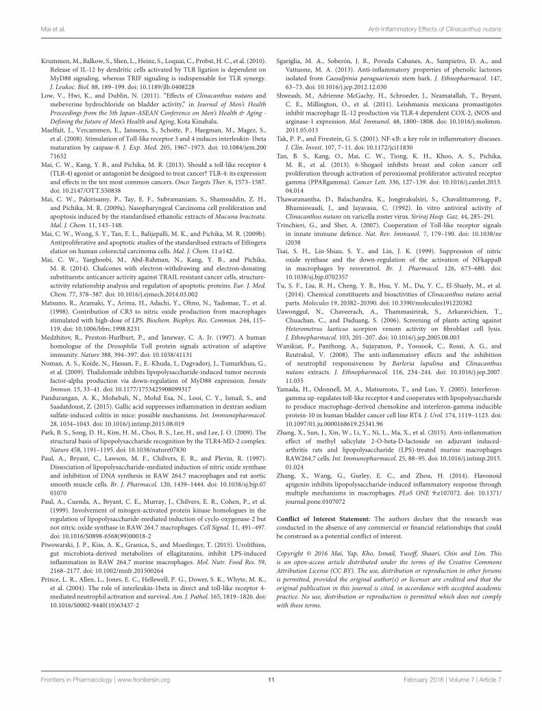
FIGURE 5 | Effect of extract (LP) on LPS-induced p65, p38, pERK1/2, pJNK 1/2, pIRF3, and β-actin in RAW264.7 macrophages. RAW264.7macrophages were pre-treated with vehicle (0.1%DMSO) or the most potent extract (20 μg/mL) of LP for 1 h before stimulation with LPS (100ng/mL) for 18 h.(A) Whole-cell extracts were assayed for its phosphorylated and non-phosphorylated p65, p38, pERK1/2, pJNK 1/2, pIRF3, and β-actin as described in Materialsand methods. (B) Quantification of each blot was performed by scanning densitometry. Each blot is representative of three others. Each value is the mean ± SD ofthree independent immunoblotting experiments, with ∗p < 0.05 from macrophages treated with LPS alone.
Frontiers in Pharmacology | www.frontiersin.org 8 February 2016 | Volume 7 | Article 7
Mai et al. Anti-Inflammatory Effects of Clinacanthus nutans
concentration-dependent manner (Figure 2B), with LP beingthe most potent (IC50NO = 18.9 ± 3.6 μg/mL; Table 1).
Toll-like receptor-4 activation is the hallmark of inflammation(Mai et al., 2013). LPS (100 ng/mL) induced significant(p < 0.05) TLR-4 activation in HEK-BlueTM hTLR4 cellscompared with cells treated DMSO or with extracts alone (LN,LP, SP, or SN, each 100 μg/mL; Figure 3A). All four extractsinhibited LPS-induced TLR-4 activation in a concentration-dependent manner (Figure 3B), with LP being the most potent(IC50TLR4 = 21.3 ± 5.00 μg/mL; Table 1).
Inhibition of TLR 4 Activation and of NOProduction by C. nutans Extracts areRelated to Its Phenolic Compounds andFlavonoidsClinacanthus nutans extracts indicated content of phenoliccompounds (TPC = 1.43 to 7.99 mg GAE/g dm) and flavonoids(TFC = 3.27 to 16.09 mg QE/g dm). The leaf extracts extractedwith methanol and dichloromethane (LP), showed the highestTPC and TFC among all the extracts. A higher correlation wasobtained between TFC and IC50NO (r2 = 0.839) compared tocorrelation between TPC and IC50NO (r2 = 0.731). Similarly,a higher correlation was obtained between TFC and IC50TLR4(r2 = 0.854) compared to correlation between TPC and IC50TLR4(r2 = 0.764). These results suggest the flavonoids in the extractsmay play a role in inhibiting LPS-induced NO production andLPS-induced TLR-4 activation.
Clinacanthus nutans Extracts Inhibit LPSInduced Cytokines ProductionPolar leaf extract and LN significantly (p < 0.05) inhibitedLPS-induced TNF-α, IFN-γ, IL-1β, IL-6, IL12p40, and IL-17production (Figure 4). The SN extract significantly inhibited theLPS-induced production of TNF-α, IFN-γ, IL-6, IL12p40, and IL-17 but not IL-1β, whereas the SP extract inhibited the productionof TNF-α, IFN-γ, IL-1β, IL-6, and IL-17 but not that of IL12p40.Four cytokines (IL-2, IL-4, IL-5, and IL-10) were below detectablelimits (results not shown).
Clinacanthus nutans extracts inhibit LPSinduced TLR-4 inflammatory proteinsBased on the results from the Griess assay and TLR-4 activationassay, the most potent anti-inflammatory extract (LP) wasselected for immunoblotting. Exposure of macrophages to100 ng/mL of LPS or combination of 0.1%DMSO and LPSled to phosphorylation of p65, p38, ERK1/2, JNK1/2, andIRF3 (Figure 5A)., The extract (20 μg/mL of LP (≈IC50NO)significantly (p< 0.05) reduced the LPS induced phosphorylationof p65, p38, ERK1/2, JNK1/2, and IRF3. In unstimulatedmacrophages cells either with LP alone or 0.1% DMSO, therewas no significant phosphorylation of p65, p38, ERK1/2, JNK1/2,and IRF3 (Figure 5B). No significant change was observed in theloading control proteins, β-actin, between treated and untreatedcells.
DISCUSSION
Although C. nutans extracts have been previously shown toreduce superoxide anion production (Tu et al., 2014), and toinhibit neutrophil responsiveness (Wanikiat et al., 2008), themechanisms underlying its anti-inflammatory effects remainunknown.
Therefore, our study was carried out to examine the effectsof extracts on inflammatory processes using the well-establishedLPS-induced inflammation macrophage model (Paul et al., 1999;Hatziieremia et al., 2006; Cheng et al., 2014; Piwowarski et al.,2015; Zhang et al., 2015). LPS produces a powerful inflammatoryresponse through activation of the TLR-4, resulting in activationof nuclear factor-κB (NF-κB) and the production of NO andinflammatory cytokines, including IFN-γ, TNF-α, IL-1β, IL-6,IL-12p40, and IL-1 (Medzhitov et al., 1997; Kawai and Akira,2007; Trinchieri and Sher, 2007). In the present study, wefound that the extracts of C. nutans produced a marked andconcentration-dependent inhibition of the production of NOand the inflammatory cytokines; this was clearly unrelated tocytotoxicity caused by the extracts when administered alone. Theroles of NO (Hatziieremia et al., 2006; Gomez et al., 2013), IL-1β (Maelfait et al., 2008), TNF-α (Prince et al., 2004; Nomanet al., 2009), IFN-γ (Yamada et al., 2005), IL-6 (Greenhill et al.,2011), IL-12 (Krummen et al., 2010), and IL-17 (Derkow et al.,2015) in the TLR-4 related inflammatory responses have beenwell established. The suppression of the production of NO andthese cytokines suggested that the anti-inflammatory effects ofthese extracts were related to inhibition of TLR-4 activation.
Unlike other TLR, TLR-4 is the only TLR that activates bothmyeloid differentiation primary response 88 (MyD88) dependentand Toll/IL-1R domain containing adapter inducing IFN-β(TRIF) dependent pathways in the presence of LPS. MyD88recruits the IL-1 receptor-associated kinases and causes theactivation of NF-κB, (such as p65), p38, ERK, and Jun N-terminalkinase (JNK) (Kawai and Akira, 2007). NF-κB activation, adownstream pathway of TLR-4, is a hallmark for inflammation,contributing to chronic diseases such as inflammatory boweldisease, systemic inflammatory response syndrome, and chronicinflammatory demyelinating polyradiculoneuritis (Tak andFirestein, 2001). In the TRIF pathway, TRIF activates tumornecrosis factor receptor-associated factor (TRAF) family memberthrough TRAF3 (Kawai and Akira, 2010). TRAF3 promotes IRF3activation and subsequently IFN-γ induction (Kawai and Akira,2010; Mai et al., 2013). The most potent of our extracts (LP)inhibited LPS-induced phosphorylation of p65, p38, ERK, JNK,and IRF3. These results correlated with the ability of the extractsto inhibit LPS induced TLR-4 activities. Moreover, the extractreduced LPS-induced production of IFN-γ and phosphorylationof IRF3. These are the first results showing C. nutans to targetboth the MyD88 dependent and TRIF dependent pathways. Thefinding that TLR-4 activation was prevented by the extracts ofC. nutans as measured by SEAP production in response to LPS,lends further support to the hypothesis that C. nutans exertsits anti-inflammatory effects by preventing the activation of theTLR-4 receptor, thus reducing the production of inflammatorycytokines and TLR-4 related inflammatory proteins. At which
Frontiers in Pharmacology | www.frontiersin.org 9 February 2016 | Volume 7 | Article 7
Mai et al. Anti-Inflammatory Effects of Clinacanthus nutans
point(s) of this pathway the extract produces its effects remain(s)to be determined.
We found good correlations (r2 > 0.7) between TPCand TFC with the potency of the extracts in inhibiting NOproduction (IC50NO) and TLR-4 activation (IC50TLR4), suggestingboth phenolic compounds and flavonoids in the extractscould mediate potential anti-inflammatory effects. The greatercorrelation between TFC of extracts and its anti-inflammatoryeffects could be largely attributed to the presence of flavonoids,as compared to phenolic compounds in consideration of itscorrelation to TPC. These results corroborate with other findingswhereby both flavonoids (schaftoside, gendarucin A, apigenin)and phenolic compounds (gallic acid, 3,3-di-O-methylellagicacid) were present in C. nutans Lindau leave extracts (Khooet al., 2015). The leaf extracts in the study by Khoo et al.(2015) had similar TPC values to those in the LP extracts inour study although different extraction methods were used.Previous studies indicated that these flavonoids (Erel et al.,2011; Funakoshi-Tago et al., 2011; Zhang et al., 2014) andphenolic compounds(Sgariglia et al., 2013; Pandurangan et al.,2015) identified by Khoo et al. (2015) exerts anti-inflammatoryeffects. Further studies should be carried out to elucidate theactive compounds in leave extracts (LP) and to consolidate thescientific evidence especially the mechanisms involved, in orderto further enhance the use of C. nutans as an anti-inflammatoryagent.
CONCLUSION
This study provides evidence that the crude extracts of C. nutansleaves exert their anti-inflammatory effects by inhibiting TLR-4 activation. These results provide a basis for understanding
the mechanisms underlying the traditionally belief of anti-inflammatory of C. nutans crude extracts. More studies usingpathway specific inhibitors or a genetic knockout of TLR-4 mayfurther elucidate the mechanism of action of the extracts inspecific pathways of TLR-4. A detailed isolation of bioactivecompounds from LP extracts would be highly warranted in viewof the potential anti-inflammatory attributes of C. nutans.
AUTHOR CONTRIBUTIONS
Cell culture experiments, inflammation studies, andimmunoblotting were conducted by CM. Cytokine assays weredesigned and conducted by KSY, MK, KY, and EL. Preparation ofplant extracts were designed and completed by NI, KS, SC, andEL. All authors contributed toward data analysis, drafting, andrevising the manuscript.
FUNDING
The study was funded by International Medical University andUniversiti Putra Malaysia.
ACKNOWLEDGMENTS
Wewould like to express our gratitude toMr.MohdNorfaizal binGhazalli fromMalaysian Agricultural Research and DevelopmentInstitute (MARDI) and Dr. Richard Chung from Forest ResearchInstitute of Malaysia (FRIM) for assistance rendered during thecourse of the study. We also appreciate Emeritus Prof. Brian L.Furman from University of Strathclyde, UK for his critical reviewof the manuscript.
REFERENCES
Cheeptham, N., and Towers, G. H. (2002). Light-mediated activities of someThai medicinal plant teas. Fitoterapia 73, 651–662. doi: 10.1016/S0367-326X(02)00224-1
Cheng, A., Yan, H., Han, C., Wang, W., Tian, Y., and Chen, X. (2014). Polyphenolsfrom blueberries modulate inflammation cytokines in LPS-induced RAW264.7macrophages. Int. J. Biol. Macromol. 69, 382–387. doi: 10.1016/j.ijbiomac.2014.05.071
Derkow, K., Kruger, C., Dembny, P., and Lehnardt, S. (2015). Microglia induceneurotoxic IL-17+ gammadelta T cells dependent on TLR2, TLR4, and TLR9activation. PLoS ONE 10:e0135898. doi: 10.1371/journal.pone.0135898
Erel, S. B., Karaalp, C., Bedir, E., Kaehlig, H., Glasl, S., Khan, S., et al. (2011).Secondary metabolites of Centaurea calolepis and evaluation of cnicin for anti-inflammatory, antioxidant, and cytotoxic activities. Pharm. Biol. 49, 840–849.doi: 10.3109/13880209.2010.551538
Funakoshi-Tago, M., Nakamura, K., Tago, K., Mashino, T., and Kasahara, T.(2011). Anti-inflammatory activity of structurally related flavonoids,Apigenin, Luteolin and Fisetin. Int. Immunopharmacol. 11, 1150–1159.doi: 10.1016/j.intimp.2011.03.012
Gomez, R., Scotece, M., Conde, J., Lopez, V., Pino, J., Lago, F., et al. (2013). Nitricoxide boosts TLR-4 mediated lipocalin 2 expression in chondrocytes. J. Orthop.Res. 31, 1046–1052. doi: 10.1002/jor.22331
Greenhill, C. J., Rose-John, S., Lissilaa, R., Ferlin, W., Ernst, M., Hertzog, P. J.,et al. (2011). IL-6 trans-signaling modulates TLR4-dependent inflammatory
responses via STAT3. J. Immunol. 186, 1199–1208. doi: 10.4049/jimmunol.1002971
Gross, S. S., andWolin, M. S. (1995). Nitric oxide: pathophysiological mechanisms.Annu. Rev. Physiol. 57, 737–769. doi: 10.1146/annurev.ph.57.030195.003513
Hatziieremia, S., Gray, A. I., Ferro, V. A., Paul, A., and Plevin, R. (2006).The effects of cardamonin on lipopolysaccharide-induced inflammatoryprotein production and MAP kinase and NFkappaB signalling pathways inmonocytes/macrophages. Br. J. Pharmacol. 149, 188–198. doi: 10.1038/sj.bjp.0706856
Kawai, T., and Akira, S. (2007). Signaling to NF-kappaB by Toll-like receptors.Trends Mol. Med. 13, 460–469. doi: 10.1016/j.molmed.2007.09.002
Kawai, T., and Akira, S. (2010). The role of pattern-recognition receptors in innateimmunity: update on Toll-like receptors. Nat. Immunol. 11, 373–384. doi:10.1038/ni.1863
Khoo, L. W., Mediani, A., Zolkeflee, N. K. Z., Leong, S. W., Ismail, I. S., Khatib, A.,et al. (2015). Phytochemical diversity of Clinacanthus nutans extracts and theirbioactivity correlations elucidated by NMR based metabolomics. Phytochem.Lett. 14, 123–133. doi: 10.1016/j.phytol.2015.09.015
Kim, H., Lee, H. S., Chang, K. T., Ko, T. H., Baek, K. J., and Kwon, N. S. (1995).Chloromethyl ketones block induction of nitric oxide synthase in murinemacrophages by preventing activation of nuclear factor-kappa B. J. Immunol.154, 4741–4748.
Kittisiripornkul, S. (1984). The Antiinflammatory Action and Toxicological Studiesof Extracts from Clinacanthus nutans. M.Sc. thesis, Mahidol University,Bangkok.
Frontiers in Pharmacology | www.frontiersin.org 10 February 2016 | Volume 7 | Article 7
Mai et al. Anti-Inflammatory Effects of Clinacanthus nutans
Krummen,M., Balkow, S., Shen, L., Heinz, S., Loquai, C., Probst, H. C., et al. (2010).Release of IL-12 by dendritic cells activated by TLR ligation is dependent onMyD88 signaling, whereas TRIF signaling is indispensable for TLR synergy.J. Leukoc. Biol. 88, 189–199. doi: 10.1189/jlb.0408228
Low, V., Hwi, K., and Dublin, N. (2011). “Effects of Clinacanthus nutans andmebeverine hydrochloride on bladder activity,” in Journal of Men’s HealthProceedings from the 5th Japan-ASEAN Conference on Men’s Health & Aging -Defining the future of Men’s Health and Aging, Kota Kinabalu.
Maelfait, J., Vercammen, E., Janssens, S., Schotte, P., Haegman, M., Magez, S.,et al. (2008). Stimulation of Toll-like receptor 3 and 4 induces interleukin-1betamaturation by caspase-8. J. Exp. Med. 205, 1967–1973. doi: 10.1084/jem.20071632
Mai, C. W., Kang, Y. B., and Pichika, M. R. (2013). Should a toll-like receptor 4(TLR-4) agonist or antagonist be designed to treat cancer? TLR-4: its expressionand effects in the ten most common cancers. Onco Targets Ther. 6, 1573–1587.doi: 10.2147/OTT.S50838
Mai, C. W., Pakirisamy, P., Tay, E. F., Subramaniam, S., Shamsuddin, Z. H.,and Pichika, M. R. (2009a). Nasopharyngeal Carcinoma cell proliferation andapoptosis induced by the standardised ethanolic extracts of Mucuna bracteata.Mal. J. Chem. 11, 143–148.
Mai, C. W., Wong, S. Y., Tan, E. L., Balijepalli, M. K., and Pichika, M. R. (2009b).Antiproliferative and apoptotic studies of the standardised extracts of Etlingeraelatior on human colorectal carcinoma cells.Mal. J. Chem. 11:e142.
Mai, C. W., Yaeghoobi, M., Abd-Rahman, N., Kang, Y. B., and Pichika,M. R. (2014). Chalcones with electron-withdrawing and electron-donatingsubstituents: anticancer activity against TRAIL resistant cancer cells, structure-activity relationship analysis and regulation of apoptotic proteins. Eur. J. Med.Chem. 77, 378–387. doi: 10.1016/j.ejmech.2014.03.002
Matsuno, R., Aramaki, Y., Arima, H., Adachi, Y., Ohno, N., Yadomae, T., et al.(1998). Contribution of CR3 to nitric oxide production from macrophagesstimulated with high-dose of LPS. Biochem. Biophys. Res. Commun. 244, 115–119. doi: 10.1006/bbrc.1998.8231
Medzhitov, R., Preston-Hurlburt, P., and Janeway, C. A. Jr. (1997). A humanhomologue of the Drosophila Toll protein signals activation of adaptiveimmunity. Nature 388, 394–397. doi: 10.1038/41131
Noman, A. S., Koide, N., Hassan, F., E.-Khuda, I., Dagvadorj, J., Tumurkhuu, G.,et al. (2009). Thalidomide inhibits lipopolysaccharide-induced tumor necrosisfactor-alpha production via down-regulation of MyD88 expression. InnateImmun. 15, 33–41. doi: 10.1177/1753425908099317
Pandurangan, A. K., Mohebali, N., Mohd Esa, N., Looi, C. Y., Ismail, S., andSaadatdoust, Z. (2015). Gallic acid suppresses inflammation in dextran sodiumsulfate-induced colitis in mice: possible mechanisms. Int. Immunopharmacol.28, 1034–1043. doi: 10.1016/j.intimp.2015.08.019
Park, B. S., Song, D. H., Kim, H. M., Choi, B. S., Lee, H., and Lee, J. O. (2009). Thestructural basis of lipopolysaccharide recognition by the TLR4-MD-2 complex.Nature 458, 1191–1195. doi: 10.1038/nature07830
Paul, A., Bryant, C., Lawson, M. F., Chilvers, E. R., and Plevin, R. (1997).Dissociation of lipopolysaccharide-mediated induction of nitric oxide synthaseand inhibition of DNA synthesis in RAW 264.7 macrophages and rat aorticsmooth muscle cells. Br. J. Pharmacol. 120, 1439–1444. doi: 10.1038/sj.bjp.0701070
Paul, A., Cuenda, A., Bryant, C. E., Murray, J., Chilvers, E. R., Cohen, P., et al.(1999). Involvement of mitogen-activated protein kinase homologues in theregulation of lipopolysaccharide-mediated induction of cyclo-oxygenase-2 butnot nitric oxide synthase in RAW 264.7 macrophages. Cell Signal. 11, 491–497.doi: 10.1016/S0898-6568(99)00018-2
Piwowarski, J. P., Kiss, A. K., Granica, S., and Moeslinger, T. (2015). Urolithins,gut microbiota-derived metabolites of ellagitannins, inhibit LPS-inducedinflammation in RAW 264.7 murine macrophages. Mol. Nutr. Food Res. 59,2168–2177. doi: 10.1002/mnfr.201500264
Prince, L. R., Allen, L., Jones, E. C., Hellewell, P. G., Dower, S. K., Whyte, M. K.,et al. (2004). The role of interleukin-1beta in direct and toll-like receptor 4-mediated neutrophil activation and survival.Am. J. Pathol. 165, 1819–1826. doi:10.1016/S0002-9440(10)63437-2
Sgariglia, M. A., Soberón, J. R., Poveda Cabanes, A., Sampietro, D. A., andVattuone, M. A. (2013). Anti-inflammatory properties of phenolic lactonesisolated from Caesalpinia paraguariensis stem bark. J. Ethnopharmacol. 147,63–73. doi: 10.1016/j.jep.2012.12.030
Shweash, M., Adrienne McGachy, H., Schroeder, J., Neamatallah, T., Bryant,C. E., Millington, O., et al. (2011). Leishmania mexicana promastigotesinhibit macrophage IL-12 production via TLR-4 dependent COX-2, iNOS andarginase-1 expression. Mol. Immunol. 48, 1800–1808. doi: 10.1016/j.molimm.2011.05.013
Tak, P. P., and Firestein, G. S. (2001). NF-κB: a key role in inflammatory diseases.J. Clin. Invest. 107, 7–11. doi: 10.1172/jci11830
Tan, B. S., Kang, O., Mai, C. W., Tiong, K. H., Khoo, A. S., Pichika,M. R., et al. (2013). 6-Shogaol inhibits breast and colon cancer cellproliferation through activation of peroxisomal proliferator activated receptorgamma (PPARgamma). Cancer Lett. 336, 127–139. doi: 10.1016/j.canlet.2013.04.014
Thawaranantha, D., Balachandra, K., Jongtrakulsiri, S., Chavalittumrong, P.,Bhumiswasdi, J., and Jayavasu, C. (1992). In vitro antiviral activity ofClinacanthus nutans on varicella zoster virus. Siriraj Hosp. Gaz. 44, 285–291.
Trinchieri, G., and Sher, A. (2007). Cooperation of Toll-like receptor signalsin innate immune defence. Nat. Rev. Immunol. 7, 179–190. doi: 10.1038/nri2038
Tsai, S. H., Lin-Shiau, S. Y., and Lin, J. K. (1999). Suppression of nitricoxide synthase and the down-regulation of the activation of NFkappaBin macrophages by resveratrol. Br. J. Pharmacol. 126, 673–680. doi:10.1038/sj.bjp.0702357
Tu, S. F., Liu, R. H., Cheng, Y. B., Hsu, Y. M., Du, Y. C., El-Shazly, M., et al.(2014). Chemical constituents and bioactivities of Clinacanthus nutans aerialparts.Molecules 19, 20382–20390. doi: 10.3390/molecules191220382
Uawonggul, N., Chaveerach, A., Thammasirirak, S., Arkaravichien, T.,Chuachan, C., and Daduang, S. (2006). Screening of plants acting againstHeterometrus laoticus scorpion venom activity on fibroblast cell lysis.J. Ethnopharmacol. 103, 201–207. doi: 10.1016/j.jep.2005.08.003
Wanikiat, P., Panthong, A., Sujayanon, P., Yoosook, C., Rossi, A. G., andReutrakul, V. (2008). The anti-inflammatory effects and the inhibitionof neutrophil responsiveness by Barleria lupulina and Clinacanthusnutans extracts. J. Ethnopharmacol. 116, 234–244. doi: 10.1016/j.jep.2007.11.035
Yamada, H., Odonnell, M. A., Matsumoto, T., and Luo, Y. (2005). Interferon-gamma up-regulates toll-like receptor 4 and cooperates with lipopolysaccharideto produce macrophage-derived chemokine and interferon-gamma inducibleprotein-10 in human bladder cancer cell line RT4. J. Urol. 174, 1119–1123. doi:10.1097/01.ju.0000168619.25341.96
Zhang, X., Sun, J., Xin, W., Li, Y., Ni, L., Ma, X., et al. (2015). Anti-inflammationeffect of methyl salicylate 2-O-beta-D-lactoside on adjuvant induced-arthritis rats and lipopolysaccharide (LPS)-treated murine macrophagesRAW264.7 cells. Int. Immunopharmacol. 25, 88–95. doi: 10.1016/j.intimp.2015.01.024
Zhang, X., Wang, G., Gurley, E. C., and Zhou, H. (2014). Flavonoidapigenin inhibits lipopolysaccharide-induced inflammatory response throughmultiple mechanisms in macrophages. PLoS ONE 9:e107072. doi: 10.1371/journal.pone.0107072
Conflict of Interest Statement: The authors declare that the research wasconducted in the absence of any commercial or financial relationships that couldbe construed as a potential conflict of interest.
Copyright © 2016 Mai, Yap, Kho, Ismail, Yusoff, Shaari, Chin and Lim. Thisis an open-access article distributed under the terms of the Creative CommonsAttribution License (CC BY). The use, distribution or reproduction in other forumsis permitted, provided the original author(s) or licensor are credited and that theoriginal publication in this journal is cited, in accordance with accepted academicpractice. No use, distribution or reproduction is permitted which does not complywith these terms.
Frontiers in Pharmacology | www.frontiersin.org 11 February 2016 | Volume 7 | Article 7