Embed Size (px)
Citation preview
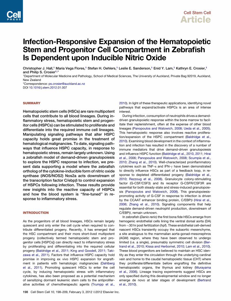
Cell Stem Cell
Article
Infection-Responsive Expansion of the HematopoieticStem and Progenitor Cell Compartment in ZebrafishIs Dependent upon Inducible Nitric OxideChristopher J. Hall,1 Maria Vega Flores,1 Stefan H. Oehlers,1 Leslie E. Sanderson,1 Enid Y. Lam,1 Kathryn E. Crosier,1
and Philip S. Crosier1,*1Department of Molecular Medicine and Pathology, School of Medical Sciences, The University of Auckland, Private Bag 92019, Auckland,
New Zealand*Correspondence: [email protected]
DOI 10.1016/j.stem.2012.01.007
SUMMARY
Hematopoietic stem cells (HSCs) are raremultipotentcells that contribute to all blood lineages. During in-flammatory stress, hematopoietic stem and progen-itor cells (HSPCs) can be stimulated to proliferate anddifferentiate into the required immune cell lineages.Manipulating signaling pathways that alter HSPCcapacity holds great promise in the treatment ofhematological malignancies. To date, signaling path-ways that influence HSPC capacity, in response tohematopoietic stress, remain largely unknown. Usinga zebrafish model of demand-driven granulopoiesisto explore the HSPC response to infection, we pre-sent data supporting a model where the zebrafishortholog of the cytokine-inducible form of nitric oxidesynthase (iNOS/NOS2) Nos2a acts downstream ofthe transcription factor C/ebpb to control expansionof HSPCs following infection. These results providenew insights into the reactive capacity of HSPCsand how the blood system is ‘‘fine-tuned’’ in re-sponse to inflammatory stress.
INTRODUCTION
As the progenitors of all blood lineages, HSCs remain largely
quiescent and only enter the cell cycle when required to con-
tribute differentiated progeny. Recently, it has emerged that
the HSC compartment and their more short-lived multipotent
progeny (collectively termed hematopoietic stem and pro-
genitor cells [HSPCs]) can directly react to inflammatory stress
by proliferating and differentiating into the required cellular
progeny (Baldridge et al., 2011; King and Goodell, 2011; Taki-
zawa et al., 2011). Factors that influence HSPC capacity hold
promise in improving ex vivo HSPC expansion for engraft-
ment in patients with hematologic malignancies (Dahlberg
et al., 2011). Promoting quiescent HSCs to enter the cell
cycle, by inducing hematopoietic stress with inflammatory
cytokines, has also been proposed as a potential mechanism
of sensitizing dormant leukemic stem cells to the antiprolifer-
ative activities of chemotherapeutic agents (Trumpp et al.,
198 Cell Stem Cell 10, 198–209, February 3, 2012 ª2012 Elsevier Inc
2010). In light of these therapeutic applications, identifying novel
pathways that expand/activate HSPCs is an area of intense
interest.
During infection, consumption of neutrophils drives a demand-
driven granulopoietic response within the bone marrow to facil-
itate their replenishment, often at the expense of other blood
lineages (Panopoulos and Watowich, 2008; Ueda et al., 2005).
This hematopoietic response also involves reactive prolifera-
tion/expansion of the HSPC compartment (Baldridge et al.,
2010). Examining blood development in the context of inflamma-
tion and infection has resulted in the discovery of a number of
immune mediators that drive demand-driven granulopoiesis
and influence HSPC function (Baldridge et al., 2010, 2011; Hirai
et al., 2006; Panopoulos and Watowich, 2008; Scumpia et al.,
2010; Zhang et al., 2010). Well-characterized proinflammatory
cytokines such as TNF-a and IFN-g have been demonstrated
to directly influence HSCs as part of a feedback loop, in re-
sponse to depleted differentiated progeny (Baldridge et al.,
2010; Rezzoug et al., 2008). Granulocyte colony-stimulating
factor (G-CSF/CSF3) and its receptor G-CSFR/CSF3R are
essential for both steady-state and stress-induced granulopoie-
sis (Panopoulos and Watowich, 2008). This granulopoiesis-
promoting activity of G-CSF in response to infection is driven
by the CCAAT enhancer binding protein, C/EBPb (Hirai et al.,
2006; Zhang et al., 2010). Signaling components that help
regulate demand-driven neutrophil production, downstream of
C/EBPb, remain unknown.
In zebrafish (Danio rerio) the first bona fide HSCs emerge from
hemogenic endothelial cells lining the ventral dorsal aorta (DA)
from�30 hr post fertilization (hpf). These endothelial cell-derived
nascent HSCs transiently occupy the subaortic mesenchyme,
a site analogous to the mammalian aorta-gonad-mesonephros
(AGM) region, where they have been observed to undergo
limited (i.e. a single), presumably symmetric cell division (Ber-
trand et al., 2010; Kissa and Herbomel, 2010; Lam et al., 2010).
These blood progenitors are believed to maintain an HSC iden-
tity as they enter the circulation through the underlying cardinal
vein and home to the caudal hematopoietic tissue (CHT) where
they proliferate/differentiate before engrafting the definitive
hematopoietic organs, the thymus and kidney (Murayama
et al., 2006). Lineage tracing experiments suggest HSCs are
only specified during this developmental window and no longer
emerge de novo at later stages of development (Bertrand
et al., 2010).
.
Cell Stem Cell
Hematopoietic Stem Cells and Infection
Employing a larval zebrafish model of demand-driven granulo-
poiesis to examine HSPC development through the ‘‘lens of
infection,’’ we provide new insight into how inflammatory stress
‘‘fine-tunes’’ hematopoiesis. We show that the zebrafish larval
hematopoietic system responds to infection through the reactive
expansion of the HSPC compartment. Further examination re-
vealed a previously unappreciated role for C/ebpb-dependent
Nos2a-generated nitric oxide (NO) during HSPC and neutrophil-
fated progenitor expansion following infection. These results
provide new mechanistic insight into how the HSPC compart-
ment responds to inflammatory stress.
RESULTS
Larval Hematopoietic Response to InfectionPrior to investigating the effect of infection on HSPCs, we exam-
ined whether larval hematopoiesis was responsive to infection.
To address whether zebrafish replenish neutrophils lost as
a result of cell death or apoptosis following infection, neutrophil
abundance within Tg(lyz:DsRED) larvae (a transgenic reporter
line that specifically marks neutrophils) was quantified through-
out the course of infection. To facilitate direct observation of
bacterial burden, a GFP-expressing Salmonella enterica serovar
Typhimurium (hereafter referred to as Sal-GFP) was selected
(Hall et al., 2007). Time-lapse imaging following injection of
Sal-GFP into the hindbrain ventricle of Tg(lyz:DsRED) larvae at
50 hpf (Figure 1A) revealed robust recruitment of neutrophils to
the midbrain and hindbrain. Highly motile neutrophils were
observed phagocytosing the injected bacteria as part of an
immediate innate immune response (Figure S1 and Movie S1,
available online). One day following bacterial injection (1 dpi
Infected), larvae demonstrated one of four phenotypes when
compared with PBS-injected controls (1 dpi PBS). Infected
larvae were: dead; overwhelmed by infection (such larvae never
survived to 2 dpi and were always removed from further anal-
ysis); possessing numbers of neutrophils similar to uninjected
controls that were dispersed throughout larval tissues (often
with low levels of bacterial burden remaining in the head); or
demonstrating a marked reduction in neutrophil numbers with
very little or no detectable bacterial burden (Figure S1; see also
Supplemental Experimental Procedures). Flow cytometry anal-
ysis revealing surviving larvae that possessed reduced neutro-
phil numbers at 1 dpi demonstrated a marked increase in
whole-larvae neutrophil numbers that peaked at 2 dpi before
gradually returning to steady-state levels, when compared with
PBS controls (Figure 1B).We next examinedwhether this neutro-
phil expansion was the result of enhanced granulopoiesis within
larval hematopoietic sites. Live imaging of infected larvae that
possessed an almost complete loss of neutrophils by 1 dpi
revealed de novo emergence of neutrophils within the AGM
and CHT regions from 1.25–1.5 dpi that was most evident at 2
dpi (Figures 1C and 1D). All further assessment of infection on
hematopoiesis focused on the AGM region (a region only
sparsely populated with neutrophils under steady-state condi-
tions) due to the confounding influence of steady-state granulo-
poiesis within the CHT. Throughout the remainder of this study
(unless otherwise stated), an optimized infection dose of
between 400 and 600 colony-forming units (cfu) was injected
into the hindbrain ventricle at 50 hpf. This gave a sufficient
Ce
proportion of larvae demonstrating neutrophil depletion at 1
dpi and subsequent demand-driven granulopoiesis, as scored
within the AGM region at 2 dpi (�55% to 60%of surviving larvae),
with survival of �85% to 90% (Figure S1). Unless otherwise
stated, data presented throughout the remainder of this study
as ‘‘Infected’’ refers to this surviving cohort.
In support of our live imaging studies, quantitative-(Q)PCR
analysis of the neutrophil-specific (lyz and mpx), pan-myeloid
(lcp1), and myeloid progenitor (spi1) genes following infection
demonstrated a peak in expression at �2 dpi (Figure S1)
(Lieschke et al., 2002; Lyons et al., 2001; Meijer et al., 2008).
The macrophage-specific marker csf1ra also increased fol-
lowing infection, suggesting that demand-driven hematopoiesis
may not be exclusively granulopoietic. To assess whether infec-
tion also resulted in expansion of themacrophage lineage, which
shares a common spi1-expressing progenitor with neutrophils,
we examined whether the macrophage-lineage marker mpeg1
was also expressed by cells within the AGM following infection
(Ellett et al., 2011). Although large numbers of macrophages
were detected within the infection site, consistent with the later
timing of macrophage infiltration during inflammation, no ex-
pression of mpeg1 was observed within the AGM region (Fig-
ure 1E). This was in contrast to spi1 expression, which strongly
marked myeloid progenitors within this domain (Figure 1E).
Expression analysis confirmed that these spi1-expressing pro-
genitor cells were fated to the neutrophil lineage, as evidenced
by lyz coexpression (Figure 1F).
To investigate whether demand-driven granulopoiesis influ-
enced the development of the lymphoid lineage, we examined
expression of the transcription factor ikaros, which typically
marks lymphoid progenitors within the AGM, following infection
at 2 dpi (Murayama et al., 2006; Willett et al., 2001) (Figure S1).
Quantifying the number of somite boundaries within the AGM
region containing ikaros+ cells revealed a significant decrease
in ikaros expression following infection (Figure S1). To examine
if this decrease in ikaros expression resulted in a decrease in
thymus-resident T cell precursors, we assessed the abundance
of lck-expressing thymocytes within infected double transgenic
Tg(lck:GFP)/Tg(lyz:DsRED) larvae at 3, 4, and 5 dpi, compared
with PBS controls (Figure S1). Coincident with initiation of
demand-driven granulopoiesis within the AGM and consistent
with a decrease in ikaros expression, a marked decrease in
lck:GFP-expressing thymocytes was observed following
infection.
These results demonstrate that the larval zebrafish hemato-
poietic system can respond to infection by specifically en-
hancing the production of neutrophils, at the expense of other
blood lineages.
Infected Larvae Possess an Expanded HSPCCompartmentTo demonstrate that demand-driven granulopoiesis within the
AGM was dependent upon definitive hematopoiesis (and by
association resident HSPCs), we assessed the ability of
Runx1-depleted embryos to replenish neutrophil numbers
following infection. Runx1 is a transcription factor that in zebra-
fish, as in mammals, is absolutely required for HSPC develop-
ment and definitive hematopoiesis (Lam et al., 2009). We have
shown that HSPC ontogeny, including their emergence within
ll Stem Cell 10, 198–209, February 3, 2012 ª2012 Elsevier Inc. 199
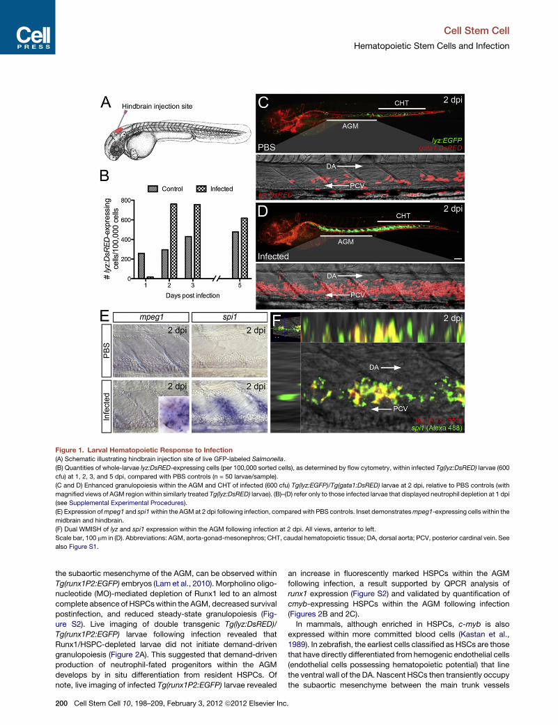
Figure 1. Larval Hematopoietic Response to Infection
(A) Schematic illustrating hindbrain injection site of live GFP-labeled Salmonella.
(B) Quantities of whole-larvae lyz:DsRED-expressing cells (per 100,000 sorted cells), as determined by flow cytometry, within infected Tg(lyz:DsRED) larvae (600
cfu) at 1, 2, 3, and 5 dpi, compared with PBS controls (n = 50 larvae/sample).
(C and D) Enhanced granulopoiesis within the AGM and CHT of infected (600 cfu) Tg(lyz:EGFP)/Tg(gata1:DsRED) larvae at 2 dpi, relative to PBS controls (with
magnified views of AGM region within similarly treated Tg(lyz:DsRED) larvae). (B)–(D) refer only to those infected larvae that displayed neutrophil depletion at 1 dpi
(see Supplemental Experimental Procedures).
(E) Expression ofmpeg1 and spi1within the AGMat 2 dpi following infection, comparedwith PBS controls. Inset demonstratesmpeg1-expressing cells within the
midbrain and hindbrain.
(F) Dual WMISH of lyz and spi1 expression within the AGM following infection at 2 dpi. All views, anterior to left.
Scale bar, 100 mm in (D). Abbreviations: AGM, aorta-gonad-mesonephros; CHT, caudal hematopoietic tissue; DA, dorsal aorta; PCV, posterior cardinal vein. See
also Figure S1.
Cell Stem Cell
Hematopoietic Stem Cells and Infection
the subaortic mesenchyme of the AGM, can be observed within
Tg(runx1P2:EGFP) embryos (Lam et al., 2010). Morpholino oligo-
nucleotide (MO)-mediated depletion of Runx1 led to an almost
complete absence of HSPCswithin the AGM, decreased survival
postinfection, and reduced steady-state granulopoiesis (Fig-
ure S2). Live imaging of double transgenic Tg(lyz:DsRED)/
Tg(runx1P2:EGFP) larvae following infection revealed that
Runx1/HSPC-depleted larvae did not initiate demand-driven
granulopoiesis (Figure 2A). This suggested that demand-driven
production of neutrophil-fated progenitors within the AGM
develops by in situ differentiation from resident HSPCs. Of
note, live imaging of infected Tg(runx1P2:EGFP) larvae revealed
200 Cell Stem Cell 10, 198–209, February 3, 2012 ª2012 Elsevier Inc
an increase in fluorescently marked HSPCs within the AGM
following infection, a result supported by QPCR analysis of
runx1 expression (Figure S2) and validated by quantification of
cmyb-expressing HSPCs within the AGM following infection
(Figures 2B and 2C).
In mammals, although enriched in HSPCs, c-myb is also
expressed within more committed blood cells (Kastan et al.,
1989). In zebrafish, the earliest cells classified as HSCs are those
that have directly differentiated from hemogenic endothelial cells
(endothelial cells possessing hematopoietic potential) that line
the ventral wall of the DA. Nascent HSCs then transiently occupy
the subaortic mesenchyme between the main trunk vessels
.
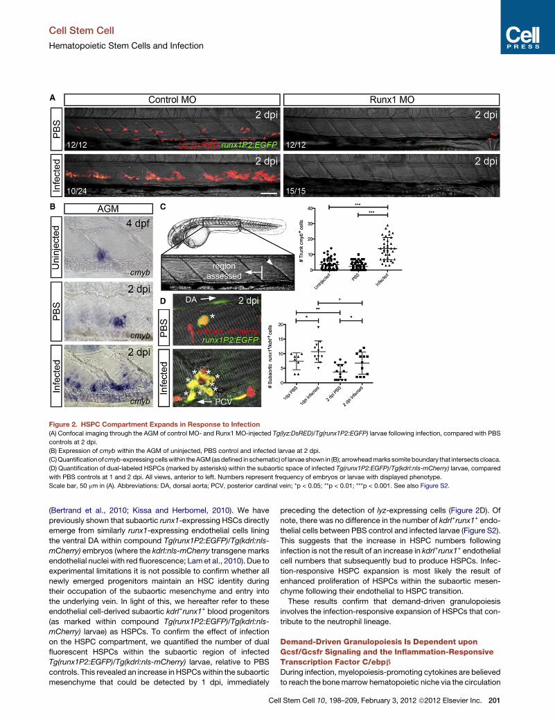
Figure 2. HSPC Compartment Expands in Response to Infection
(A) Confocal imaging through the AGM of control MO- and Runx1 MO-injected Tg(lyz:DsRED)/Tg(runx1P2:EGFP) larvae following infection, compared with PBS
controls at 2 dpi.
(B) Expression of cmyb within the AGM of uninjected, PBS control and infected larvae at 2 dpi.
(C)Quantificationofcmyb-expressingcellswithin theAGM(asdefined in schematic) of larvae shown in (B); arrowheadmarks somiteboundary that intersects cloaca.
(D) Quantification of dual-labeled HSPCs (marked by asterisks) within the subaortic space of infected Tg(runx1P2:EGFP)/Tg(kdrl:nls-mCherry) larvae, compared
with PBS controls at 1 and 2 dpi. All views, anterior to left. Numbers represent frequency of embryos or larvae with displayed phenotype.
Scale bar, 50 mm in (A). Abbreviations: DA, dorsal aorta; PCV, posterior cardinal vein; *p < 0.05; **p < 0.01; ***p < 0.001. See also Figure S2.
Cell Stem Cell
Hematopoietic Stem Cells and Infection
(Bertrand et al., 2010; Kissa and Herbomel, 2010). We have
previously shown that subaortic runx1-expressing HSCs directly
emerge from similarly runx1-expressing endothelial cells lining
the ventral DA within compound Tg(runx1P2:EGFP)/Tg(kdrl:nls-
mCherry) embryos (where the kdrl:nls-mCherry transgene marks
endothelial nuclei with red fluorescence; Lamet al., 2010). Due to
experimental limitations it is not possible to confirm whether all
newly emerged progenitors maintain an HSC identity during
their occupation of the subaortic mesenchyme and entry into
the underlying vein. In light of this, we hereafter refer to these
endothelial cell-derived subaortic kdrl+runx1+ blood progenitors
(as marked within compound Tg(runx1P2:EGFP)/Tg(kdrl:nls-
mCherry) larvae) as HSPCs. To confirm the effect of infection
on the HSPC compartment, we quantified the number of dual
fluorescent HSPCs within the subaortic region of infected
Tg(runx1P2:EGFP)/Tg(kdrl:nls-mCherry) larvae, relative to PBS
controls. This revealed an increase in HSPCswithin the subaortic
mesenchyme that could be detected by 1 dpi, immediately
Ce
preceding the detection of lyz-expressing cells (Figure 2D). Of
note, there was no difference in the number of kdrl+runx1+ endo-
thelial cells between PBS control and infected larvae (Figure S2).
This suggests that the increase in HSPC numbers following
infection is not the result of an increase in kdrl+runx1+ endothelial
cell numbers that subsequently bud to produce HSPCs. Infec-
tion-responsive HSPC expansion is most likely the result of
enhanced proliferation of HSPCs within the subaortic mesen-
chyme following their endothelial to HSPC transition.
These results confirm that demand-driven granulopoiesis
involves the infection-responsive expansion of HSPCs that con-
tribute to the neutrophil lineage.
Demand-Driven Granulopoiesis Is Dependent uponGcsf/Gcsfr Signaling and the Inflammation-ResponsiveTranscription Factor C/ebpbDuring infection, myelopoiesis-promoting cytokines are believed
to reach the bonemarrow hematopoietic niche via the circulation
ll Stem Cell 10, 198–209, February 3, 2012 ª2012 Elsevier Inc. 201
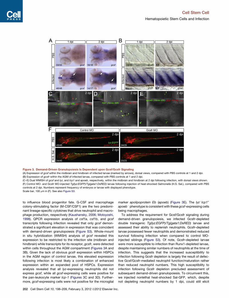
Figure 3. Demand-Driven Granulopoiesis Is Dependent upon Gcsf/Gcsfr Signaling
(A) Expression of gcsf within the midbrain and hindbrain of infected larvae (marked by arrows), dorsal views, compared with PBS controls at 1 and 2 dpi.
(B) Expression of gcsfr within the AGM of infected larvae, compared with PBS controls at 1 and 2 dpi.
(C–E) Dual WMISH of gcsf and lyz, and lcp1 and apoeb, respectively, within the midbrain and hindbrain at 2 dpi following infection, with dorsal views shown.
(F) Control MO- and Gcsfr MO-injected Tg(lyz:EGFP)/Tg(gata1:DsRED) larvae following injection of heat-shocked Salmonella (H.S. Sal.), compared with PBS
controls at 2 dpi. Numbers represent frequency of embryos or larvae with displayed phenotype.
Scale bar, 100 mm in (F). See also Figure S3.
Cell Stem Cell
Hematopoietic Stem Cells and Infection
to influence blood progenitor fate. G-CSF and macrophage
colony-stimulating factor (M-CSF/CSF1) are the two predomi-
nant lineage-specific cytokines that drive neutrophil and macro-
phage production, respectively (Kaushansky, 2006; Motoyoshi,
1998). QPCR expression analysis of csf1a, csf1b, and gcsf
transcripts following infection revealed that only gcsf demon-
strated a significant elevation in expression that was coincident
with demand-driven granulopoiesis (Figure S3). Whole-mount
in situ hybridization (WMISH) analysis of gcsf revealed this
expression to be restricted to the infection site (midbrain and
hindbrain) while transcripts for its receptor, gcsfr, were detected
within cells throughout the AGM compartment (Figures 3A and
3B). Given the lack of obvious gcsfr expression within HSPCs
in the AGM region of control larvae, this elevated expression
following infection is most likely a combination of enhanced
expression within an expanded pool of HSPCs. Expression
analysis revealed that all lyz-expressing neutrophils did not
express gcsf, while all gcsf-expressing cells were positive for
the pan-leukocyte marker lcp-1 (Figures 3C and 3D). Further-
more, gcsf-expressing cells were not positive for the microglial
202 Cell Stem Cell 10, 198–209, February 3, 2012 ª2012 Elsevier Inc
marker apolipoprotein Eb (apoeb) (Figure 3E). The lyz�lcp1+
apoeb� phenotype is consistent with these gcsf-expressing cells
being macrophages.
To address the requirement for Gcsf/Gcsfr signaling during
demand-driven granulopoiesis, we infected Gcsfr-depleted
double transgenic Tg(lyz:EGFP)/Tg(gata1:DsRED) larvae and
assessed their ability to replenish neutrophils. Gcsfr-depleted
larvae possessed fewer neutrophils and demonstrated reduced
survival following infection when compared to control MO-
injected siblings (Figure S3). Of note, Gcsfr-depleted larvae
were more susceptible to infection than Runx1-depleted larvae,
despite maintaining similar numbers of neutrophils at the time of
infection. This suggests that the increased susceptibility to
infection following Gcsfr depletion is largely the result of defec-
tive Gcsf/Gcsfr-mediated neutrophil function/maturation rather
than reduced neutrophil numbers. This high susceptibility to
infection following Gcsfr depletion precluded assessment of
subsequent demand-driven granulopoiesis. To circumvent this,
we injected nonlethal heat-shocked Sal-GFP, which, despite
not depleting neutrophil numbers by 1 dpi, could still elicit
.
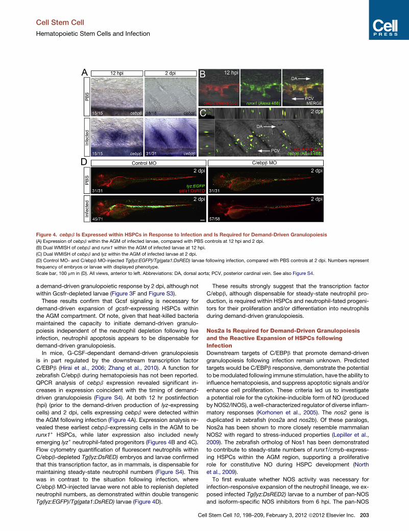
Figure 4. cebpb Is Expressed within HSPCs in Response to Infection and Is Required for Demand-Driven Granulopoiesis
(A) Expression of cebpb within the AGM of infected larvae, compared with PBS controls at 12 hpi and 2 dpi.
(B) Dual WMISH of cebpb and runx1 within the AGM of infected larvae at 12 hpi.
(C) Dual WMISH of cebpb and lyz within the AGM of infected larvae at 2 dpi.
(D) Control MO- and C/ebpb MO-injected Tg(lyz:EGFP)/Tg(gata1:DsRED) larvae following infection, compared with PBS controls at 2 dpi. Numbers represent
frequency of embryos or larvae with displayed phenotype.
Scale bar, 100 mm in (D). All views, anterior to left. Abbreviations: DA, dorsal aorta; PCV, posterior cardinal vein. See also Figure S4.
Cell Stem Cell
Hematopoietic Stem Cells and Infection
a demand-driven granulopoietic response by 2 dpi, although not
within Gcsfr-depleted larvae (Figure 3F and Figure S3).
These results confirm that Gcsf signaling is necessary for
demand-driven expansion of gcsfr-expressing HSPCs within
the AGM compartment. Of note, given that heat-killed bacteria
maintained the capacity to initiate demand-driven granulo-
poiesis independent of the neutrophil depletion following live
infection, neutrophil apoptosis appears to be dispensable for
demand-driven granulopoiesis.
In mice, G-CSF-dependant demand-driven granulopoiesis
is in part regulated by the downstream transcription factor
C/EBPb (Hirai et al., 2006; Zhang et al., 2010). A function for
zebrafish C/ebpb during hematopoiesis has not been reported.
QPCR analysis of cebpb expression revealed significant in-
creases in expression coincident with the timing of demand-
driven granulopoiesis (Figure S4). At both 12 hr postinfection
(hpi) (prior to the demand-driven production of lyz-expressing
cells) and 2 dpi, cells expressing cebpb were detected within
the AGM following infection (Figure 4A). Expression analysis re-
vealed these earliest cebpb-expressing cells in the AGM to be
runx1+ HSPCs, while later expression also included newly
emerging lyz+ neutrophil-fated progenitors (Figures 4B and 4C).
Flow cytometry quantification of fluorescent neutrophils within
C/ebpb-depleted Tg(lyz:DsRED) embryos and larvae confirmed
that this transcription factor, as in mammals, is dispensable for
maintaining steady-state neutrophil numbers (Figure S4). This
was in contrast to the situation following infection, where
C/ebpb MO-injected larvae were not able to replenish depleted
neutrophil numbers, as demonstrated within double transgenic
Tg(lyz:EGFP)/Tg(gata1:DsRED) larvae (Figure 4D).
Ce
These results strongly suggest that the transcription factor
C/ebpb, although dispensable for steady-state neutrophil pro-
duction, is required within HSPCs and neutrophil-fated progeni-
tors for their proliferation and/or differentiation into neutrophils
during demand-driven granulopoiesis.
Nos2a Is Required for Demand-Driven Granulopoiesisand the Reactive Expansion of HSPCs followingInfectionDownstream targets of C/EBPb that promote demand-driven
granulopoiesis following infection remain unknown. Predicted
targets would be C/EBPb responsive, demonstrate the potential
to bemodulated following immune stimulation, have the ability to
influence hematopoiesis, and suppress apoptotic signals and/or
enhance cell proliferation. These criteria led us to investigate
a potential role for the cytokine-inducible form of NO (produced
byNOS2/INOS), a well-characterized regulator of diverse inflam-
matory responses (Korhonen et al., 2005). The nos2 gene is
duplicated in zebrafish (nos2a and nos2b). Of these paralogs,
Nos2a has been shown to more closely resemble mammalian
NOS2 with regard to stress-induced properties (Lepiller et al.,
2009). The zebrafish ortholog of Nos1 has been demonstrated
to contribute to steady-state numbers of runx1/cmyb-express-
ing HSPCs within the AGM region, supporting a proliferative
role for constitutive NO during HSPC development (North
et al., 2009).
To first evaluate whether NOS activity was necessary for
infection-responsive expansion of the neutrophil lineage, we ex-
posed infected Tg(lyz:DsRED2) larvae to a number of pan-NOS
and isoform-specific NOS inhibitors from 6 hpi. The pan-NOS
ll Stem Cell 10, 198–209, February 3, 2012 ª2012 Elsevier Inc. 203
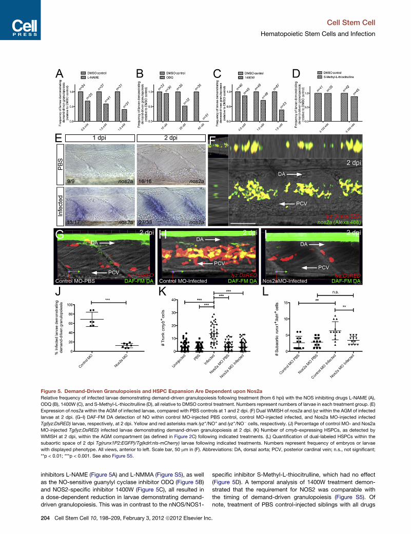
Figure 5. Demand-Driven Granulopoiesis and HSPC Expansion Are Dependent upon Nos2a
Relative frequency of infected larvae demonstrating demand-driven granulopoiesis following treatment (from 6 hpi) with the NOS inhibiting drugs L-NAME (A),
ODQ (B), 1400W (C), and S-Methyl-L-thiocitrulline (D), all relative to DMSO control treatment. Numbers represent numbers of larvae in each treatment group. (E)
Expression of nos2a within the AGM of infected larvae, compared with PBS controls at 1 and 2 dpi. (F) Dual WMISH of nos2a and lyz within the AGM of infected
larvae at 2 dpi. (G–I) DAF-FM DA detection of NO within control MO-injected PBS control, control MO-injected infected, and Nos2a MO-injected infected
Tg(lyz:DsRED) larvae, respectively, at 2 dpi. Yellow and red asterisks mark lyz+/NO+ and lyz+/NO� cells, respectively. (J) Percentage of control MO- and Nos2a
MO-injected Tg(lyz:DsRED) infected larvae demonstrating demand-driven granulopoiesis at 2 dpi. (K) Number of cmyb-expressing HSPCs, as detected by
WMISH at 2 dpi, within the AGM compartment (as defined in Figure 2C) following indicated treatments. (L) Quantification of dual-labeled HSPCs within the
subaortic space of 2 dpi Tg(runx1P2:EGFP)/Tg(kdrl:nls-mCherry) larvae following indicated treatments. Numbers represent frequency of embryos or larvae
with displayed phenotype. All views, anterior to left. Scale bar, 50 mm in (F). Abbreviations: DA, dorsal aorta; PCV, posterior cardinal vein; n.s., not significant;
**p < 0.01; ***p < 0.001. See also Figure S5.
Cell Stem Cell
Hematopoietic Stem Cells and Infection
inhibitors L-NAME (Figure 5A) and L-NMMA (Figure S5), as well
as the NO-sensitive guanylyl cyclase inhibitor ODQ (Figure 5B)
and NOS2-specific inhibitor 1400W (Figure 5C), all resulted in
a dose-dependent reduction in larvae demonstrating demand-
driven granulopoiesis. This was in contrast to the nNOS/NOS1-
204 Cell Stem Cell 10, 198–209, February 3, 2012 ª2012 Elsevier Inc
specific inhibitor S-Methyl-L-thiocitrulline, which had no effect
(Figure 5D). A temporal analysis of 1400W treatment demon-
strated that the requirement for NOS2 was comparable with
the timing of demand-driven granulopoiesis (Figure S5). Of
note, treatment of PBS control-injected siblings with all drugs
.
Cell Stem Cell
Hematopoietic Stem Cells and Infection
used, at all doses, resulted in no observable developmental
defects, including steady-state neutrophil production (data not
shown). These results prompted us to evaluate the expression
of nos2a within the AGM compartment following infection.
Presumptive HSPCs expressing nos2a were detected as early
as 1 day following infection (Figure 5E). Expression analysis at
2 dpi revealed that the majority of nos2a-expressing cells were
lyz+ neutrophil-fated progenitors (Figure 5F).
Next we exploited the live imaging potential of the cell-
permeable 4-amino-5-methylamino-20,70-difluororescein diace-
tate (DAF-FM DA) fluorescent NO probe to examine whether
this infection-responsive nos2a expression within the AGM cor-
related with active NO production. Consistent with the expres-
sion analysis, NO was detected within lyz:DsRED-expressing
cells within the AGM compartment, specifically following infec-
tion (Figures 5G and 5H). To confirm a role for Nos2a during
NOproduction and demand-driven granulopoiesis, we assessed
NO production within Nos2a-depleted infected Tg(lyz:DsRED)
larvae and the potential of Nos2a-depleted compound
Tg(lyz:EGFP)/Tg(gata1:DsRED) larvae to replenish depleted neu-
trophil numbers following infection (Figures 5G–5J and Fig-
ure S5). This revealed a requirement for Nos2a during NO pro-
duction and demand-driven granulopoiesis within the AGM
following infection (Figures 5I and 5J and Figure S5). Flow cytom-
etry quantification of fluorescent neutrophils within Nos2a-
depleted Tg(lyz:DsRED) embryos and larvae confirmed that
Nos2a, like C/ebpb, was dispensable for maintaining homeo-
static neutrophil numbers (Figure S5). No significant reduction
in survival was observed within Nos2a-depleted larvae following
infection, suggesting that Nos2a, although required for neutro-
phil replenishment/expansion in response to infection, was not
required to survive the infection doses used in this study (Fig-
ure S5). Given the early expression of both cebpb and nos2a
by HSPCs within the AGM when HSPC numbers within this
region are expanding, we investigated whether Nos2a was
required for HSPC expansion following infection. This analysis
revealed that the infection-responsive increases of both cmyb-
and runx1P2:EGFP/kdrl:nsl-mCherry-expressing HSPCs within
the subaortic mesenchyme are Nos2a-dependent (Figures 5K
and 5L). These results confirm a role for Nos2a-generated NO
during infection-responsive expansion of subaortic HSPCs.
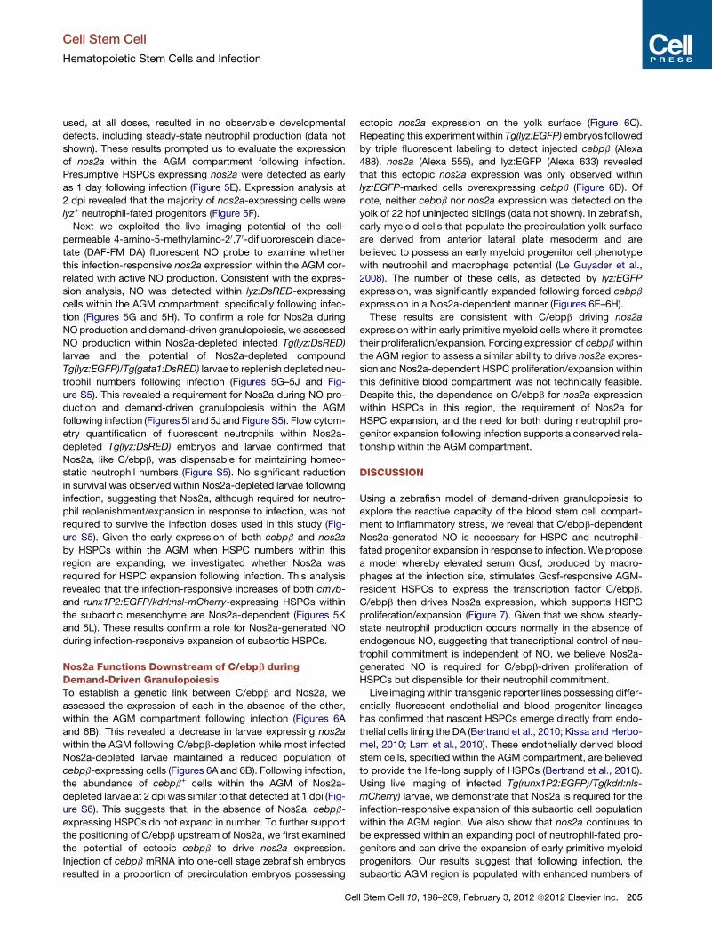
Nos2a Functions Downstream of C/ebpb duringDemand-Driven GranulopoiesisTo establish a genetic link between C/ebpb and Nos2a, we
assessed the expression of each in the absence of the other,
within the AGM compartment following infection (Figures 6A
and 6B). This revealed a decrease in larvae expressing nos2a
within the AGM following C/ebpb-depletion while most infected
Nos2a-depleted larvae maintained a reduced population of
cebpb-expressing cells (Figures 6A and 6B). Following infection,
the abundance of cebpb+ cells within the AGM of Nos2a-
depleted larvae at 2 dpi was similar to that detected at 1 dpi (Fig-
ure S6). This suggests that, in the absence of Nos2a, cebpb-
expressing HSPCs do not expand in number. To further support
the positioning of C/ebpb upstream of Nos2a, we first examined
the potential of ectopic cebpb to drive nos2a expression.
Injection of cebpb mRNA into one-cell stage zebrafish embryos
resulted in a proportion of precirculation embryos possessing
Ce
ectopic nos2a expression on the yolk surface (Figure 6C).
Repeating this experiment within Tg(lyz:EGFP) embryos followed
by triple fluorescent labeling to detect injected cebpb (Alexa
488), nos2a (Alexa 555), and lyz:EGFP (Alexa 633) revealed
that this ectopic nos2a expression was only observed within
lyz:EGFP-marked cells overexpressing cebpb (Figure 6D). Of
note, neither cebpb nor nos2a expression was detected on the
yolk of 22 hpf uninjected siblings (data not shown). In zebrafish,
early myeloid cells that populate the precirculation yolk surface
are derived from anterior lateral plate mesoderm and are
believed to possess an early myeloid progenitor cell phenotype
with neutrophil and macrophage potential (Le Guyader et al.,
2008). The number of these cells, as detected by lyz:EGFP
expression, was significantly expanded following forced cebpb
expression in a Nos2a-dependent manner (Figures 6E–6H).
These results are consistent with C/ebpb driving nos2a
expression within early primitive myeloid cells where it promotes
their proliferation/expansion. Forcing expression of cebpbwithin
the AGM region to assess a similar ability to drive nos2a expres-
sion and Nos2a-dependent HSPC proliferation/expansion within
this definitive blood compartment was not technically feasible.
Despite this, the dependence on C/ebpb for nos2a expression
within HSPCs in this region, the requirement of Nos2a for
HSPC expansion, and the need for both during neutrophil pro-
genitor expansion following infection supports a conserved rela-
tionship within the AGM compartment.
DISCUSSION
Using a zebrafish model of demand-driven granulopoiesis to
explore the reactive capacity of the blood stem cell compart-
ment to inflammatory stress, we reveal that C/ebpb-dependent
Nos2a-generated NO is necessary for HSPC and neutrophil-
fated progenitor expansion in response to infection. We propose
a model whereby elevated serum Gcsf, produced by macro-
phages at the infection site, stimulates Gcsf-responsive AGM-
resident HSPCs to express the transcription factor C/ebpb.
C/ebpb then drives Nos2a expression, which supports HSPC
proliferation/expansion (Figure 7). Given that we show steady-
state neutrophil production occurs normally in the absence of
endogenous NO, suggesting that transcriptional control of neu-
trophil commitment is independent of NO, we believe Nos2a-
generated NO is required for C/ebpb-driven proliferation of
HSPCs but dispensible for their neutrophil commitment.
Live imagingwithin transgenic reporter lines possessing differ-
entially fluorescent endothelial and blood progenitor lineages
has confirmed that nascent HSPCs emerge directly from endo-
thelial cells lining the DA (Bertrand et al., 2010; Kissa and Herbo-
mel, 2010; Lam et al., 2010). These endothelially derived blood
stem cells, specified within the AGM compartment, are believed
to provide the life-long supply of HSPCs (Bertrand et al., 2010).
Using live imaging of infected Tg(runx1P2:EGFP)/Tg(kdrl:nls-
mCherry) larvae, we demonstrate that Nos2a is required for the
infection-responsive expansion of this subaortic cell population
within the AGM region. We also show that nos2a continues to
be expressed within an expanding pool of neutrophil-fated pro-
genitors and can drive the expansion of early primitive myeloid
progenitors. Our results suggest that following infection, the
subaortic AGM region is populated with enhanced numbers of
ll Stem Cell 10, 198–209, February 3, 2012 ª2012 Elsevier Inc. 205
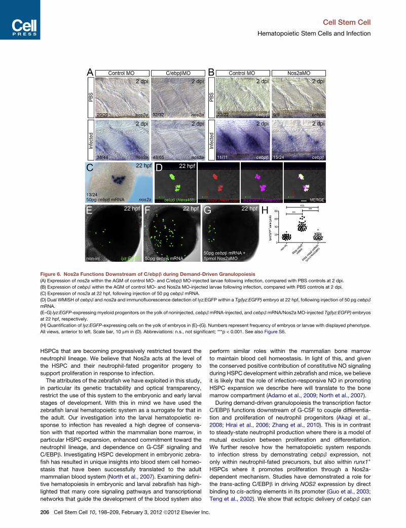
Figure 6. Nos2a Functions Downstream of C/ebpb during Demand-Driven Granulopoiesis
(A) Expression of nos2a within the AGM of control MO- and C/ebpb MO-injected larvae following infection, compared with PBS controls at 2 dpi.
(B) Expression of cebpb within the AGM of control MO- and Nos2a MO-injected larvae following infection, compared with PBS controls at 2 dpi.
(C) Expression of nos2a at 22 hpf, following injection of 50 pg cebpb mRNA.
(D) Dual WMISH of cebpb and nos2a and immunofluorescence detection of lyz:EGFP within a Tg(lyz:EGFP) embryo at 22 hpf, following injection of 50 pg cebpb
mRNA.
(E–G) lyz:EGFP-expressing myeloid progenitors on the yolk of noninjected, cebpbmRNA-injected, and cebpbmRNA/Nos2a MO-injected Tg(lyz:EGFP) embryos
at 22 hpf, respectively.
(H) Quantification of lyz:EGFP-expressing cells on the yolk of embryos in (E)–(G). Numbers represent frequency of embryos or larvae with displayed phenotype.
All views, anterior to left. Scale bar, 10 mm in (D). Abbreviations: n.s., not significant; ***p < 0.001. See also Figure S6.
Cell Stem Cell
Hematopoietic Stem Cells and Infection
HSPCs that are becoming progressively restricted toward the
neutrophil lineage. We believe that Nos2a acts at the level of
the HSPC and their neutrophil-fated progenitor progeny to
support proliferation in response to infection.
The attributes of the zebrafish we have exploited in this study,
in particular its genetic tractability and optical transparency,
restrict the use of this system to the embryonic and early larval
stages of development. With this in mind we have used the
zebrafish larval hematopoietic system as a surrogate for that in
the adult. Our investigation into the larval hematopoietic re-
sponse to infection has revealed a high degree of conserva-
tion with that reported within the mammalian bone marrow, in
particular HSPC expansion, enhanced commitment toward the
neutrophil lineage, and dependence on G-CSF signaling and
C/EBPb. Investigating HSPC development in embryonic zebra-
fish has resulted in unique insights into blood stem cell homeo-
stasis that have been successfully translated to the adult
mammalian blood system (North et al., 2007). Examining defini-
tive hematopoiesis in embryonic and larval zebrafish has high-
lighted that many core signaling pathways and transcriptional
networks that guide the development of the blood system also
206 Cell Stem Cell 10, 198–209, February 3, 2012 ª2012 Elsevier Inc
perform similar roles within the mammalian bone marrow
to maintain blood cell homeostasis. In light of this, and given
the conserved positive contribution of constitutive NO signaling
during HSPC development within zebrafish andmice, we believe
it is likely that the role of infection-responsive NO in promoting
HSPC expansion we describe here will translate to the bone
marrow compartment (Adamo et al., 2009; North et al., 2007).
During demand-driven granulopoiesis the transcription factor
C/EBPb functions downstream of G-CSF to couple differentia-
tion and proliferation of neutrophil progenitors (Akagi et al.,
2008; Hirai et al., 2006; Zhang et al., 2010). This is in contrast
to steady-state neutrophil production where there is a model of
mutual exclusion between proliferation and differentiation.
We further resolve how the hematopoietic system responds
to infection stress by demonstrating cebpb expression, not
only within neutrophil-fated precursors, but also within runx1+
HSPCs where it promotes proliferation through a Nos2a-
dependent mechanism. Studies have demonstrated a role for
the trans-acting C/EBPb in driving NOS2 expression by direct
binding to cis-acting elements in its promoter (Guo et al., 2003;
Teng et al., 2002). We show that ectopic delivery of cebpb can
.
Figure 7. Proposed Model Illustrating the Reactive
Capacity of the Zebrafish Blood System to Inflam-
matory Stress
In response to bacterial challenge, neutrophils infiltrate the
infection foci to help clear bacteria before undergoing
apoptosis. Macrophages then arrive to remove neutrophil
debris and express the granulopoiesis-promoting cyto-
kine Gcsf (1). Gcsf reaches the AGM compartment via
the circulation to support the proliferation/expansion of
HSPCs and neutrophil-fated progenitors by promoting
C/ebpb-dependent nos2a expression (2).
Cell Stem Cell
Hematopoietic Stem Cells and Infection
drive nos2a expression within zebrafish embryos. Analysis of the
promoter region immediately upstream of the zebrafish nos2a
transcription start site revealed a number of C/EBPb consensus
motifs (data not shown).
NO is a pleiotropic biomodulator in a number of systems,
including the hematopoietic system. Positive and negative roles
for NO during cell proliferation and steady-state blood develop-
ment have been reported (Guthrie et al., 2005; Krasnov et al.,
2008; Krsti�c et al., 2009; Michurina et al., 2004; North et al.,
2009; Villalobo, 2006). In zebrafish, blood-flow-responsive
nos1 (nnos/enos)-derived NO has been shown to contribute to
runx1+cmyb+ HSPC development within the AGM region such
that NO-donors enhance, while NO-inhibitors (and inhibitors
of downstream components of NO signaling, such as ODQ)
deplete, HSPC numbers (North et al., 2009). In mice, intrauterine
delivery of the pan-NOS-inhibitor L-NAME blocks hematopoietic
cluster formation within the AGM, strongly arguing for a con-
served role for NO signaling as a positive regulator of blood
stem cell development. We propose a role for infection-
responsive NO, generated by Nos2a, during expansion of zebra-
fish HSPCs in response to inflammatory stress. Given the
expression of nos2a within HSPCs in the AGM from 1 dpi, this
source of NO likely influences neighboring blood progenitors in
a paracrine fashion. We were unable to confirm these early
nos2a-expressing cells as HSPCs by dual staining with HSPC
markers due to technical limitations. However, given that these
nos2a-expressing cells occupy the AGM, prior to the emergence
of lyz-expressing neutrophil-fated progenitors at a time when
HSPC numbers are expanding in a Nos2a-dependent manner,
suggests that they are HSPCs. Despite studies examining
the role of NOS2 during the immune response, a role during
demand-driven granulopoiesis or infection-responsive HSPC
expansion has not been described.
This study proposes a role for Nos2a-generated NO, down-
stream of C/ebpb, in supporting HSPC proliferation/expansion
Cell Stem Cell 10, 19
following infection. To date, the ability to expand
HSPCs ex vivo remains limited (Dahlberg et al.,
2011). As such, identifying novel mechanisms
that influence HSPC proliferative capacity is an
area of intense interest heightened by their
therapeutic relevance to both malignant and
nonmalignant diseases. Additional studies are
required to determine whether manipulating
C/EBPb and/or NOS2 activity influences the
capacity of mammalian HSPCs and whether
this newly identified pathway can be targeted for therapeutic
benefit.
EXPERIMENTAL PROCEDURES
Refer to the Supplemental Information for fully detailed experimental
procedures.
Zebrafish Maintenance
Zebrafish embryos obtained from natural spawnings were raised at 28�C in E3
Medium (Westerfield, 2000) and were developmentally staged as described
(Kimmel et al., 1995). Transgenic reporter lines used in this study are summa-
rized in Table S1. Research was conducted with approval from The University
of Auckland Animal Ethics Committee.
QPCR
QPCR was performed with Platinum SYBR Green qPCR SuperMix-UDG with
ROX (Invitrogen) using an ABI PRISM7900HT Fast sequence detection system
(Applied Biosystems) essentially as described (Oehlers et al., 2010). Gene-
specific oligonucleotides were designed using Primer Express software
(Table S2).
Statistical Analysis
Statistical analyses were performed using Prism 5.0 (GraphPad Software,
Inc.). Statistical significance was assessed using unpaired, two-tailed t tests,
and all data was presented in scatter plots with means and standard
deviations.
WMISH
WMISHswere performed as described (Jowett and Lettice, 1994). Fluorescent
in situ hybridization was adapted from Clay and Ramakrishnan (2005).
RT-PCR
Total RNAs were extracted from embryos and larvae using TRIzol reagent
(Invitrogen) as per the manufacturer’s instructions. cDNAs were then gener-
ated using Superscript III reverse transcriptase (Invitrogen). These cDNAs
were used to generate gene-specific amplicons using the primer pairs listed
in Table S2 (all primer pairs were designed to span at least 1 intron to control
for contaminating genomic DNA).
8–209, February 3, 2012 ª2012 Elsevier Inc. 207
Cell Stem Cell
Hematopoietic Stem Cells and Infection
MO Injection
MOs (Gene Tools, Philomath, OR) were resuspended and injected as
described (Nasevicius and Ekker, 2000). Efficacious doses for all MOs were
determined empirically. RT-PCR was used to determine MO specificity where
splice-blocking MOs were used (see Table S3 for oligonucleotide sequences
and doses).
Chemical Treatments
Unless otherwise stated compounds were resuspended according to the
manufacturer’s instructions and diluted to the indicated working concentra-
tions such that the final DMSO concentration was 1%. Full details of the chem-
icals used, their targets, and manufacturer’s and working concentrations are
provided in Table S4.
Infection of Zebrafish Embryos and Larvae
Embryos and larvae were infected at the indicated ages with either GFP-
labeled or nonlabeled Salmonella enterica serovar Typhimurium (Hall et al.,
2007). Bacteria were delivered into the hindbrain ventricle of anaesthetized
larvae, typically at 50 hpf. For a detailed description of bacteria culture condi-
tions, injection procedure and actual cfu dose calculation, see Supplemental
Experimental Procedures.
Capped RNA Synthesis
Synthetic capped RNA was synthesized using the mMESSAGE mMACHINE
SP6 kit (Ambion) as per the manufacturer’s instructions.
DAF-FM DA Staining of Larvae
The NO-specific fluorescent dye DAF-FM DA (Molecular Probes, Invitrogen)
was used to detect endogenous NO as previously described (Lepiller et al.,
2007).
Immunofluorescence
Immunofluorescence detection of fluorescent reporters was performed essen-
tially as described (Hall et al., 2009a). For detection of EGFP, a chicken anti-
GFP (Abcam) primary and goat anti-chicken Alexa Fluor 488-conjugated
secondary antibody (Invitrogen) or a rabbit anti-GFP primary (Invitrogen) and
goat anti-rabbit Alexa Fluor 633-conjugated secondary antibody (Invitrogen)
were used.
Flow Cytometry
Flow cytometry was performed as previously described (Hall et al., 2009a).
Confocal Imaging
Live embryos and larvae were mounted for confocal imaging as previously
described (Hall et al., 2009b). Images collected by confocal microscopy
were processed and analyzed using Volocity 5.5 (Perkin Elmer).
SUPPLEMENTAL INFORMATION
Supplemental Information for this article includes Figures S1–S6, Tables
S1–S4, and Supplemental Experimental Procedures and can be found with
this article online at doi:10.1016/j.stem.2012.01.007.
ACKNOWLEDGMENTS
We thank Annie Chien, Sophie Wicker, Pauline Misa, Alisha Malik, and Alhad
Mahagaonkar for excellent technical assistance. We also thank Nick Trede
and Len Zon for supplying reporter lines and Graham Lieschke for supplying
probe templates. Funding for this work was provided by a grant awarded to
P.C. from the Ministry of Science & Innovation, New Zealand.
Received: August 17, 2011
Revised: December 10, 2011
Accepted: January 17, 2012
Published: February 2, 2012
208 Cell Stem Cell 10, 198–209, February 3, 2012 ª2012 Elsevier Inc
REFERENCES
Adamo, L., Naveiras, O., Wenzel, P.L., McKinney-Freeman, S., Mack, P.J.,
Gracia-Sancho, J., Suchy-Dicey, A., Yoshimoto, M., Lensch, M.W., Yoder,
M.C., et al. (2009). Biomechanical forces promote embryonic haematopoiesis.
Nature 459, 1131–1135.
Akagi, T., Saitoh, T., O’Kelly, J., Akira, S., Gombart, A.F., and Koeffler, H.P.
(2008). Impaired response to GM-CSF and G-CSF, and enhanced apoptosis
in C/EBPbeta-deficient hematopoietic cells. Blood 111, 2999–3004.
Baldridge, M.T., King, K.Y., Boles, N.C., Weksberg, D.C., and Goodell, M.A.
(2010). Quiescent haematopoietic stem cells are activated by IFN-gamma in
response to chronic infection. Nature 465, 793–797.
Baldridge, M.T., King, K.Y., and Goodell, M.A. (2011). Inflammatory signals
regulate hematopoietic stem cells. Trends Immunol. 32, 57–65.
Bertrand, J.Y., Chi, N.C., Santoso, B., Teng, S., Stainier, D.Y., and Traver, D.
(2010). Haematopoietic stem cells derive directly from aortic endothelium
during development. Nature 464, 108–111.
Clay, H., and Ramakrishnan, L. (2005). Multiplex fluorescent in situ hybridiza-
tion in zebrafish embryos using tyramide signal amplification. Zebrafish 2,
105–111.
Dahlberg, A., Delaney, C., and Bernstein, I.D. (2011). Ex vivo expansion of
human hematopoietic stem and progenitor cells. Blood 117, 6083–6090.
Ellett, F., Pase, L., Hayman, J.W., Andrianopoulos, A., and Lieschke, G.J.
(2011). mpeg1 promoter transgenes direct macrophage-lineage expression
in zebrafish. Blood 117, e49–e56.
Guo, Z., Shao, L., Feng, X., Reid, K., Marderstein, E., Nakao, A., and Geller,
D.A. (2003). A critical role for C/EBPbeta binding to the AABS promoter
response element in the human iNOS gene. FASEB J. 17, 1718–1720.
Guthrie, S.M., Curtis, L.M., Mames, R.N., Simon, G.G., Grant, M.B., and Scott,
E.W. (2005). The nitric oxide pathway modulates hemangioblast activity of
adult hematopoietic stem cells. Blood 105, 1916–1922.
Hall, C., Flores, M.V., Storm, T., Crosier, K., and Crosier, P. (2007). The zebra-
fish lysozyme C promoter drives myeloid-specific expression in transgenic
fish. BMC Dev. Biol. 7, 42.
Hall, C., Flores, M.V., Chien, A., Davidson, A., Crosier, K., and Crosier, P.
(2009a). Transgenic zebrafish reporter lines reveal conserved Toll-like receptor
signaling potential in embryonic myeloid leukocytes and adult immune cell
lineages. J. Leukoc. Biol. 85, 751–765.
Hall, C., Flores, M.V., Crosier, K., and Crosier, P. (2009b). Live cell imaging of
zebrafish leukocytes. Methods Mol. Biol. 546, 255–271.
Hirai, H., Zhang, P., Dayaram, T., Hetherington, C.J., Mizuno, S., Imanishi, J.,
Akashi, K., and Tenen, D.G. (2006). C/EBPbeta is required for ‘emergency’
granulopoiesis. Nat. Immunol. 7, 732–739.
Jowett, T., and Lettice, L. (1994). Whole-mount in situ hybridizations on zebra-
fish embryos using a mixture of digoxigenin- and fluorescein-labelled probes.
Trends Genet. 10, 73–74.
Kastan, M.B., Slamon, D.J., and Civin, C.I. (1989). Expression of protoonco-
gene c-myb in normal human hematopoietic cells. Blood 73, 1444–1451.
Kaushansky, K. (2006). Lineage-specific hematopoietic growth factors.
N. Engl. J. Med. 354, 2034–2045.
Kimmel, C.B., Ballard, W.W., Kimmel, S.R., Ullmann, B., and Schilling, T.F.
(1995). Stages of embryonic development of the zebrafish. Dev. Dyn. 203,
253–310.
King, K.Y., and Goodell, M.A. (2011). Inflammatory modulation of HSCs:
viewing the HSC as a foundation for the immune response. Nat. Rev. 11,
685–692.
Kissa, K., and Herbomel, P. (2010). Blood stem cells emerge from aortic endo-
thelium by a novel type of cell transition. Nature 464, 112–115.
Korhonen, R., Lahti, A., Kankaanranta, H., andMoilanen, E. (2005). Nitric oxide
production and signaling in inflammation. Curr. Drug Targets Inflamm. Allergy
4, 471–479.
.
Cell Stem Cell
Hematopoietic Stem Cells and Infection
Krasnov, P., Michurina, T., Packer, M.A., Stasiv, Y., Nakaya, N., Moore, K.A.,
Drazan, K.E., and Enikolopov, G. (2008). Neuronal nitric oxide synthase
contributes to the regulation of hematopoiesis. Mol. Med. 14, 141–149.
Krsti�c, A., Ili�c, V., Mojsilovi�c, S., Jovci�c, G., Milenkovi�c, P., and Bugarski, D.
(2009). p38 MAPK signaling mediates IL-17-induced nitric oxide synthase
expression in bone marrow cells. Growth Factors 27, 79–90.
Lam, E.Y., Chau, J.Y., Kalev-Zylinska, M.L., Fountaine, T.M., Mead, R.S., Hall,
C.J., Crosier, P.S., Crosier, K.E., and Flores, M.V. (2009). Zebrafish runx1
promoter-EGFP transgenics mark discrete sites of definitive blood progeni-
tors. Blood 113, 1241–1249.
Lam, E.Y., Hall, C.J., Crosier, P.S., Crosier, K.E., and Flores, M.V. (2010). Live
imaging of Runx1 expression in the dorsal aorta tracks the emergence of blood
progenitors from endothelial cells. Blood 116, 909–914.
Le Guyader, D., Redd, M.J., Colucci-Guyon, E., Murayama, E., Kissa, K.,
Briolat, V., Mordelet, E., Zapata, A., Shinomiya, H., and Herbomel, P. (2008).
Origins and unconventional behavior of neutrophils in developing zebrafish.
Blood 111, 132–141.
Lepiller, S., Laurens, V., Bouchot, A., Herbomel, P., Solary, E., and Chluba, J.
(2007). Imaging of nitric oxide in a living vertebrate using a diamino-fluorescein
probe. Free Radic. Biol. Med. 43, 619–627.
Lepiller, S., Franche, N., Solary, E., Chluba, J., and Laurens, V. (2009).
Comparative analysis of zebrafish nos2a and nos2b genes. Gene 445, 58–65.
Lieschke, G.J., Oates, A.C., Paw, B.H., Thompson, M.A., Hall, N.E., Ward,
A.C., Ho, R.K., Zon, L.I., and Layton, J.E. (2002). Zebrafish SPI-1 (PU.1) marks
a site of myeloid development independent of primitive erythropoiesis:
implications for axial patterning. Dev. Biol. 246, 274–295.
Lyons, S.E., Shue, B.C., Oates, A.C., Zon, L.I., and Liu, P.P. (2001). A novel
myeloid-restricted zebrafish CCAAT/enhancer-binding protein with a potent
transcriptional activation domain. Blood 97, 2611–2617.
Meijer, A.H., van der Sar, A.M., Cunha, C., Lamers, G.E., Laplante, M.A.,
Kikuta, H., Bitter, W., Becker, T.S., and Spaink, H.P. (2008). Identification
and real-time imaging of a myc-expressing neutrophil population involved in
inflammation and mycobacterial granuloma formation in zebrafish. Dev.
Comp. Immunol. 32, 36–49.
Michurina, T., Krasnov, P., Balazs, A., Nakaya, N., Vasilieva, T., Kuzin, B.,
Khrushchov, N., Mulligan, R.C., and Enikolopov, G. (2004). Nitric oxide is
a regulator of hematopoietic stem cell activity. Mol. Ther. 10, 241–248.
Motoyoshi, K. (1998). Biological activities and clinical application of M-CSF.
Int. J. Hematol. 67, 109–122.
Murayama, E., Kissa, K., Zapata, A., Mordelet, E., Briolat, V., Lin, H.F., Handin,
R.I., and Herbomel, P. (2006). Tracing hematopoietic precursor migration to
successive hematopoietic organs during zebrafish development. Immunity
25, 963–975.
Nasevicius, A., and Ekker, S.C. (2000). Effective targeted gene ‘knockdown’ in
zebrafish. Nat. Genet. 26, 216–220.
Ce
North, T.E., Goessling, W., Walkley, C.R., Lengerke, C., Kopani, K.R., Lord,
A.M., Weber, G.J., Bowman, T.V., Jang, I.H., Grosser, T., et al. (2007).
Prostaglandin E2 regulates vertebrate haematopoietic stem cell homeostasis.
Nature 447, 1007–1011.
North, T.E., Goessling, W., Peeters, M., Li, P., Ceol, C., Lord, A.M., Weber,
G.J., Harris, J., Cutting, C.C., Huang, P., et al. (2009). Hematopoietic stem
cell development is dependent on blood flow. Cell 137, 736–748.
Oehlers, S.H., Flores, M.V., Hall, C.J., O’Toole, R., Swift, S., Crosier, K.E., and
Crosier, P.S. (2010). Expression of zebrafish cxcl8 (interleukin-8) and its recep-
tors during development and in response to immune stimulation. Dev. Comp.
Immunol. 34, 352–359.
Panopoulos, A.D., and Watowich, S.S. (2008). Granulocyte colony-stimulating
factor: molecular mechanisms of action during steady state and ‘emergency’
hematopoiesis. Cytokine 42, 277–288.
Rezzoug, F., Huang, Y., Tanner, M.K., Wysoczynski, M., Schanie, C.L.,
Chilton, P.M., Ratajczak, M.Z., Fugier-Vivier, I.J., and Ildstad, S.T. (2008).
TNF-alpha is critical to facilitate hemopoietic stem cell engraftment and func-
tion. J. Immunol. 180, 49–57.
Scumpia, P.O., Kelly-Scumpia, K.M., Delano, M.J., Weinstein, J.S., Cuenca,
A.G., Al-Quran, S., Bovio, I., Akira, S., Kumagai, Y., and Moldawer, L.L.
(2010). Cutting edge: bacterial infection induces hematopoietic stem and
progenitor cell expansion in the absence of TLR signaling. J. Immunol. 184,
2247–2251.
Takizawa, H., Regoes, R.R., Boddupalli, C.S., Bonhoeffer, S., and Manz, M.G.
(2011). Dynamic variation in cycling of hematopoietic stem cells in steady state
and inflammation. J. Exp. Med. 208, 273–284.
Teng, X., Li, D., Catravas, J.D., and Johns, R.A. (2002). C/EBP-beta mediates
iNOS induction by hypoxia in rat pulmonary microvascular smooth muscle
cells. Circ. Res. 90, 125–127.
Trumpp, A., Essers, M., and Wilson, A. (2010). Awakening dormant haemato-
poietic stem cells. Nat. Rev. 10, 201–209.
Ueda, Y., Kondo, M., and Kelsoe, G. (2005). Inflammation and the reciprocal
production of granulocytes and lymphocytes in bone marrow. J. Exp. Med.
201, 1771–1780.
Villalobo, A. (2006). Nitric oxide and cell proliferation. FEBS J. 273, 2329–2344.
Westerfield, M. (2000). The zebrafish book. A guide for the laboratory use of
zebrafish (Danio rerio), Fourth Edition (Eugene: Univ. of Oregon Press).
Willett, C.E., Kawasaki, H., Amemiya, C.T., Lin, S., and Steiner, L.A. (2001).
Ikaros expression as amarker for lymphoid progenitors during zebrafish devel-
opment. Dev. Dyn. 222, 694–698.
Zhang, H., Nguyen-Jackson, H., Panopoulos, A.D., Li, H.S., Murray, P.J., and
Watowich, S.S. (2010). STAT3 controls myeloid progenitor growth during
emergency granulopoiesis. Blood 116, 2462–2471.
ll Stem Cell 10, 198–209, February 3, 2012 ª2012 Elsevier Inc. 209