Embed Size (px)
Citation preview
LP 2B Brain structures 1 10/01/18
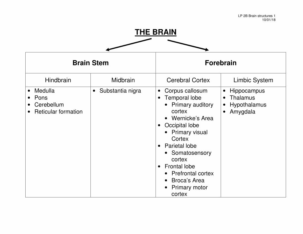
THE BRAIN
Brain Stem Forebrain
Hindbrain Midbrain Cerebral Cortex Limbic System
• Medulla
• Pons
• Cerebellum
• Reticular formation
• Substantia nigra • Corpus callosum
• Temporal lobe
• Primary auditory cortex
• Wernicke’s Area
• Occipital lobe
• Primary visual Cortex
• Parietal lobe
• Somatosensory cortex
• Frontal lobe
• Prefrontal cortex
• Broca’s Area
• Primary motor cortex
• Hippocampus
• Thalamus
• Hypothalamus
• Amygdala
LP 2B Brain structures 2 10/01/18
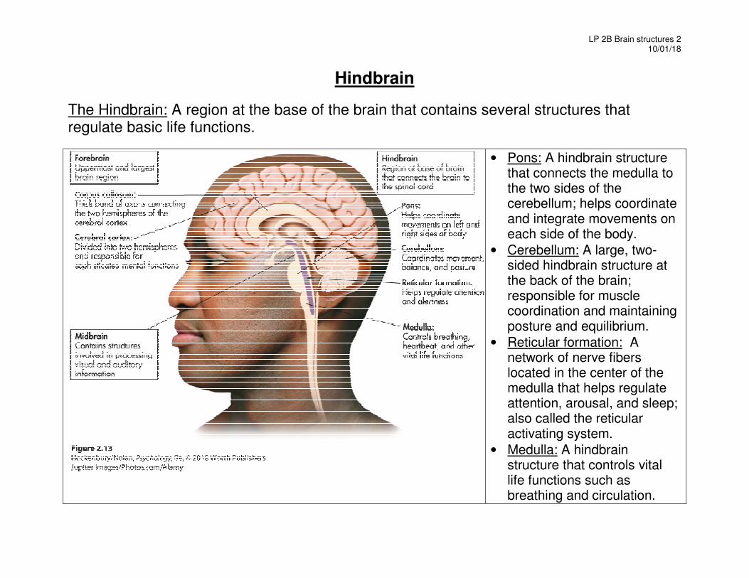
Hindbrain
The Hindbrain: A region at the base of the brain that contains several structures that regulate basic life functions.
• Pons: A hindbrain structure that connects the medulla to the two sides of the cerebellum; helps coordinate and integrate movements on each side of the body.
• Cerebellum: A large, two-sided hindbrain structure at the back of the brain; responsible for muscle coordination and maintaining posture and equilibrium.
• Reticular formation: A network of nerve fibers located in the center of the medulla that helps regulate attention, arousal, and sleep; also called the reticular activating system.
• Medulla: A hindbrain structure that controls vital life functions such as breathing and circulation.
LP 2B Brain structures 3 10/01/18
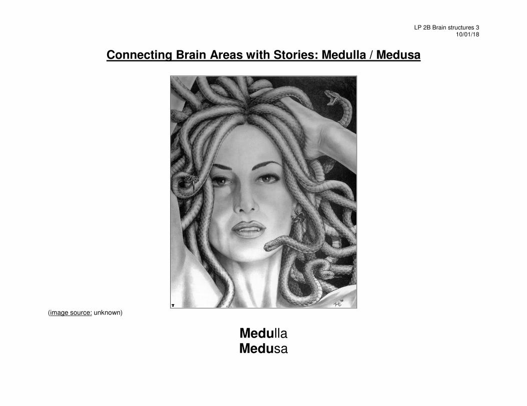
Connecting Brain Areas with Stories: Medulla / Medusa
(image source: unknown)
Medulla Medusa
LP 2B Brain structures 4 10/01/18
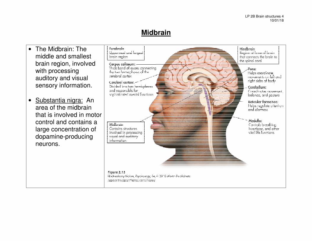
Midbrain
• The Midbrain: The middle and smallest brain region, involved with processing auditory and visual sensory information.
• Substantia nigra: An area of the midbrain that is involved in motor control and contains a large concentration of dopamine-producing neurons.
LP 2B Brain structures 5 10/01/18
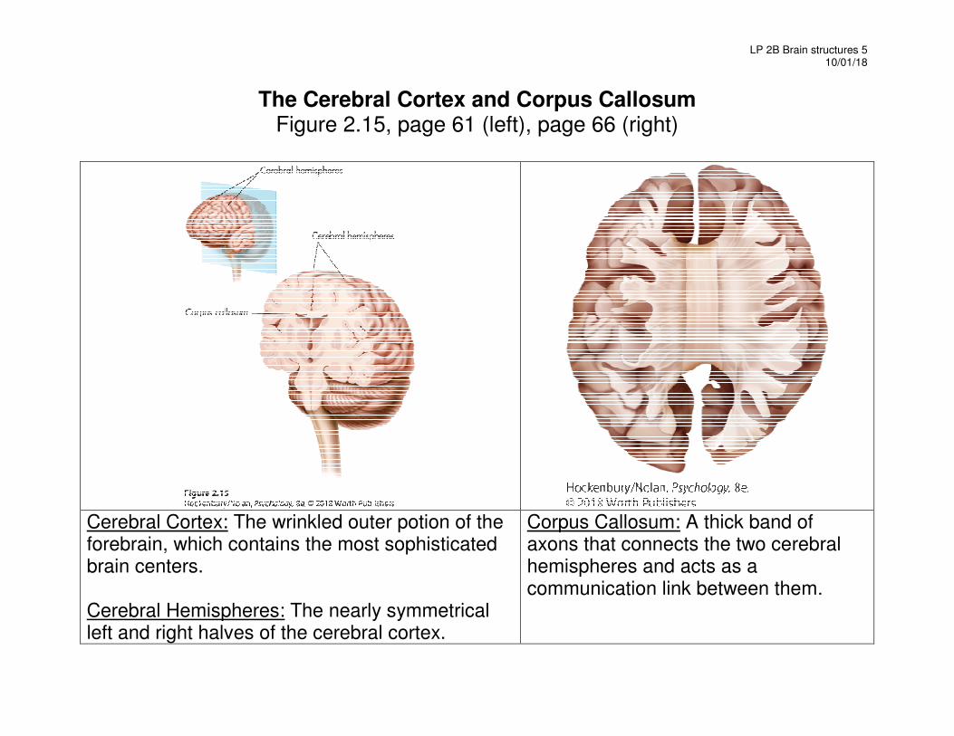
The Cerebral Cortex and Corpus Callosum Figure 2.15, page 61 (left), page 66 (right)
Cerebral Cortex: The wrinkled outer potion of the forebrain, which contains the most sophisticated brain centers. Cerebral Hemispheres: The nearly symmetrical left and right halves of the cerebral cortex.
Corpus Callosum: A thick band of axons that connects the two cerebral hemispheres and acts as a communication link between them.
LP 2B Brain structures 6 10/01/18
Forebrain and Cortical Structures
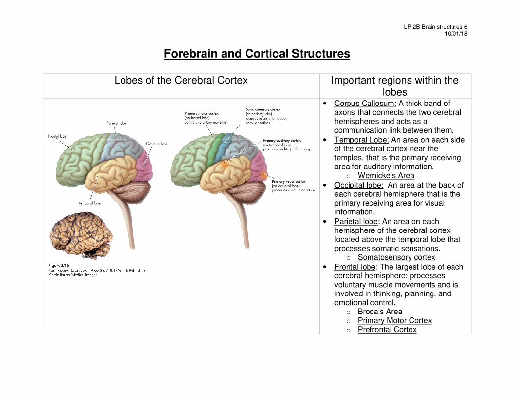
Lobes of the Cerebral Cortex Important regions within the
lobes
• Corpus Callosum: A thick band of axons that connects the two cerebral hemispheres and acts as a communication link between them.
• Temporal Lobe: An area on each side of the cerebral cortex near the temples, that is the primary receiving area for auditory information.
o Wernicke’s Area
• Occipital lobe: An area at the back of each cerebral hemisphere that is the primary receiving area for visual information.
• Parietal lobe: An area on each hemisphere of the cerebral cortex located above the temporal lobe that processes somatic sensations.
o Somatosensory cortex
• Frontal lobe: The largest lobe of each cerebral hemisphere; processes voluntary muscle movements and is involved in thinking, planning, and emotional control.
o Broca’s Area o Primary Motor Cortex o Prefrontal Cortex
LP 2B Brain structures 7 10/01/18
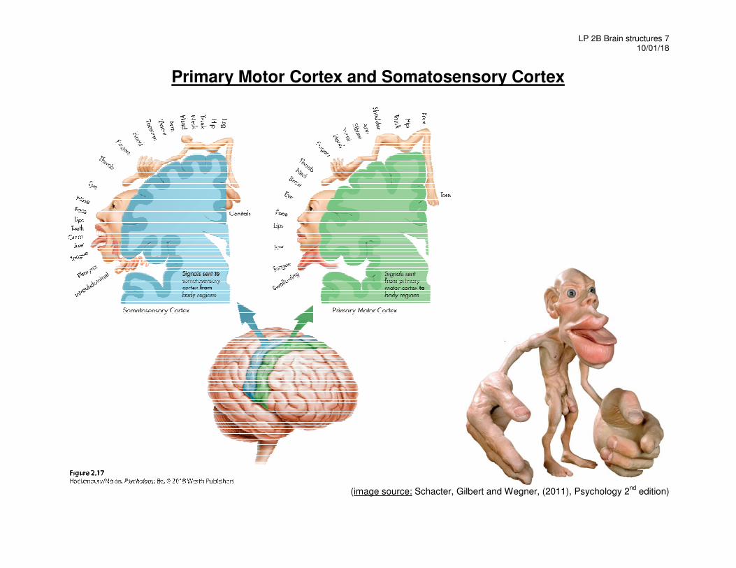
Primary Motor Cortex and Somatosensory Cortex
(image source: Schacter, Gilbert and Wegner, (2011), Psychology 2
nd edition)
LP 2B Brain structures 8 10/01/18
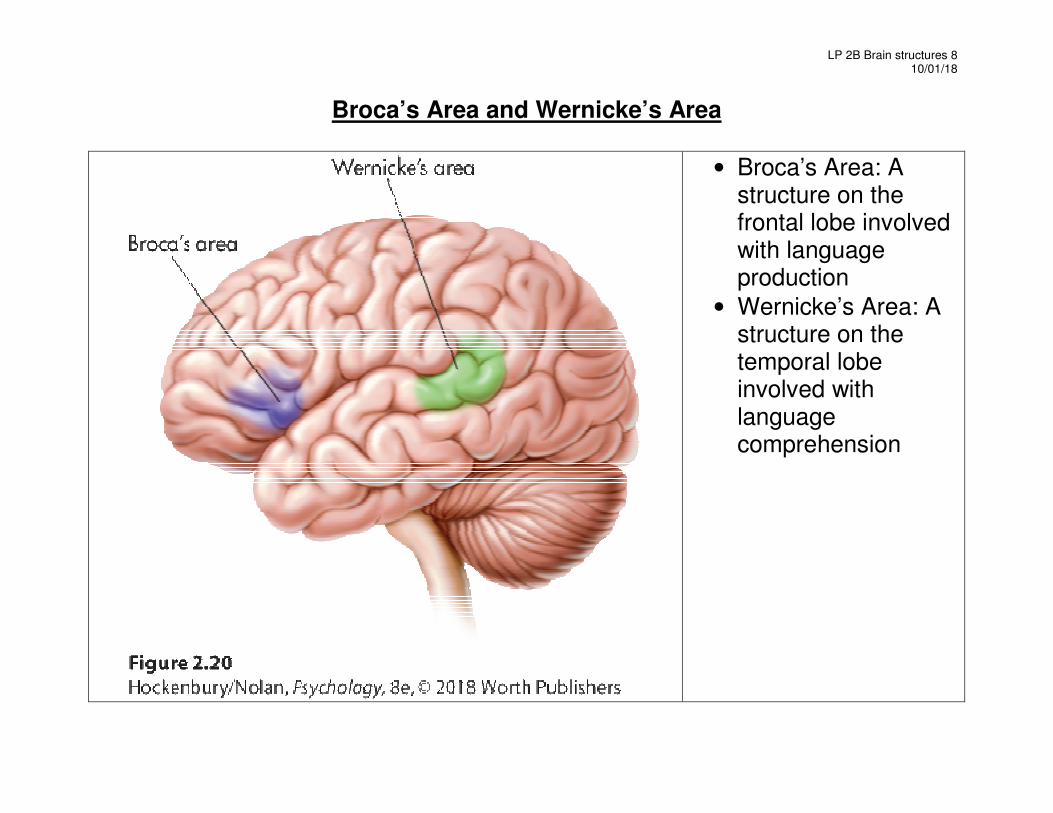
Broca’s Area and Wernicke’s Area
• Broca’s Area: A structure on the frontal lobe involved with language production
• Wernicke’s Area: A structure on the temporal lobe involved with language comprehension
LP 2B Brain structures 9 10/01/18
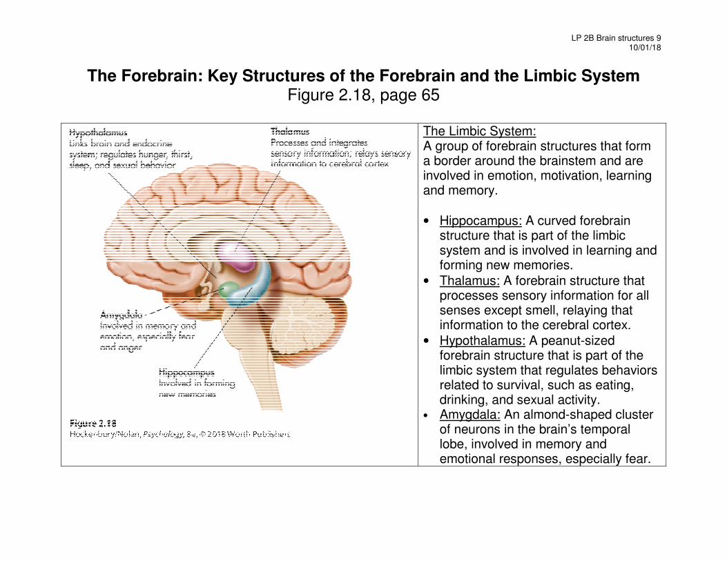
The Forebrain: Key Structures of the Forebrain and the Limbic System Figure 2.18, page 65
The Limbic System: A group of forebrain structures that form a border around the brainstem and are involved in emotion, motivation, learning and memory.
• Hippocampus: A curved forebrain structure that is part of the limbic system and is involved in learning and forming new memories.
• Thalamus: A forebrain structure that processes sensory information for all senses except smell, relaying that information to the cerebral cortex.
• Hypothalamus: A peanut-sized forebrain structure that is part of the limbic system that regulates behaviors related to survival, such as eating, drinking, and sexual activity.
• Amygdala: An almond-shaped cluster of neurons in the brain’s temporal lobe, involved in memory and emotional responses, especially fear.
LP 2B Brain structures 10 10/01/18
Chapter 6: Memory Using elaborative rehearsal to remember information (page 248, 249): But if you elaborated on the information in some meaningful way, you would be more likely to recall it. For example, you could think about the limbic system’s involvement in emotions, memory, and motivation by constructing a simple story.
• “I knew it was lunchtime because my hypothalamus told me I was hungry, thirsty and cold.
• My hippocampus helped me remember a new restaurant that opened on campus,
• but when I got there, I had to wait in line and my amygdala reacted with anger.

LP 2B Brain structures 11 10/01/18
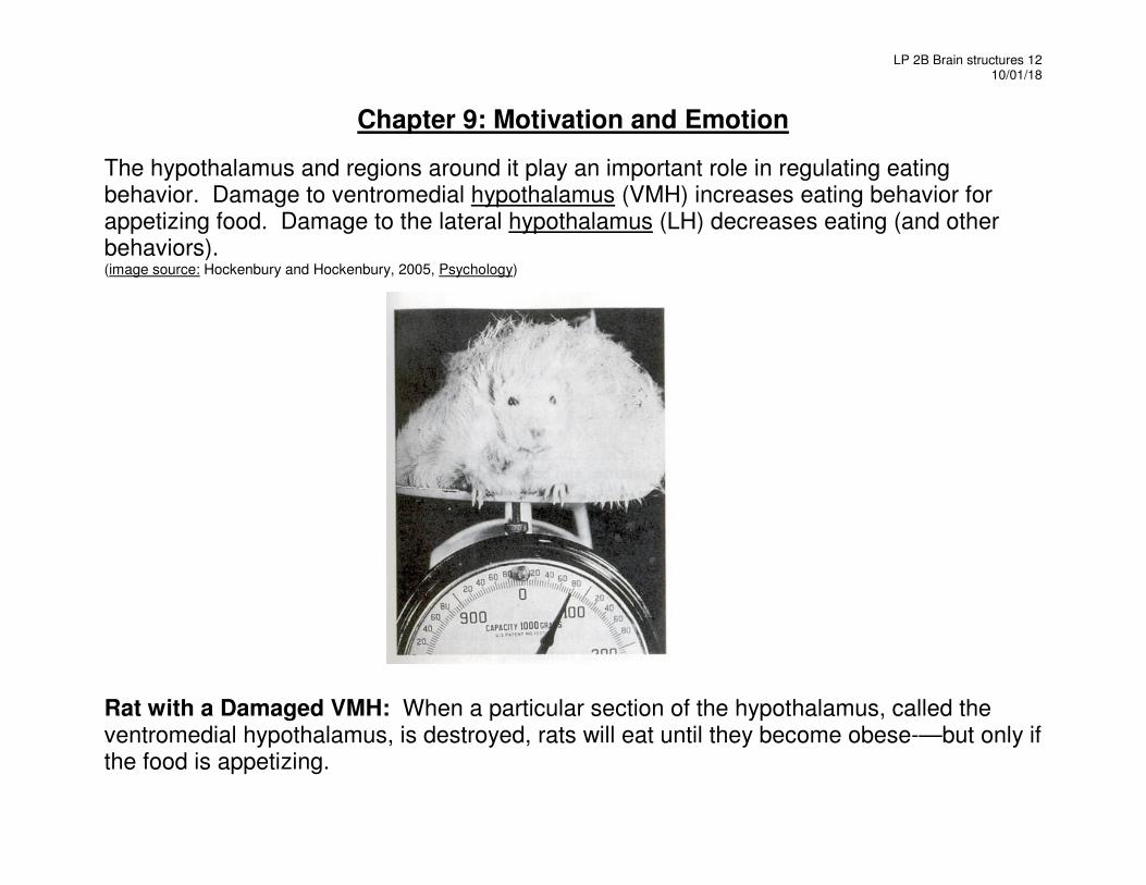
LP 2B Brain structures 12 10/01/18
Chapter 9: Motivation and Emotion
The hypothalamus and regions around it play an important role in regulating eating behavior. Damage to ventromedial hypothalamus (VMH) increases eating behavior for appetizing food. Damage to the lateral hypothalamus (LH) decreases eating (and other behaviors). (image source: Hockenbury and Hockenbury, 2005, Psychology)
Rat with a Damaged VMH: When a particular section of the hypothalamus, called the ventromedial hypothalamus, is destroyed, rats will eat until they become obese-—but only if the food is appetizing.