Embed Size (px)
Citation preview
REVIEWpublished: 03 August 2017
doi: 10.3389/fnana.2017.00064
Midbrain-Hindbrain BoundaryMorphogenesis: At the Intersectionof Wnt and Fgf SignalingHolly C. Gibbs1, Ana Chang-Gonzalez1, Wonmuk Hwang1,2,3, Alvin T. Yeh1
and Arne C. Lekven4*1Department of Biomedical Engineering, Texas A&M University, College Station, TX, United States, 2Department of MaterialsScience and Engineering, Texas A&M University, College Station, TX, United States, 3School of Computational Sciences,Korea Institute for Advanced Study, Seoul, South Korea, 4Department of Biology, Texas A&M University, College Station, TX,United States
Edited by:Sandra Blaess,
University of Bonn, Germany
Reviewed by:Gokul Kesavan,
Technische Universität Dresden,Germany
Steffen Scholpp,University of Exeter, United Kingdom
*Correspondence:Arne C. Lekven
Received: 02 June 2017Accepted: 17 July 2017
Published: 03 August 2017
Citation:Gibbs HC, Chang-Gonzalez A,
Hwang W, Yeh AT and Lekven AC(2017) Midbrain-Hindbrain BoundaryMorphogenesis: At the Intersection of
Wnt and Fgf Signaling.Front. Neuroanat. 11:64.
doi: 10.3389/fnana.2017.00064
A constriction in the neural tube at the junction of the midbrain and hindbrain isa conserved feature of vertebrate embryos. The constriction is a defining feature ofthe midbrain-hindbrain boundary (MHB), a signaling center that patterns the adjacentmidbrain and rostral hindbrain and forms at the junction of two gene expressiondomains in the early neural plate: an anterior otx2/wnt1 positive domain and a posteriorgbx/fgf8 positive domain. otx2 and gbx genes encode mutually repressive transcriptionfactors that create a lineage restriction boundary at their expression interface. Wntand Fgf genes form a mutually dependent feedback system that maintains theirexpression domains on the otx2 or gbx side of the boundary, respectively. Constrictionmorphogenesis occurs after these conserved gene expression domains are establishedand while their mutual interactions maintain their expression pattern; consequently,mutant studies in zebrafish have led to the suggestion that constriction morphogenesisshould be considered a unique phase of MHB development. We analyzed MHBmorphogenesis in fgf8 loss of function zebrafish embryos using a reporter driven by theconserved wnt1 enhancer to visualize anterior boundary cells. We found that fgf8 loss offunction results in a re-activation of wnt1 reporter expression posterior to the boundarysimultaneous with an inactivation of the wnt1 reporter in the anterior boundary cells, andthat these events correlate with relaxation of the boundary constriction. In considerationof other results that correlate the boundary constriction with Wnt and Fgf expression,we propose that the maintenance of an active Wnt-Fgf feedback loop is a key factor indriving the morphogenesis of the MHB constriction.
Keywords: MHB, mes/r1, Wnt, Fgf, constriction morphogenesis, two-photon fluorescence, image analysis,zebrafish
INTRODUCTION
The midbrain-hindbrain boundary (MHB), also called the isthmic organizer, has piqued theinterest of developmental biologists for decades. Characterized by a conspicuous constrictionin the developing neural tube, the MHB, located at the interface of the midbrain and hindbrainneuromeres, is well known to function as a signaling center responsible for patterning cell fatesanteriorly in the midbrain and posteriorly in the cerebellum (Wurst and Bally-Cuif, 2001; Raibleand Brand, 2004; Dworkin and Jane, 2013). The constriction is particularly evident in the dorsalneural tube and defines the posterior midbrain tectum and the hindbrain cerebellum. The MHBconstriction also separates ventricular regions within the neural tube lumen, with the midbrain
Frontiers in Neuroanatomy | www.frontiersin.org 1 August 2017 | Volume 11 | Article 64
Gibbs et al. Wnt and Fgf at the Midbrain-Hindbrain Boundary
ventricle anterior to the constriction and the hindbrain ventriclebehind. The MHB thus represents a crucial dividing pointin the developing brain with characteristic morphologicalfeatures which are critical for several MHB functions: asa signaling center, as a guide for neuronal migration andaxon pathfinding (Volkmann et al., 2010), and as a physicalseparation of brain ventricles (Lowery et al., 2009). Whatis less well understood is the link between the mechanismsresponsible for MHB specification and patterning, and betweenthe signaling molecules that provide its signaling centeractivity and the constriction morphology that invariantlyaccompanies vertebrate MHB development. In other words,why is there always a neural tube constriction at the MHB,and is this morphology a cause, or consequence, of MHBfunction?
The mechanisms behind MHB specification and functionare of interest on multiple levels. First, model organism studieshave shown that defects in specification and patterning of theMHB lead to major deficiencies in the brain, such as theabsence of midbrain, loss of cerebellum, and overgrowth ofthe midbrain tectum (McMahon and Bradley, 1990; Thomasand Capecchi, 1990; Buckles et al., 2004). Second, advances inmagnetic resonance imaging (MRI) have enabled new analysesof human midbrain-hindbrain malformations (Doherty et al.,2013). These new imaging studies are revealing a surprisingnumber of human central nervous system deficits that likelyare caused by aberrant developmental patterning, such asthe association of septo-optic dysplasia with chromosome14 deletions, which include the neural patterning gene, otx2(Severino et al., 2014). Identifying potential causes of these severenervous system diseases requires a thorough understandingof the developmental mechanisms behind midbrain-hindbraindevelopment.
As demonstrated by mouse mutants and zebrafish reporterlines, the MHB is specifically positioned within a domain ofthe early neural plate referred to as mes/r1 in mouse (Zervaset al., 2004) or the midbrain hindbrain domain (MH) inzebrafish (Tallafuss and Bally-Cuif, 2003). These studies showthat the early mesencephalon (mes) and rhombomere 1 (r1)in the anterior hindbrain are genetically co-specified, and theMHB defines a balance point between these midbrain andhindbrain divisions. Besides positioning the future MHB, thebalance point also represents an interface between Wnt ligandexpressing progenitors of the posterior mesencephalon and Fgfligand expressing progenitors of the anterior rhombencephalon,which interact in multiple ways throughout the specification andmorphogenesis of mes/r1 and the MHB. Thus, an importantquestion that is not yet sufficiently answered is what isthe significance of the Wnt-Fgf interface at the MHB tomes/r1 development?
MIDBRAIN HINDBRAIN DOMAINMORPHOGENESIS AND PATTERNING
To appreciate the difficulty of dissecting the role of Wnt and Fgfsignaling families in the morphogenesis and patterning of theMH by the isthmic organizer, and to begin to identify processes
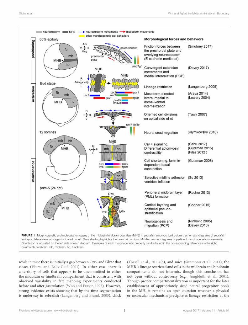
whose disruption would result in neurological disorder, it ishelpful to first have a clear picture of how the MH takes shape.A model of the current morphological and molecular ontogenyof the MH region in zebrafish is shown in Figure 1.
PositioningThe first critical step inMHmorphogenesis is correct positioningof the neural primordium on the body axes such thatspatiotemporal positioning cues can properly ‘‘posteriorize’’the nervous system, that is, establish anteroposterior positionalinformation after neural induction. Neural primordiumpositioning is mediated in part by the mechanical coupling ofthe presumptive neurectoderm to the involuted, anteriorly-migrating prechordal plate of the axial mesoderm via frictionforces generated by the cell adhesion molecule E-cadherin,which is coupled on its cytoplasmic end to the canonicalWnt effector molecule, β-catenin (Smutny et al., 2017).In this study, uncoupled ectodermal cells anterior to thepresumptive neurectoderm ‘‘flowed’’ laterally, posteriorly,and then medially. These complex ‘‘vortex’’ migrations ofpresumptive neurectodermal cells are presumably part ofconvergent extension movements that rely on non-canonicalWnt/PCP signaling to facilitate medial intercalation andanterior-posterior axis lengthening (Heisenberg et al., 2000;Davey and Moens, 2017). What is remarkable is that ontheir tumultuous journey, presumptive neurectodermal cellsare precisely exposed to a complex molecular program thatincludes posteriorizing Wnt and Fgf signals emanating from theblastoderm margin (reviewed in Green et al., 2015; Tuazon andMullins, 2015), resulting in a correctly patterned neural plate.One particularly remarkable and unknown aspect of this processis how early cell responses to Wnt and Fgf ligands occur duringthe complex morphogenetic changes of gastrulation.
Several lines of evidence suggest that Wnt and Fgf ligandsexpressed in the nascent paraxial mesoderm function asmorphogens by establishing concentration or activity gradientsthat generate anteroposterior positional information that isinterpreted into patterned cell fates (e.g., Cox and Hemmati-Brivanlou, 1995; McGrew et al., 1995). A crucial function inparticular for Wnt ligands in polarizing the neural plate hasbeen established from experiments in zebrafish, Xenopus, andchick (Kiecker and Niehrs, 2001; Nordström et al., 2002; Dorskyet al., 2003; Rhinn et al., 2005). The mechanism by whichgraded Wnt activity is established is not yet known, thoughrecent results suggest delivery of Wnt ligand via filopodiafrom paraxial mesoderm progenitors may be a major mode oftransport (Stanganello et al., 2015). This differs from Fgf in thiscontext, which has been shown to freely diffuse from its paraxialmesoderm source and form a gradient through a source-sinkmechanism (Scholpp and Brand, 2004; Yu et al., 2009).
Of importance to the position of the MHB organizer,specifically, is the activation of the transcription factor gbx1 inthe posterior neural plate by Wnt8a (Rhinn et al., 2005, 2009),which antagonizes independently activated otx2 expressed in theforebrain and midbrain (Kurokawa et al., 2012). In the zebrafish,these domains overlap slightly at 60% epiboly but subsequentlybecome mutually exclusive by 80% epiboly (Rhinn et al., 2003),
Frontiers in Neuroanatomy | www.frontiersin.org 2 August 2017 | Volume 11 | Article 64
Gibbs et al. Wnt and Fgf at the Midbrain-Hindbrain Boundary
FIGURE 1 | Morphogenetic and molecular ontogeny of the midbrain hindbrain boundary (MHB) in zebrafish embryos. Left column: schematic diagrams of zebrafishembryos, lateral view, at stages indicated on left. Gray shading highlights the brain primordium. Middle column: diagrams of pertinent morphogenetic movements.Orientation is indicated on the left side of each diagram. Examples of each morphogenetic property can be found in the corresponding references in the rightcolumn. fb, forebrain; mb, midbrain; hb, hindbrain.
while in mice there is initially a gap between Otx2 and Gbx2 thatcloses (Wurst and Bally-Cuif, 2001). In either case, there isa territory of cells that appears to be uncommitted to eitherthe midbrain or hindbrain compartment that is consistent withobserved variability in fate mapping experiments conductedbefore and after gastrulation (Woo and Fraser, 1995). However,strong evidence exists showing that by the time segmentationis underway in zebrafish (Langenberg and Brand, 2005), chick
(Tossell et al., 2011a,b), and mice (Sunmonu et al., 2011), theMHB is lineage restricted and cells in themidbrain and hindbraincompartments do not intermix, though this conclusion hasnot been without controversy (e.g., Jungbluth et al., 2001).Though proper compartmentalization is important for the laterestablishment of appropriately sized neural progenitor poolsin the MH, it remains an open question whether a physicalor molecular mechanism precipitates lineage restriction at the
Frontiers in Neuroanatomy | www.frontiersin.org 3 August 2017 | Volume 11 | Article 64
Gibbs et al. Wnt and Fgf at the Midbrain-Hindbrain Boundary
MHB. A report from chick suggests there is a posterior shiftin the position of the Otx/Gbx interface, such that it onlycoincides with the physical MHB constriction at later stages indevelopment (Hidalgo-Sánchez et al., 2005). In zebrafish andin mice, however, the consensus is that the initial Otx/Gbxboundary definitively marks the future MHB constriction priorto when it becomes morphologically visible. Notch signaling,which classically causes cells to make such boundary decisions byamplification of small stochastic differences in gene expressionfollowed by cell sorting, has been implicated in sorting Otxand Gbx cells at the MHB in chick (Tossell et al., 2011b).Reports frommice andmedaka suggest other genetic interactionsmay refine the boundary, as Gbx2 interacts with Grouchorepressors and can directly compete with POU transcriptionalactivators of Otx2 (Heimbucher et al., 2007; Inoue et al., 2012).Intercellular actinomyosin networks that have been shown todrive morphogenesis, such as during mesoderm invagination inDrosophila, may also play a physical role in lineage restriction atthe MH (Kasza and Zallen, 2011) or possibly regional changesin cortical actin tension cause cells to sort to one side of theboundary or another (Heisenberg and Bellaïche, 2013).
ActivationShortly after the positioning phase during gastrulation, a suite ofMHB genes are activated in distinct domains around the otx2/gbxboundary as the neural plate undergoes neurulation to form theneural tube. Expression of wnt1 anteriorly and fgf8a posteriorlyto the presumptive MHB (that is still not morphologicallyobvious) reinforce the otx2/gbx interface while her5, eng2a, andpax2a are expressed on both sides of the boundary (Rhinn andBrand, 2001; Buckles et al., 2004).Whichmolecules and/or forcesactivate these core members of the more extensive MHB geneticprogram remains poorly characterized, and, surprisingly, thisactivation program can occur in the absence of at least parts ofthe positioning machinery (Su et al., 2014). Once activated, thespecific roles of each gene in promoting subsequent developmentwithin the MH (beyond providing spatial cues) is also not wellunderstood, though several components of the MHB programappear to have roles in both fate specification andmorphogenesiswithin the MH (Dworkin and Jane, 2013). For instance, her5 isknown to inhibit neurogenesis during segmentation (Tallafussand Bally-Cuif, 2003; Ninkovic et al., 2005) and to subsequentlypromote neural stem cell identity in adult zebrafish (Chapoutonet al., 2006).
The establishment of the Wnt/Fgf signaling interface,however, is certainly crucial to the development of the MH.Both Wnt1−/− and Fgf8−/− mice fail to develop the entireMH region (McMahon and Bradley, 1990; Chi et al., 2003). Inzebrafish, loss of several redundant Wnts (wnt3, wnt3a, wnt1and wnt10b) recapitulates a similar phenotype (Lekven et al.,2003; Buckles et al., 2004) and the zebrafish fgf8a mutant acelacks a cerebellum and MHB constriction, though the midbrainis present but unpolarized, resulting in aberrant retinotectalprojections (Picker et al., 1999). Fgf8 has been deemed themost important ‘‘organizing molecule’’ based on results fromimplanting Fgf8-soaked beads at sites anterior and posterior tothe MHB. In these experiments, Fgf8 was sufficient to induce
tectal and cerebellar structures and an underlying Otx/Gbxboundary, while similar experiments for Wnt1 showed nosignificant re-patterning of the surrounding tissues (Martinezet al., 1999). Indeed, no gain of function analysis for all theother major MHB molecules in any organism has yielded suchstriking results. However, a study in which Otx2 and Fgf8 weresimultaneously knocked down has challenged the idea thatFgf8 is required to pattern cell fates in the MHB. Foucheret al. (2006) showed that in the absence of Fgf8, if Otx2 levelswere depleted, cerebellar neurons were able to successfullydifferentiate, though MHB morphology was abnormal in theseembryos. Recent analysis of otx;gbx;fgf embryos also suggeststhat robust cerebellar differentiation requires Fgf (Su et al., 2014).
During MHB program activation, the process of primaryneurulation, in which the neural plate coalesces on the dorsalmidline, is ongoing (Lowery and Sive, 2004). During this process,the medial-lateral organization of the neural plate is transformedto a ventral-dorsal orientation (Schmitz et al., 1993). It is worthmentioning that although the subsequently developing MHBconstriction has been studied primarily in reference to the A/Paxis, it is not uniform on the D/V axis of the neural tube, whichmay reflect graded or inhomogeneous Wnt/Fgf activity along theD/V axis and integration with dorsoventral patterning signalingactivities (Lekven et al., 2003; Puelles et al., 2003). Thus, theMHBliterature is largely focused on organizer activity in the alar regionof the MH with relatively little known of the basal tegmentum.
Shortly after the neural tube is formed and the neuralcrest begins to migrate, the MHB constriction becomes avisible morphological feature as brain ventricles begin to form.In zebrafish, MHB constriction requires cell shortening andsubsequent laminin-dependent basal constriction of a smallring of cells at the boundary (Gutzman et al., 2008). The cellshape changes involved in MHB constriction morphogenesisrequire non-muscle myosin II, and recent results show that cellshortening required at the MHB constriction is a consequenceof calcium transient regulation of myosin light chain kinase(Gutzman and Sive, 2010; Gutzman et al., 2015; Sahu et al., 2017).
MaintenanceOnce the neural tube is formed and the MHB constriction hasbeen initiated, the genetic program within the MH subsequentlytransitions to the maintenance phase accompanied by continuedreshaping of the brain tissue and ventricular system, as wellas production of cerebrospinal fluid that may itself contributeto MHB regulation (Parada et al., 2005; Gato and Desmond,2009). Computational modeling and experimentation in chickindicate importance of differential myosin-mediated contractilityto produce brain ventricle geometry and suggest strategiesmay differ from compartment to compartment depending onthe end fate of the junction, as some are only transientstructures (rhombomere boundaries, for example) while others,such as the MHB constriction, persist as structures in theadult brain that must resist increasing fluid pressure from theventricular system (Filas et al., 2012). One function the MHBconstriction may play, thus, is as a point of transition betweendifferent anterior and posterior brain ventricle morphogenesisprograms converging at the boundary. Such a structure would
Frontiers in Neuroanatomy | www.frontiersin.org 4 August 2017 | Volume 11 | Article 64
Gibbs et al. Wnt and Fgf at the Midbrain-Hindbrain Boundary
need to maintain cell adhesion at the boundary until brainventricle morphology was established on either side to preventmisspecification of the surrounding tissues. The constrictioncould also mediate the timing of signaling between anterior andposterior brain compartments in the case of signaling moleculessecreted in the cerebrospinal fluid. Such phenomena are notwithout precedent; for example, in mouse embryos it is wellknown that left/right asymmetry is broken by cilia- directedfluid flow in the node, though it is not known if the signalmediated through the unidirectional fluid flow is mechanicalor chemical in nature (Yoshiba and Hamada, 2014). Brainventricles have been shown to have cilia, and in zebrafish ciliain the developing telencephalon were shown to direct neuronalmigration (Kishimoto et al., 2011). Some such mechanism mayaccount for the evolution of the closed primary neurulationstrategy seen in zebrafish compared to neural tube infolding seenin other vertebrates.
In the maintenance phase, several sub-regions of the MHemerge that execute their own morphogenetic programs inanticipation of neurogenesis. In the midbrain, the optic tectumis shaped by the formation of a tight sheet of cells calledthe peripheral midbrain layer (PML) harboring slow-cyclingneural progenitor cells that will give rise to columns of neuronsorganized by alternating protocadherin expression that populatethemore anterior tectum in a cortical fashion (Recher et al., 2013;Cooper et al., 2015; Rapaciolii et al., 2016). Posterior to the MHB,the cerebellar rhombic lip and ventricular zones form, fromwhich granule and Purkinje progenitor cells are later derived,respectively, before their neural derivatives organize into thedorsoventral layers and mediolateral compartments that providethe foundation of the cerebellar circuitry (Hashimoto and Hibi,2012; Millen et al., 2014). The MH tegmentum has almost noovert morphological landmarks apart from a relatively shallowconstriction, but its correct patterning and morphogenesis iscritical to the proper formation of serotonergic and cholinergicnuclei implicated in important behavioral functions (Parkeret al., 2013).
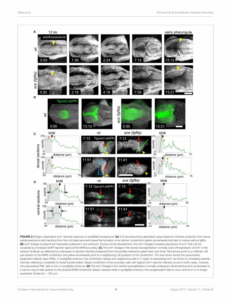
The correct establishment and maintenance of each of theaforementioned pro-neural sub-regions of the MH requires atminimum aWnt and Fgf signaling feedback loop to establish theproper unique molecular and mechanical microenvironments(Carletti and Rossi, 2008). In zebrafish ace(fgf8a) mutants, theexpression of wnt1 and several other genes in the boundaryregion including her5, pax2a, and eng2/3 are activated but theirexpression fades as Fgf-dependent feedback fails in early- tomid-somitogenesis (Reifers et al., 1998). Similarly, the combinedloss of wnt1/wnt10b/wnt3a in zebrafish results in transientexpression of fgf8a, pax2a, and eng2/3 in the early MHB(Buckles et al., 2004). We have recently found using livemultiphoton imaging (Gibbs et al., 2014a) that acemutants forma constriction that fails to mature properly in the maintenancephase (Figure 2), amorphological transient output of amoleculartransience (Gibbs et al., 2013). The failure of the constrictionto continue morphogenesis in the maintenance phase is dueto aberrant cell behaviors in two groups of cells. By imaginga transgenic wnt1 reporter line (Gibbs et al., 2014b) in theace(fgf8a) background, we identified one group of cells that fails
to maintain wnt1 expression in the posterior midbrain, andto subsequently coordinate the proper morphogenesis of thePML and boundary tegmentum, and another group that fails tosuppress wnt1 expression in the dorsal part of r1 to correctlyspecify the cerebellar plate (Figures 2, 3). This observation, basedon identification of individual cells, supports previous reportsof an isthmo/cerebellar-to-tectal transformation in molecularidentity of the presumptive cerebellum that occurs with geneticreprogramming during themaintenance phase (Jászai et al., 2003;Gibbs, 2014), though with live imaging we observed that thisreprogramming caused by a lack of fgf8a does not preclude theprevious initiation of the morphogenesis of the MHB duringthe activation phase. Thus, a mechanism independent of fgf8apositions and initializes this physical boundary, while an fgf8a-dependentmechanism (either directly or indirectly) maintains itscontinued morphogenesis.
Wnt and Fgf signaling may also have more direct roles inshaping the MH during neurogenesis. As mentioned previously,during early stages of MHB formation, neurogenesis is activelyinhibited by her5 in zebrafish (Tallafuss and Bally-Cuif, 2003)but subsequently neurons are born as her5 expression recedesto a narrow ring at the constriction. Wnt1 has recently beenproposed to mediate the timing of neurogenesis in the midbrainby driving Fgf8 expression at the boundary and graduallysuppressing it away from the boundary by inducing Sproutyexpression so that Fgf dependent her5 also recedes (Dyer et al.,2014). Wnt1 may also function to promote neural stem cellidentity in the dorsal midbrain and MHB (Miyake et al., 2012;Lin and Lee, 2016), possibly regulated by Fgf3/8-dependentFgf22 signaling (Miyake and Itoh, 2013) and may contributeto shaping the MH by regulating the cytoskeleton duringaxon guidance (Ciani and Salinas, 2005). In the hindbrain,differentiation of unique tegmentum nuclei identities happensin spatiotemporal waves emanating from the upper rhombiclip. Recently, these migrations were shown to be conserved inmice and zebrafish, with discrete Wnt1 populations in the upperrhombic lip sequentially migrating anteriorly toward the MHBand turning ventrally to their final positions in the hindbraintegmentum (Volkmann et al., 2010). Fgf9/Fgfr2 signaling isimportant for differentiation of Bergmann glial cells in thecerebellum of mice (Meier et al., 2014), a cell type conservedin the zebrafish cerebellum (although zebrafish do not appearto have an fgf9, this function could be attributed to anotherredundantly functioning Fgf; Bae et al., 2009).
MORPHOGENETIC ROLES FOR WNT ANDFGF SIGNALING DURING CONSTRICTIONFORMATION
The Wnt and Fgf signaling pathways are expansive coredevelopmental pathways that play a variety of context-dependent roles. In this section, we further examine the conceptthat a Wnt/Fgf signaling loop is required for proper MHmorphogenesis and discuss potential points of crosstalk betweenthese signaling pathways and cell adhesion and cytoskeletalmachinery, based on studies both in the MH and other systems.
Frontiers in Neuroanatomy | www.frontiersin.org 5 August 2017 | Volume 11 | Article 64
Gibbs et al. Wnt and Fgf at the Midbrain-Hindbrain Boundary
FIGURE 2 | Region dependent wnt1 reporter response in ace(fgf8a) background. (A) 3-D reconstructions generated using maximum intensity projection from tissueautofluorescence (with sections from the roof plate removed) reveal the formation of an isthmic constriction (yellow arrowheads) that fails to mature without fgf8a.(B) wnt1 lineage is present but improperly polarized in ace embryos. During normal development, the wnt1 lineage increases expression of wnt1 that can bevisualized by increased eGFP reporter signal at the MHB boundary. (C) The wnt1 lineage in the dorsal neuroepithelium normally turns off expression of wnt1 in theanterior hindbrain as reflected by a decrease in reporter intensity (measured from the profiles marked by green lines over time). Red arrows point to a midbrain celljust anterior to the MHB constriction and yellow arrowheads point to a neighboring cell posterior to the constriction. The blue arrow shows the presumptiveperipheral midbrain layer (PML). In ace(fgf8a) embryos, the constriction relaxes and neighboring cells in r1 begin re-expressing wnt1 as shown by increasing reporterintensity, reflecting a cerebellar-to-tectal transformation. Basal constriction of the boundary cells with highest wnt1 reporter intensity occurs in both cases, however,the presumptive PML fails to form in ace(fgf8a) embryos. (D) The wnt1 lineage in the ventral neuroepithelium normally undergoes cell shortening and compresses toa narrow ring of cells anterior to the physical MHB constriction (black markers) while in ace(fgf8a) embryos, this reorganization fails to occur and wnt1 is no longerexpressed. Scale bar = 100 µm.
Frontiers in Neuroanatomy | www.frontiersin.org 6 August 2017 | Volume 11 | Article 64
Gibbs et al. Wnt and Fgf at the Midbrain-Hindbrain Boundary
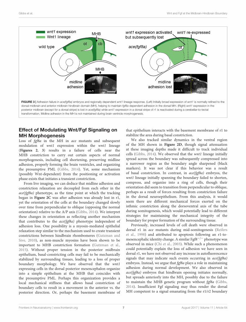
FIGURE 3 | Adhesion failure in ace(fgf8a) embryos and regionally dependent wnt1 lineage response. (Left) Initially broad expression of wnt1 is normally refined to thedorsal midbrain and anterior midbrain hindbrain domain (MH), helping to maintain fgf8a dependent adhesion in the dorsal MH. (Right) wnt1 expression in theposterior midbrain (except for a dorsal stripe) is lost in ace(fgf8a) while wnt1 expression in a dorsal stripe of r1 is reactivated during a cerebellar-to-tectaltransformation. Midline adhesion in the MH is not maintained during brain ventricle morphogenesis.
Effect of Modulating Wnt/Fgf Signaling onMH MorphogenesisLoss of fgf8a in the MH in ace mutants and subsequentmodulation of wnt1 expression within the wnt1 lineage(Figures 2, 3) results in a failure of cells near theMHB constriction to carry out certain aspects of normalmorphogenesis, including cell shortening, preserving midlineadhesion, properly forming the brain ventricles, and organizingthe presumptive PML (Gibbs, 2014). Yet, some mechanism(possibly Wnt-dependent) from the positioning or activationphase exists that initiates a transient constriction.
From live imaging, we can deduce that midline adhesion andconstriction relaxation are decoupled from each other in theace(fgf8a) phenotype, as the time point at which the trackingbegan in Figure 2C was after adhesion was already lost in r1,yet the orientation of the cells at the boundary changed slowlyover time from perpendicular to oblique (opposing the normalorientation) relative to the A/P axis (Gibbs, 2014). We interpretthese changes in orientation as reflecting another mechanismthat contributes to the ace(fgf8a) phenotype independent ofadhesion loss. One possibility is a myosin-mediated epithelialrelaxation step similar to the mechanism used to create transientconstrictions between hindbrain rhombomeres (Gutzman andSive, 2010), as non-muscle myosins have been shown to beimportant to MHB constriction formation (Gutzman et al.,2015). Without proper tension in the posterior midbrainepithelium, basal-constricting cells may fail to be mechanicallystabilized by surrounding tissues, leading to a loss of properboundary morphology. We have observed that the wnt1expressing cells in the dorsal posterior mesencephalon organizeinto a simple epithelium at the MHB that coincides withthe presumptive PML. Perhaps this organization provides alocal mechanical stiffness that allows basal constriction ofboundary cells to result in a movement in the anterior vs. theposterior direction. Or, perhaps the basement membrane of
that epithelium interacts with the basement membrane of r1 tostabilize the area during basal constriction.
We also tracked similar dynamics in the ventral regionof the MH shown in Figure 2D, though signal attenuationat these imaging depths made it difficult to track individualcells (Gibbs, 2014). We observed that the wnt1 lineage initiallyspread across the boundary was subsequently compressed intoa narrower region as the boundary angle sharpened (blackmarkers). It was not clear if this behavior was a resultof basal constriction. In contrast, in ace(fgf8a) embryos, thewnt1 lineage initially spanning the boundary failed to shorten,compress, and organize into a ring of cells, though theirorientation did seem to transition from perpendicular to oblique,perhaps as a result of forces resulting from constriction failurein the dorsal neuroepithelium. From this analysis, it wouldseem there are different mechanical forces exerted on theisthmic constriction along the dorsoventral axis of the tubeduring somitogenesis, which would potentially lead to differentstrategies for maintaining the mechanical integrity of theboundary for proper formation of the surrounding tissue.
Previously, increased levels of cell death were observed indorsal r1 in ace mutants during mid-somitogenesis (Reiferset al., 1998) and attributed to apoptosis following an r1-to-mesencephalic identity change. A similar Fgf8−/− phenotype wasobserved in mice (Chi et al., 2003). While such a phenomenoncould potentially explain the loss of adhesion we have seen indorsal r1, we have not observed any increase in autofluorescencesignals that may indicate such events occurring in ace(fgf8a)embryos. Instead, we argue that fgf8a plays a role in maintainingadhesion during normal development. We also observed inace(fgf8a) embryos that hindbrain opening initiates normally,but spreads anteriorly into the MH, possibly due to the failureto maintain the MHB genetic program without fgf8a (Gibbs,2014). Insufficient Fgf signaling may thus render the dorsalMH competent to a signal emanating from the r1/r2 boundary
Frontiers in Neuroanatomy | www.frontiersin.org 7 August 2017 | Volume 11 | Article 64
Gibbs et al. Wnt and Fgf at the Midbrain-Hindbrain Boundary
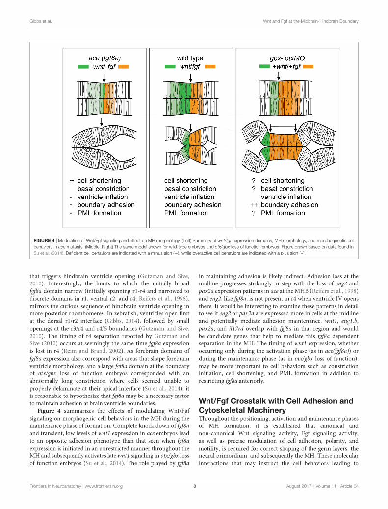
FIGURE 4 | Modulation of Wnt/Fgf signaling and effect on MH morphology. (Left) Summary of wnt/fgf expression domains, MH morphology, and morphogenetic cellbehaviors in ace mutants. (Middle, Right) The same model shown for wild-type embryos and otx/gbx loss of function embryos. Figure drawn based on data found inSu et al. (2014). Deficient cell behaviors are indicated with a minus sign (−), while overactive cell behaviors are indicated with a plus sign (+).
that triggers hindbrain ventricle opening (Gutzman and Sive,2010). Interestingly, the limits to which the initially broadfgf8a domain narrow (initially spanning r1-r4 and narrowed todiscrete domains in r1, ventral r2, and r4; Reifers et al., 1998),mirrors the curious sequence of hindbrain ventricle opening inmore posterior rhombomeres. In zebrafish, ventricles open firstat the dorsal r1/r2 interface (Gibbs, 2014), followed by smallopenings at the r3/r4 and r4/5 boundaries (Gutzman and Sive,2010). The timing of r4 separation reported by Gutzman andSive (2010) occurs at seemingly the same time fgf8a expressionis lost in r4 (Reim and Brand, 2002). As forebrain domains offgf8a expression also correspond with areas that shape forebrainventricle morphology, and a large fgf8a domain at the boundaryof otx/gbx loss of function embryos corresponded with anabnormally long constriction where cells seemed unable toproperly delaminate at their apical interface (Su et al., 2014), itis reasonable to hypothesize that fgf8a may be a necessary factorto maintain adhesion at brain ventricle boundaries.
Figure 4 summarizes the effects of modulating Wnt/Fgfsignaling on morphogenic cell behaviors in the MH during themaintenance phase of formation. Complete knock down of fgf8aand transient, low levels of wnt1 expression in ace embryos leadto an opposite adhesion phenotype than that seen when fgf8aexpression is initiated in an unrestricted manner throughout theMH and subsequently activates latewnt1 signaling in otx/gbx lossof function embryos (Su et al., 2014). The role played by fgf8a
in maintaining adhesion is likely indirect. Adhesion loss at themidline progresses strikingly in step with the loss of eng2 andpax2a expression patterns in ace at the MHB (Reifers et al., 1998)and eng2, like fgf8a, is not present in r4 when ventricle IV opensthere. It would be interesting to examine these patterns in detailto see if eng2 or pax2a are expressed more in cells at the midlineand potentially mediate adhesion maintenance. wnt1, eng1.b,pax2a, and il17rd overlap with fgf8a in that region and wouldbe candidate genes that help to mediate this fgf8a dependentseparation in the MH. The timing of wnt1 expression, whetheroccurring only during the activation phase (as in ace(fgf8a)) orduring the maintenance phase (as in otx/gbx loss of function),may be more important to cell behaviors such as constrictioninitiation, cell shortening, and PML formation in addition torestricting fgf8a anteriorly.
Wnt/Fgf Crosstalk with Cell Adhesion andCytoskeletal MachineryThroughout the positioning, activation and maintenance phasesof MH formation, it is established that canonical andnon-canonical Wnt signaling activity, Fgf signaling activity,as well as precise modulation of cell adhesion, polarity, andmotility, is required for correct shaping of the germ layers, theneural primordium, and subsequently the MH. These molecularinteractions that may instruct the cell behaviors leading to
Frontiers in Neuroanatomy | www.frontiersin.org 8 August 2017 | Volume 11 | Article 64
Gibbs et al. Wnt and Fgf at the Midbrain-Hindbrain Boundary
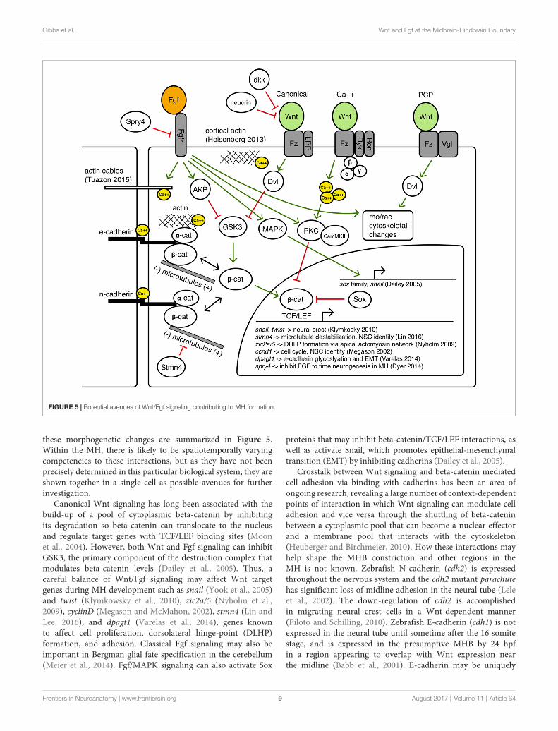
FIGURE 5 | Potential avenues of Wnt/Fgf signaling contributing to MH formation.
these morphogenetic changes are summarized in Figure 5.Within the MH, there is likely to be spatiotemporally varyingcompetencies to these interactions, but as they have not beenprecisely determined in this particular biological system, they areshown together in a single cell as possible avenues for furtherinvestigation.
Canonical Wnt signaling has long been associated with thebuild-up of a pool of cytoplasmic beta-catenin by inhibitingits degradation so beta-catenin can translocate to the nucleusand regulate target genes with TCF/LEF binding sites (Moonet al., 2004). However, both Wnt and Fgf signaling can inhibitGSK3, the primary component of the destruction complex thatmodulates beta-catenin levels (Dailey et al., 2005). Thus, acareful balance of Wnt/Fgf signaling may affect Wnt targetgenes during MH development such as snail (Yook et al., 2005)and twist (Klymkowsky et al., 2010), zic2a/5 (Nyholm et al.,2009), cyclinD (Megason and McMahon, 2002), stmn4 (Lin andLee, 2016), and dpagt1 (Varelas et al., 2014), genes knownto affect cell proliferation, dorsolateral hinge-point (DLHP)formation, and adhesion. Classical Fgf signaling may also beimportant in Bergman glial fate specification in the cerebellum(Meier et al., 2014). Fgf/MAPK signaling can also activate Sox
proteins that may inhibit beta-catenin/TCF/LEF interactions, aswell as activate Snail, which promotes epithelial-mesenchymaltransition (EMT) by inhibiting cadherins (Dailey et al., 2005).
Crosstalk between Wnt signaling and beta-catenin mediatedcell adhesion via binding with cadherins has been an area ofongoing research, revealing a large number of context-dependentpoints of interaction in which Wnt signaling can modulate celladhesion and vice versa through the shuttling of beta-cateninbetween a cytoplasmic pool that can become a nuclear effectorand a membrane pool that interacts with the cytoskeleton(Heuberger and Birchmeier, 2010). How these interactions mayhelp shape the MHB constriction and other regions in theMH is not known. Zebrafish N-cadherin (cdh2) is expressedthroughout the nervous system and the cdh2 mutant parachutehas significant loss of midline adhesion in the neural tube (Leleet al., 2002). The down-regulation of cdh2 is accomplishedin migrating neural crest cells in a Wnt-dependent manner(Piloto and Schilling, 2010). Zebrafish E-cadherin (cdh1) is notexpressed in the neural tube until sometime after the 16 somitestage, and is expressed in the presumptive MHB by 24 hpfin a region appearing to overlap with Wnt expression nearthe midline (Babb et al., 2001). E-cadherin may be uniquely
Frontiers in Neuroanatomy | www.frontiersin.org 9 August 2017 | Volume 11 | Article 64
Gibbs et al. Wnt and Fgf at the Midbrain-Hindbrain Boundary
responsible for maintaining cell adhesion to help stabilizecytoskeletal rearrangements at the midline in the MH regionor during the formation of the PML. One example linking Fgfsignaling with morphogenetic remodeling of the cytoskeletonhas been proposed to work through Fgfr-Ras-MAPK signalingin the formation of the lateral line sensory system in zebrafish(Harding and Nechiporuk, 2012). In this study, authors foundthat Ras-MAPK signaling activated by Fgfr was required forthe formation of rosettes by localizing Rho-associated kinase(Rock) to the apical surface to drive its constriction. Fgfsignaling has also been shown to have a role in otic vesicleformation, which requires apical constriction mediated bylocal increases in actin. In the otic vesicle, Fgf signalingactivates phospholipase-C (PLC) which triggers non-canonicalmyosin-II activity (Sai and Ladher, 2008). Classically, myosin-IIis understood to ratchet along actin filaments to promotecontraction, however, upon phosphorylation by PLC, myosin-IIpromoted the degradation of basal actin (resulting in enrichedapical actin and otic cup invagination). Wnt signaling has alsobeen implicated in cytoskeletal remodeling via the planar-cell-polarity pathway and perhaps also canonical signaling pathways(Lapebie et al., 2011). Once neurogenesis begins in the MHregion, it is possible that Wnt/Fgf signaling may modulate neuralmigration (Knosp et al., 2015).Wnt and Fgf have been implicatedin changes in epithelial cell adhesion in neurogenic cranialplacodes (Lassiter et al., 2014) and it has been shown that novelWnt receptors Ryk and Ror can interact with the cytoskeleton topromote axon guidance (Clark et al., 2012).
Balance of intra- and intercellular calcium is anotherinteresting candidate target bridging Wnt/Fgf signaling withcell adhesion and cytoskeletal dynamics (Kim et al., 2011; Tsaiet al., 2015). Fgf signaling can promote intracellular calciumrelease and affect cytoskeletal organization through calcium andcalmodulin dependent protein kinases (Schlessinger, 2000), afunction that can also be accomplished byWnt (Babb et al., 2001;Cohen et al., 2008) and may be combinatorial in the formation ofthe MHB constriction. The findings of Gutzman et al. of calciumtransients that appear to drive myosin-dependent cell shorteningin the posterior midbrain highlight the potential of such amorphogenetic role for Wnt/Fgf in the MH (Gutzman et al.,2015; Sahu et al., 2017). Additionally, how extrinsic and intrinsicphysical forces triggering mechanotransduction pathways mayintersect with tissue patterning pathways continues to be anactive area of research (Heisenberg and Bellaïche, 2013).
As Figure 4 summarizes, several cell behaviors in the MHregion during the maintenance phase appear to depend onWnt/Fgf signaling, but studies are just beginning to identifywhichmolecular components of the cell adhesion, cytoskeletal, ormechanotransduction machinery are responsible for particularbehaviors and have not yet enumerated the direct pathwaysdownstream of Wnt or Fgf that act in this region. Loss ofFgf8a signaling and resulting failure to maintain wnt1 expressionin ace(fgf8a) mutants result in the failure of cells to properlyshorten at the constriction, aberrant ventricle inflation andmorphology in the posterior midbrain, loss of midline adhesionat the constriction, and the failure of the PML to begin toform; yet, basal constriction of the boundary cells does occur.
Expanded Wnt/Fgf signaling (with Wnt signaling not becomingactive until the maintenance phase) in gbx1/2 mutants withotx2 knockdown results in a large constriction due to excessivemidline adherence, apparently normal ventricle inflation andunknown effect on cell shortening, basal constriction, andPML formation. Figure 5 depicts potential avenues connectingWnt/Fgf signaling and possible downstream effectors of thesecell behaviors that continue to require further investigation asit remains difficult to study or visualize how multiple signalinginputs are transduced by secondary molecules and downstreameffectors to regulate gene transcription, cytoskeletal dynamics,cell adhesion, and subsequent cell behavior in vivo.
NEW TOOLS FOR ADDRESSING AN OLDMODEL ORGANIZER
Despite all we know about Wnt and Fgf signaling andMHB development, significant questions, such as how tissuepatterning via cell signaling intersects with the generation ofmorphogenetic force and cell shape change, remain unanswered.One roadblock to significant further progress has been thelack of appropriate enabling technologies to visualize both cellidentity and cytoskeletal changes. Classically, morphogenesis andpatterning have been studied in a relatively disconnected mannerdue to technological limits regarding the scale and precision ofgenetic and embryological manipulations and molecular labels,the spatiotemporal and spectral resolution of imaging systems,difficulty automating sophisticated image processing tasks,and minimal collaboration among developmental biologists,physicists, and engineers. These limitations are all manifestlyhighlighted by the gaps in understanding regarding themechanistic patterning and morphogenesis of the MH region.In this section, we briefly discuss recent advances in imagingand image processing technologies we hope will help enable theassembly of a more spatiotemporally comprehensive model ofMH patterning and morphogenesis.
ImagingThe images presented in Figure 2 were acquired with a home-built, ultrashort pulse microscopy (UPM) system configured torender intensity images using two-photon excited fluorescence.In our UPM system, sub-10-femtosecond pulses from a passivelymode-locked Ti:Sapphire oscillator are coupled by a dual-axis, galvanometer-driven scanner into an upright microscope.The zebrafish embryos were mounted in agarose wells andsubmerged for coupling with water immersion objectives. Theupright geometry is advantageous for manually aligning theregion of interest in the embryo with the optical axis ofthe microscope. Generated two-photon excited fluorescence iscollected in back-reflected geometry by the microscope objectiveand directed to photon-counting photomultiplier tubes for imagerendering. In this configuration, our UPM system is point-scanning wherein images are rendered digitally pixel-by-pixel(Gibbs et al., 2014b).
The experimental configuration of our point-scanning UPMsystem is similar conceptually to laser scanning confocal
Frontiers in Neuroanatomy | www.frontiersin.org 10 August 2017 | Volume 11 | Article 64
Gibbs et al. Wnt and Fgf at the Midbrain-Hindbrain Boundary
microscopy (LSCM) systems, and comparisons between LSCMand two-photon laser scanning fluorescence microscopy (2PM)have been well discussed (Gao et al., 2012). Here, we emphasizetwo points, those of signal generation and photobleaching.Fluorescence signal used in LSCM is generated following thelinear absorption of incident photons; linear absorption may beverified by a linear relationship between incident laser intensityand fluorescence signal. Indeed, ‘‘one-photon’’ fluorescence isa readily observable phenomenon such that signal is generatedthroughout the excitation beam path within the sample. Thus inLSCM, a pinhole confocal with the object plane is placed in frontof the detector to discriminate against out-of-focus signal so thatthin optical images may be rendered.
Fluorescence signal used in 2PM is generated following thenonlinear absorption of incident photons, i.e., simultaneousabsorption of two photons. Two-photon absorption may beverified by a quadratic relationship between incident laserintensity and fluorescence signal. This nonlinear relationshipbetween incident laser intensity and fluorescence manifests inlimiting signal generation to the focus of the beam. Thus in2PM, fluorescence detection may be optimized for collectionbecause the signal (point) source is assumed to be the focusof the incident beam. This optimization of collection combinedwith near-infrared excitation leads to a general advantage of 2PMover LSCM to acquire images from greater depths within thick,biological samples (e.g., theMHB throughout the entire DV axis).
Every absorptive event, whether linear or nonlinear, ispotentially catastrophic to the emission properties of thefluorophore. Photobleaching is facilitated by the absorbed energythrough which photo-induced damage, chemical modification,and environmental factors contribute to fluorophore fading.Since photon energy absorption is essential to the fluorescenceprocess, the potential for photobleaching is unavoidable. In thisrespect, 2PM ismore frugal than LSCM in its use of fluorophores,which can be an important consideration in live cell imagingstudies over developmental time periods (e.g., to visualize thedevelopment of the MHB lineage restriction boundary).
Recent advances in live cell imaging have led to thedevelopment of systems that can comprehensively capturemorphogenetic movements and divisions over multipledevelopmental stages. These recent advances in ‘‘in toto’’imaging have been aided by the capability to sensitize highresolution microscopy techniques to single nuclei usinggenetically-encoded fluorescent markers such as fusion ofhistone and green fluorescent protein, though endogenoussignals have been used to image and track every cell in thezebrafish embryo to create a lineage tree through its first 9 celldivisions (Olivier et al., 2010). Localized to nuclei, fluorescentmarkers of adjacent cells are well separated which is importantfor their delineation within the crowded environment of theembryo. Lineage tracing in this context then becomes an exercisein tracking progenitors and their progeny, albeit a challenginginformatics exercise, especially in tissues with high cell densitysuch as the forming MHB. This challenge has driven advances inimaging technology and computer aided analyses (Peng, 2008)that have revealed, with high spatial and temporal resolution,collective cell migrations (McMahon et al., 2008) and even
divisions and movements of every cell within a developingzebrafish embryo over a 24 h period (Keller et al., 2008).
Light sheet microscopy, which has been developed utilizingfluorescence from linear and nonlinear absorption, has recentlyemerged in the developmental biology community (Keller et al.,2010; Santi, 2011; Weber and Huisken, 2011; Huisken, 2012)and goes by several names including SPIM (selective planeillumination microscopy), mSPIM (m for multidirectional), andDSLM (digital scanned laser light sheet microscopy), denotingdifferences in configuration and formation of the light sheet(Keller and Stelzer, 2008). The basic principle of SPIM wasdeveloped in 1903 by Siedentopf and Zsigmondy (Huisken,2012), but as with LSCM, the technique did not impact thebiological community until much later when in 2004, SPIM wasused in vivo to image both the relatively transparent medakaembryo and more opaque Drosophila embryo (Huisken et al.,2004). To create a sheet of light, Huisken et al. (2004) used acylindrical lens, which focuses light along one axis instead oftwo, as a spherical lens does, creating a sheet of light ratherthan a line. The sheet is scanned through the sample and thesignal is detected by an objective lens placed at a 90◦ anglethat images onto a charged-coupled device (CCD) array. Thisparallelized excitation and detection renders an entire image ontothe CCD camera, greatly increasing acquisition speed especiallywhen compared with point-scanning microscopies.
More recently, light sheets have been created by fast-scanninga laser beam with a long depth of field or confocal parameteralong one axis (DSLM), providing significantly higher signalto noise ratios than previous approaches (Keller and Stelzer,2008). In demonstrating DSLM, Keller et al. (2008) characterizednuclear movements in zebrafish over the first 24 h ofdevelopment for both wild type and Mzoep mutant embryos.Keller et al. (2008) found that the mechanism of hypoblastformation during epiboly varied by position, with dorsalmesendoderm forming by ingression and ventral mesendodermby involution. Mzoep mutants failed to internalize cells to formthe hypoblast.
In the light sheet configuration, the excitation beam path is inthe object plane of detection and, thus, generation of fluorescencesignal used to render images is confined to the plane ofimaging. Therefore, whether fluorescence is generated by linearor nonlinear absorption, excited fluorophores contribute signalto image rendering before its unavoidable loss to photobleaching.This economical use of fluorophores compares favorably withLSCM that generates signal widely and then discriminatesagainst out of focus fluorescence. In optimizing light sheetmicroscopy for live cell imaging and minimizing photobleachingin particular, different configurations have been developed tomaximize acquisition speed and resolution while homogenizingillumination and minimizing peak intensities, e.g., lattice lightsheet microscopy (Chen et al., 2014).
It is now possible to image with high (sub-cellular)resolution and to track the divisions and morphogeneticmovements of every cell within a developing embryo overmultiple developmental stages. With these technologicaldevelopments, and with developments in genetic andembryological manipulations to generate genetically encoded
Frontiers in Neuroanatomy | www.frontiersin.org 11 August 2017 | Volume 11 | Article 64

Gibbs et al. Wnt and Fgf at the Midbrain-Hindbrain Boundary
molecular labels, constituent specific imaging and characterizingthe interactions of multiple constituents in the developingembryo are now possible and may enable multicomponentanalysis of complex biological systems. With fluorescence-basedmicroscopy techniques, multiconstituent imaging will requiredifferent labels, excitation of those labels, their detection andimage segmentation.
Technical hurdles to multiconstituent imaging exist butare rapidly being addressed. One issue is excitation anddetection of multiple fluorophores in a single sample. Theemission maximum of fluorescence from linear absorption islower in energy than the absorbed photon and, therefore,spectrally shifted from its excitation wavelength. The detectionof fluorescence signal may thus be achieved with the spectralrejection of the excitation laser from the detection path. Withmultiple fluorophores, it may be possible to tune the excitation towithin overlapping absorption spectra of the fluorophores, tunean excitation laser for each fluorophore, or some combinationthereof. However, with multiple fluorophores, one or moreexcitation laser wavelengths may overlap with fluorescencesignal. Therefore, in spectrally rejecting the excitation lasers,some signal may be rejected as well. One approach to avoidrejection of signal is to sequentially excite the fluorophores,which also aids in image segmentation, albeit at the cost of imageacquisition speed (Valm et al., 2017).
The emission maximum of two-photon excited fluorescenceis higher in energy than the absorbed photons. In fact, theexcitation wavelength is usually far removed spectrally fromthe detection window, i.e., near-infrared excitation wavelengthsfor fluorescence in the visible region. Thus, to excite multiplefluorophores, the central wavelength of the excitation laser pulsesmay be tuned to within overlapping nonlinear absorption spectraof the fluorophores, one may tune an excitation laser oscillatorfor each fluorophore (though not cost effective), or somecombination thereof. Nevertheless, simultaneous excitation anddetection of multiple fluorophores is achievable without anyloss of signal from rejecting the excitation laser pulses. Oneapproach is to use an ultrashort laser pulse that has a broadexcitation spectrum to nonlinearly excite multiple fluorophoressimultaneously (Gibbs et al., 2014a). Segmentation of the imagesmay be achieved through spectral unmixing. This approach hasan added advantage of image co-registration because a singlelaser is used to acquire the multicomponent images. Otherschemes have, for example, elegantly used nonlinear opticsto generate multicolor images of neural circuit formation incombination with the ‘‘brainbow’’ labeling system (Mahou et al.,2012).
Rapid advances in imaging technologies coupled with newmethods for chromosome engineering via CRISPR/Cas9 (forexample, see Sander and Joung, 2014) promise to usher ina new period of rapid advances based on these technologiesto understand MHB development. For instance, the ability toengineer specific genomic loci to express fluorescent reportersof developmentally important genes will allow a greaterunderstanding of cell lineage behaviors in imaging-friendlyorganisms. As one example, Ota et al. (2016) used CRISPR/Castargeting to generate an eGFP expressing allele of zebrafish pax2a
(Ota et al., 2016). By combining lineage and cytoskeletal reportersin multiconstituent imaging experiments, one could monitorthe contributions of specific cell types to MHB morphogenesis.Alternatively, new high resolution labeling and imaging methodscould enable the visualization of specific chromosomal loci andenhancers within defined cell lineages to understand the genomiccontrol of cell fates within the MHB. These new tools are rapidlyexpanding our ability to visualize aspects of MHB developmentthat were previously obscured from view.
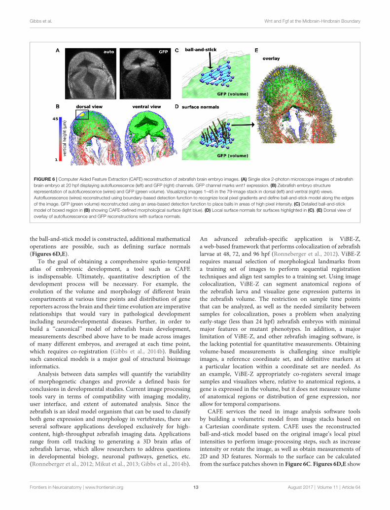
Image AnalysisAnother current limitation to characterizing dynamic changes inthe developing MHB is the state of image analysis software. Thenumber and breadth of image analysis tools are increasing. Useof these tools ranges from general application programs such asImageJ and Icy (de Chaumont et al., 2012; Schindelin et al., 2015)to application-specific tools (Peng, 2008; Eliceiri et al., 2012).Development of these tools is driven by the need to rapidly andquantitatively analyze large, multi-modal image datasets. Suchdata vary in image types, time-series of 2D or 3D images, andmulti-channel measurements. There are also many aspects ofimage analysis, such as visualizing pixel data in more informativeways, making automated measurements when images havedistinct features or landmarks, and building structural modelsof the system being imaged. Despite the plethora of availablesoftware, visualizing zebrafish morphogenesis, especially duringearly stages of development, poses a considerable challenge sincedistinct landmarks (Mikut et al., 2013) may have not developedyet in these systems. Currently, there is no single software thatcan process an image stack and render a 3D reconstructionmodel of the imaging volume. Remarkably, even though theearly-stage zebrafish embryo is relatively featureless (landmark-free), humans can easily recognize its shape. The fact that humanbrains can process the image and construct a mental map ofthe zebrafish embryo suggests that it is in principle possible tocomputationally generate a similar 3D model. A 3D model refersto surfaces and anatomical volumes ‘‘recognized’’ or ‘‘identified’’by a software program and mathematically described so thatmultidimensional parameterization is possible. This refers toobtaining values for 2- and 3-dimensional features, such asmeasuring the volume of the brain ventricles and compartments,distribution of gene expression and spatiotemporal evolution,as well as co-registration across different samples. To achievethis goal, we have been developing a program named CAFE(Computer Aided Feature Extraction). It originated from anapplication mainly to quantify filaments in 2D images (Hwangand Eryilmaz, 2014). A distinct feature of CAFE is its synergybetween image analysis and model building. Since pixel dataare affected by experimental conditions, CAFE initially usespixel data to build a coarse-grained, ball-and-stick model ofthe system being imaged (Figure 6). Working on geometricelements instead of pixel data increases calculation speed andallows for mathematical descriptions of the model. For example,for the ball-and-stick model of a zebrafish brain, another modelfrom a different imaging channel (e.g., fluorescence image stacksof gene expression) can be overlaid, which will be useful foranalyzing fluorophore spatial relation (Figures 6A–C). Once
Frontiers in Neuroanatomy | www.frontiersin.org 12 August 2017 | Volume 11 | Article 64
Gibbs et al. Wnt and Fgf at the Midbrain-Hindbrain Boundary
FIGURE 6 | Computer Aided Feature Extraction (CAFE) reconstruction of zebrafish brain embryo images. (A) Single slice 2-photon microscope images of zebrafishbrain embryo at 20 hpf displaying autofluorescence (left) and GFP (right) channels. GFP channel marks wnt1 expression. (B) Zebrafish embryo structurerepresentation of autofluorescence (wires) and GFP (green volume). Visualizing images 1–45 in the 79-image stack in dorsal (left) and ventral (right) views.Autofluorescence (wires) reconstructed using boundary-based detection function to recognize local pixel gradients and define ball-and-stick model along the edgesof the image. GFP (green volume) reconstructed using an area-based detection function to place balls in areas of high pixel intensity. (C) Detailed ball-and-stickmodel of boxed region in (B) showing CAFE-defined morphological surface (light blue). (D) Local surface normals for surfaces highlighted in (C). (E) Dorsal view ofoverlay of autofluorescence and GFP reconstructions with surface normals.
the ball-and-stick model is constructed, additional mathematicaloperations are possible, such as defining surface normals(Figures 6D,E).
To the goal of obtaining a comprehensive spatio-temporalatlas of embryonic development, a tool such as CAFEis indispensable. Ultimately, quantitative description of thedevelopment process will be necessary. For example, theevolution of the volume and morphology of different braincompartments at various time points and distribution of genereporters across the brain and their time evolution are imperativerelationships that would vary in pathological developmentincluding neurodevelopmental diseases. Further, in order tobuild a ‘‘canonical’’ model of zebrafish brain development,measurements described above have to be made across imagesof many different embryos, and averaged at each time point,which requires co-registration (Gibbs et al., 2014b). Buildingsuch canonical models is a major goal of structural bioimageinformatics.
Analysis between data samples will quantify the variabilityof morphogenetic changes and provide a defined basis forconclusions in developmental studies. Current image processingtools vary in terms of compatibility with imaging modality,user interface, and extent of automated analysis. Since thezebrafish is an ideal model organism that can be used to classifyboth gene expression and morphology in vertebrates, there areseveral software applications developed exclusively for high-content, high-throughput zebrafish imaging data. Applicationsrange from cell tracking to generating a 3D brain atlas ofzebrafish larvae, which allow researchers to address questionsin developmental biology, neuronal pathways, genetics, etc.(Ronneberger et al., 2012; Mikut et al., 2013; Gibbs et al., 2014b).
An advanced zebrafish-specific application is ViBE-Z,a web-based framework that performs colocalization of zebrafishlarvae at 48, 72, and 96 hpf (Ronneberger et al., 2012). ViBE-Zrequires manual selection of morphological landmarks froma training set of images to perform sequential registrationtechniques and align test samples to a training set. Using imagecolocalization, ViBE-Z can segment anatomical regions ofthe zebrafish larva and visualize gene expression patterns inthe zebrafish volume. The restriction on sample time pointsthat can be analyzed, as well as the needed similarity betweensamples for colocalization, poses a problem when analyzingearly-stage (less than 24 hpf) zebrafish embryos with minimalmajor features or mutant phenotypes. In addition, a majorlimitation of ViBE-Z, and other zebrafish imaging software, isthe lacking potential for quantitative measurements. Obtainingvolume-based measurements is challenging since multipleimages, a reference coordinate set, and definitive markers ata particular location within a coordinate set are needed. Asan example, ViBE-Z appropriately co-registers several imagesamples and visualizes where, relative to anatomical regions, agene is expressed in the volume, but it does not measure volumeof anatomical regions or distribution of gene expression, norallow for temporal comparisons.
CAFE services the need in image analysis software toolsby building a volumetric model from image stacks based ona Cartesian coordinate system. CAFE uses the reconstructedball-and-stick model based on the original image’s local pixelintensities to perform image-processing steps, such as increaseintensity or rotate the image, as well as obtain measurements of2D and 3D features. Normals to the surface can be calculatedfrom the surface patches shown in Figure 6C. Figures 6D,E show
Frontiers in Neuroanatomy | www.frontiersin.org 13 August 2017 | Volume 11 | Article 64
Gibbs et al. Wnt and Fgf at the Midbrain-Hindbrain Boundary
these surface normals. For volumetric analysis, CAFE can detectthe boundary surrounding a user-defined open region and defineits general geometry. This parameter is the 3D complementof the midbrain ventricle area parameter derived from 2-Dcross sections (Gibbs et al., 2014b). Pixel-based algorithmshave been developed and implemented into several image-processing platforms to perform image analysis calculations.CAFE’s ability to produce 2D and 3D ball-and-stick modelsand use these representations to obtain quantitative calculationswill enhance the ability to extract measurements from multipleimage samples. Researchers will be able to quantitatively classifyspatiotemporal changes in a developing embryo and likewisemark characteristic effects of missing gene reporters, such as inthe ace phenotype.
CONCLUSION
Crucial to understand for its role in brain development andpotential contributions to human brain patterning birth defects,the MHB is also a remarkable model for the dissection ofsignaling control of cell fate and tissue morphogenesis. Inthis review, we present evidence that the Wnt-Fgf signalinginterface is correlated with specific morphogenetic changesthat drive MHB morphogenesis. The complexity of MHBpatterning and morphogenesis creates an imperative to applynew methodologies and approaches to uncover its underlyingmolecular nature. The simultaneous generation of correctbrain morphology, cell types, and neural circuitry is adaunting challenge but is a very robust and adaptable process.Cell behaviors such as differential adhesion, growth andapoptosis, migration and cytoskeletal remodeling must beprecisely coordinated over large regions within the developingneuroepithelium and this achievement is so astounding that itis understandable we have been both fascinated and frustrated
with our attempts to understand the process as a whole.Moving forward to a more complete mechanistic understandingconnecting the earliest patterning events with eventual brainarchitecture and cell fates will require enhanced cooperationbetween disciplines so that the best possible models can beformulated and thoroughly tested. There are still many openquestions regarding how correct brain architecture is formedand cell types designated during neural development, thougha thorough understanding of these processes is of extremeimportance from a basic science perspective and also foradvancement of regenerative medicine in neural disease.
AUTHOR CONTRIBUTIONS
All authors developed and wrote the manuscript.
FUNDING
The work was supported by National Institutes of Health(Grant R01NS088564). AC-G was funded by the NationalScience Foundation Graduate Research Fellowship under GrantNo. DGE 1252521. Any opinion, findings, and conclusions orrecommendations expressed in this material are those of theauthors and do not necessarily reflect the views of the NationalScience Foundation.
ACKNOWLEDGMENTS
The authors thank David Green, Saurav Mohanty, andAmy Whitener for comments on the manuscript. Some ofthe information included in this manuscript was previouslypublished in the doctoral dissertation of Gibbs (2014), availableonline.
REFERENCES
Babb, S. G., Barnett, J., Doedens, A. L., Cobb, N., Liu, Q., Sorkin, B. C., et al.(2001). Zebrafish E-cadherin: expression during early embryogenesis andregulation during brain development. Dev. Dyn. 221, 231–237. doi: 10.1002/dvdy.1132
Bae, Y. K., Kani, S., Shimizu, T., Tanabe, K., Nojima, H., Kimura, Y.,et al. (2009). Anatomy of zebrafish cerebellum and screen for mutationsaffecting its development. Dev. Biol. 330, 406–426. doi: 10.1016/j.ydbio.2009.04.013
Buckles, G. R., Thorpe, C. J., Ramel, M. C., and Lekven, A. C. (2004).Combinatorial Wnt control of zebrafish midbrain-hindbrain boundaryformation.Mech. Dev. 121, 437–447. doi: 10.1016/s0925-4773(04)00074-7
Carletti, B., and Rossi, F. (2008). Neurogenesis in the cerebellum. Neuroscientist14, 91–100. doi: 10.1177/1073858407304629
Chapouton, P., Adolf, B., Leucht, C., Tannhäuser, B., Ryu, S., Driever, W., et al.(2006). her5 expression reveals a pool of neural stem cells in the adult zebrafishmidbrain. Development 133, 4293–4303. doi: 10.1242/dev.02573
Chen, B. C., Legant, W. R., Wang, K., Shao, L., Milkie, D. E., Davidson, M. W.,et al. (2014). Lattice light-sheet microscopy: imaging molecules to embryosat high spatiotemporal resolution. Science 346:1257998. doi: 10.1126/science.1257998
Chi, C. L., Martinez, S.,Wurst,W., andMartin, G. R. (2003). The isthmic organizersignal FGF8 is required for cell survival in the prospective midbrain andcerebellum. Development 130, 2633–2644. doi: 10.1242/dev.00487
Ciani, L., and Salinas, P. C. (2005). WNTs in the vertebrate nervous system:from patterning to neuronal connectivity. Nat. Rev. Neurosci. 6, 351–362.doi: 10.1038/nrn1665
Clark, C. E. J., Nourse, C. C., and Cooper, H. M. (2012). The tangled web ofnon-canonical wnt signalling in neural migration. Neurosignals 20, 202–220.doi: 10.1159/000332153
Cohen, E. D., Tian, Y., and Morrisey, E. E. (2008). Wnt signaling: anessential regulator of cardiovascular differentiation, morphogenesis andprogenitor self-renewal. Development 135, 789–798. doi: 10.1242/dev.016865
Cooper, S. R., Emond, M. R., Duy, P. Q., Liebau, B. G., Wolman, M. A.,and Jontes, J. D. (2015). Protocadherins control the modular assembly ofneuronal columns in the zebrafish optic tectum. J. Cell Biol. 211, 807–814.doi: 10.1083/jcb.201507108
Cox, W. G., and Hemmati-Brivanlou, A. (1995). Caudalization of neural fate bytissue recombination and bFGF. Development 121, 4349–4358.
Dailey, L., Ambrosetti, D., Mansukhani, A., and Basilico, C. (2005). Mechanismsunderlying differential responses to FGF signaling. Cytokine Growth FactorRev. 16, 233–247. doi: 10.1016/j.cytogfr.2005.01.007
Davey, C. F., and Moens, C. B. (2017). Planar cell polarity in moving cells:think globally, act locally. Development 144, 187–200. doi: 10.1242/dev.122804
de Chaumont, F., Dallongeville, S., Chenouard, N., Herve, N., Pop, S., Provoost, T.,et al. (2012). Icy: an open bioimage informatics platform for extendedreproducible research. Nat. Methods 9, 690–696. doi: 10.1038/nmeth.2075
Frontiers in Neuroanatomy | www.frontiersin.org 14 August 2017 | Volume 11 | Article 64
Gibbs et al. Wnt and Fgf at the Midbrain-Hindbrain Boundary
Doherty, D., Millen, K. J., and Barkovich, A. J. (2013). Midbrain and hindbrainmalformations: advances in clinical diagnosis, imaging, and genetics. LancetNeurol. 12, 381–393. doi: 10.1016/s1474-4422(13)70024-3
Dorsky, R. I., Itoh, M., Moon, R. T., and Chitnis, A. (2003). Two tcf3 genescooperate to pattern the zebrafish brain. Development 130, 1937–1947.doi: 10.1242/dev.00402
Dworkin, S., and Jane, S. M. (2013). Novel mechanisms that pattern andshape the midbrain-hindbrain boundary. Cell. Mol. Life Sci. 70, 3365–3374.doi: 10.1007/s00018-012-1240-x
Dyer, C., Blanc, E., Hanisch, A., Roehl, H., Otto, G. W., Yu, T., et al. (2014). Abi-modal function ofWnt signalling directs an FGF activity gradient to spatiallyregulate neuronal differentiation in the midbrain. Development 141, 63–72.doi: 10.1242/dev.099507
Eliceiri, K. W., Berthold, M. R., Goldberg, I. G., Ibáñez, L., Manjunath, B. S.,Martone, M. E., et al. (2012). Biological imaging software tools. Nat. Methods9, 697–710. doi: 10.1038/nmeth.2084
Filas, B. A., Oltean, A., Majidi, S., Bayly, P. V., Beebe, D. C., and Taber, L. A. (2012).Regional differences in actomyosin contraction shape the primary vesiclesin the embryonic chicken brain. Phys. Biol. 9:066007. doi: 10.1088/1478-3975/9/6/066007
Foucher, I., Mione, M., Simeone, A., Acampora, D., Bally-Cuif, L., and Houart, C.(2006). Differentiation of cerebellar cell identities in absence of Fgf signallingin zebrafish Otx morphants. Development 133, 1891–1900. doi: 10.1242/dev.02352
Gao, L., Shao, L., Higgins, C. D., Poulton, J. S., Peifer, M., Davidson, M. W.,et al. (2012). Noninvasive imaging beyond the diffraction limit of 3D dynamicsin thickly fluorescent specimens. Cell 151, 1370–1385. doi: 10.1016/j.cell.2012.10.008
Gato, A., and Desmond, M. E. (2009). Why the embryo still matters: CSF andthe neuroepithelium as interdependent regulators of embryonic brain growth,morphogenesis and histiogenesis.Dev. Biol. 327, 263–272. doi: 10.1016/j.ydbio.2008.12.029
Gibbs, H. C. (2014).Digitizing Vertebrate Brain Development with Ultrashort PulseMicroscopy. Texas A&M University: Dissertation College Station (TX).
Gibbs, H. C., Bai, Y. Q., Lekven, A. C., and Yeh, A. T. (2014a). Imagingembryonic development with ultrashort pulse microscopy. Opt. Eng.53:051506. doi: 10.1117/1.oe.53.5.051506
Gibbs, H. C., Dodson, C. R., Bai, Y. Q., Lekven, A. C., and Yeh, A. T. (2014b).Combined lineage mapping and gene expression profiling of embryonic brainpatterning using ultrashort pulse microscopy and image registration. J. Biomed.Opt. 19:126016. doi: 10.1117/1.jbo.19.12.126016
Gibbs, H. C., Dodson, C. R., Bai, Y., Lekven, A. C., and Yeh, A. T. (2013).‘‘Combined lineagemapping and fate specification profiling withNLOM-OCMusing sub-10-fs pulses,’’ in Proceedings SPIE 8593, Optical Methods inDevelopmental Biology. doi:10.1117/12.2004330
Green, D., Whitener, A. E., Mohanty, S., and Lekven, A. C. (2015). Vertebratenervous system posteriorization: grading the function of wnt signaling. Dev.Dyn. 244, 507–512. doi: 10.1002/dvdy.24230
Gutzman, J. H., Graeden, E. G., Lowery, L. A., Holley, H. S., and Sive, H. (2008).Formation of the zebrafish midbrain-hindbrain boundary constriction requireslaminin-dependent basal constriction.Mech. Dev. 125, 974–983. doi: 10.1016/j.mod.2008.07.004
Gutzman, J. H., Sahu, S. U., and Kwas, C. (2015). Non-muscle myosin IIA and IIBdifferentially regulate cell shape changes during zebrafish brainmorphogenesis.Dev. Biol. 397, 103–115. doi: 10.1016/j.ydbio.2014.10.017
Gutzman, J. H., and Sive, H. (2010). Epithelial relaxation mediated by the myosinphosphatase regulator Mypt1 is required for brain ventricle lumen expansionand hindbrain morphogenesis. Development 137, 795–804. doi: 10.1242/dev.042705
Harding, M. J., and Nechiporuk, A. V. (2012). Fgfr-Ras-MAPK signaling isrequired for apical constriction via apical positioning of Rho-associatedkinase during mechanosensory organ formation.Development 139, 3130–3135.doi: 10.1242/dev.082271
Hashimoto, M., and Hibi, M. (2012). Development and evolution of cerebellarneural circuits.Dev. Growth Differ. 54, 373–389. doi: 10.1111/j.1440-169x.2012.01348.x
Heimbucher, T., Murko, C., Bajoghli, B., Aghaallaei, N., Huber, A., Stebegg, R.,et al. (2007). Gbx2 and Otx2 interact with the WD40 domain of Groucho/Tlecorepressors.Mol. Cell. Biol. 27, 340–351. doi: 10.1128/mcb.00811-06
Heisenberg, C. P., and Bellaïche, Y. (2013). Forces in tissue morphogenesis andpatterning. Cell 153, 948–962. doi: 10.1016/j.cell.2013.05.008
Heisenberg, C. P., Tada, M., Rauch, G. J., Saúde, L., Concha, M. L., Geisler, R., et al.(2000). Silberblick/Wnt11 mediates convergent extension movements duringzebrafish gastrulation. Nature 405, 76–81. doi: 10.1038/35011068
Heuberger, J., and Birchmeier, W. (2010). Interplay of cadherin-mediated celladhesion and canonical wnt signaling. Cold Spring Harb. Perspect. Biol.2:a002915. doi: 10.1101/cshperspect.a002915
Hidalgo-Sánchez, M., Millet, S., Bloch-Gallego, E., and Alvarado-Mallart, R. M. (2005). Specification of the meso-isthmo-cerebellar region:the Otx2/Gbx2 boundary. Brain Res. Rev. 49, 134–149. doi: 10.1016/j.brainresrev.2005.01.010
Huisken, J., Swoger, J., Del Bene, F., Wittbrodt, J., and Stelzer, E. H. K. (2004).Optical sectioning deep inside live embryos by selective plane illuminationmicroscopy. Science 305, 1007–1009. doi: 10.1126/science.1100035
Huisken, J. (2012). Slicing embryos gently with laser light sheets. Bioessays 34,406–411. doi: 10.1002/bies.201100120
Hwang, W., and Eryilmaz, E. (2014). Kinetic signature of fractal-like filamentnetworks formed by orientational linear epitaxy. Phys. Rev. Lett. 113:025502.doi: 10.1103/physrevlett.113.025502
Inoue, F., Kurokawa, D., Takahashi, M., and Aizawa, S. (2012). Gbx2 directlyrestricts Otx2 expression to forebrain and midbrain, competing withclass III POU factors. Mol. Cell. Biol. 32, 2618–2627. doi: 10.1128/MCB.00083-12
Jászai, J., Reifers, F., Picker, A., Langenberg, T., and Brand, M. (2003). Isthmus-to-midbrain transformation in the absence of midbrain-hindbrain organizeractivity. Development 130, 6611–6623. doi: 10.1242/dev.00899
Jungbluth, S., Larsen, C., Wizenmann, A., and Lumsden, A. (2001). Cell mixingbetween the embryonic midbrain and hindbrain. Curr. Biol. 11, 204–207.doi: 10.1016/s0960-9822(01)00049-5
Kasza, K. E., and Zallen, J. A. (2011). Dynamics and regulation of contractileactin-myosin networks in morphogenesis. Curr. Opin. Cell Biol. 23, 30–38.doi: 10.1016/j.ceb.2010.10.014
Keller, P. J., Schmidt, A. D., Santella, A., Khairy, K., Bao, Z. R., Wittbrodt, J., et al.(2010). Fast, high-contrast imaging of animal development with scanned lightsheet-based structured-illumination microscopy. Nat. Methods 7, 637–642.doi: 10.1038/nmeth.1476
Keller, P., Schmidt, A., Wittbrodt, J., and Stelzer, E. (2008). Reconstruction ofzebrafish early embryonic development by scanned light sheet microscopy.Science 322, 1065–1069. doi: 10.1126/science.1162493
Keller, P. J., and Stelzer, E. H. K. (2008). Quantitative in vivo imaging of entireembryos with digital scanned laser light sheet fluorescence microscopy. Curr.Opin. Neurobiol. 18, 624–632. doi: 10.1016/j.conb.2009.03.008
Kiecker, C., and Niehrs, C. (2001). A morphogen gradient of Wnt/beta-catenin signalling regulates anteroposterior neural patterning in Xenopus.Development 128, 4189–4201.
Kim, S. A., Tai, C. Y., Mok, L. P., Mosser, E. A., and Schuman, E. M.(2011). Calcium-dependent dynamics of cadherin interactions at cell-celljunctions. Proc. Natl. Acad. Sci. U S A 108, 9857–9862. doi: 10.1073/pnas.1019003108
Kishimoto, N., Alfaro-Cervello, C., Shimizu, K., Asakawa, K., Urasaki, A.,Nonaka, S., et al. (2011). Migration of neuronal precursors from thetelencephalic ventricular zone into the olfactory bulb in adult zebrafish.J. Comp. Neurol. 519, 3549–3565. doi: 10.1002/cne.22722
Klymkowsky, M. W., Rossi, C. C., and Artinger, K. B. (2010). Mechanisms drivingneural crest induction and migration in the zebrafish and Xenopus laevis. CelllAdhes. Migr. 4, 595–608. doi: 10.4161/cam.4.4.12962
Knosp, W. M., Knox, S. M., Lombaert, I. M. A., Haddox, C. L., Patel, V. N.,and Hoffman, M. P. (2015). Submandibular parasympathetic gangliogenesisrequires sprouty-dependent Wnt signals from epithelial progenitors. Dev. Cell32, 667–677. doi: 10.1016/j.devcel.2015.01.023
Kurokawa, D., Ohmura, T., Akasaka, K., and Aizawa, S. (2012). A lineage specificenhancer drives Otx2 expression in teleost organizer tissues. Mech. Dev. 128,653–661. doi: 10.1016/j.mod.2011.11.001
Langenberg, T., and Brand, M. (2005). Lineage restriction maintains a stableorganizer cell population at the zebrafish midbrain-hindbrain boundary.Development 132, 3209–3216. doi: 10.1242/dev.01862
Lapebie, P., Borchiellini, C., and Houliston, E. (2011). Dissecting thePCP pathway: one or more pathways? Does a separate Wnt-Fz-Rho
Frontiers in Neuroanatomy | www.frontiersin.org 15 August 2017 | Volume 11 | Article 64
Gibbs et al. Wnt and Fgf at the Midbrain-Hindbrain Boundary
pathway drive morphogenesis? Bioessays 33, 759–768. doi: 10.1002/bies.201100023
Lassiter, R. N. T., Stark, M. R., Zhao, T. Y., and Zhou, C. J. J. (2014). Signalingmechanisms controlling cranial placode neurogenesis and delamination. Dev.Biol. 389, 39–49. doi: 10.1016/j.ydbio.2013.11.025
Lekven, A. C., Buckles, G. R., Kostakis, N., and Moon, R. T. (2003). wnt1 andwnt10b function redundantly at the zebrafish midbrain-hindbrain boundary.Dev. Biol. 254, 172–187. doi: 10.1016/s0012-1606(02)00044-1
Lele, Z., Folchert, A., Concha, M., Rauch, G. J., Geisler, R., Rosa, F., et al. (2002).Parachute/n-cadherin is required for morphogenesis and maintained integrityof the zebrafish neural tube. Development 129, 3281–3294.
Lin, M. J., and Lee, S. J. (2016). Stathmin-like 4 is critical for the maintenance ofneural progenitor cells in dorsal midbrain of zebrafish larvae. Sci. Rep. 6:36188.doi: 10.1038/srep36188
Lowery, L. A., De Rienzo, G., Gutzman, J. H., and Sive, H. (2009). Characterizationand classification of zebrafish brainmorphologymutants.Anat. Rec. (Hoboken)292, 94–106. doi: 10.1002/ar.20768
Lowery, L. A., and Sive, H. (2004). Strategies of vertebrate neurulation and are-evaluation of teleost neural tube formation. Mech. Dev. 121, 1189–1197.doi: 10.1016/j.mod.2004.04.022
Mahou, P., Zimmerley, M., Loulier, K., Matho, K. S., Labroille, G., Morin, X.,et al. (2012). Multicolor two-photon tissue imaging by wavelength mixing.Nat.Methods 9, 815–818. doi: 10.1038/nmeth.2098
Martinez, S., Crossley, P. H., Cobos, I., Rubenstein, J. L. R., and Martin, G. R.(1999). FGF8 induces formation of an ectopic isthmic organizer andisthmocerebellar development via a repressive effect on Otx2 expression.Development 126, 1189–1200.
McGrew, L. L., Lai, C.-J., and Moon, R. T. (1995). Specification of theanteroposterior neural axis through synergistic interaction of theWnt signalingcascade with noggin and follistatin. Dev. Biol. 172, 337–342. doi: 10.1006/dbio.1995.0027
McMahon, A. P., and Bradley, A. (1990). The Wnt-1 (int-1) proto-oncogeneis required for development of a large region of the mouse brain. Cell 62,1073–1085. doi: 10.1016/0092-8674(90)90385-r
McMahon, A., Supatto, W., Fraser, S. E., and Stathopoulos, A. (2008).Dynamic analyses of Drosophila gastrulation provide insights intocollective cell migration. Science 322, 1546–1550. doi: 10.1126/science.1167094
Megason, S. G., andMcMahon, A. P. (2002). A mitogen gradient of dorsal midlineWnts organizes growth in the CNS. Development 129, 2087–2098.
Meier, F., Giesert, F., Delic, S., Faus-Kessler, T., Matheus, F., Simeone, A.,et al. (2014). FGF/FGFR2 signaling regulates the generation and correctpositioning of bergmann glia cells in the developing mouse cerebellum. PLoSOne 9:e101124. doi: 10.1371/journal.pone.0101124
Mikut, R., Dickmeis, T., Driever, W., Geurts, P., Hamprecht, F. A., Kausler, B. X.,et al. (2013). Automated processing of zebrafish imaging data: a survey.Zebrafish 10, 401–421. doi: 10.1089/zeb.2013.0886
Millen, K. J., Steshina, E. Y., Iskusnykh, I. Y., and Chizhikov, V. V. (2014).Transformation of the cerebellum into more ventral brainstem fates causescerebellar agenesis in the absence of ptf1a function. Proc. Natl. Acad. Sci. U S A111, E1777–E1786. doi: 10.1073/pnas.1315024111
Miyake, A., and Itoh, N. (2013). Fgf22 regulated by Fgf3/Fgf8 signaling is requiredfor zebrafish midbrain development. Biol. Open 2, 515–524. doi: 10.1242/bio.20134226
Miyake, A., Nihno, S., Murakoshi, Y., Satsuka, A., Nakayama, Y., and Itoh, N.(2012). Neucrin, a novel secreted antagonist of canonical Wnt signaling, playsroles in developing neural tissues in zebrafish. Mech. Dev. 128, 577–590.doi: 10.1016/j.mod.2012.01.001
Moon, R. T., Kohn, A. D., De Ferrari, G. V., and Kaykas, A. (2004). WNT andbeta-catenin signalling: diseases and therapies. Nat. Rev. Genet. 5, 689–701.doi: 10.1038/nrg1427
Ninkovic, J., Tallafuss, A., Leucht, C., Topczewski, J., Tannhäuser, B., Solnica-Krezel, L., et al. (2005). Inhibition of neurogenesis at the zebrafishmidbrain-hindbrain boundary by the combined and dose-dependent activityof a new hairylE(spl) gene pair. Development 132, 75–88. doi: 10.1242/dev.01525
Nordström, U., Jessell, T. M., and Edlund, T. (2002). Progressive induction ofcaudal neural character by graded Wnt signaling. Nat. Neurosci. 5, 525–532.doi: 10.1038/nn854
Nyholm, M. K., Abdelilah-Seyfried, S., and Grinblat, Y. (2009). A novel geneticmechanism regulates dorsolateral hinge-point formation during zebrafishcranial neurulation. J. Cell Sci. 122, 2137–2148. doi: 10.1242/jcs.043471
Olivier, N., Luengo-Oroz, M. A., Duloquin, L., Faure, E., Savy, T., Veilleux, I., et al.(2010). Cell lineage reconstruction of early zebrafish embryos using label-freenonlinear microscopy. Science 329, 967–971. doi: 10.1126/science.1189428
Ota, S., Taimatsu, K., Yanagi, K., Namiki, T., Ohga, R., Higashijima, S., et al.(2016). Functional visualization and disruption of targeted genes usingCRISPR/Cas9-mediated eGFP reporter integration in zebrafiah. Sci. Rep.6:34991. doi: 10.1038/srep34991
Parada, C., Martin, C., Alonso, M. I., Moro, J. A., Bueno, D., and Gato, A.(2005). Embryonic cerebrospinal fluid collaborates with the isthmic organizerto regulate mesencephalic gene expression. J. Neurosci. Res. 82, 333–345.doi: 10.1002/jnr.20618
Parker, M. O., Brock, A. J., Walton, R. T., and Brennan, C. H. (2013). The roleof zebrafish (Danio rerio) in dissecting the genetics and neural circuits ofexecutive function. Front. Neural Circuits 7:13. doi: 10.3389/fncir.2013.00063
Peng, H. C. (2008). Bioimage informatics: a new area of engineering biology.Bioinformatics 24, 1827–1836. doi: 10.1093/bioinformatics/btn346
Picker, A., Brennan, C., Reifers, F., Clarke, J. D. W., Holder, N., andBrand, M. (1999). Requirement for the zebrafish mid-hindbrain boundary inmidbrain polarisation, mapping and confinement of the retinotectal projection.Development 126, 2967–2978.
Piloto, S., and Schilling, T. F. (2010). Ovo1 links Wnt signaling with N-cadherinlocalization during neural crest migration. Development 137, 1981–1990.doi: 10.1242/dev.048439
Puelles, E., Acampora, D., Lacroix, E., Signore, M., Annino, A., Tuorto, F., et al.(2003). Otx dose-dependent integrated control of antero-posterior and dorso-ventral patterning of midbrain. Nat. Neurosci. 6, 453–460. doi: 10.1038/nn1037
Raible, F., and Brand, M. (2004). Divide et Impera—the midbrain-hindbrainboundary and its organizer. Trends Neurosci. 27, 727–734. doi: 10.1016/j.tins.2004.10.003
Rapaciolii, M., Palma, V., and Flores, V. (2016). Morphogenetic and histogeneticroles of the temporal-spatial organization of cell proliferation in the vertebratecorticogenesis as revealed by inter-specific analyses of the optic tectum cortexdevelopment. Front. Cell. Neurosci. 10:112. doi: 10.3389/fncel.2016.00112
Recher, G., Jouralet, J., Brombin, A., Heuzé, A., Mugniery, E., Hermel, J. M.,et al. (2013). Zebrafish midbrain slow-amplifying progenitors exhibit highlevels of transcripts for nucleotide and ribosome biogenesis. Development 140,4860–4869. doi: 10.1242/dev.099010
Reifers, F., Böhli, H., Walsh, E. C., Crossley, P. H., Stainier, D. Y. R., andBrand, M. (1998). Fgf8 is mutated in zebrafish acerebellar (ace) mutants and isrequired for maintenance of midbrain-hindbrain boundary development andsomitogenesis. Development 125, 2381–2395.
Reim, G., and Brand, M. (2002). Spiel-ohne-grenzen/pou2 mediates regionalcompetence to respond to Fgf8 during zebrafish early neural development.Development 129, 917–933.
Rhinn, M., and Brand, M. (2001). The midbrain—hindbrain boundary organizer.Curr. Opin. Neurobiol. 11, 34–42. doi: 10.1016/s0959-4388(00)00171-9
Rhinn, M., Lun, K., Ahrendt, R., Geffarth, M., and Brand, M. (2009). Zebrafishgbx1 refines the Midbrain-Hindbrain Boundary border and mediates theWnt8 posteriorization signal. Neural Dev. 4:12. doi: 10.1186/1749-8104-4-12
Rhinn, M., Lun, K., Amores, A., Yan, Y. L., Postlethwait, J. H., and Brand, M.(2003). Cloning, expression and relationship of zebrafish gbx1 and gbx2 genesto Fgf signaling.Mech. Dev. 120, 919–936. doi: 10.1016/s0925-4773(03)00135-7
Rhinn, M., Lun, K., Luz, M., Werner, M., and Brand, M. (2005). Positioning ofthe midbrain-hindbrain boundary organizer through global posteriorization ofthe neuroectodermmediated byWnt8 signaling. Development 132, 1261–1272.doi: 10.1242/dev.01685
Ronneberger, O., Liu, K., Rath, M., Ruess, D., Mueller, T., Skibbe, H., et al. (2012).ViBE-Z: a framework for 3D virtual colocalization analysis in zebrafish larvalbrains. Nat. Methods 9, 735–742. doi: 10.1038/nmeth.2076
Sahu, S. U., Visetsouk, M. R., Garde, R. J., Hennes, L., Kwas, C., and Gutzman, J. H.(2017). Calcium signals drive cell shape changes during zebrafish midbrain-hindbrain boundary formation. Mol. Biol. Cell 28, 875–882. doi: 10.1091/mbc.E16-08-0561
Sai, X., and Ladher, R. K. (2008). FGF signaling regulates cytoskeletal remodelingduring epithelial morphogenesis. Curr. Biol. 18, 976–981. doi: 10.1016/j.cub.2008.05.049
Frontiers in Neuroanatomy | www.frontiersin.org 16 August 2017 | Volume 11 | Article 64
Gibbs et al. Wnt and Fgf at the Midbrain-Hindbrain Boundary
Sander, J. D., and Joung, J. K. (2014). CRISPR-Cas systems for editing, regulatingand targeting genomes. Nat. Biotechnol. 32, 347–355. doi: 10.1038/nbt.2842
Santi, P. A. (2011). Light sheet fluorescence microscopy: a review. J. Histochem.Cytochem. 59, 129–138. doi: 10.1369/0022155410394857
Schindelin, J., Rueden, C. T., Hiner, M. C., and Eliceiri, K. W. (2015). The ImageJecosystem: an open platform for biomedical image analysis. Mol. Reprod. Dev.82, 518–529. doi: 10.1002/mrd.22489
Schlessinger, J. (2000). Cell signaling by receptor tyrosine kinases. Cell 103,211–225. doi: 10.1016/s0092-8674(00)00114-8
Schmitz, B., Papan, C., and Campos-Ortega, J. A. (1993). Neurulation in theanterior trunk region of the zebrafish brachydanio-rerio. Rouxs Arch. Dev. Biol.202, 250–259. doi: 10.1007/BF00363214
Scholpp, S., and Brand, M. (2004). Endocytosis controls spreading and effectivesignaling range of Fgf8 protein. Curr. Biol. 14, 1834–1841. doi: 10.1016/j.cub.2004.09.084
Severino, M., Allegri, A. E. M., Pistorio, A., Roviglione, B., Di Iorgi, N.,Maghnie, M., et al. (2014). Midbrain-hindbrain involvement in septo-opticdysplasia. Am. J. Neuroradiol. 35, 1586–1592. doi: 10.3174/ajnr.A3959
Smutny, M., Ákos, Z., Silvia, G., Shamipour, S., Ruprecht, V., Capek, D., et al.(2017). Friction forces position the neural anlage. Nat. Cell Biol. 19, 306–317.doi: 10.1038/ncb3492
Stanganello, E., Hagemann, A. I. H., Mattes, B., Sinner, C., Meyen, D.,Weber, S., et al. (2015). Filopodia-basedWnt transport during vertebrate tissuepatterning. Nat. Commun. 6:5846. doi: 10.1038/ncomms6846
Su, C.-Y., Kemp, H. A., and Moens, C. B. (2014). Cerebellar development in theabsence of Gbx function in zebrafish. Dev. Biol. 386, 181–190. doi: 10.1016/j.ydbio.2013.10.026
Sunmonu, N. A., Li, K. R., Guo, Q. X., and Li, J. Y. H. (2011). Gbx2 and Fgf8 aresequentially required for formation of the midbrain-hindbrain compartmentboundary. Development 138, 725–734. doi: 10.1242/dev.055665
Tallafuss, A., and Bally-Cuif, L. (2003). Tracing of her5 progeny in zebrafishtransgenics reveals the dynamics of midbrain-hindbrain neurogenesis andmaintenance. Development 130, 4307–4323. doi: 10.1242/dev.00662
Thomas, K. R., and Capecchi, M. R. (1990). Targeted disruption of the murineint-1 proto-oncogene resulting in severe abnormalities in midbrain andcerebellar development. Nature 346, 847–850. doi: 10.1038/346847a0
Tossell, K., Andreae, L. C., Cudmore, C., Lang, E., Muthukrishnan, U.,Lumsden, A., et al. (2011a). Lrrn1 is required for formation of the midbrain-hindbrain boundary and organiser through regulation of affinity differencesbetween midbrain and hindbrain cells in chick. Dev. Biol. 352, 341–352.doi: 10.1016/j.ydbio.2011.02.002
Tossell, K., Kiecker, C., Wizenmann, A., Lang, E., and Irving, C. (2011b). Notchsignalling stabilises boundary formation at the midbrain-hindbrain organiser.Development 138, 3745–3757. doi: 10.1242/dev.070318
Tsai, F. C., Kuo, G. H., Chang, S. W., and Tsai, P. J. (2015). Ca2+ signaling incytoskeletal reorganization, cell migration, and cancer metastasis. Biomed Res.Int. 2015:409245. doi: 10.1155/2015/409245
Tuazon, F. B., and Mullins, M. C. (2015). Temporally coordinated signalsprogressively pattern the anteroposterior and dorsoventral body axes. Semin.Cell Dev. Biol. 42, 118–133. doi: 10.1016/j.semcdb.2015.06.003
Valm, A., Cohen, S., Legant, W., Melunis, J., Hershberg, U., Wait, E., et al. (2017).Applying systems-level spectral imaging and analysis to reveal the organelleinteractome. Nature 546, 162–167. doi: 10.1038/nature22369
Varelas, X., Bouchie, M. P., and Kukuruzinska, M. A. (2014). ProteinN-glycosylation in oral cancer: dysregulated cellular networks amongDPAGT1, E-cadherin adhesion and canonical Wnt signaling. Glycobiology 24,579–591. doi: 10.1093/glycob/cwu031
Volkmann, K., Chen, Y. Y., Harris, M. P., Wullimann, M. F., and Köster, R. W.(2010). The zebrafish cerebellar upper rhombic lip generates tegmentalhindbrain nuclei by long-distance migration in an evolutionary conservedmanner. J. Comp. Neurol. 518, 2794–2817. doi: 10.1002/cne.22364
Weber, M., and Huisken, J. (2011). Light sheet microscopy for real-timedevelopmental biology.Curr. Opin. Genet. Dev. 21, 566–572. doi: 10.1016/j.gde.2011.09.009
Woo, K., and Fraser, S. E. (1995). Order and coherence in the fate map of thezebrafish nervous-system. Development 121, 2595–2609.
Wurst, W., and Bally-Cuif, L. (2001). Neural plate patterning: upstreamand downstream of the isthmic organizer. Nat. Rev. Neurosci. 2, 99–108.doi: 10.1038/35053516
Yook, J. I., Li, X. Y., Ota, I., Fearon, E. R., and Weiss, S. J. (2005). Wnt-dependentregulation of the E-cadherin repressor snail. J. Biol. Chem. 280, 11740–11748.doi: 10.1074/jbc.m413878200
Yoshiba, S., and Hamada, H. (2014). Roles of cilia, fluid flow, and Ca2+ signalingin breaking of left-right symmetry. Trends Genet. 30, 10–17. doi: 10.1016/j.tig.2013.09.001
Yu, S. R., Burkhardt, M., Nowak, M., Ries, J., Petrásek, Z., Scholpp, S.,et al. (2009). Fgf8 morphogen gradient forms by a source-sink mechanismwith freely diffusing molecules. Nature 461, 533–536. doi: 10.1038/nature08391
Zervas, M., Millet, S., Ahn, S., and Joyner, A. L. (2004). Cell behaviors andgenetic lineages of themesencephalon and rhombomere 1.Neuron 43, 345–357.doi: 10.1016/j.neuron.2004.07.010
Conflict of Interest Statement: The authors declare that the research wasconducted in the absence of any commercial or financial relationships that couldbe construed as a potential conflict of interest.
Copyright © 2017 Gibbs, Chang-Gonzalez, Hwang, Yeh and Lekven. This is anopen-access article distributed under the terms of the Creative Commons AttributionLicense (CC BY). The use, distribution or reproduction in other forums is permitted,provided the original author(s) or licensor are credited and that the originalpublication in this journal is cited, in accordance with accepted academic practice.No use, distribution or reproduction is permitted which does not comply with theseterms.
Frontiers in Neuroanatomy | www.frontiersin.org 17 August 2017 | Volume 11 | Article 64