Embed Size (px)
Citation preview

Khairun Nisa Berawi, dr., Mkes., AIFO

SPECIAL SENSES
1. Vision
2. Hearing and Equilibrium
3. Smell
4. Taste


ANATOMY

LENS AND CILIARY MUSCLE

THE RETINA

THE ROD AND CONE
•Rhodopsin
•Retinal (11-cis retinal)
•Opsin :
-Scotopsin in rods
-Photopsin in cones
•Nyctalopia

ACCESSORY STRUCTURES OF THE EYE
1. Orbit
2. Eyelids, eye lashes, eyebrows
3. Conjunctiva
4. Lacrimal apparatus
5. Muscle of the eye and eyelid
Please re-read by yourself !!!

PHYSIOLOGY OF VISION
1. Refraction of the light rays entering the eye
2. Focusing of images on the retina by accommodation of the lens and convergence of the images
3. Conversion of light waves by photochemical activity into neural impulses
4. Processing of neural activity in the retina and transmission of coded impulses through the optic nerve
5. Processing in the brain, culminating in perception – the object is “seen”
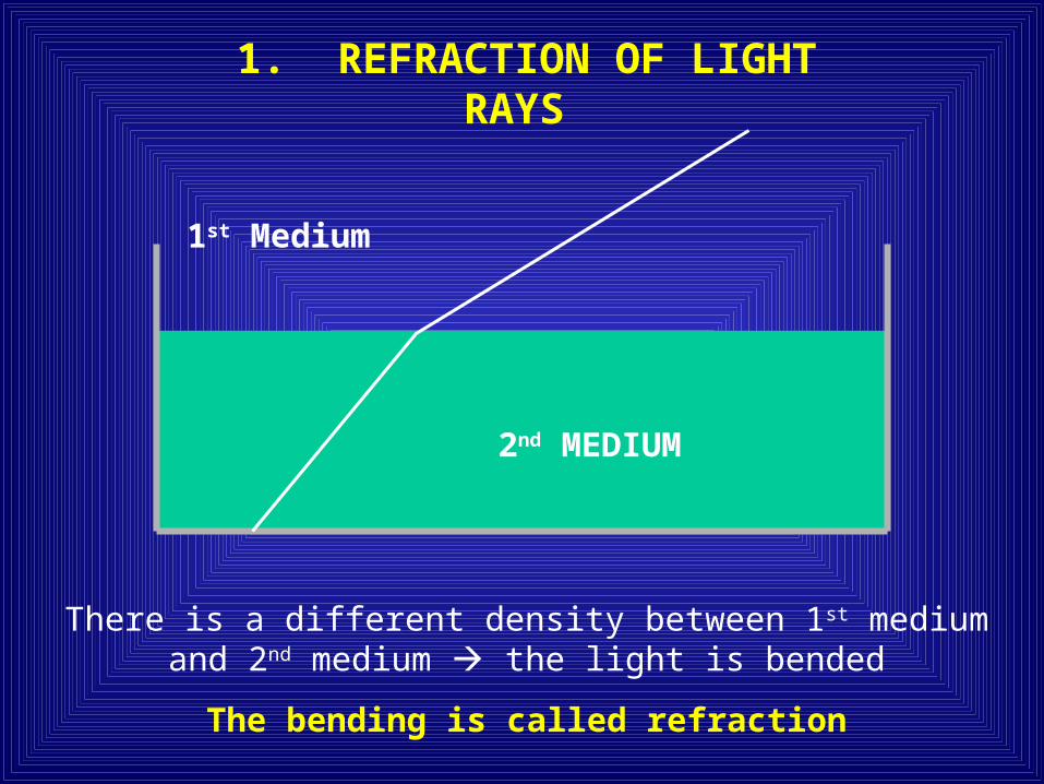
1. REFRACTION OF LIGHT RAYS
1st Medium
2nd MEDIUM
There is a different density between 1st medium and 2nd medium the light is bended
The bending is called refraction

REFRACTION IN NORMAL EYE
In normal eye (emmetropia), light rays come to focal exactly on the retina
Refraction

REFRACTION IN MYOPIA
In myopia (nearsighted eye), the focal point falls in front of the retina

REFRACTION IN HYPERMETROPIA
In hypermetropia (farsighted eye), the focal point falls behind the retina

2. ACCOMMODATION
The role of the adjustable lens
A reflex process
Bring images into perfect focus on the retina

Nearby Object
Distant Object
The lens becomes round
The lens becomes flattened

1. Ciliary muscle contract
2. Ciliary body pull forward and inward
3. Tension on suspensory ligaments of lens reduced
4. Lens becomes thicker (rounder) due to its elasticity
MKING IT POSSIBLE TO FOCUS A NEARBY OBJECT
TO FOCUS ON NEARBY OBJECT
TO FOCUS ON A DISTANT OBJECT
1. Ciliary muscle relax
2. Ciliary body returns to resting satae
3. Tension on suspensory ligaments of the lens increased
4. Lens becomes thinner (flatter)
MAKING IT POSSIBLE TO FOCUS ON A DISTANT
OBJECT

PURKINYE IMAGES
Object
IIIIII
Accommodation
Cornea
Lens
Phacoscope
To see Purkinye II and III through the pupil
Pupil

CONVERGENCE
Near Object
•Binocular visionPerceive on image
Distance
Depth
Three – dimensionality
•Stereoscopic vision
The two eyeballs turn slightly inward

3. CONVERSION OF LIGHT WAVES BY PHOTOCHEMICAL ACTIVITY INTO
NEURAL IMPULSES
1. Photochemical activity in rods
2. Photochemical activity in cones

PHOTOCHEMICAL ACTIVITY IN RODS
11-cis retinal in rhodopsin
absorb a photon
11-cis retinal converted into all-trans retinal, which
activates scotopsin to act as an enzyme
Transducin is produced, and activate
phosphodiesterase
Phosphodiestarease hydrolysis cGMP
Plasma membrane channel close, Na+ are prevented from
entering rod, and hyperpolarization occurs
11-cis retinal and scotopsin combine to synthesis rhodopsin
Neural signals are processed by bipolar, amacrine, horizontal,
and ganglion cells
Action potentials of ganglion cells are
conveyed to brain via optic nerve
Light is perceived as visual images
PhotonRhodopsin molecule
(11-cis retinal + scotopsin

BLEACHING AND REGENERATION OF
PHOTOPIGMENT
What is the conversion of cis- to trans-retinal called ?.

PHOTOCHEMICAL ACTIVITY IN CONE
The Events of Visual Excitation is Similar as in Rod
Rods contain : Scotopsin
Cones contain : Photopsin
3 types of cones: Red, Green, and Blue Cones
Color perception depend on which cones are stimulated and their combinations
The same retinal absorbs a different frequency of light•Red cones: the long wave
•Green cones: the middle wave
•Blue cones: the short wave

VISUAL ADAPTATION
Bright light “bleaches” rhodopsin
The light-sensitive rods become overloaded
Rods are no more sensitive
Light adaptation occurs when eyes adjust to bright light
Dark adaptation occurs as eyes adapt slowly to darkness
Light adaptationEnter darkened room
Resynthesize rhodopsin Dark adaptation
(5 minutes)
(20 – 30 minutes)
Rods become more senstive

NEURAL PATHWAYS FOR VISION

LIGHT REFLEX
(PUPIL REFLEX)
LIGHT RETINA IMPULS N. OPTICUS CHIASMA OPTICUS
TRACTUS OPTICUS
CORPUS GENICULATUM
LATERALE
COLLICULUS SUPERIORN. III
GANGLION CILIARIS
N. CILIARISM. CIRCULARIS
IRIDIS
PUPIL CONSTRICT


![Public Speaking Pizzazz[PSP]-MD INDERA](https://img.pdfslide.us/doc/110x75/586fb84a1a28abe57d8b8093/public-speaking-pizzazzpsp-md-indera.jpg)