Embed Size (px)
Citation preview

Abstract—Anogeissus leiocarpus (Combretaceae) is well known
for its medicinal uses in African traditional medicine, for treating
many human diseases mainly skin diseases and infections. Mycetoma
disease is a fungal and/ or bacterial skininfection, mainly cause by
Madurella mycetomatis fungus. This study was carried out in vitro to
investigate the antifungal activity of Anogeissus leiocarpus leaf
extracts against the isolated pathogenic Madurella mycetomatis, by
using the NCCLS modified method compared to Ketoconazole
standard drug, and MTT assay. The bioactive fraction was subjected
to chemical analysis implementing different chromatographic
analytical methods (TLC, HPLC, and LC-MS/MS). The results
showed significance antifungal activity of A. leiocarpus leaf extracts
against the isolated pathogenic M. mycetomatis, compared to negative
and positive controls. The chloroform fraction showed the highest
antifungal activity. The chromatographic analysis of the chloroform
fraction with the highest activity showed the presence of important
bioactive compounds such as ellagic and flavellagic acids derivatives,
flavonoids and stilbenoid, which are well known for their antifungal
activity.
Keywords—Anogeissus leiocarpus, crude extracts and fractions
of Anogeissus leiocarpus, in vitro susceptibility of Madurella
mycetomatis, Madurella mycetomatis.
I. INTRODUCTION
NOGEISSUS LEIOCARPUS (Combretaceae), is an
evergreen tree widely distributed in Africa [1], [2] and
well known in African traditional medicine for treating many
diseases mainly skin diseases and infections, wounds
infections, sore feet, boils, cysts, syphilitic and diabetic ulcers
[3]-[5].
Leaves are widely used against skin diseases and infections,
jaundice, hepatitis, haemorrhoids, respiratory diseases,
headache and toothache, as antimalarial, leprotic, laxative and
anthelmintic [1], [3], [6]-[10].
A. leiocarpus showed strong antibacterial and antifungal
activity against pathogenic microorganisms [11]-[17].
Ikram Mohamed Eltayeb Elsiddig is with the Department of
Pharmacognosy, Faculty of Pharmacy, University of Medical Sciences and Technology/ Khartoum/Sudan (corresponding author: phone +249912987518;
fax +249/83/224799; e-mail: kramela_07 @yahoo.com).
Abdel Khalig Muddather is with the Department of Pharmacognosy, Faculty of Pharmacy, University of Khartoum/ Khartoum/Sudan (e-mail:
Hiba Abdel Rahman Ali is with the Commission of Biotechnology and Genetic Engineering, National Center for Research/ Khartoum/Sudan (e-mail:
Saad Mohamed Hussein Ayoub is with the Department of Pharmacognosy, Faculty of Pharmacy, University of Medical Science and Technology/
Khartoum/Sudan (e-mail: [email protected]).
Mycetoma is a chronic subcutaneous and deep tissues
granulomatous skin disease or a group of skin infections
caused by several fungi (eumycetoma) mainly Madurella
mycetomatis fungus, or by bacteria (actinomycetoma).
Progressive destruction of tissues leads to loss of function and
impaired the affected site. Serious cases require amputation
leading to loss of numerous infected limbs [18].
In Sudan, mycetoma is a serious common disease leading to
loss of numerous limbs. The incidence of mycetoma in Sudan
has not change and around 400 new cases are seen in hospital
and outpatient clinics every year [18], [19].
There are no 100% effective drugs for eumycetoma
infection, and adequate treatment requires a prolonged
antifungal drug combined with extensive surgical treatment
[18].
Meager data is available for susceptibility of M.
mycetomatis to plant secondary metabolites [20]-[22].
II. MATERIALS AND METHODS
A. Plant Material Collection and Preparation
A. leiocarpus leaves were collected from El Damazeine
region, Sudan, identified by taxonomist in the department of
silviculture, Faculty of Forestry, University of Khartoum,
andthe voucher specimen, IKR2, May - 2008 was kept in the
Herbarium of the Department of Biochemistry, Commission
of Biotechnology and Genetic Engineering, National Centre
for Research. The plant material was air dried under shade at
room temperature, then ground into powder using pestle and
mortar.
B. Preparation of the Extract
Powdered leaves were extracted by maceration overnight in
80% alcohol, and then the extract was fractionated by using
solvents with increasing polarities: petroleum (PE),
chloroform (CHCl3) and ethyl acetate (EtOAc). The solvents
were evaporated to dryness under reduced pressure using
rotary evaporator.
C. Collection and Culture of Madurella mycetomatis
Fungus
Isolated M. mycetomatis fungus was collected in mycetoma
research center at Soba hospital whereas, black grains were
exuded from open sinuses and surgical biopsy from the lesion,
freed from tissues and carried by forceps in sterile container
(Fig. 1), then washed with saline for several times.
In vitro Susceptibility of Madurella mycetomatis to
the Extracts of Anogeissus leiocarpus Leaves Ikram Mohamed Eltayeb Elsiddig, Abdel Khalig Muddather, Hiba Abdel Rahman Ali, Saad Mohamed Hussein
Ayoub
A
World Academy of Science, Engineering and TechnologyInternational Journal of Biological, Biomolecular, Agricultural, Food and Biotechnological Engineering Vol:9, No:12, 2015
1189International Scholarly and Scientific Research & Innovation 9(12) 2015 scholar.waset.org/1999.1/10002992
Inte
rnat
iona
l Sci
ence
Ind
ex, B
ioen
gine
erin
g an
d L
ife
Scie
nces
Vol
:9, N
o:12
, 201
5 w
aset
.org
/Pub
licat
ion/
1000
2992

D. RPMI 1640 Medium Preparation
RPMI 1640 with L- glutamine medium, prepared by
dissolving 0.3g RPMI 1640 with L- glutamine powder (PM
Biomedical Inc. France) and 0.02g MOPHS buffer (3, 4-
morpholinopropane sulfonic acid) in one liter distilled water
and sterilized by autoclaving at 151bs pressure and121°C for
15 minutes.
Fig. 1 Mycetoma pathogen collection
E. Preparation of Fungal Suspension
The isolated grains of M. mycetomatis were firstly cultured
in blood agar media, then subculture in Sabouraud dextrose
agar and incubated at 37°C for 8 days.
The isolate strains were subcultured again to maintain pure
isolate of hyphae. The subculture of hyphae was repeated for
two weeks to maintain pure hyphae which were harvested in
mycological peptone (BDH) water broth medium with
chloroamphenicol. The harvested mycelia or hyphal was
washed for two to three times with RPMI 1640 with L-
glutamine medium, then incubated for 24 hours. The
harvesting mycelia, was sonicated for 2 mins until
homogenous suspension of mycelia obtained.
F. Antifungal Procedure
1. NCCLS Modified Assay for Antifungal Activity and
Determination of MIC Value
One ml of RPMI medium containing serially diluted
extracts (10-0.31mg/ml) in sterile test tubes, then 1ml of
prepared suspension was added. Two sets of control tubes
were added to the experiment, one is growth (-ve) control
tubes contained 1ml of RPMI medium without any treatment
and 1mlof prepared suspension, other is standard drug (+ve)
control tubes contained 1ml of RPMI medium with serially
diluted ketoconazole (5-0.31mg/ml). The optical density of
prepared suspension (growth control) before incubation was
measured by a spectrophotometer at 680 nm red filter and
taken as initial reading. Then all test tubes were incubated at
37°C for a week. After a week the optical density was
measured spectrophotometerically at 680 nm.[20],[21].
MIC value is the least concentration before the
spectrophotometer transmission reading is the same as or more
than the initial reading [22].
2) MTT Assay
A quick sensitive colorimetric method utilizes tetrazolium
salt as indicator of microbial metabolism for evaluation of cell
death [23].
This assay based on the reduction of the yellow MTT
[tetrazolium salt (3-{4, 5-dimethylthiazole-2-yl}-2, 5-diphenyl
tetrazolium bromide)] by the mitochonderial dehydrogenase,
present only in the living cells and hence released to the
supernatant. MTT salt converted to the violet blue or green
blue colored formazan. The colour intensity is directly
proportional to the living cell numbers in the culture.
One drop of the indicator was added to the all tested tubes
after measuring the final optical density by a
spectrophotometer [24], [25].
G. Reverse Phase High Performance Liquid
Chromatography (RHPLC)
Reversed-phase HPLC system was equipped with: RP-C18
HPLC column and Diode array UV detector (DAD) recorded
at 320 – 380 nm for the detection of compounds.
H. HPLC-Triple Quadruple Spectrometric Analysis (LC-
MS/MS)
RP-HPLC was joined with a Finnigan LCQ ion trap mass
spectrometer with the Electrospray Ionization (ESI) interface
at negative ion mode.
Collision induced dissociation (CID) experiment was
performed for fragmentation of glycoside.
III. RESULTS AND DISCUSSIONS
1
3 4
2
Fig. 2 In vitro susceptibility of M. mycetomatis to A. leiocarpus leaf
extracts (1: alch; 2: pet; 3: ch; 4: ethy)
Fig. 3 In vitro susceptibility of M. mycetomatis to ketoconazole drug
As appeared in Fig. 2, the extracts inhibited the fungal
growth compared to the standard drug (Ketoconazole) in Fig.
3. The result was shown in Table I and Fig. 4. The extracts
possessed significant activity against M. mycetomatis
compared to standard drug (ketoconazole). In addition to the
chloroform fraction showed the higher activity.
World Academy of Science, Engineering and TechnologyInternational Journal of Biological, Biomolecular, Agricultural, Food and Biotechnological Engineering Vol:9, No:12, 2015
1190International Scholarly and Scientific Research & Innovation 9(12) 2015 scholar.waset.org/1999.1/10002992
Inte
rnat
iona
l Sci
ence
Ind
ex, B
ioen
gine
erin
g an
d L
ife
Scie
nces
Vol
:9, N
o:12
, 201
5 w
aset
.org
/Pub
licat
ion/
1000
2992
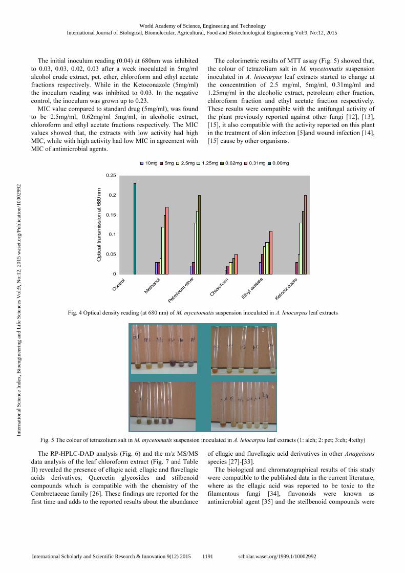
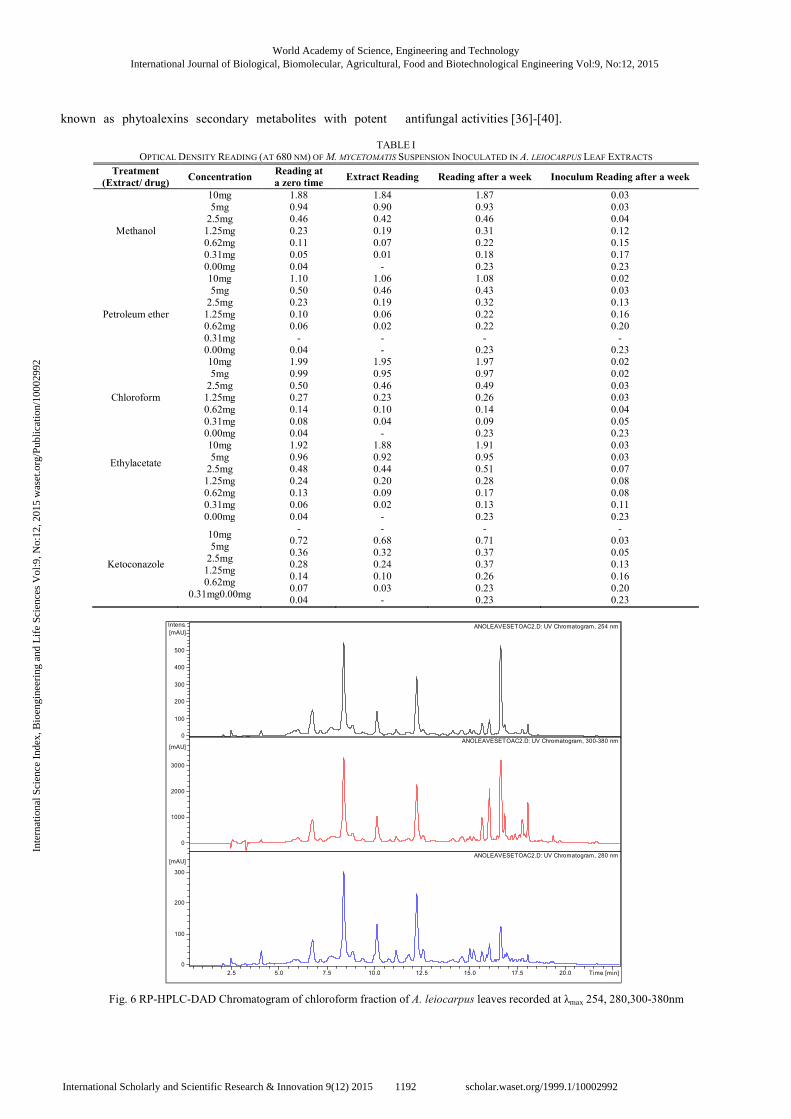
The initial inoculum reading (0.04) at 680nm was inhibited
to 0.03, 0.03, 0.02, 0.03 after a week inoculated in 5mg/ml
alcohol crude extract, pet. ether, chloroform and ethyl acetate
fractions respectively. While in the Ketoconazole (5mg/ml)
the inoculum reading was inhibited to 0.03. In the negative
control, the inoculum was grown up to 0.23.
MIC value compared to standard drug (5mg/ml), was found
to be 2.5mg/ml, 0.62mg/ml 5mg/ml, in alcoholic extract,
chloroform and ethyl acetate fractions respectively. The MIC
values showed that, the extracts with low activity had high
MIC, while with high activity had low MIC in agreement with
MIC of antimicrobial agents.
The colorimetric results of MTT assay (Fig. 5) showed that,
the colour of tetrazolium salt in M. mycetomatis suspension
inoculated in A. leiocarpus leaf extracts started to change at
the concentration of 2.5 mg/ml, 5mg/ml, 0.31mg/ml and
1.25mg/ml in the alcoholic extract, petroleum ether fraction,
chloroform fraction and ethyl acetate fraction respectively.
These results were compatible with the antifungal activity of
the plant previously reported against other fungi [12], [13],
[15], it also compatible with the activity reported on this plant
in the treatment of skin infection [5]and wound infection [14],
[15] cause by other organisms.
Fig. 4 Optical density reading (at 680 nm) of M. mycetomatis suspension inoculated in A. leiocarpus leaf extracts
1
3
2
4
Fig. 5 The colour of tetrazolium salt in M. mycetomatis suspension inoculated in A. leiocarpus leaf extracts (1: alch; 2: pet; 3:ch; 4:ethy)
The RP-HPLC-DAD analysis (Fig. 6) and the m/z MS/MS
data analysis of the leaf chloroform extract (Fig. 7 and Table
II) revealed the presence of ellagic acid; ellagic and flavellagic
acids derivatives; Quercetin glycosides and stilbenoid
compounds which is compatible with the chemistry of the
Combretaceae family [26]. These findings are reported for the
first time and adds to the reported results about the abundance
of ellagic and flavellagic acid derivatives in other Anageissus
species [27]-[33].
The biological and chromatographical results of this study
were compatible to the published data in the current literature,
where as the ellagic acid was reported to be toxic to the
filamentous fungi [34], flavonoids were known as
antimicrobial agent [35] and the steilbenoid compounds were
0
0.05
0.1
0.15
0.2
0.25
Con
trol
Methan
ol
Pet
roleum
eth
er
Chlorof
orm
Eth
yl a
cetate
Ket
ocon
azo
le
Optical transm
issio
n a
t 680 n
m
10mg 5mg 2.5mg 1.25mg 0.62mg 0.31mg 0.00mg
World Academy of Science, Engineering and TechnologyInternational Journal of Biological, Biomolecular, Agricultural, Food and Biotechnological Engineering Vol:9, No:12, 2015
1191International Scholarly and Scientific Research & Innovation 9(12) 2015 scholar.waset.org/1999.1/10002992
Inte
rnat
iona
l Sci
ence
Ind
ex, B
ioen
gine
erin
g an
d L
ife
Scie
nces
Vol
:9, N
o:12
, 201
5 w
aset
.org
/Pub
licat
ion/
1000
2992
known as phytoalexins secondary metabolites with potent antifungal activities [36]-[40].
TABLE I OPTICAL DENSITY READING (AT 680 NM) OF M. MYCETOMATIS SUSPENSION INOCULATED IN A. LEIOCARPUS LEAF EXTRACTS
Treatment
(Extract/ drug) Concentration
Reading at
a zero time Extract Reading Reading after a week Inoculum Reading after a week
Methanol
10mg
5mg 2.5mg
1.25mg
0.62mg 0.31mg
0.00mg
1.88
0.94 0.46
0.23
0.11 0.05
0.04
1.84
0.90 0.42
0.19
0.07 0.01
-
1.87
0.93 0.46
0.31
0.22 0.18
0.23
0.03
0.03 0.04
0.12
0.15 0.17
0.23
Petroleum ether
10mg 5mg
2.5mg
1.25mg 0.62mg
0.31mg 0.00mg
1.10 0.50
0.23
0.10 0.06
- 0.04
1.06 0.46
0.19
0.06 0.02
- -
1.08 0.43
0.32
0.22 0.22
- 0.23
0.02 0.03
0.13
0.16 0.20
- 0.23
Chloroform
10mg
5mg
2.5mg 1.25mg
0.62mg
0.31mg 0.00mg
1.99
0.99
0.50 0.27
0.14
0.08 0.04
1.95
0.95
0.46 0.23
0.10
0.04 -
1.97
0.97
0.49 0.26
0.14
0.09 0.23
0.02
0.02
0.03 0.03
0.04
0.05 0.23
Ethylacetate
10mg
5mg 2.5mg
1.25mg
0.62mg 0.31mg
0.00mg
1.92
0.96 0.48
0.24
0.13 0.06
0.04
1.88
0.92 0.44
0.20
0.09 0.02
-
1.91
0.95 0.51
0.28
0.17 0.13
0.23
0.03
0.03 0.07
0.08
0.08 0.11
0.23
Ketoconazole
10mg 5mg
2.5mg
1.25mg 0.62mg
0.31mg0.00mg
-
0.72
0.36 0.28
0.14
0.07 0.04
-
0.68
0.32 0.24
0.10
0.03 -
-
0.71
0.37 0.37
0.26
0.23 0.23
-
0.03
0.05 0.13
0.16
0.20 0.23
Fig. 6 RP-HPLC-DAD Chromatogram of chloroform fraction of A. leiocarpus leaves recorded at λmax 254, 280,300-380nm
ANOLEAVESETOAC2.D: UV Chromatogram, 254 nm
ANOLEAVESETOAC2.D: UV Chromatogram, 300-380 nm
ANOLEAVESETOAC2.D: UV Chromatogram, 280 nm
0
100
200
300
400
500
Intens.
[mAU]
0
1000
2000
3000
[mAU]
0
100
200
300
[mAU]
2.5 5.0 7.5 10.0 12.5 15.0 17.5 20.0 Time [min]
World Academy of Science, Engineering and TechnologyInternational Journal of Biological, Biomolecular, Agricultural, Food and Biotechnological Engineering Vol:9, No:12, 2015
1192International Scholarly and Scientific Research & Innovation 9(12) 2015 scholar.waset.org/1999.1/10002992
Inte
rnat
iona
l Sci
ence
Ind
ex, B
ioen
gine
erin
g an
d L
ife
Scie
nces
Vol
:9, N
o:12
, 201
5 w
aset
.org
/Pub
licat
ion/
1000
2992

TABLE II
RP-HPLC DATA (PEAK NO. & RTT), MS/MS DATA (M/Z) AND ASSIGNED STRUCTURES OF A. LEIOCARPUS LEAF CHLOROFORM FRACTION
Compoun
d Peak
(Rt)
(min) M-H (m/z) CID Mn main fraction ions(m/z) Expected compound
1 6.8 541 425, 377, 301, 275, 271, 229, 201,173 Di- hydroxyl-tri-O- methylellagic acid-7-O-β-glucoside
2 8.5 552 481, 301, 275, 271, 243 Di- hydroxyl-tri-O- methylellagic acid-7-O-β-glucosidederevative
3 8.8 541 459, 425, 377, 301, 275, 271, 257, 227, 185,117 Di- hydroxyl-tri-O- methylellagic acid-7-O-β-glucoside
4 10.2 467 458, 436, 419, 401, 382, 351, 313,301, 275, 229 Ellagic acid-7-O-β-glucoside
5 12.4 617 601, 541, 522, 481,301, 299, 275, 271, 243 Di- hydroxyl-tri-O- methylellagic acid-7-O-β-glucosidederevative
6 12.4 628 623, 552, 481, 301,275, 271, 243,187 Di- hydroxyl-tri-O- methylellagic acid-7-O-β-glucosidederevative
7 12.7 453 312.7, 252.7, 222.7, 168.7, 168.7,150.7,124.8,124.8 E-Viniferin
8 12.7 490 453, 312.7, 252.7,222.7,168.7, 168.7, 150.7.8 Methyl E-Viniferin
9 15.7 447 365, 300, 283, 271, 257, 243,
229,170, 185,157,145,89 Ellagic acid-4'-O-β- rhamnoside
10 15.7 615 463, 301,300, 271, 255, 229, 193,178,151,107 Quercetin-3-O-galloyl- 7-O-β-glucoside
11 16.8 301 283, 271, 257, 240, 229, 228, 217, 202, 185, 173,139,
89 Ellagic acid
12 16.8 463 381, 301, 300, 271, 255, 229, 214,179,175, 151,107 Quercetin-7-O-β-glucopyranoside
13 17.9 447 365, 327, 285, 255,227, 211, 201,167,151,119 Kampefrol-7-O-β-glucopyranoside
14 18 477 449, 360, 301, 285, 271, 255, 243, 239,
211,123,179,163, 151,107 Quercetin3-methoxy-7-O- β –glucopyranoside
15 18.2 447 365,301,300,283,271,255,229,211,179,151,107 Quercetin-7-O-β-rhamnoside
Fig. 7 (a) MS/MS (m/z) and assigned structures of compound (1) in the chloroform fraction of A. leiocarpus leaf extract
World Academy of Science, Engineering and TechnologyInternational Journal of Biological, Biomolecular, Agricultural, Food and Biotechnological Engineering Vol:9, No:12, 2015
1193International Scholarly and Scientific Research & Innovation 9(12) 2015 scholar.waset.org/1999.1/10002992
Inte
rnat
iona
l Sci
ence
Ind
ex, B
ioen
gine
erin
g an
d L
ife
Scie
nces
Vol
:9, N
o:12
, 201
5 w
aset
.org
/Pub
licat
ion/
1000
2992

Fig. 7 (b) MS/MS (m/z) and assigned structures of compounds (2&3) in the chloroform fraction of A. leiocarpus leaf extract
World Academy of Science, Engineering and TechnologyInternational Journal of Biological, Biomolecular, Agricultural, Food and Biotechnological Engineering Vol:9, No:12, 2015
1194International Scholarly and Scientific Research & Innovation 9(12) 2015 scholar.waset.org/1999.1/10002992
Inte
rnat
iona
l Sci
ence
Ind
ex, B
ioen
gine
erin
g an
d L
ife
Scie
nces
Vol
:9, N
o:12
, 201
5 w
aset
.org
/Pub
licat
ion/
1000
2992

Fig. 7 (c) MS/MS (m/z) and assigned structures of compounds (4&5) in the chloroform fraction of A. leiocarpus leaf extract
World Academy of Science, Engineering and TechnologyInternational Journal of Biological, Biomolecular, Agricultural, Food and Biotechnological Engineering Vol:9, No:12, 2015
1195International Scholarly and Scientific Research & Innovation 9(12) 2015 scholar.waset.org/1999.1/10002992
Inte
rnat
iona
l Sci
ence
Ind
ex, B
ioen
gine
erin
g an
d L
ife
Scie
nces
Vol
:9, N
o:12
, 201
5 w
aset
.org
/Pub
licat
ion/
1000
2992
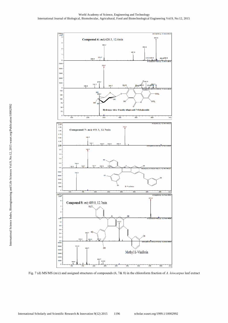
Fig. 7 (d) MS/MS (m/z) and assigned structures of compounds (6, 7& 8) in the chloroform fraction of A. leiocarpus leaf extract
World Academy of Science, Engineering and TechnologyInternational Journal of Biological, Biomolecular, Agricultural, Food and Biotechnological Engineering Vol:9, No:12, 2015
1196International Scholarly and Scientific Research & Innovation 9(12) 2015 scholar.waset.org/1999.1/10002992
Inte
rnat
iona
l Sci
ence
Ind
ex, B
ioen
gine
erin
g an
d L
ife
Scie
nces
Vol
:9, N
o:12
, 201
5 w
aset
.org
/Pub
licat
ion/
1000
2992

Fig. 7 (e) MS/MS (m/z) and assigned structures of compounds (9, 10& 11) in the chloroform fraction of A. leiocarpus leaf extract
World Academy of Science, Engineering and TechnologyInternational Journal of Biological, Biomolecular, Agricultural, Food and Biotechnological Engineering Vol:9, No:12, 2015
1197International Scholarly and Scientific Research & Innovation 9(12) 2015 scholar.waset.org/1999.1/10002992
Inte
rnat
iona
l Sci
ence
Ind
ex, B
ioen
gine
erin
g an
d L
ife
Scie
nces
Vol
:9, N
o:12
, 201
5 w
aset
.org
/Pub
licat
ion/
1000
2992
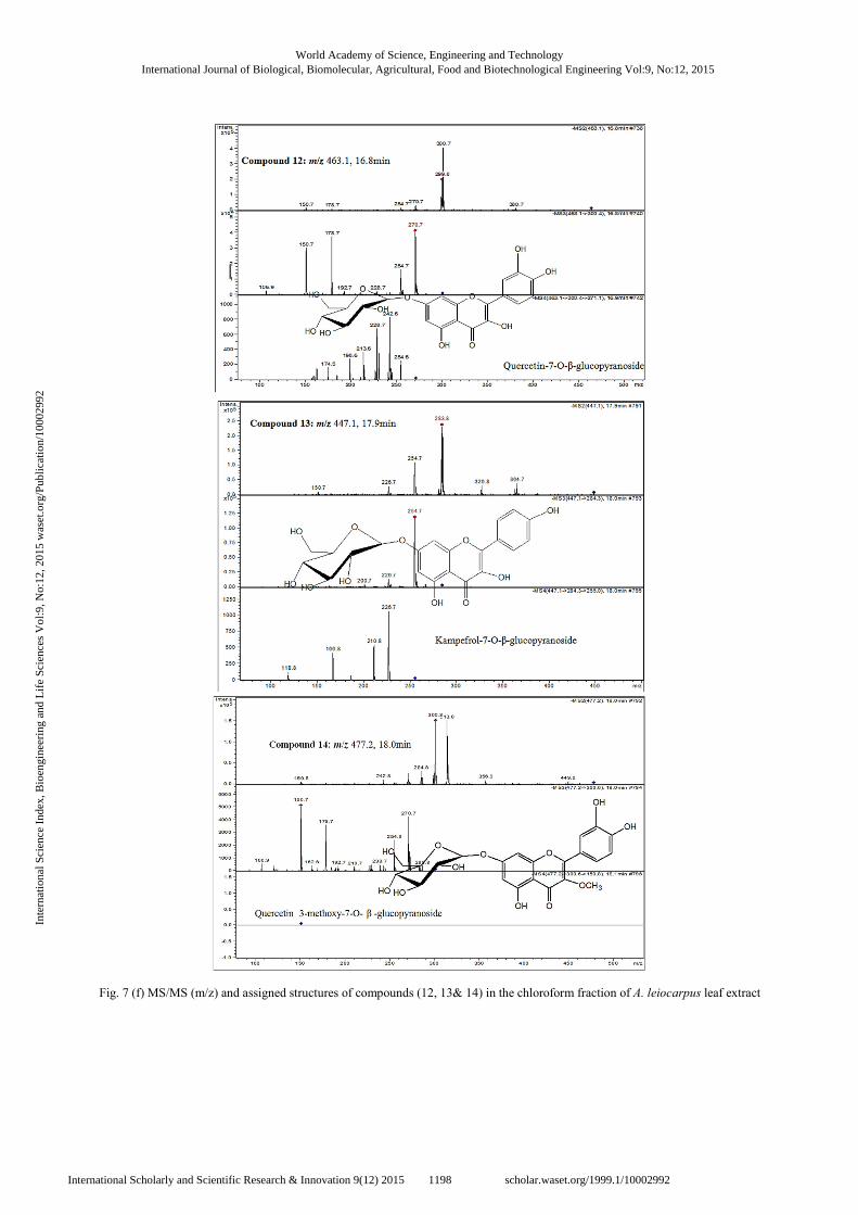
Fig. 7 (f) MS/MS (m/z) and assigned structures of compounds (12, 13& 14) in the chloroform fraction of A. leiocarpus leaf extract
World Academy of Science, Engineering and TechnologyInternational Journal of Biological, Biomolecular, Agricultural, Food and Biotechnological Engineering Vol:9, No:12, 2015
1198International Scholarly and Scientific Research & Innovation 9(12) 2015 scholar.waset.org/1999.1/10002992
Inte
rnat
iona
l Sci
ence
Ind
ex, B
ioen
gine
erin
g an
d L
ife
Scie
nces
Vol
:9, N
o:12
, 201
5 w
aset
.org
/Pub
licat
ion/
1000
2992

Fig. 7 (g) MS/MS (m/z) and assigned structures of compound (15) in the chloroform fraction of A. leiocarpus leaf extract
IV. CONCLUSIONS
In conclusion, the results of the in vitro susceptibility of M.
mycetomatis to the A. leiocarpus leaf extracts showed the
potent antifungal activity of the extracts against mycetoma
causing pathogen. These results confirmed the previous
antimicrobial activity of A. leiocarpus [14] and justifying its
traditional uses as a medicinal plant for treatment of skin
infections.
REFERENCES
[1] Agaie, B.M.; Onyeyili, P.A.; Muhammad, B.Y. and Landan, M.J., 2007.
Some Toxic Effects of Aqueous Leaf Extract of Anogeissus leiocarpus
in rats. Journal of Pharmacology and Toxicology, 2(4): 396-401. [2] Adejumobi, J.A.; Ogundiya, M. O.; Kolapo, A.andkunade, M.B., 2008.
Phytochemical composition and in vitro antimicrobial activity of
Anogeissus leiocarpus on some common oral pathogens. Journal of Medicinal Plants Research, 2(8):193-196.
[3] Okpekon, T.; Yolou, S.; Gleye, C.; Roblot, F.; Loiseau, P.; Bories, C.;
Grellier, P.; Frappier F.; Laurens, A. and Hocquemiller, R., 2004. Antiparasitic activities of medicinal plants used in Ivory Coast. Journal
of Ethnopharmacology, 90: 91-97.
[4] Agaie, B. M. and Onyeyili, P. A., 2007. Anthelmintic activity of the crude aqueous leaf extracts of Anogeissus leiocarpus in sheep. African
Journal of Biotechnology, 6(13):1511-1515. [5] Adeleye, I.A.; Ogunniyi, A.A. and Omonigbehin, E. A., 2003.
Antimicrobial activity of some local herbs on common skin pathogens.
Bioscience Research Communication, 15(3): 231-236.
[6] Vonthron-Sénécheau, C.; Weniger, B.; Ouattara, M.; Tra Bi, F.; Kamenan, A.; Lobstein, A.; Brun, R. and Anton, R., 2003. In vitro
antiplasmodial activity and cytotoxicity of ethnobotanically selected
Ivorian plants. Journal of Ethnopharmacology, 87: 221-225. [7] Mustofa, V. A.; Benoˆıt-Vical, F.; Pellissier, Y.; Kone-Bamba, D. and
Mallié, M., 2000. Antiplasmodial activity of plants extracts used in West
African traditional Medicine. Journal of Ethnopharmacology, 73: 145-151.
[8] Chaabi, M.; Benayache, S.; Vonthron-Sénécheau, C.; Weniger, B.;
Anton, R. andLobstein, A., 2006. Antiprotozoal activity of saponins from Anogeissus leiocarpus (Combretaceae). Planta Med., 72: 7.
[9] Almagboul, A. Z.; Basher, A. and Salih, A.K. M., 1988. Antimicrobial activity of certain Sudanese plants used in folkloric medicine. Screening
for antifungal activity. Fitoterapia, 59: 393-396.
[10] Mann, A.; Amupitan, J.O.; Oyewale, A.O.; Okogun, J.I.; Ibrahim, K.; Oladosu, P.; Lawson, L.; Olajide, I. and Nnamdi, A., 2008. Evaluation
of in vitro antimycobacterial activity of Nigerian plants used for treatment of respiratory diseases. African Journal of Biotechnology,
7(11): 1630-1636.
[11] Ibrahim, M.B.; Owonubi, M.O.; Onaopo, J.A., 1997. Antibacterial effect of extract of leaf, stem and root bark of Anogeissus leiocarpus on some
bacterial organisms. J. Pharm. Res. Dev., 2(1): 20-23.
[12] Sanogo, R., 2005. Antifungal and Antioxidant Activities of 14 plants used in the treatment of sexually transmitted infections. Afr. J. Trad,
Complem. Alter. Med.,2(2): 177- 205.
[13] Batawila, K.; Kokou, K.; Koumaglo, K.; Gbéassor, M.; de Foucault,B.; Bouchet, Ph. and Akpagana, K., 2005. Antifungal activities of five
Combretaceae used in Togolese traditional medicine. Fitoterapia, 76:
264-268. [14] Mann, A.; Yahaya Y.; Banso, A. and Ajayi, G.O., 2008.Phytochemical
and antibacterial screening of Anogeissus leiocarpus against some
microorganisms associated with infectious wounds. African Journal of Microbiology Research, 2: 60-62.
[15] Mann, A.; Banso, A. and Clifford, L.C., 2008. An antifungal property of
crude plant Extracts from Anogessius leiocarpus and Terminalia avicennioides, Tanzania Journal of Health Researc.,10(1): 34-38.
[16] Mann, A.; Amupitan, J.O.; Oyewale, A.O.; Okogun, J.I. and Ibrahim,
K., 2009. Antibacterial activity of terpenoidal fractions from Anogeissus leiocarpus and Terminalia avicennioides against community acquired
infections. African Journal of Pharmacy and Pharmacology, 3(1): 22-25.
[17] Mann, A.; Amupitan, J.O.; Oyewale, A.O.; Okogun, J.I. and Ibrahim, K., 2009. Chemistry of secondary metabolites and their antimicrobial
activity in the drug development process: A review of the genus
Anogeissus. Medicinal Plants-International Journal of Phytomedicines and Related Industries, 1(2): 6.
[18] Gumaa, S.A., 1994. The aetiology and epidemiology of mycetoma.
Sudan medical journal, 32(2): 14-22. [19] Mahgoub, E.S., 1994. Medical treatment of mycetoma. Sudan medical
journal, 32(2): 88-97. [20] NCCLS, 2002. National Committee for Clinical Laboratory Standards.
2002. Reference method for broth dilution antifungal susceptibility
testing of filamentous fungi. Approved standard NCCLS Document M38-A.9. National Committee for Clinical Laboratory Standards,
Wayne, USA.
[21] Ahmed, A.O.; Van de Sande, W.W.; Van Vianen, W.; Belkum, Van Alex; Fahal, A.H.; Verbrug, H.A.; Irma, A. and Bakker-Woudenber,
J.M., 2004. In vitro Susceptibility of Madurella mycetomatis to
Itraconazole and Amphotericin B assed by a Modified NCCLS Method and a viability based 2,3-bis(2-methoxy 040nitro-5-sulfophenyl)-5-
{(phenylamino) carbonyl}-2H-tetrazplium hydroxide (XTT) assay and a
modified NCCLS method. Journal of Antimicrobial Chemotherapy, 48(7): 2742-2746.
World Academy of Science, Engineering and TechnologyInternational Journal of Biological, Biomolecular, Agricultural, Food and Biotechnological Engineering Vol:9, No:12, 2015
1199International Scholarly and Scientific Research & Innovation 9(12) 2015 scholar.waset.org/1999.1/10002992
Inte
rnat
iona
l Sci
ence
Ind
ex, B
ioen
gine
erin
g an
d L
ife
Scie
nces
Vol
:9, N
o:12
, 201
5 w
aset
.org
/Pub
licat
ion/
1000
2992
[22] Van de Sande, W.W.J.; Lujendijk, A. and Ahmed, A.O., 2005.Testing of
the in-vitro susceptibility of Madurella mycetomatis to six antifungal agents by using the sensititer system in comparison with viability based
2,3-bis(2-methoxy040nitro-5-sulfophenyl)-5-{(phenylamino) carbonyl}-
2H-tetrazplium hydroxide (XTT) assay and a modified NCCLS method. Journal of Antimicrobial Chemotherapy, 49:1364-1368.
[23] Muraina, I.A.; Picard, J.A. and Eloff, J.N., 2009. Development of a
reproducible method to determine minimum inhibitory concentration (MIC) of plant extract against a slow-growing mycoplasmas organism.
Phytomedicine, 16(2-3): 262-264.
[24] Kuo-Ching Wen, I-Chen Shih, Jhe-Cyuan Hu, Sue-Tsai Liao, Tsung-Wei Su, and Hsiu-Mei Chiang, 2011. Inhibitory Effects of Terminalia
catappa on UVB-Induced Photodamage in Fibroblast Cell Line. Evid
Based Complement Alternat Med.: 904532. [25] Ten-Ning, C.; Guan-Jhong, H.; Yu-Lin, H.; Shgh-Shgun, H.; Heng-Yua,
C.; Yuan-Shium, C., 2009. Antioxidant and Antiproliferative Activities
of Crossostephium chinenis. The American Journal of Chinese Medicine, 37(4): 797-814.
[26] Eloff, J. N.; Katere, D. R.; McGaw, L.J., 2008. The biological activity
and chemistry of the Southern African Combretaceae Journal of Ethnopharmacology, 119: 686-699.
[27] Reddy, K.K.; Rajadurai, S.; Sastry, K.N.S. and Nayudamma, Y., 1964.
Studies on dhava tannins: Part I. The isolation and constitution of a gallotannin from dhava (Anogeissus latifolia). Australian Journal of
Chemistry, 17(2): 238-245.
[28] Reddy, K.K.; Rajadurai, S. and Nayudamma,Y., 1965. Studies on Dhava (Anogeis suslatifolia) Tannins: Part III. Polyphenols of bark, sapwood
and heartwood of Dhava. Indian. J. Chem., 27: 308-310. [29] Deshpande, V.H.; Patil, A.D.; Rama Roa, A.V. and Venkataraman.,
1976. Methylellagic acid and methylflavellagic acid from Anogeis
suslatifolia bark, Ind. J. Chem., 14B: 641-643. [30] Govindarajan, R.; Vijayakumar, M.; Rao, Ch.V.;Shirwaikar, A.;Rawat,
A.K.S.; Mehrotra, S. and Pushpangadan, P., 2004. Antioxidant potential
of Anogeissus. Biol. Pharm. Bull., 27(8): 1266-1269. [31] Govindarajan, R.; Vijayakumar, M.; Rao, Ch.V.; Shirwaikar, A.;
Pushpangadan, P. andMehrotra, S., 2004. Healing potential of
Anogeissus latifolia for dermal wounds in rats. Acta. Pharmaceutica., 54(4): 331-338.
[32] Govindarajan, R.; Vijayakumar, M.; Shirwaikar, A.; Rawat A.K.S.;
Mehrotra, S.and Pushpangadan, P., 2005. Activity Guided Isolation of Antioxidant Tannoid Principles from Anogeissus latifolia. Natural
Product Sciences, 11(3):174-178. [33] Pradeep, H.A.; Khan, S.; Ravikumar, K.; Ahmed, M.F.; Rao, M.S.;
Kiranmai, M.; Reddy, D.S.; Ahamed, Sh.R.;and Ibrahim, M., 2009.Hepatoprotective evaluation of Anogeissus latifolia: In vitro and in
vivo studies. World. J. Gastroenterol., 15(38): 4816-4822. [34] Scalbert, A., 1991. Antimicrobial properties of tannins. Phytochemistry,
30: Pp 3875-3883.
[35] Cowan, M. M., 1999. Plant Products as Antimicrobial Agents. Clinical microbiology reviews, 12 (4): Pp 564-582.
[36] Langcake P., 1981. Disease resistance of Vitis spp. and the production of
the stress metabolites resveratrol, e-viniferin, a-viniferin and pterostilbene. Physiological Plant Pathology,18: 312–226.
[37] Alessandro, M.; Marco, S.D.; Osti, F. and Cesari, A., 2000. Bioassays
on the activity of resveratrol, pterostilbene and phosphorous acid towards fungal associated with esca of grape vine. Phytopathol.
Mediterr., 39: 357-365.
[38] Alessandro, M.; Marco, S.D.; Osti, F. and Cesari, A., 2000. Bioassays on the activity of resveratrol, pterostilbene and phosphorous acid
towards fungal associated with esca of grape vine. Phytopathol.
Mediterr., 39: 357-365. [39] Suh, N.; Paul, S.; Hao, X.; Simi, B.; Xiao, H.; Rimando, A.M. and
Reddy, B.S., 2007. Pterostilbene, an active constituent of Blueberries,
suppresses aberrant crypt foci formation in the azoxymethane-induced colon carcinogenesis model in rats. Clin. Cancer Res., 13(1): 350-355.
[40] Rimando, A.M. and Suh, N., 2008. Biological/Chemopreventive Activity of Stilbenes and their Effect on Colon Cancer. Planta Med., 74(13): 1635-1643.
World Academy of Science, Engineering and TechnologyInternational Journal of Biological, Biomolecular, Agricultural, Food and Biotechnological Engineering Vol:9, No:12, 2015
1200International Scholarly and Scientific Research & Innovation 9(12) 2015 scholar.waset.org/1999.1/10002992
Inte
rnat
iona
l Sci
ence
Ind
ex, B
ioen
gine
erin
g an
d L
ife
Scie
nces
Vol
:9, N
o:12
, 201
5 w
aset
.org
/Pub
licat
ion/
1000
2992