Embed Size (px)
Citation preview
IN VITRO ASSEMBLY OF AN INFECTIOUS cDNA CLONE OF INFECTIOUS
BRONCHITIS VIRUS AND ITS APPLICATION AS A GENE TRANSFER
VECTOR
A Dissertation
by
SOONJEON YOUN
Submitted to the Office of Graduate Studies of
Texas A&M University in partial fulfillment of the requirements for the degree of
DOCTOR OF PHILOSOPHY
December 2003
Major Subject: Veterinary Microbiology
IN VITRO ASSEMBLY OF AN INFECTIOUS cDNA CLONE OF INFECTIOUS
BRONCHITIS VIRUS AND ITS APPLICATION AS A GENE TRANSFER
VECTOR
A Dissertation
by
SOONJEON YOUN
Submitted to Texas A&M University in partial fulfillment of the requirements
for the degree of
DOCTOR OF PHILOSOPHY
Approved as to style and content by:
Ellen W. Collisson (Chair of Committee)
Julian Leibowitz (Member)
Judith M. Ball (Member)
Karen-Beth Scholthof (Member)
Ann Kier (Head of Department)
December 2003
Major Subject: Veterinary Microbiology
iii
ABSTRACT
In vitro Assembly of an Infectious cDNA Clone of Infectious Bronchitis Virus
and Its Application as a Gene Transfer Vector. (December 2003)
Soonjeon Youn, B.S.; M.S.; D. V. M., Konkuk University
Chair of Advisory Committee: Dr. Ellen W. Collisson
An infectious cDNA clone of Vero cell adapted Beaudette strain of IBV was
constructed using in vitro assembly of cDNA fragments. The entire genome of IBV was
RT-PCR amplified into seven fragments, with each piece overlapping about 10
nucleotides. The fragments were ligated and transcribed to synthesize RNA, which was
transfected into BHK-21 cells. These cells were then overlaid onto IBV susceptible Vero
cells. After five days transfection, the virus was successfully rescued from the
transfected cells. The cDNA clone from our laboratory strain has a five nucleotide
insertion not present in the originally sequenced virus, resulting in total genome size of
27,613 nucleotides. The infectious cDNA clone was further manipulated to demonstrate
its potential as a gene transfer vector, by replacing the ORF5a open reading frame with
enhanced green fluorescent protein. The recombinant infectious cDNA clone was also
successfully rescued after three days transfection of BHK-21 cells followed by co-
culturing with Vero cells. This study showed that the 5a protein, whose function is not
known, is not necessary for in vitro IBV replication. This study also showed that the 5a
ORF is a good candidate for an insertion site of recombinant genes for the development
of IBV infectious cDNA clone as a gene transfer vector.

iv
ACKNOWLEDGEMENTS
I thank Dr. Ellen W. Collisson for her trust and enthusiasm toward my study. I
also appreciate her giving me the wonderful opportunity to work with her at Texas A&M
University.
I thank Dr. Ann Kier for her financial support that helped me to complete my
study in department of Veterinary Pathobiology in Texas A&M University.
I thank Dr. Julian Leibowitz for his guidance, encouragement and advice.
I thank Dr. Karen Scholthof for her encouragement and careful proofreading of
this dissertation.
I thank Dr. Tres Camacho for his constant encouragement, inspiration, patience
and tremendous help with my English.
I thank Jyothi Jayaram for her help with my English and friendship.
I thank Tootie Youn for her patience and being such good company during hard
times.
v
TABLE OF CONTENTS
Page
ABSTRACT…………………………………………………………………. iii
ACKNOWLEDGEMENTS………………………………………………... iv
TABLE OF CONTENTS……………………………………………………. V
LIST OF TABLES…………………………………………………………... vii
LIST OF FIGURES………………………………………………………….. viii
CHAPTER
I INTRODUCTION……………………………………………………. 1
Infectious bronchitis…………………………………………………... 1Classification………………………………………………………….. 3Molecular biology of coronavirus…………………………………… 4Replication of coronavirus…………………………………………… 18Infectious cDNA clones of coronavirus……………………………… 28
I II SYSTEMIC ASSEMBLY OF A FULL-LENGTH INFECTIOUS
cDNA OF A BEAUDETTE STRAIN OF INFECTIOUS BRONCHITIS
VIRUS (IBV)…………………………………………………….……….. 31
Introduction………………………………………………………….... 31Materials and Methods………………………………………………... 34Results………………………………………………………….……... 51Discussion………………………………………………………….…. 69
III EXPRESSION OF HETEROLOGOUS PROTEIN, GREEN
FLUORESCENT PROTEIN USING INFECTIOUS cDNA CLONE
OF INFECTIOUS BRONCHITIS VIRUS (IBV)…………………… 73
vi
CHAPTER Page
Introduction………………………………………………………….... 73Materials and Methods………………………………………………... 76Results………………………………………………………….……... 92Discussion………………………………………………………….…. 99
IV RECOMBINANT SEMLIKI FOREST VIRUS REPLICON CAN
BE PACKAGED INTO IBV VIRIONS WITHOUT IBV CIS
SIGNAL SEQUENCE………………………………………………... 102
Introduction…………………………………………………………. 102Materials and Methods……………………………………………… 106Results………………………………………………………………. 118Discussion…………………………………………………………... 131
V CONCLUSION………………………………………………………. 136
REFERENCES………………………………………………………………. 141
VITA………………………………………………………………………… 163
vii
LIST OF TABLES
TABLE Page
1 Primer pairs used for cloning of the individual IBV amplicons…... 39
2
Summary of nucleotide and amino acid differences between IBV
Beaudette used in this study and published sequence…………….. 56
3 Sequences of primer pairs used for construction of IBVG………... 79
4
Primer pairs used for RT-PCR or PCR amplification and
sequencing of recombinant SFV replicons………………………… 109
viii
LIST OF FIGURES
FIGURE Page
1 Schematic of the IBV genome and subgenomic RNA organization……. 6
2 Comparison of genome organization of coronaviruses……….………... 8
3 Schematic of coronavirus gene 1 organization………………………….. 10
4 The leader-primed model of coronavirus transcription…………………. 23
5 Discontinuous extension of negative strands model of transcription…… 25
6 Strategy for orderly assembly of an IBV infectious cDNA clone………. 37
7 No see’m technology using BsmBI restriction enzyme properties……... 38
8 RT-PCR amplification of the entire IBV genome………………………. 52
9 Preparation of insert DNAs from each amplicon……………………….. 60
10 Systemic assembly of IBV full-length cDNA…………………………... 62
11 In vitro transcribed RNA from in vitro assembled cDNA template
encompassing the entire genome of IBV………………………………... 64
12 Sequence comparison of molecularly cloned IBV with the wild type
IBV Beaudette strain…………………………………………………….. 66
13 Molecularly cloned virus plaque morphology…………………………... 67
14 Growth kinetic comparison of wild type IBV and molecularly cloned
IBV……………………………………………………………………… 68
15 Schematic of cloning strategy used to replace enhanced green
fluorescent protein with ORF5a…………………………………………. 77
ix
FIGURE Page
16 “No see’m technology” used to replace EGFP with ORF5a……………. 80
17 Site directed mutagenesis by PCR………………………………………. 93
18 Sequence verification of EGFP ORF and ORF5a replacement…………. 95
19 Recombinant infectious cDNA clone which 5a ORF was replaced with
EGFP ORF was viable in infected cells and expressed EGFP………….. 98
20 Sequence alignment of MHV and BoCV packaging signal sequences…. 120
21 Sequence alignment of BoCV packaging signal sequence and the
putative IBV homologue………………………………………………… 121
22 Schematics of pSFV-1 eukaryotic expression vector and recombinant
SFV replicons constructed using pSFV-1 vector………………………... 123
23 Schematic of the overall experimental design…………………………... 126
24 Recombinant SFV replicons can be packaged into IBV virions with or
without IBV cis sequences………………………………………………. 128
25 Without helper virus, recombinant SFV replicons cannot be passaged
from passage 0 to passage 1…………………………………………….. 129
1
CHAPTER I
INTRODUCTION
INFECTIOUS BRONCHITIS
Avian infectious bronchitis is one of the most important poultry diseases in the
world, causing great economic losses to the poultry industry (39). It is also considered
the most common disease in both broilers and layers. Its causative agent is infectious
bronchitis virus (IBV). IBV was first described in 1931 by Schalk and Hawn in North
Dakota as an “apparently new respiratory disease of baby chicks” (158) and then
subsequently isolated by Beaudette and Hudson in 1937 (14). Initially IBV caused
respiratory disease in chickens but later additional types of IBV emerged. IBV can be
divided into three different forms based on the primary symptoms in infected birds. The
respiratory form of IBV primarily results in respiratory illness such as gasping, tracheal
rales, coughing and nasal discharge (6). This form is caused by IBV strains that include
Massachusetts, Arkansas, and Connecticut. Second is the nephropathogenic form of
IBV, which causes not only respiratory illness but also causes nephritis by certain stains
of IBV such as Gray, Holts, Australian T strain and PA/Wolg/98 (1, 208). Often the
respiratory form is more severe in young chicks and the nephritic form is predominantly
seen in older chicks up to 10 weeks of age. The third form of IBV is a reproductive form
This dissertation follows the format of Journal of Virology.
2
in which the virus directly affects the reproductive organs of birds such as ovaries and
reproductive tracts (41). The reproductive form of IBV infection causes not only poor
egg production in infected birds but also poor egg quality. The most economically
detrimental consequences for the poultry industry of infection of IBV are poor egg
quality and markedly poor growth in chicks that survive causing vast economic loss in
the poultry industry (14). Chicks infected with IBV show varying mortality depending
on strains of virus that infect flocks, illness ranges from asymptomatic infection to very
high mortality with no lesions because of rapid disease progression (39). A problem in
controlling of IBV is the increased susceptibility to bacterial infection, such as E. coli
and this complication exacerbating chronic respiratory disease and airsacculitis (6).
The incubation time of the virus is dose dependent and ranges from 18 to 36
hours after transmission by inhalation. The virus is not vertically transmitted through
eggs (39). In experimental infection studies, IBV can be isolated from the trachea, lungs,
kidney, and bursa of chickens from 24 hr to the 7th day after aerosol exposure (76).
The control of IBV depends mainly on vaccination. However, concurrent
infection of the flocks with more than one serotype and vaccine breaks by field strains
differing from the vaccine have been reported. With live vaccines, it may be possible to
prevent disease but not prevent infection (65, 87). Usually a vaccine regimen requires
using an inactived IBV strain given as an oil-in-water emulsion to chickens that have
already been primed by vaccination with a live attenuated IBV vaccine.
3
CLASSIFICATION
Avian infectious bronchitis virus is a member of the Coronaviridae family that
belongs to the order of Nidovirales. Nidovirales contains four families, the Toroviridae,
Arteriviridae, Roniviridae as well as Coronaviridae (34, 168). The classification is based
on polycistronic genome organization and regulation of gene expression from a nested
set of subgenomic mRNAs, despite significant size differences between genuses ranging
from 13 to 32 kb. Coronaviruses, as well as the other nidoviruses, have a unique,
discontinuous transcription strategy that produces several subgenomic mRNAs, as well
as the genomic RNA packaged into its virions. The subgenomic mRNAs form 3’ co-
terminal nested sets in which each mRNA contains a common 3’ untranslated region and
5’ leader sequence (90).
Coronaviruses infect various animal species, causing respiratory, gastrointestinal,
cardiovascular, and neurological disease (126). Human coronaviruses have been
associated with mild common colds, diarrhea, possibly multiple sclerosis, as well as
severe acute respiratory syndrome (SARS) (70). Coronaviruses have been divided into
three distinct groups by antigenic cross-reactivity and sequence analysis of structural
protein genes. Group I coronaviruses include human coronavirus 229E (HCoV-229E),
transmissible gastroenteritis virus (TGEV), canine coronavirus (CCV), feline
coronavirus (FECV), and feline infectious peritonitis virus (FIPV). Group II includes
human coronavirus OC43 (HCoV-OC43), murine hepatitis virus (MHV), and bovine
coronavirus (BoCV). Group III includes IBV with some turkey coronaviruses (TCV) and
pheasant coronavirus. The recently identified coronavirus that is the causative agent of
4
severe acute respiratory syndrome (SARS) in humans is distinct from other
coronaviruses based on its genomic organization and structural genes (186). However,
there was a report that SARS is a Group II coronaviruses based on phylogenetic study
comparing the replicase gene with other coronaviruses and torovirus (167).
MOLECULAR BIOLOGY OF CORONAVIRUS
Morphology
Coronavirus virions are pleomorphic in shape with a diameter of 80 to 160 nm and
are composed of four major structural proteins, which include the nucleocapsid protein
(N), the integral membrane protein (M), the spike protein (S) and the small envelope
protein (E) (90). Petal-like spikes that appear as a corona give the virus its name and
have a distinct morphology by electron microscopy (EM) (15). These prominent surface
projections of up to 20 nm in length cover the entire virion surface (15). Some species of
coronaviruses, such as TCV, have smaller spikes on the virion surface. The N proteins
interact with viral genomic RNA to form helical ribonucleoprotein complexes (RNP)
that are similar in morphology to paramyxovirus RNPs (49). Not only genomic RNA but
also every subgenomic RNA interacts with N protein forming RNP in the infected cells.
In the case of TGEV, an icosahedral core shell structure has been shown by EM (142).
One report indicated that two different forms of IBV virions could be made in infected
cells (119). One form is the infectious virus particle, which has viral RNP, the other
form has a lower density and has no RNP. This finding is consistent with the observation
5
that co-expression of E and M protein produce virus like particles (VLPs) without
internal virion structures (193).
Genome Organization
The genomes of the coronaviruses ranging from 27.6 to 32 kb are the largest
genomes of all the known RNA viruses. Coronaviruses have a linear, positive sensed
polyadenylated RNA that is infectious when transfected into susceptible cells (112, 161).
In infected cells, coronaviruses produce from five to eight subgenomic RNAs in addition
to genomic RNA depending on the virus (165). Each RNA is numbered according to its
size. Genomic and subgenomic RNA organization of IBV is illustrated in Figure 1. IBV
has six subgenomic RNAs including its genomic RNA with RNA 1 being the largest,
and RNA 6 being the smallest (35). The genomic RNA, as well as every subgenomic
RNA, is 5’ end capped and has a polyadenylated tail of about 100 nucleotides (nts) at the
3’ end (112, 113, 161, 196). Subgenomic RNAs range from 2 to 8 kb and with genomic
RNA share a common 3’ terminus forming a nested set (172, 173). Genomic and
subgenomic RNAs have 3’ and 5’ untranslated regions (UTR) believed to contain cis-
signal sequences for viral replication and transcription (48). The 5’ end of each mRNA
has leader sequence about 60 to 72 nts (91, 92, 171). Most coronavirus ORFs are
preceded by an untranslated region of varying length containing a conserved sequence,
which has been given the term intergenic sequence (IG) or transcription regulatory
sequence (TRS) (28, 90).
6
1 3 5 7 9 11 13 15 17 19 21 23 25 27
1 3 5 7 9 11 13 15 17 19 21 23 25 27
RNA 1
RNA 3
RNA 6
RNA 5
RNA 4
RNA 2
5a and b
M ORF
ORF1a
ORF1bS ORF N
3 a,b, E
FIG. 1. Schematic of the IBV genomic and subgenomic RNA organization. S represents the
spike glycoprotein; M, the membrane protein; E, the small envelope protein; N, the
nucleocapsid protein. RNA1 encodes replicase gene, pp1a and pp1ab; RNA2 encodes S
protein, RNA3 encodes 3a, 3b and the E proteins, RNA4 encodes the M protein, RNA5
encodes 5a and 5b whose functions are not known and RNA6 encodes the N protein. The
black box represents poly (A) tails.
7
The overall IBV genome organization is similar to other prototype coronaviruses such as
MHV and TGEV, having the “replicase gene-S-E-M-N” gene order. ORFs encoding
small nonstructural group specific proteins, such as 3a, b and 5a, b of IBV lie between
the structural genes (103). Comparisons of the different genomic organizations of
coronaviruses are illustrated in Figure 2. However, with a smaller genome size of 27.6
kb compare to other coronaviruses, such as MHV with a genome of 31 kb, IBV genomic
usage is more compact. ORFs of IBV are overlapped. For example, the 3’ end of the 1b
genome overlaps with the S ORF by about 50 nts (30).
IBV RNA 1, which encodes the replicase gene, is indistinguishable from
genomic RNA and contains the ORF for replicase genes, pp1a and pp1ab, and ORFs for
the structural proteins. Like most coronaviruses, every subgenomic RNA of IBV only
expresses the extreme 5’ ORF except for RNA 3 and RNA 5, which encode 3a, 3b, 3c
(E), 5a and 5b ORFs respectively. IBV has several nonstructural proteins of which
functions are not well understood. The exception is gene 3c, which encodes an important
structural membrane protein, E protein (104, 203). Two thirds of RNA 1, about 20 kb, of
IBV genome encoding two large ORF1a and 1b, that express polyproteins of pp1a, 441
kDa and pp1b, 300 kDa, respectively. pp1ab is produced via -1 ribosomal frame shifting
upstream of a psuedoknot structure between ORF1a and ORF1b (25, 26). Polyproteins
encoded by gene 1 are cleaved by viral proteinases during posttranslational processing
(57).
RNA 2 encodes the spike (S) protein of IBV. As mentioned previously, the 3’
end of 1b ORF overlaps with the ORF of S by about 50 nts in the Beaudette strain. It is
8
SCoV
TGEV
FIG 2. Comparison of genome organization of coronaviruses. ORF1a and
ORF1b are interrupted and shortened to highlight the remaining genes. Boxes
represent ORFs.
E 3
5 4
5 3 S M N
S M N
S M N
MHV
IBV
BCV pp1b
ppla 2 H S 4
5M N E
a b 2 H S M NE
9
not certain if this is a common feature of IBV. RNA 3 encodes three ORFs, 3a, 3b, and
3c. Nucleotide sequencing and expression studies indicated that gene 3 was tricistronic
and that ORF3a and 3b produced proteins in small amounts in IBV infected cells (103),
but their functions are a subject of further study. The ORF3c encodes the small envelope
protein (103, 104). An unusual folding region (UFR) of 265 nts containing the 3a and 3b
coding region upstream of the E ORFs was noticed (95). This UFR consists of five RNA
stem-loops that could be modeled into a superstructure by the interaction of two putative
pseudoknots. It was predicted that this region could serve as an internal ribosomal entry
site (IRES) for the cap independent expression of E protein. RNA 4 of IBV encodes M
protein. Just as RNA 3 is multicistronic, nucleotide sequencing and expression studies
showed that RNA 5 is dicistronic and encodes two nonstructural proteins, 5a and 5b.
Expression of these two proteins has been shown in IBV infected cells, but the function
of these proteins and mechanism of expression also needs to be determined (105).
RNA6, the smallest but the most abundant RNA in IBV infected cells encodes the N
proteins.
Coronavirus Non-Structural Proteins
RNA 1 encodes the coronavirus replicase gene. The schematic of genomic RNA
organization of coronaviruses is illustrated in Figure 3. The replicase genes of
coronaviruses consist of two large open reading frames, ORF1a and ORF1b, the latter of
which is expressed by ribosomal frame shifting (26). The ORF1a and ORF1ab
translation products are large nonstructural polyproteins encoding polyproteins pp1a and
10
FIG. 3. Schematic of coronavirus gene 1 organization. PL1 pro represents a papain-like
proteinase 1; PL2 pro, a papain-like proteinase 2; 3CLpro, a 3C-like proteinase; MP1 and
MP2 represent the hydrophobic domains; RdRp, RNA-dependent RNA polymerase;
HEL, helicase and the black box indicates a domain with an unknown function in
coronaviruses. The small green box between RdRp and HEL represent a cysteine-rich
domain. IBV and SCoV have only one PL2 pro domain instead of two PL pro.
HD
RdRp HD
3CLpro
ORF 1a HEL
HD
x
PL1pro PL2pro
11
pp1ab, respectively. Polyprotein precursors of the nidovirus ORF1a and ORF1ab
translation products are cleaved by viral proteinases at a minimum of 13 sites (207).
ORF1b encodes RNA dependent RNA polymerase (RdRp), helicase (HEL) and
methyltransferase (56). Functions of the ORF1a-encoded polyproteins include
proteinases that process viral nonstructural proteins and proteins that are membrane
associated (100-102, 133). Most nidovirus replicase subunits are thought to assemble
into an RNA-protein complex, which is presumably held together by protein-protein and
/or protein-RNA interactions. Some of replicase genes have been shown to co-localize
with viral RNA (19, 20, 133, 159, 164, 192). Putative hydrophobic domains in the
ORF1a protein have been proposed to mediate the membrane association of this
complex (19, 164). The N terminal half of the ORF1a protein encodes multiple papain-
like cysteine protease domains (PLpro). The proteolytic activity of PLpro has been
demonstrated and characterized in detail (73, 97, 99, 207). Compared to group I and
group II coronaviruses that encode two PLpro, IBV has two overlapping PLpro and only
the first PLpro is functional. SARS also encodes only one PL2pro (97, 186). Another
functional coronaviral proteinase, the 3C-like cysteine protease (3CLpro) is located in the
central region of the ORF 1a polyprotein (114). This 3CLpro belongs to the
chymotrypsin-like protease superfamily and is flanked by hydrophobic domains (207).
Proteolytic activity of the protein has been demonstrated and characterized in detail in
MHV, HCoV and IBV (69, 74, 115, 206). Several studies have shown that this
proteinase cleaves pp1ab fusion polyprotein at the QS dipeptide bonds to the 100-kDa
protein species in IBV (101, 102, 107).
12
Compared to other positive strand RNA viruses, distinct features of a coronavirus
replicase include a helicase domain downstream of the RdRp motif in ORF1b and the
classic GDD core motif in the RdRp is substituted by an SDD (68). In addition,
compared to a putative catalytic triad, such as cysteine, histidine and glutamic acid, for
the proteinase activity, cysteine and histidine are sufficient enough for coronavirus
3CLpro protease activity (102).
Coronavirus Structural Proteins
Coronaviruses have four essential structural proteins. These include spike
glycoprotein (S), small envelope protein (E), integral membrane protein (M), and
nucleocapsid protein (N). In addition to these elements, some strains of group I and II
coronaviruses have an additional glycoprotein, the hemagglutinin/esterase (HE) protein
which is assumed to be acquired from influenza virus C and is not essential for viral
replication in vivo or in vitro (4, 16, 37, 119).
The S protein is the most prominent virion protein that can be seen by EM, forming a
corona around the virions. This type I glycoprotein passes through the host derived lipid
bilayer membrane once, with most of the molecule forming an amino-terminal
ectodomain (18). The S protein can be divided into three different parts other than the
signal sequence that targets the proteins to the ER (117). The first part is the ectodomain
of S, which is largely exposed outer surface of the virion. The second part is the
transmembrane domain (TM), which consists of hydrophobic amino acids that are
embedded in the virus envelope. The third part of the S protein is the endodomain that
13
has a C terminal tail inside of the virions. The TM and endodomain are believed to
confer the stability of the S proteins on the virion.
S proteins form homotrimers (33) and are co-translationally N-glycosylated, after
which the carbohydrates are processed in the endoplasmic reticulum and in the Golgi
apparatus (55, 134). The majority of the S protein in virus-infected cells localizes in the
ER and Golgi intermediate compartment (ERGIC) but when the S protein is expressed
by itself, it is transported to the plasma membrane. This implies that some other viral
protein interacts with S protein and localizes to the budding sites. In addition, an excess
amount of S protein expressed in infected cells is transported to the plasma membrane
inducing cell-cell fusion of neighboring cells thus promoting virus spreading.
Newly synthesized proglycoprotein S is cleaved by carboxypeptidase-like
activity in vivo or by trypsin in vitro into S1, the N-terminal product and S2, the C-
terminal product. The cleavage site lies near the center of the protein at a cluster of basic
amino acids (37). This cleavage has been shown to be required for the activation of viral
infectivity and cell fusion in MHV Sturman, 1985 #136, 181). IBV-Beaudette S1 and
S2 are comprised of 514 amino acid residues (56.2 kDa), and 625 amino acid residues
(69.2 kDa), respectively. The amino acid cleavage sites of IBV and MHV S1 and S2
were identified by partial amino terminal sequencing of spike polypeptide, S2, which
was identified as RRFRR for IBV and RRAR(H)R for MHV (37, 117).
However, in the case of FIPV, TGEV and likely SARS, the S protein dose not have a
cleavage site (78). Major neutralization epitopes, which determine the serotypes of IBV,
map to the S1 protein (36). This protein interacts with cellular receptors, whereas the S2
14
is responsible for anchoring S1 into the viral envelope and for cell membrane fusion.
Cellular receptors for several coronaviruses have been identified. Two cellular receptors
have been identified for MHV. MHVRI and MHVR2, which are translated from the
same mRNA through alternate splicing and are expressed in different tissues (46) (58).
Integral membrane glycoprotein (M) is the most abundant envelope glycoprotein of
coronaviruses (152). It is composed of 225 amino acids in IBV. Its predicted molecular
weight, based on amino acid sequence data is about 25 kDa. However, the actual
molecular weight in infected cells is about 30 kDa. M protein expressed in infected cells
or expressed from cDNA transcripts has several isotypes. The MHV has five different
isotypes that can be distinguished electrophoretically ranging from M0 to M5 (83, 150,
191. This molecular weight discrepancy is based on different glycosylation of the M
protein depending on the location of the protein in different intracellular compartments
Locker, 1992 #332)(106). M proteins of coronaviruses also differ in glycosylation and
localization. MHV has O-linked glycosylation and targets to the trans-Golgi apparatus
(110, 135, 178) whereas IBV has N linked glycosylation (32, 108) and targets to cis-
Golgi (118).
The M protein spans the membrane bilayer three times, having a short glycosylated
amino terminal domain on the exterior of the virus and a large carboxyl terminus,
containing more than half the mass of the molecule, in the virion interior (5, 111, 151).
In contrast to other viral glycoproteins, M protein doesn’t have a signal sequence at its N
terminal but has internal signal sequence in the membrane-spanning hydrophobic
regions (148). In IBV, the first transmembrane domain appears to be sufficient for
15
retention of the protein in the Golgi (182), whereas the cytoplasmic tail and at least one
transmembrane domain of the MHV-M protein are necessary for viral retention at the
trans-Golgi (109).
The M protein is an important viral structural protein. Its interaction with S protein is
important for the incorporation of S protein into a virus particle. S protein can be
expressed on plasma membranes without the presence of M protein expression, however
if the S protein is co-expressed with M protein, it localizes to the ER-Golgi intermediate
compartment which is suggested as a virus budding site (118, 149) (189). M and S
protein interaction in MHV has been shown (137). Transmembrane domains and
amphipathic domains of the M protein are necessary for interaction with S in MHV (52).
Targeted mutagenesis of MHV and FIPV has shown that without the proper TM and
endodomain of the S protein, the ectodomain of different species S protein cannot be
incorporated into virus particles (53, 66). This study suggested that TM and endodomain
of the S protein might interact with the M protein. The M protein is also important for
virus budding and assembly with E protein. The M protein has been shown to have an
affinity for RNA even though the domain of M that interacts with the RNA or RNP is
not known (179). Recently, Narayanan et al. (128) suggested that M protein provides the
specificity of viral genomic RNA packaging by interacting directly with the packaging
signal sequence of MHV genomic RNA. Monoclonal antibodies specific for the M
protein do not neutralize virus infectivity, suggesting that M protein is not involved in
virus attachment to receptors.
16
The third glycoprotein on the virion surface is HE. It is present in BCV, TCV,
HCoV, and some strains of MHV, but is not present in IBV or TGEV. It is also an N-
linked glycoprotein. This glycoprotein shares some sequence homology with the
hemagglutinin protein of influenza C virus, and it is proposed that HE protein of these
coronaviruses is derived from the influenza virus by non-homologous recombination
(116). HE protein of BCV exhibits hemagglutinin and esterase activity and some strains
of MHV also show esterase activity. Monoclonal antibodies specific for the HE protein
of BoCV can inhibit virus-induced hemagglutination and neutralize viral infectivity
implying that at least in BoCV, HE is involved with S protein for viral attachment.
The E protein was recently recognized as a structural protein (104, 203). This protein
in IBV is comprised of 108 amino acids are about 10 kDa molecular weight. In contrast
to other coronavirus structural proteins, E protein is expressed by cap-independent
translation from an IRES which is located in the ORF3a and 3b regions or in the 5a
region in MHV (106, 188). E protein localizes in the perinuclear region with a small
amount migrating to the cell surface (203). The E protein is anchored in the membrane
by a sequence in the N-terminal half of the protein and is largely embedded within the
viral membrane with only a hydrophilic C-terminus protruding inside the virion (121).
Co-expression of E and M proteins produces virus like particles (VLP) without viral
RNP. However, expression of the E proteins of MHV or IBV produces vesicles that can
be released from the cells in the absence of other viral proteins (47, 120). Co-expression
of M and E are necessary in TGEV and bovine coronavirus (BoCV) to make virus-like
particles (VLP) (13, 17, 193). E protein has been suggested to “pinch off the neck” of the
17
assembled virus particle from cells during the final stages of budding (193), implying
that an E protein is essential for VLP formation in every coronavirus. However, Kuo et
al. (85) has shown that an E deletion mutant of MHV is still viable, although it grow
poorly and with delayed kinetics. The exact role of E protein in the virus assembly
process and budding is not yet clear. Direct interaction between the E and M proteins has
not been demonstrated. It is possible that such an interaction would serve to facilitate the
budding of virus particles.
The N protein is the only phosphorylated structural protein in the coronavirus (198).
N protein is composed of 409 amino acids having a molecular weight of about 50 kDa in
IBV (22). Coronavirus nucleocapsid sequences vary among major antigenic groups but
are highly conserved within groups (199). The N protein has basic residues that occur in
clusters depending on the virus, serine residues account for 8 to 10% of the total amino
acids in N proteins and the abundance of serine residues count for the specific
phosphorylation on serine residues in MHV (170).
N protein is considered a multifunctional protein playing important roles not only as a
structural protein but also as a protein involved in viral replication and transcription. N
protein interacts with viral genomic RNA forming a helical nucleocapsid (RNP) and
interacts with subgenomic RNAs in the cytoplasm. MHV N protein has been shown to
interact specifically with the 5’ leader containing sequences (10). MHV and IBV-N
proteins can be divided into three structural domains. The middle domain is an RNA-
binding domain that binds to both coronavirus and non-coronavirus RNA sequences in
vitro in MHV (125, 130). However, there is no known RNA binding motif in
18
coronavirus N protein. In the case of IBV, C terminal and N terminal regions of N
protein but not the middle region interact with the 3’ end of the non-coding region of
IBV genomic RNA (43, 205). Antiserum raised against N protein inhibited the synthesis
of the genome-sized RNA by 90% in an in vitro replication system implying N protein is
involved in transcription or/and translation of the viral genome (45). Several reports
implied that the phosphorylation status of the N protein might be involved in the control
of viral transcription and replication.
REPLICATION OF CORONAVIRUSES
Coronaviruses replicate in the cytoplasm of infected cells, preferentially in epithelial
cells. It has been shown that the MHV can replicate inside the enucleated cell implying
that the host nucleus and/or nuclear factors are not involved in virus replication.
However, studies with IBV and a human coronavirus have shown that the replication of
these viruses is hindered by α-amanitin treatment or in enucleated cells (61). This
suggests that maybe some host nuclear factor(s) are involved in virus replication, at least
for IBV and HCoV. Coronavirus replication does not completely shut down the cell
replication cycle and continuous host translation is necessary for viral replication (154).
The replication complex of coronaviruses has been described to be associated with late
endosomal membranes by colocalization of viral replicase and newly synthesized RNA
with a late endosomal marker by immunofluorescence assay (IFA) and electron
microscopy (EM) (166, 192). This replication complex translocates to the virus budding
site, ERGIC, at late times post infection (20).
19
Coronavirus replication also needs continuous viral protein translation and N protein
(45). Inhibitors of protein synthesis, such as cycloheximide, inhibit RNA synthesis
(122). Dependency of RNA synthesis on protein synthesis continues throughout the viral
replication cycle. The RNA polymerase or some cofactors have to be continuously
synthesized. Negative-stranded and positive-stranded coronavirus RNA synthesis have
different sensitivities to inhibition by cycloheximide (154), implying that the negative
and positive-stranded RNAs are likely synthesized by two different polymerase
activities. Two enzymatically distinct RdRp activities, one acting early and the other
during late infection, have been described (24) even though there are some
inconsistencies between studies (45, 122).
Coronaviruses employ several different strategies to translate their structural or non-
structural proteins in infected cells. First, several subgenomic RNAs are produced that
are functionally monocistronic, expressing only the extreme 5’ ORF. Second, it exploits
ribosomal frameshifting and post-translational cleavage of polyproteins. Finally, an
IRES is used to translate proteins in a cap-independent manner from polycistronic ORFs.
Attachment and Penetration
Attachment and penetration are complex processes that have not been
characterized in detail for most coronaviruses. The virion binds to the cell membrane
receptors by either the S or HE glycoproteins (42). IBV entry into susceptible cells
occurs at both at 4°C and 37°C (138). Conformational changes in either of the viral
proteins S or HE may be induced by virus binding to the cellular receptor and lead to
20
fusion of the virus envelope with host cell membranes (46). MHV-A59-induced fusion is
optimal at neutral or mildly alkaline pH (180). Other coronaviruses, such as HCV-229E,
IBV or some strains of MHV do not fuse at neutral pH and the fusion occurs in
endosomal membranes rather than the plasma membrane (64). Coronaviruses generally
are highly species specific in vivo and in vitro. The species specificity appears to be
mediated at the site of entry, since MHV genomic RNA is infectious and MHV
replicates efficiently in nonpermissive hosts that express the MHV receptor. In addition,
a recombinant FIPV with the MHV S protein can replicate in murine cell lines (71).
Receptors for the MHV virus have been identified. MHV receptor (MHVR) is a member
of the carcinoembryonic antigen-cell adhesion molecule (CEACAMs) family of
glycoproteins (200). Subsequently, MHVR (2d) was found as a splice variant of MHVR
(4d) (58). Membrane expression of this molecule renders MHV resistant cells, such as
human and hamster cells, susceptible to MHV infection (58, 59). Aminopetidase N
(APN), a membrane bound metalloprotease, has been identified as a receptor for TGEV
and HCV-229E. In humans, hAPN is also known as CD13 (54). TGEV and HCV-229E
both utilize only species-specific APN glycoproteins as receptors. This indicates that not
only the attachment to receptors, but also possibly some other host factor(s) are involved
in host specific virus infection (45). Several studies implicate the involvement of HE
protein in virus attachment in MHV and even more so in BCV. However, these studies
need to be expanded. After virion attachment onto its cellular receptor, virus will be
internalized and targeted to the cellular organelles where the virus will replicate.
Mechanisms of replication are not yet well studied in coronavirus.
21
Viral Transcription
Similar to most positive stranded RNA viruses, after the release of the genomic
viral RNA into host cells, replication begins with the viral RNA serving as the
messenger for RdRp synthesis. The polymerase transcribes the genomic RNA into a
negative stranded full length RNA and the negative stranded subgenomic RNA serve as
templates for the synthesis of subgenomic and genomic RNAs (93). The presence of
multiple subgenomic mRNAs is a characteristic feature of coronaviruses. Depending on
the coronavirus, five to eight subgenomic RNAs are transcribed in infected cells. They
have 5’ and 3’ untranslated regions (UTR) and a poly(A) tail of about 100 nts. The
extreme of 5’ end of each subgenomic RNA has a stretch of 65-98 nucleotides, referred
to as the leader sequence. The leader sequence is identical to the sequence at the 5’ end
of the genomic RNA and is not found elsewhere in the genome (89, 91). However, the
sequences between each gene, termed the intergenic sequences (IG) or transcription
regulatory sequence (TRS), have a sequence homology of 7 to 18 nucleotides with the 3’
end of the leader sequence (28). The core sequence of the IG sequence for MHV is
AAUCUAAAC, whereas the 3’ end of the leader consists of several tandem repeats of
UCUAA (88). The consensus sequence for IBV is CU(U/G)AACAA, AUCUAAAC is
the consensus sequence for BoCV, and AACUAAAC is the consensus sequence for
FIPV and TGEV (170). Because of the characteristics of coronavirus transcription that
produces subgenomic RNAs, and function of leader sequences, several models have
been proposed to explain the generation of subgenomic RNAs for coronavirus mRNA
transcription. Two facts exclude splicing as the mechanism to produce subgenomic
22
RNA. First, in ultraviolet (UV) transcriptional mapping studies, the UV target size of
each mRNA is equivalent to its physical size, indicating that each mRNA is transcribed
independently, and is not derived from the process of a large precursor RNA (79, 174).
Second, because coronaviruses replicate solely in the cytoplasm, the possibility of
splicing as a mechanism for coronavirus transcription mechanism is excluded.
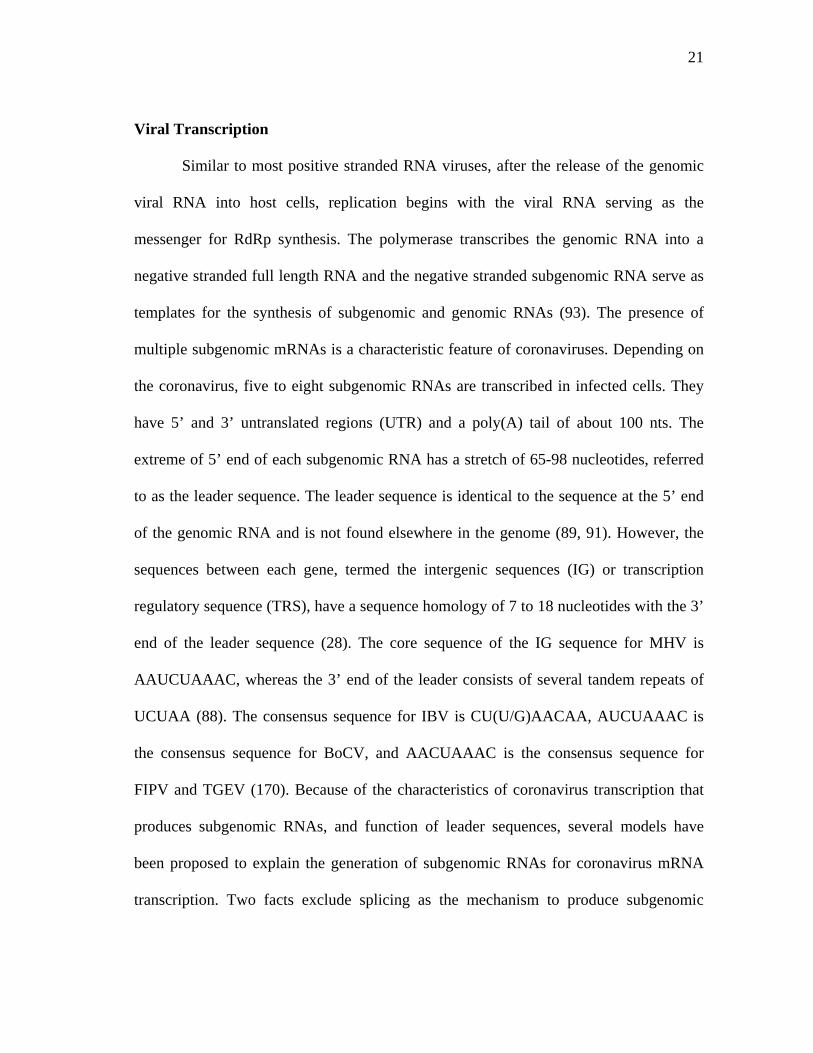
One coronavirus transcription model is the leader-primed model. This model is
illustrated in Figure 4. According to this model, discontinuous transcription occurs
during positive stranded RNA synthesis. Leader RNAs are transcribed at the 3’ end of
the negative stranded RNA template, before dissociating from the template, and
subsequently binding to any IG or TRS on the negative stranded template. The leader
RNA serves as the primer for subgenomic RNA transcription, resulting in a subgenomic
RNA that contains a leader RNA fused to the mRNA. This model is based on the
presence of free leaders in infected cells. Several small leader sequence-related RNA
species have been detected in the cytoplasm of MHV-infected cells (11). These leader
RNAs are distinct in size and reproducible in different cell types. However, most of
these leader RNAs are either larger or smaller than the leader sequence present at the 5’
end of the mRNAs, implying that additional processing of these leader RNAs may occur
during mRNA transcription. A temperature-sensitive (ts) mutant has been isolated that
synthesizes only the small leader-related RNAs, but not mRNAs, at the nonpermissive
23
FIG. 4. The leader-primed model of coronavirus transcription
5’
Negative sense RNA
Genomic RNA
Anti-TRS TRS Anti-leader Leader
3’ UUUUUUUU
5’ AAAAAAAAA
3’ UUUUUUUU
3’ UUUUUUUU
3’ UUUUUUUU
AAAAAAAAA
24
temperature (11). Thus, the possibility that the synthesis of the leader RNA and mRNAs
are two separate and discontinuous events is supported by in vivo and in vitro studies.
When different strains of MHV were coinfected into cells or exogenous leader RNA was
introduced into virus infected cells, heterologous MHV leader sequence was
incorporated in trans into subgenomic mRNAs at the precise leader-mRNA junction
sites (7). As mentioned before, seven nucleotides are conserved in the TRS in most
coronaviruses.
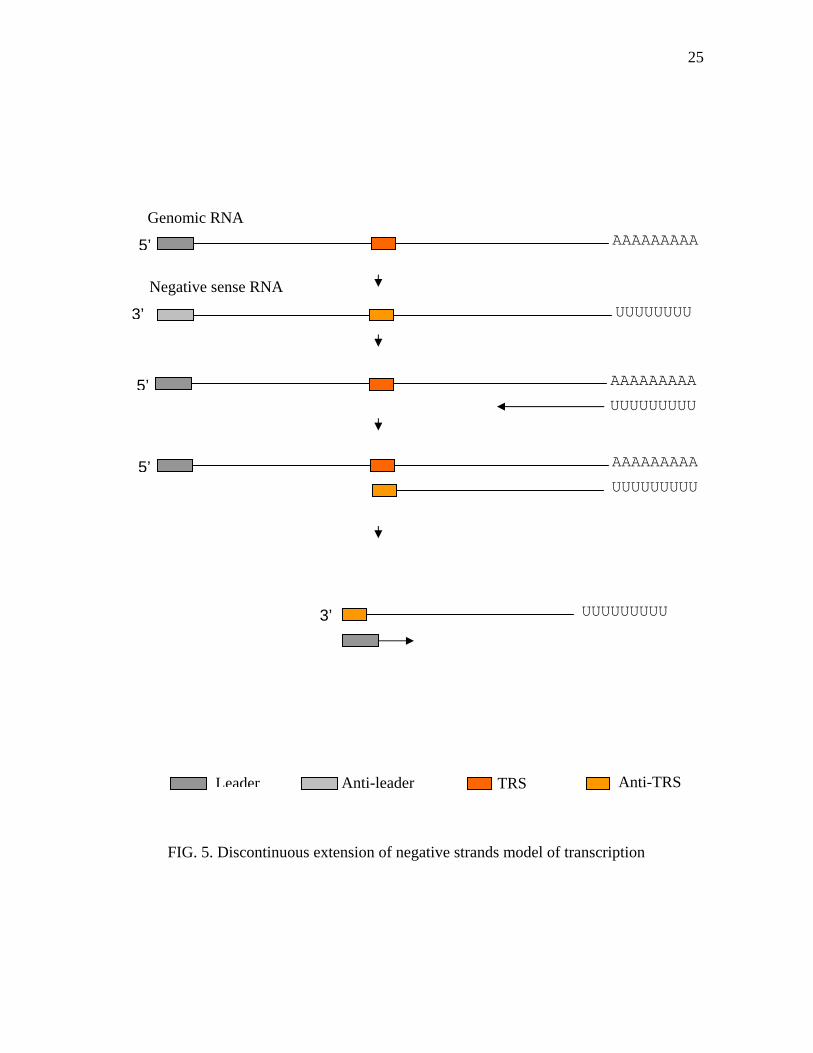
The second model is the discontinuous extension of negative strand transcription
(156). This model is illustrated in Figure 5. Discontinuous transcription could potentially
occur during negative-strand RNA synthesis. During negative strand RNA synthesis
from the full-length genomic RNA template, RdRp pauses at one of the IG or TRS
sequences and then jumps to the 3’ end of the leader sequence in the genomic RNA
template, generating a subgenomic negative strand RNA with an antisense leader
sequence at its 3’ end. This subgenomic negative strand RNA then serves as a template
for synthesis of mRNAs. Several studies have shown that both genomic and subgenomic
sized negative stranded RNAs are present even though positive strand RNA is present at
a higher amount (12, 155, 157, 162). All the negative strand RNAs were in double-
stranded form and no free negative stranded RNA was detected.
Coronavirus RNA synthesis takes place on membranous structures associated
with late endosomes. Colocalization of newly synthesized viral RNA and most of the
gene 1 products have been shown. N protein involvement in RNA transcription has been
suggested because antibody against N protein inhibits MHV RNA synthesis in vitro.
25
FIG. 5. Discontinuous extension of negative strands model of transcription
Negative sense RNA
Genomic RNA
3’
Anti-TRS TRS Anti-leaderLeader
5’ AAAAAAAAA
3’ UUUUUUUU
5’ AAAAAAAAA
UUUUUUUUU
5’ AAAAAAAAA
UUUUUUUUU
UUUUUUUUU
26
Recombination
One of unique features of coronaviruses is the high frequency of RNA
recombination (123). The high rate of recombination in coronaviruses has been
demonstrated not only experimentally in mixed infections, but also in infected animals
and in natural infection (82, 86). A calculation of recombination frequencies suggests
that the entire genome of MHV will have a recombination frequency of roughly 25% (9).
The recombination frequency is so high that no selection pressure was needed for the
isolation of recombinants (82, 123). The possible sites of recombination between
different MHV strains appear to be distributed over the entire genome, even though a
recombination hot spot was identified near a hypervariable region in MHV and IBV (8,
195). The high frequency of coronavirus recombination is probably caused by the
nonprocessive nature of the coronavirus RNA polymerase. Coronavirus replication of
the genome, except for the leader sequence, may proceed in a discontinuous and
nonprocessive manner (11). RNA synthesis pauses at sites of extensive secondary
structure of the RNA template and the incomplete RNA products dissociate from the
template because of a nonprocessive nature of the coronavirus RNA polymerase. These
dissociated RNA products then rebind to the RNA template to continue RNA synthesis
(96).
Viral Assembly
The genomic RNA synthesis and encapsidation are coupled in MHV-infected
cells. Because encapsidation requires N proteins, the accumulation of the N protein
27
could possibly tip the balance of RNA synthesis from transcription towards RNA
replication. The binding of the N protein to RNA has been shown in vitro for many
coronaviruses (10, 176). The encapsidation by N proteins is necessary, but not sufficient
for RNA packaging into mature virions. The N protein binding to viral RNA could be a
regulatory mechanism of RNA synthesis rather than a virion assembly process. Without
viral infection, coexpression of the M, E, and S proteins results in the assembly of
coronavirus-like particles that are released from cells (17, 193). The VLPs produced in
this manner form a homogeneous population that is morphologically indistinguishable
from normal virions. Thus, coronavirus virion assembly does not require the
participation of the nucleocapsid, defining a new mode of virion budding. In addition,
the reverse genetic approach was used to show that S protein is also dispensable in the
viral assembly, only the M and E proteins without RNP are required for VLP. It has been
suggested that in MHV, N protein interacts with viral genomic RNA producing helical
nucleocapsids and then the M protein and packaging signal sequence of genomic RNA
interact to confer specificity on packaging of the viral genomic RNA into the virion
(128).
S protein and M protein interactions are important for assembly to produce
infectious virus that can function in the virus and receptor interaction. Assembly of S
protein into the coronavirus envelope is governed by the M and S protein interaction. S
protein is expressed on the plasma membranes without M protein, but when it is co-
expressed with M protein, it is localized in the ER-Golgi intermediate compartment, also
known as cis-Golgi, where the virus obtains its lipid bilayer as it buds (83). The carboxyl-
28
terminal domain of the S, that includes the S transmembrane domain and an endodomain
of 64 residues, is sufficient to interact with the M protein and draw the mature S protein
into particles (66).
Coronaviruses are assembled at perinuclear membranes by budding into the ER-
Golgi intermediate compartment. From there, the particles are transported in vesicles
through the secretory pathway to the plasma membrane domain where they are released
by exocytosis (190). TGEV enters and is released apically (146). MHV-A59 enters
apically but is released both apically and basolaterally depending on the cell line (145,
147). HCoV-22E also infects the apical surface and is released apically (194). It is
believed that polarity of the viral release from the infected cell can affect tropism and
spread of the viruses whether it will cause systematic infection or local infection. It is
interesting that coronaviruses causing local infection, such as gastroenteritis and upper
respiratory disease, are released apically, but the MHV that causes more systemic
disease such as hepatitis and encephalitis is released mainly basolaterally.
INFECTIOUS cDNA CLONES OF CORONAVIRUS
Because of its large size limitation, and regions toxic to E. coli, until recently there
was no infectious cDNA clone of a coronavirus. Recently, several groups used different
approaches to overcome the size limitation of coronaviruses and have cloned the
approximately 30 kb entire genomes of several coronaviruses. The first breakthrough was
by Almazan et al. (2), using bacterial artificial chromosomes (BAC) to construct an
infectious cDNA clone of TGEV. Yount et al. (201) used a novel approach to produce an
29
infectious cDNA clone of TGEV. Instead of cloning the entire TGEV genome into a
cloning vector, they used in vitro assembly using unique restriction enzyme systems. The
type II restriction enzymes, such as BsmBI and SapI, recognize non-palindromic
sequences, and digest the DNA outside of the restriction enzyme recognition sequence
resulting in 3-4 nucleotide overhangs. These nucleotide overhangs are non-palindromic
and have unique sequence composition. Each DNA fragment digested with these
restriction enzymes can be religated with compatible overhangs produced by digestion
with the same enzyme. Subsequently, Thiel and Casais (31, 185) made infectious cDNA
clones of human coronavirus, HCoV 229E and IBV respectively using vaccinia virus as a
cloning vector. The entire coronavirus genomes were cloned into vaccinia virus. The
recombinant virus will infect cells and produce both coronavirus and vaccinia virus, but
because of their size difference, recombinant vaccinia virus was filtered out.
Each approach used to develop infectious cDNA clones of coronaviruses has
advantages and disadvantages. In addition to the large size of the genome being an obstacle
developing infectious clones, regions in the polymerase genes are very unstable in bacterial
plasmid vectors or BAC systems. These problems have been solved by adding an
additional intron into the BAC system or by breaking down the coronavirus genome into
smaller fragments for cloning into separate plasmids. These infectious cDNA clones have
been used to express heterologous genes, demonstrating their potential as expression
vectors or gene delivery systems (3, 169, 187). Using characteristics of coronaviruses,
which produce several subgenomic RNAs to express several different proteins, Thiel et al.
(187) has developed a multigene RNA vector with HcoV-229E. In this study, they
30
expressed three different proteins, chloramphenicol acetyltransferase (CAT), the firefly
luciferase (LUC) and the green fluorescent protein (GFP) genes by using independent
TRSs for each gene (187). This study shows the possibility of coronavirus infectious
cDNA clones as an alternative for gene delivery systems or expression vectors.
31
CHAPTER II
SYSTEMATIC ASSEMBLY OF A FULL-LENGTH INFECTIOUS cDNA OF A
BEAUDETTE STRAIN OF INFECTIOUS BRONCHITIS VIRUS (IBV)
INTRODUCTION
Infectious bronchitis virus (IBV) is one of the most economically important
diseases in the poultry industry (39). The mortality of the disease varies, depending on
the viral strain, but the acute and chronic effect of disease such as the retarded growth of
broilers and a decrease in egg production in layers causes tremendous losses in the
industry (6).
IBV belongs to the order of Nidovirales, genus Coronavirinae. The genomes of
these viruses are 5’ capped, 3’ polyadenylated, and are positive sense RNA (34, 161). As
with other positive strand RNA viruses, after infecting susceptible cells, the IBV genome
is used as an mRNA to express its replicase. IBV expresses its proteins using
subgenomic RNAs (sgRNA) and an internal ribosomal entry site (IRES) (95, 106). IBV
sgRNAs are structurally polycistronic, but functionally monocistronic with the exception
of RNA 3 and RNA 5. RNA 3 is a tricistronic mRNA which expresses 3a, 3b, and 3c
proteins (103). Among these, 3c is a structural protein, the small envelope protein, E
(104, 203). RNA 5 expresses two proteins, 5a and 5b. Even though expression of these
proteins in infected cells has been shown (105), the function of these proteins have not
been addressed. Expression of E protein from RNA 3 uses an IRES, but the mechanism
32
of 3b and 5b expression is speculated as a leaky scanning mechanism but has not yet
been shown experimentally.
IBV can be classified into several different serotypes depending on the S1
protein of the virus (65). Since some vaccine strains of IBV cannot cross protect against
IBV strains with distinct serotypes, problems in controlling the disease are continuing to
arise in field situations. In addition, because of the innate nature of the polymerase gene
in RNA viruses, the IBV polymerase does not have proof reading ability. In addition,
frequent recombination between different strains of viruses causes the emergence of
naturally occurring new types of recombinant viruses in the field causing vaccine breaks
(86).
Molecular biology studies of many viruses, including both positive and negative
strand viruses, have been accelerated by a reverse genetic systems (23, 62, 141). Infectious
cDNA clones have been used to address mechanisms of viral replication, pathogenesis,
virus packaging and in the development of vaccines not only for single stranded genomic
RNA viruses, but also for segmented viruses, such as Influenza virus (72, 75, 131, 132).
Infectious cDNA clones of coronaviruses were difficult to generate because of their
large genome size and the instability of its genes in bacterial plasmids. However, several
groups have recently developed infectious cDNA clones of coronaviruses (2, 31, 201,
202). Several approaches were used to overcome the size limitation of the coronavirus
genome to successfully clone the entire genomes. One of the first breakthroughs was by
Almazan et al. (2) using bacterial artificial chromosomes (BACs) as a cloning vectors to
construct an infectious cDNA clone of TGEV. Yount et al. (201) used a unique approach
33
to produce an infectious cDNA clone of TGEV. Instead of cloning the entire TGEV
genome into cloning vectors, they used in vitro assembly of several cDNAs using a unique
type II restriction enzyme system that recognize non-palindromic sequences and digested
the DNA outside of the restriction enzyme recognition sequences. By using enzymes such
as BglI and BsmBI, each DNA fragment was religated with neighboring fragments that
had compatible sequence composition. Following this procedure, Thiel and Casais (31,
185) made infectious cDNA clones of human coronavirus, HcoV 229E and IBV
respectively, using vaccinia virus as a cloning vector. Yount et al. also developed a new
strategy to construct an MHV infectious cDNA clone (202). By introducing restriction
enzyme recognition sequences in MHV genomes by RT-PCR that can be used for in vitro
assembly without introducing nucleotide sequence changes, Yount et al. used the
characteristics of the restriction enzyme and coined the term "no see’m technology”. After
digestion of the amplicons with the restriction enzyme and in vitro ligation, the restriction
enzyme recognition sequences introduced by PCR disappear.
We used this in vitro assembly strategy to construct an infectious cDNA clone of
IBV using restriction enzymes BsmBI and SapI. Our aim was to develop a rapid process to
make IBV vaccines that can effectively control the outbreak of new IBV strains in the
field. To develop an effective vaccine against IBV, it is critical to know the biology of the
virus, especially with regards to the genes of the virus involved in viral attenuation and
pathogenicity. As mentioned before, to understand the molecular biology of RNA viruses,
the development of an infectious cDNA clone is very useful. In this study, we developed
34
an infectious cDNA clone of IBV using the cell adapted Beaudette strain which is easily
manipulated in a short time frame.
MATERIALS AND METHODS
Viruses and Cells
A Vero cell adapted Beaudette strain of IBV was obtained from American Type
Culture Collection (ATCC, Manassas, VA) and then plaque purified three times. The
virus was propagated in the African green monkey kidney Vero cell line, obtained from
the ViroMed Laboratory (Minnetonka, MN) and maintained in Dubecco’s modified
Eagle medium (DMEM) containing 5% fetal bovine serum (FBS) supplemented with
penicillin G (100 unit/ml) and streptomycin (100 ug/ml). The baby hamster kidney cell
line, BHK-21, was also obtained from Viromed and maintained in DMEM containing
10% FBS supplemented with antibiotics as described for the Vero cells.
RNA Preparation
Viral RNA was prepared from the supernatant of IBV infected Vero cells.
Briefly, Vero cells were infected with IBV at 0.1 multiplicity of infection (m.o.i) and
incubated in a CO2 incubator at 37°C until the cells showed more than 80%
cytopathogenic effect (CPE), such as syncytia formation. The cells showing extensive
CPE were frozen and thawed three times and then cell debris was pelleted by
35
centrifugation at 3,000 rpm using a tabletop centrifuge (CR 412; Jouan Inc. Winchester,
VA). The supernatants were then ultracentrifuged at 35,000 rpm for 2 hours at 4°C using
an L7-55 ultracentrifuge (Beckman, Palo Alto, CA) with an SW55Ti swinging bucket
rotor to pellet the virus. The viral pellet was re-suspended in small volumes of TEN (10
mM Tris-Cl, pH 8.0, 1 mM ethylendiaminotetraacetate (EDTA), pH 8.0 and 0.1 M
NaCl) buffer.
TRIZOL reagent (Invitrogen, Carlsbad, CA) was used to extract the RNA from
the re-suspended viral pellet following the manufacturer’s instruction. Briefly, 1 ml of
TRIZOL reagent was added into 200 ul of viral pellet suspension in TEN buffer and then
mixed thoroughly by inverting the microfuge tube several times. The mixture was
incubated for 5 minutes at room temperate and then 200 ul of chloroform was added
before mixing thoroughly by inversion. The mixture was then incubated at room
temperature for 3 minutes. To separate the aqueous and the organic phases, the mixture
was centrifuged at 12,000 rpm for 15 minutes at 4°C using a microcentrifuge (5417 R ;
Eppendorf, Hamburg, Germany). The upper (aqueous) phase was then transferred to a
new nuclease free microfuge tube and 0.5 volume of isopropanol was added to
precipitate the RNA. After 10 minutes incubation at room temperature, the RNA was
pelleted by centrifugation at 12,000 rpm for 15 minutes at 4°C. Before suspending the
RNA pellet in nuclease free-water, the RNA pellet was washed with 70% ethanol, and
briefly air-dried. The RNA solution was quantified using a SmartSpecTM 3000
spectrophotometer (BIO-RAD, Hercules, CA) and stored at –70°C until further use as a
template for RT-PCR or as a size marker for electrophoresis.
36
Reverse Transcription (RT) Reaction
The cloning strategy for a full-length infectious cDNA clone of IBV is illustrated
in Figure 6. This cloning strategy uses SapI or BsmBI restriction endonuclease to cleave
at specific sequences (Figure 7) leaving highly variable three or four nucleotide
overhangs that do not randomly self-ligate, but do ligate with the fragments containing
the complementary nucleotide overhangs generated with the identical restriction
enzyme.
The first cDNA reaction was done using the SUPERSCRIPTTM First-Stranded
Synthesis System for RT-PCR (Invitrogen, Carlsbad, CA) according to the
manufacture’s directions. Briefly, 1-2 ug of RNA in 8 ul of nuclease free water with 1 ul
of 10 mM deoxynucleotide-triphosphate (dNTP) and 10 pmol of each reverse primer
P1R, P2R, P3R, P4R and GNR (Table 1) were incubated in a 70°C water bath for 5
minutes. Reactions were chilled on ice for 5 minutes before adding 2 ul of 10X first
strand buffer, 4 ul of 25 mM MgCl2, 2 ul of 0.1 M dithiothreitol (DTT), 1 ul of
recombinant RNase inhibitor (50 unit/ul, Invitrogen) and 1 ul of Superscript reverse
transcriptase (50 Unit/ul) and then incubated at 42°C for 50 minutes. The reaction was
terminated by incubating at 75°C for 15 minutes before degrading the RNA template
with 1 ul of E. coli RNase H (2 Units/ul) with 20 minutes incubation at 37°C.
37
1 3 5 7 9 11 13 15 17 19 21 23 25 27
21mer oligo(A)
P1
P2
P3
P4
P5
P2-1
P2-2
P3-1
P3-2
T7 promoter
P1 P2 P3 P4 P5
FIG. 6. Strategy for orderly assembly of an IBV infectious cDNA clone. Using RT-
PCR and unique oligonucleotide primer mutagenesis, seven clones spanning the
entire IBV genome were amplified using standard recombinant DNA techniques.
Between P2-1 and P2-2, P3-1 and P3-2, unique BsmBI restriction sites were inserted
at the 3’ and 5’ end of each junction. The approximate locations of each RT-PCR
products is shown. T7 RNA polymerase promoter was inserted in front of the 5’ end
of P1 by PCR mutagenesis. In the same way, twenty nucleotides poly (A) tail was
inserted at the 3’ end of P5.
5a and b
M ORF
ORF1a
ORF1bS ORF N
3 a,b, E
38
BsmBI restriction enzyme recognition sequence
5’...CGTCTC(N)1 ⇓...3’ 3’...GCAGAG(N)5 ⇑...5’
Sequence of P2 between P2-1 and P2-2 junction
GCTAGTGTTAAGAGTGTTGTCGCTAGCTATAAGACCGTGTTATGTAAGGT CGATCACAATTCTCACAACAGCGATCGATATTCTGGCACAATACATTCCA NHE R 5’ GCTAGTGTTAAGAGTGTT GTCGCGAGACG 3’ 3’ CGATCACAATTCTCACAACAGC GCTCTGC 5’
NHE f 5’ CGTCTCT GTCGCTAGCTATAAGACCGTGTTA 3’ GCAGAGACAGC GATCGATATTCTGGCACAAT
5’ GCTAGTGTTAAGAGTGTT GTCGCTAGCTATAAGACCGTGTTATGT 3’ CGATCACAATTCTCACAACAGC GATCGATATTCTGGCACAATACA
FIG. 7. No see’m technology using BsmBI restriction enzyme properties. Cloning and
assembly strategy of P2-1 and P2-2 is illustrated. BsmBI site was inserted using PCR
mutagenesis at the 3’ end of P2-1, 5’ end of P2-2. Cleavage with BsmBI restriction
enzyme will create a novel four nucleotides overhang that specifically anneal with the
complementary four nucleotides overhang generated by an identical BsmBI. Upon
religation, BsmBI is deleted from the clone and reforms original sequence connecting
two pieces intact.
39
TABLE 1. Primer pairs used for cloning of the individual IBV amplicons
Primer Nucleotide Sequence Genome location
T7P1F 5’-TAATACGACTCACTATAGGACTTAAGATAGATATTAATATA-3’ 1-22
P1R 5’-CCTTTCCAGAAGAGCAAATCTCC-3’ 2254-2276
P1F 5’-GGAGATTTGCTCTTCTGGAAAGG-3 2254-2276
P2F 5’-GATCTTGCTGCGAAGAGCACTTTTG-3’ 8611-8635
NHER 5’-CGTCTCGCGACAACACTCTTAACACTAGC-3’ 5731-5753
NHEF 5’-CGTCTCTGTCGCTAGCTATAAGACCGTGTT-3’ 5748-5771
P2R 5’-CAAAAGTGCTCTTCGCAGCAAGATC-3’ 8611-8635
P3F 5’-GTAGTGATTCATTGAGACGTTTTG-3’ 15508-15531
SACR 5’-CGTCTCGGGATCTACTGCAAATGAACATAG-3’ 11926-11949
SACF 5’-CGTCTCGATCCCCGCGGACACATATTGTAAATATG-3’ 11944-11971
P3R 5’-CAAAACGTCTCAATGAATCACTAC-3’ 15508-11949
P4F 5’-CAGGCTCTTCATCAGGGTGTACTG-3’ 20543-20566
P4R 5’-CAGTACACCCTGATGAAGAGCCTG-3’ 20543-20566
GNR 5’-GCTCTAACTCTATACTAGCC-3’ 27593-27613
GNRT 5’-TTTTTTTTTTTTTTTTTTTGCTCTAACTCTATACTAGCC-3’ 27593-27613
Numbers followed the sequence data obtained from Vero cell adapted Beaudette sequence from our laboratory. Bold letter is T7 RNA polymerase promoter seq. Underlined letter is BsmBI restriction enzyme recognition sequence.
40
Polymerase Chain Reaction (PCR) for Amplification of Target Sequences
Two ul of each completed cDNA reaction was used to amplify the target DNA
by PCR. PCR was done using the Expand Long Template PCR system (Roche,
Indianapolis, IN) following the manufacturer’s directions using a GeneAmp PCR system
2400 thermocycler (PerkinElmer, Boston, MA). Sequences of the primer pairs used for
RT-PCR reaction are listed in Table 1.
cDNA synthesized with P1R primer was used as a template for P1 PCR
amplification. A 50 ul reaction included 5 ul of 10X PCR buffer number 3, containing
22.5 mM MgCl2, 2 ul of 10 mM dNTP, 1 ul of forward primer (T7P1F), 1 ul of reverse
primer (P1R), 2 ul of a cDNA reaction and 0.75 ul DNA polymerase enzyme mix.
Distilled waster was used to bring the total volume to 50 ul. Reaction conditions for the
P1 PCR included an initial denaturation at 94°C for 2 minutes, followed by 30 cycles of
denaturation at 94°C for 10 seconds, annealing at 55°C for 30 seconds and elongation at
68°C for 2 minutes. The final step was an elongation step at 68°C for 7 minutes.
P1F and NHER primers were used for P2-1 amplification. NHEF and P2R primers
were used for P2-2 amplification. P2F and SACR primers were used for P3-1
amplification. SACF and P3R primers were used for P3-2 amplification. Because P2-1,
P2-2, P3-1 and P3-2 have a similar target size of about 3.5 kb, the same reaction
condition was used for these amplifications.
The conditions for P2-1, P2-2, P3-1 and P3-2 amplification included an initial
denaturation at 94°C for 2 minute. First round PCR reaction conditions included 10
cycles at 94°C for 10 second, 58°C for 30 seconds and 68°C for 2 minutes 20 seconds.
41
Conditions for the second round of amplification were 20 cycles at 94°C for 10 seconds,
58°C for 30 seconds and 68°C for 2 minute 20 seconds, with a 20 second additional
elongation time in every cycle. A final incubation of 10 minutes at 68°C was included
for each reaction.
P3F and P4R primers were used for P4 amplification. Conditions for P4 were an
initial denaturation at 94°C for 2 minutes followed by 10 cycles at 94°C for 10 seconds,
58°C for 30 seconds and 68°C for 4 minutes 20 seconds and the second round
amplification conditions were 18 cycles at 94°C for 10 seconds, 58°C for 30 seconds and
68 °C for 4 minutes and 20 seconds, with 20 seconds additional elongation time added
each cycle. This was followed by 10 minutes incubation at 68°C for final elongation.
P4F and GNR or P4F and GNRT primers were used for P5 amplification. PCR
reactions included an initial denaturation at 94°C for 2 minutes. First round amplification
reactions included 10 cycles at 94°C for 10 seconds, 58°C for 30 seconds and 68°C for 6
minutes 20 seconds and second round amplification, 18 cycles at 94°C for 10 seconds,
58°C for 30 seconds and at 68°C for 6 minutes and 20 seconds, with 20 seconds
additional elongation time in every cycle. A final 10 minute 68°C elongation completed
the amplification.
Cloning of Amplicons
Following amplification, 2 ul of each PCR product was electrophoresed in a
0.7% agarose gel, containing Tris-acetate-EDTA (TAE) buffer and ethidium bromide
42
(EtBr). The electrophoresis results were recorded using a FluoChem TM gel analysis
system (Alpha Innotech Corporation, San Leandro, CA). Each PCR product with the
correct size was purified from a 0.7% agarose gel containing 100 ul of crystal violet (2
mg/ml) per 100 ml of agarose solution by electrophoresis. DNA bands were excised
from the agarose gel and then purified using a QIAquick Gel Extraction Kit (QIAGEN
Inc., Valencia, CA) following the manufacture’s directions. The concentration of gel
purified PCR products were measured with a SmartSpec 3000 spectrophotometer (BIO-
RAD). Each PCR product was ligated into a cloning vector. P1 was ligated into a
pSTBLUE vector (Novagen, Darmstadt, Germany) by incubating an equal molar ratio of
vector and insert at 16°C for 2 hours. P2-1 though P3-2 were ligated into a pSMART HC
vector (Lucigen, Middleton, WI) according to the manufacturer’s instructions. Briefly,
PCR products were treated with T4 DNA polymerase (Promega, Madison WI) filling in
any 3’ and 5’ end nucleotide overhangs. After T4 DNA polymerase treatment, PCR
products were treated with T4 Kinase (Promega) to phosphorylate the 5’ ends. The
products were further purified by phenol/chloroform extraction followed by ethanol
precipitation. Equal molar ratios of PCR product and pSMART-HC vector were ligated
for 2 hours at 16°C and then transformed into chemically competent NovaBlue
(Novagen, Darmstadt, Germany) cells. P4 and P5 PCR products were directly ligated
into pCR-XL-TOPO cloning vector (Invitrogen) for 5 minutes at room temperature and
transformed into chemically competent Oneshot cells (Invitrogen). Transformed bacteria
were plated on Luria-Bertani (LB) medium agar plates containing kanamycin (5 ug/ml)
and grown overnight at 37°C.
43
Cloning of a Probe
To prepare a probe for northern and Southern blot analysis that was specific for
the 3’ end of IBV, a region containing the entire N ORF through the 3’ UTR of IBV was
RT-PCR amplified and cloned into a pGEM-3Zf(-) vector (Promega, Madison, WI).
Briefly, the cDNA was prepared using the GNR primer and Superscript reverse
transcriptase (Invitrogen) as described above. Two ul of a cDNA reaction was used for
PCR. Taq polymerase (Promega) was used for PCR amplification following the
manufacture’s directions. 10 pM each of BNF (5’ ATG GCA AGC GGT AAA GCA GC
3’) and GNR (5’ GCT CTA ACT CTA TAC TAG CC 3’) oligos were added as the
forward and reverse primer, respectively. The direction of N ORF cloned into pGEM-
3Zf (-) vectors was determined by restriction enzyme digestion using BamHI (Promega),
and confirmed by sequencing, using M13 forward and M13 reverse primers (Invitrogen).
Both reverse and forward directions of ORF N were cloned as pGEBN-1 and pGEBN-2,
respectively.
Nucleotide Sequencing
Each amplicon DNA was prepared from transformed E. coli cultures grown
overnight in LB medium, and DNA templates were prepared using a QIAGEN mini prep
kit (QIAGEN). PCR products purified with QIAquick PCR purification kit (QIAGEN,
Santa Clarita, CA) were used for direct PCR product sequencing. Sequencing was
performed using the ABI prism BigDye Terminator Cycle Sequencing Ready Reaction
Kit (Applied Biosystems, Foster City, CA). The sequencing reaction included 350-500
44
ng of plasmid DNAs or 300 ng of PCR products mixed with 2 ul of BigDye mixture
(ABI) and 10 pM of primers (synthesized by Integrated DNA Technologies, Inc,
Coralville, IA) and the reaction mixtures were adjusted to 6 ul with distilled water. The
conditions for the sequencing reactions included an initial denaturation at 96°C for 2
minutes followed by 30 cycles at 96°C for 30 seconds, 52°C for 15 seconds and 60°C for
4 minutes.
Three independent clones for each amplicon were isolated with plasmid mini-
preparations as described above and sequenced using a panel of primers located about
400 nts apart on the IBV genome. Unincorporated nucleotides and primers were
removed using micro Bio-spin chromatography columns (BIO-RAD, Hercules, CA) and
the sequencing reaction was completely dried using a Speed-Vac Concentrator (Thermo
Savant, Holbrook, NY). Completed sequencing reactions were sent to a core facility
sequencing laboratory at the Department of Biology, Texas A&M University. Sequences
were read using an ABI automatic sequencer 3100 (ABI).
Analysis of Sequences
Sequence data were analyzed using SDS Biology Workbench
(http://workbench.sdsc.edu) (San Diego Supercomputer Center, University of California,
San Diego, CA). For DNA and amino acid sequence comparisons, the LALIGN program
was used. For cloning purposes, the TACG program was used to search for restriction
enzyme digestion profiles. SIXFRAME was used to convert DNA sequences into amino
acid sequences.
45
Assembly of a Full-length IBV Infectious Construct
Insert DNAs from each amplicon were digested with the appropriate restriction
enzymes. P1 was digested with XhoI (Promega, Madison, WI) and then treated with calf
intestine phosphatase (CIP) (Promega) to inhibit self-ligation. CIP treated DNA was
phenol:chloroform extracted, ethanol precipitated and then digested with restriction
enzyme SapI (New England Biolabs Inc., Beverly, MA). P2–1, P2-2 and P3-1 amplicons
were double digested with restriction enzymes SapI and BsmBI. When the buffer
conditions were not compatible, after one restriction enzyme digestion, the reactions
were ethanol precipitated and then the DNA was reconstituted with the appropriate
buffer and in the appropriate volume for the second enzyme digestion. Except for the
BsmBI restriction enzyme digestion that needed to be incubated at 55°C, the restriction
enzymes were incubated at 37°C. P3-2 and P4 replicons were single digested with
restriction enzyme BsmBI. The P5 amplicon was digested with restriction enzyme
EcoRI (Promega) then CIP treated and digested with BsmBI. Each DNA insert was run
on a TAE agarose gel (0.8 to 1%) containing 2.4 ug/ml crystal violet to prevent UV-
induced DNA damage that could impact subsequent manipulations of the DNA
fragment. Target size inserts were excised from the agarose gel and the DNA inserts
were extracted using a QIAquick Gel Extraction kit (QIAGEN Inc, Valencia, CA)
following the manufacturer’s instructions.
A high concentration of T4 DNA ligase (20 U/ul) was used to assemble
infectious cDNA of IBV. Ligation reactions were incubated at 16°C overnight before
inactivating the ligation reaction mixes by incubating at 65°C for 10 minutes. Aliquots
46
of the ligation reactions were electrophoresed on an agarose gel to confirm the ligation
reaction and size of the ligation products. The correct sizes of larger ligation fragments
were purified from agarose gel and then ligated step wise with other fragments. The final
ligation reaction of P1 through P3-1 and P3-2 though P5 were pooled without further
purification and then ligated overnight.
Northern Blot Analysis
Preparation of a random primed isotope labeled probe.
To prepare a random primed probe against the 3’ end of IBV, pGEBN-2 was
used as a template. The template DNA was digested with PstI restriction enzyme and
then phenol:chloroform extracted followed by ethanol precipitation to remove
contaminating RNase. One ug of template was used for an in vitro transcription and
labeling reaction using T7 RNA polymerase (Promega). Briefly, 4 ul of 5X transcription
buffer, 2 ul of 100 mM DTT, 1 ul of RNase inhibitor (Promega, 30 unit/ul), 4 ul of rNTP
mix containing 2.5 mM rCTP, rATP, and rGTP, 1.2 ul of 200 uM rUTP, 5 ul of [α-32P]
rUTP (Perkin-Elmer), 1.3 ul of T7 RNA polymerase (20 unit/ul) were used in a 20 ul
reaction. The reaction was incubated at 37°C for one hour and then ethanol precipitated.
The probe was resuspended in deionized formamide and stored at –20°C. Activity of the
probe was measured with a liquid scintillation system (Beckman Coulter Inc, Fullerton,
CA) before using for hybridization reactions.
47
RNA transfer
To transfer RNA from an agarose gel to a nylon membrane for northern blot
hybridization, 1 ul of each in vitro transcribed RNA was electrophoresed in a denaturing
agarose gel containing formaldehyde following standard method (153) with
modifications. RNA was denatured with sample buffer containing 1/10 v/v
formaldehyde gel–running buffer (0.2 M 3-(N-norpholino) propanesulfonic acid
(MOPS) (pH 7.0), 80 mM sodium acetate and 10 mM EDTA (pH 8.0)) with 1/5 v/v of
formaldehyde, 1/2 v/v of deionized formamide, 2 ug of EtBr, and 1/10 v/v of
formaldehyde gel-loading buffer (50% glycerol, 0.25% bromophenol blue, 0.25% xylene
cyanol FF) per RNA sample containing total volume of 15 ul of reaction volume. RNA
and sample buffer were mixed, denatured at 70°C for 5 minutes and chilled on ice. A 1%
denaturing agarose gel containing formaldehyde was prepared with a 1/10 volume of
10X formaldehyde gel running buffer and formaldehyde to give final concentration of
2.2 M formaldehyde. Each RNA sample was loaded on the agarose gel and run at 90 V
in 1X formaldehyde gel-running buffer. After every one hour, the buffer from each
reservoir was mixed and returned to the gel apparatus for further electrophoresis. When
the xylene cyanol dye front was at the bottom of the gel, electrophoresis was terminated.
The gel was washed several times with distilled water and then soaked in 0.05 N NaOH
for 20 minutes and exposed to long wavelength UV light for 60 seconds. The gel was
then washed with distilled water and soaked for 20 minutes in 10 X sodium chloride
sodium citrate (SSC) buffer. RNA from the gel was transferred to nylon membrane by
capillary transfer in 10 X SSC overnight. RNA transfer was confirmed by EtBr staining
48
of the nylon membrane and molecular weight markers were indicated with pencil. The
membrane was then fixed by UV cross-linking for 30 seconds using GS gene linker UV
chamber (BIO-RAD).
Hybridization reaction
The membrane containing transferred RNA was hybridized with a probe against
the 3’ end of the IBV gene with specific activity of 2 x 10⁷ cpm/ug that was labeled with
α-32P by random priming. Hybridization reactions were done using an autoblot
hybridization oven (Bellco Glass Inc., Vineland, NJ). Hybridization conditions included
incubation at 55°C for 12 hours in hybridization buffer containing 50% deionized
formaldehyde, 2 X Denhardt’s reagent, 5 X SSC and 0.1% SDS. After hybridization,
excess probe was removed by washing with 1 X SSC, 0.1% SDS, followed by three
washes for 20 minutes each at 68°C in 0.2 X SSC and 0.1% SDS. After hybridization,
the nylon membrane was exposed to an image plate (IP) for 20 minutes to one hour,
depending on signal intensity. The image was analyzed using a BAS-1800 II
phosphoimage reader (Fusifilm Electronic Imaging Ltd. Tokyo, Japan) and analyzed
using the Image Gauge V3.12 and L processor V1.72 program.
In vitro Transcription and Electroporation
Capped runoff T7 transcripts were synthesized in vitro from in vitro ligated IBV
cDNA template using an mMESSAGE mMACHINE TM T7 Ultra kit (Ambion, Austin,
49
TX) as described by the manufacturer with some modification. 20 ul of IBV RNA
transcription reaction mixture was prepared containing 10 ul of 2 X NTP mixture, 3 ul of
30 mM GTP, 2 ul of 10 X transcription buffer, 2 ul of enzyme mixture and remainder
was filled with nuclease free water and incubated at 37C° for 2 hours. Template DNA
was removed by incubating the transcription reaction with 1 ul of DNase I (2 units/ul)
for 20 minutes at 37 C°
To increase the efficiency of virus rescue after transfection, N transcript was also
prepared and co-transfected with the IBV transcript. pGEMN-1 was used as a template
for N transcripts. The amplicon was digested with a PstI restriction enzyme (Promega) to
linearize the template and then phenol:chloroform extracted followed by ethanol
precipitation to remove any RNase contamination. The DNA was resuspended with
diethyl pyrocarbonate (DEPC) treated distilled water. 1 ug of the template was used for
an in vitro transcription reaction using mMESSAGE mMACHINE TM T7 Ultra kit
(Ambion, Austin, TX.). A poly (A) tail was added to the transcript with E. coli Poly(A)
polymerase. Transcripts were precipitated with 7.5 M lithium chloride containing 50
mM EDTA before electroporation and transcripts were used with or without
precipitation.
BHK-21 cells were grown to about 70% subconfluence before electroporation.
BHK-21 cells were treated with trypsin to detach them from flasks. The cells were
washed twice with ice cold DEPC treated PBS and then resuspended at 2 x 10⁷ cells per
ml in the cold PBS. One third of the transcription reaction using IBV template and one
fifth of N transcript were mixed with 400 ul of BHK-21 cell suspension in a 1.5 ml
50
microcentrifuge tube. The cells and RNA mixture were immediately transferred to 2 mm
gap electrophoration cuvettes (electroporation cuvettes plus model No. 620, BTX,
Genetronics Inc., San Diego, California) and then elecrophorated at 850 V, 25 mΩ and
at resistance 720 Ω using an electro-cell manipulator 600 (BTX, Genetronics, Inc. San
Diego, California) with three consecutive pulses. After electroporation, cells were
incubated at room temperature for 10 minutes and then the transfectant was diluted 1 to
20 in complete DMEM containing 10% FBS before resuspending with 1 x 10⁶ Vero cells
per 100 mm cell culture petridish.
Plaque Staining and Plaque Purification
Virus was serially diluted 10 fold with DMEM without FBS. Then, 200 ul of
virus dilution was inoculated per well of six well cell culture plates that had been seeded
with monolayers of Vero cells. After one hour incubation, residual virus was removed by
washing the cells twice with PBS before overlaying the cells with 0.8% agarose
containing 1X DMEM, 1% FBS and antibiotics. The cells were incubated until plaques
were observed. For plaque staining, the agarose overlay was removed using a spatula,
the cell monolayers were air dried briefly and overlaid with 2 ml of crystal violet
staining solution per well. After 10 minutes, excessive crystal violet was removed and
the cells were washed several times with PBS to remove residual crystal violet. For
plaque purification, the agarose over plaques was picked with a 1000 lambda
micropipette and suspended in 1 ml DMEM without FBS. The plaque suspension was
51
incubated at 4°C overnight and then the viral suspensions were inoculated onto Vero cell
in 6 well plates as described before.
Kinetics of Viral Replication
Vero cells were infected with wild type IBV Beaudette strain and IBV Beaudette
strain derived from an infectious cDNA clone and plaque purified three times. The Vero
cells were seeded in 35 mm petridishes at a concentration of 106 per dish. The cells were
infected with three plaques forming units per cell for an hour and then the media were
replaced with DMEM containing 1% FBS with antibiotics, and 5, 10, 12, 19, and 21
hours after infection, the cells were harvested and stored at –70°C for subsequent plaque
assays.
RESULTS
RT-PCR and Cloning of Amplicons
Fragments covering the entire genome of IBV Beaudette were amplified by RT-
PCR. Each fragment overlapped by about 10 nucleotides. The RT-PCR products were
cloned into cloning vectors and labeled as P1, P2, P3, P4 and P5 and their sizes are 2.2
kb, 6.3 kb, 6.8 kb, 5 kb and 7.2 kb, respectively, as shown in Figure 8. Some parts of the
coronavirus genome have been reported to be toxic to bacterial host cells when they
were transformed into bacterial host cells. This has also been reported during the process
52
3 kb
5 kb
7 kb
1.5 kb
M 1 2 3 4 5
FIG. 8. RT-PCR amplification of the entire IBV genome. The entire genome of IBV
was RT-PCR amplified into five pieces. The RT-PCR products were separated in a
0.8% agarose gel and stained with ethidium bromide. M, represents the 1 kb DNA
ladder; Lane 1, RT-PCR product from nt 1 to 2269; Lane 2, RT-PCR product from
nt 2269 to 8635; Lane 3, RT-PCR product from nt 8625 to 15520; Lane 4, RT-PCR
product from nt 15520 to 20550; Lane 5, RT-PCR product from nt 20550 to 17613.
53
of construction of the infectious cDNA clones of TGEV, MHV and IBV (31, 201, 202).
In this lab, several efforts were made to clone the large RT-PCR products of P2 and P3
into the pCR-XL-TOPO TA vector (Invitrogen) but without success. Since regions
within these sequences of IBV were also reported to be toxic to E. coli (31), P2 and P3
were each divided into 2 smaller fragments. Because there was no SapI or BsmBI
restriction enzyme recognition sequence inside these regions of the IBV genome, I
decided to use the “no see’m technology” developed by Dr. Baric’s laboratory (202) to
insert the BsmBI restriction enzyme recognition sequences. The NHER, NHEF, SACR,
and SACF primers were designed to hold a BsmBI restriction enzyme recognition
sequence at the 5’ or 3’ end of each primer as shown in Table 1. In addition, to further
overcome the toxicity of the IBV gene products in bacterial hosts after transformation,
we used pSMARTHC-Kan vector (Lucigen) that was developed to avoid transcription of
plasmid genes in bacterial hosts. The overall strategy used to develop an infectious
cDNA clone of IBV is illustrated in Figure 6 and 7. After breaking down the P2 and P3
each into two fragments, these PCR products were successfully cloned into the
pSMARTHC-Kan vector.
Compared to MHV amplicons that are known to be relatively unstable (personal
communication, J. Leibowitz), IBV amplicons are considerably stable. After two or three
rounds of 200 ml midi-culture preparations, and subsequent subcloning procedures, no
mutations were found in P2, 3, and P5 amplicons. At nt 6385, nt 11137 and nt 13387,
there were mutations that affected the amino acid sequence. At nt 6385, and nt 11137, a
T to C transition resulted in a tyrosine to histidine substitution. At nt 13387, an A to G
M 1 2 3 4 5M 1 2 3 4 5 6 7 8
54
transition resulted in a glutamic acid to glutamine substitution. These mutations were
corrected by replacing the region containing the mutation with the same region in other
clones having correct sequences.
At nt 25793 in ORF 5b, there was one nucleotide change from A to G resulting in
an amino acid sequence change from glutamine to arginine. Since the function of the 5b
is not known, this mutation was not changed to the wild type sequence and served as a
marker for the infectious cDNA clone.
Sequencing Analysis
Each amplicon was sequenced from at least three independent clones to exclude
inherent enzymatic errors that can occur from RT and PCR reactions. Several differences
were identified in every amplicon. These aberrations were resolved by direct PCR
product sequencing. Some of the sequence discrepancies were not corrected if the
sequence differences did not result in a change in the amino acid sequence. If the
nucleotide change resulted in amino acid sequence changes, these mutations were
corrected using other amplicons that had the correct sequence. To replace these changes
with the correct sequences, inserts from independent clones were swapped using unique
restriction enzyme with correct amplicons. Some regions of the genome do not encode
proteins but have important secondary structures. This is especially true for the 3’UTR
region within P5. When there were sequence differences between independent clones for
the same region, the sequence was corrected according to the direct RT-PCR
sequencing.
55
Except for the sequence discrepancies between clones for the same amplicons, there
were 76 genuine sequence differences from the published sequence data (22, 31).
Sequence differences between the IBV strain used in this study, which has been
passaged and adapted in Vero cells and the sequence published in 1987 (22) are
summarized in Table 2. These changes occurred in the entire genome except for the
RdRp region which was totally conserved indicating that it is critical for viral survival.
An interesting observation in our study shows a 5 nts insertion in the intergenic region of
ORF 4 and ORF 5. At this time, the relevance of the 5 nts insertion in the virus is
unknown. Thus, the Beaudette strain of IBV in our study is 27613 nts instead of 27608
nts.
Systemic Assembly of Infectious cDNA by In vitro Ligation Reaction
To assemble a full-length cDNA of IBV, each amplicon was digested with
appropriate restriction enzyme and insert DNAs from amplicons were purified by
electrophoresis and gel purified as described in materials and methods (Figure 9).
Initially, ligation of all the fragments in a single reaction was attempted because
each fragment, P1 through P5, has a different sequence overhang at the 3’ and 5’ ends
after digestion with SapI or BsmBI. Theoretically, each fragment cannot be randomly
ligated to each other, but only with the neighboring sequences, that share compatible
ends. Therefore, it was hypothesized that it would be possible to connect each fragment
into an infectious clone in one ligation reaction. However, with the random ligation and
in vitro transcription reactions, only a shorter RNA transcript was obtained. To increase
56
TABLE 2. Summary of nucleotide and amino acid differences between IBV Beaudette used in this study and published sequence
Location Nucleotide change
Amino acid change Gene affected
1 32 C-T - 5’UTR
2 222 C-T -
3 282 G-A -
4 490 T-C -
5 841 C-T S-P pp1a
6 1374 G-A -
7 1845 T-C -
8 2000 C-T A-V
9 2015 A-G C-G
10 2439 A-T G-E
11 2651 C-T T-M
12 3169 A-T -
13 3210 A-G -
14 3283 A-G K-E
15 3322 C-A L-I
16 3428 C-A A-D
17 3926 T-C L-S
18 4481 C-A T-N
19 4791 A-G -
57
TABLE 2 continued
Location Nucleotide change Amino acid change Genes
20 5456 T-C L-H
21 5785 C-T L-F
22 5828 C-T P-L
23 6231 G-A
24 7293 A-C
25 7755 A-G
26 9372 A-G
27 10754 T-C V-A
28 10781 T-C I-T
29 11780 T-A V-D
30 12136 A-G S-G
31 13106 C-T pp1b
32 13178 A-C Q-H
33 13646 A-G
34 15302 A-G
35 15594 C-A
36 16286 C-T
37 16718 A-G
38 17546 T-C
58
TABLE 2. Continued
Location Nucleotide change
Amino acid change Genes
39 18195 T-C Y-H
40 20731 T-A L-I S
41 20755 C-T L-F
42 21149 A-C N-T
43 21357 A-T K-N
44 21457 A-G
45 21459 A-T
46 21628 A-C N-H
47 21711 G-A
48 22347 T-C
49 22433 A-G K-R
50 22415 A-C N-T
51 22441 C-G L-V
52 22492 A-G
53 22535 T-C F-L
54 22590 C-T
55 23402 G-T S-I
56 23724 T-A
57 24338 T-C
59
TABLE 2. Continued
Location Nucleotide change
Amino acid change Genes
58 24508 C-T P-S
59 24661 A-G I-V
60 24716 C-T T-I
61 25342 G-T
62 25426 G-A
63 25458 T-C
64 25465 C-T
65 25793 A-G Q-R
66 25901 A-G
67 25904 A-G
68 25928 T-C
69 26054 C-T N
70 26208 G-T D-Y
71 26403 G-T A-S
72 26501 G-T Q-H
73 27048 T-G Y-D
74 27051 G-T D-Y
75 27069 A-G N-D
76 27466 A-C
60
7 kb
5 kb
3 kb
1.5 kb
FIG. 9. Preparation of insert DNAs from each amplicon. Each amplicon was
digested with appropriate restriction enzymes and inserts were separated from the
vectors using agarose gel electrophoresis and DNA gel purification. M, 1 kb DNA
ladder; lane 1, P1 insert prepared by XhoI digestion, CIP treated and then digested
with SapI restriction enzyme to cut the insert out from the vector; lane 2, P2-1
insert digested with BsmBI and SapI; lane 3, P2-2 insert prepared by digestion with
BsmBI and SapI restriction enzyme; lane 4, P3-1 prepared by BsmBI and SapI
digestion; lane 5, P3-2 prepared by BsmBI digestion; lane 6, P4 insert prepared by
BsmBI digestion; lane 7, P5 large fragment prepared by EcoRI digestion followed
by BsmBI digestion; lane 8, small fragment of P5.
M 1 2 3 4 5 6 7 8
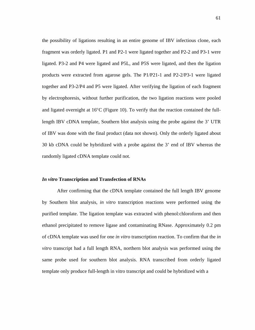
61
the possibility of ligations resulting in an entire genome of IBV infectious clone, each
fragment was orderly ligated. P1 and P2-1 were ligated together and P2-2 and P3-1 were
ligated. P3-2 and P4 were ligated and P5L, and P5S were ligated, and then the ligation
products were extracted from agarose gels. The P1/P21-1 and P2-2/P3-1 were ligated
together and P3-2/P4 and P5 were ligated. After verifying the ligation of each fragment
by electrophoresis, without further purification, the two ligation reactions were pooled
and ligated overnight at 16°C (Figure 10). To verify that the reaction contained the full-
length IBV cDNA template, Southern blot analysis using the probe against the 3’ UTR
of IBV was done with the final product (data not shown). Only the orderly ligated about
30 kb cDNA could be hybridized with a probe against the 3’ end of IBV whereas the
randomly ligated cDNA template could not.
In vitro Transcription and Transfection of RNAs
After confirming that the cDNA template contained the full length IBV genome
by Southern blot analysis, in vitro transcription reactions were performed using the
purified template. The ligation template was extracted with phenol:chloroform and then
ethanol precipitated to remove ligase and contaminating RNase. Approximately 0.2 pm
of cDNA template was used for one in vitro transcription reaction. To confirm that the in
vitro transcript had a full length RNA, northern blot analysis was performed using the
same probe used for southern blot analysis. RNA transcribed from orderly ligated
template only produce full-length in vitro transcript and could be hybridized with a
62
20, 40 kb
15 kb
5 kb
M 1 2 3 4 5 6 7
FIG 10. Systemic assembly of IBV full–length cDNA. Each amplicon was
digested with the appropriate enzymes and inserts were purified from agarose gels.
The inserts were orderly ligated and then larger ligated fragment were purified
from agarose gels before ligated to neighboring fragments. M represents 1 kb
DNA extension ladder, Lane 1 ligation reaction P1 with P2-1, lane 2, ligation of
P2-2 and P3-1, lane 3, P4, purified ligation product P3-2 with P4, lane 4, purified
ligation product P5, lane 5, ligation products from P1 to P3-1., lane 6, ligation
products P3-2 through P5, lane 7, final ligation product P1 through P5.
63
probe against 3’UTR of IBV (Figure 11). Because it has been shown that the N
transcript facilitates coronavirus transcription and increases the efficiency of virus rescue
after viral RNA transfection (56, 184, 201), we co-transfected IBV N ORF transcripts
with IBV transcript. Because we created clones of the N protein without a poly(A) tail,
poly(A) was added using E. coli Poly (A)-polymerase after in vitro transcription. In vitro
transcripts of IBV cDNA and the N ORF were elecrophorated into BHK 21 cells. Before
using in vitro transcribed RNA, electroporation conditions were optimized using total
cellular RNA extracted from IBV infected Vero cells. The transfection efficiency was
higher when more than 2 ug of RNA and lower voltage was used with consecutive
pulses rather than high voltage with a single pulse. Because BHK 21 cells are not
susceptible to IBV infection, Vero cells were overlaid with electroporated BHK 21 cells.
After 3 days transfection, the cells were overgrown and there was no evidence of CPE
except apparent ballooning of some Vero cells. Subsequently, the cells were frozen and
thawed three times and then passaged into new Vero cells. Cytopathogenic effect (CPE)
was observed in the cells infected with supernatant from the cells transfected with IBV
viral RNA 2 days after infection. CPE was noticed in the cells infected with supernatant
from the cells transfected with in vitro transcribed RNA 3 days after infection. Both wild
type IBV and molecularly cloned IBV showed the same syncytia morphologies.
To verify whether the virus was derived from the infectious cDNA clone or was
just contamination, we did direct PCR product sequencing. The infectious cDNA clone
has one nucleotide sequence difference from the wild type Beaudette strain in ORF 5b.
Both the positive control, transfected with viral RNA, and a sample that is transfected
1 2 3
64
RNA 3
RNA 2
RNA 1
FIG. 11. In vitro transcribed RNA from in vitro assembled cDNA template
encompassing the entire genome of IBV. (A) In vitro transcribed products and control
RNA, total cellular RNA from IBV infected Vero cells were electrophoresed on
denaturing agarose gel. (B) Northern blot of the RNA transferred to nylon membrane
and then hybridized with a probe against 3’ UTR of IBV. Lane 1 represents
intracellular RNA from IBV infected Vero cells; lane 2, in vitro transcription reaction
using orderly assembled in vitro ligation reaction, lane 3, in vitro transcription reaction
A B
1 2 3 1 2 3
65
with in vitro transcribed cDNA’s total cellular RNAs were extracted and used for RT-
PCR. ORF5a region containing the mutation was RT-PCR amplified then the PCR
products were used for direct PCR sequencing. As shown in Figure 12, wild type IBV
and IBV rescued from the infectious cDNA clone showed sequence difference in the
marker mutation.
Gross Plaque Morphology
To examine whether the virus from the infectious cDNA clone has the same
properties as the wild type virus, we decided to look at plaque morphology. The virus
derived form the infectious cDNA clone showed a higher titer than the control wild type
virus prepared in the similar way. The gross morphology of each virus plaque, however,
is similar (Figure 13).
Kinetics of Viral Replication
Vero cells were infected with wild type IBV Beaudette and molecularly cloned
virus from the wild type IBV to compare its growth kinetics. As shown in Figure 14,
wild type virus and cloned virus showed similar growth kinetics.
66
B
FIG. 12. Sequence comparison of (A) molecularly cloned IBV with (B) the wild type
IBV Beaudette strain. One nucleotide change was made as a marker for the molecular
clone of IBV in ORF5b changing the codon from CAA to CGA which confer the
amino acid change from Gln to Arg. This mutation was confirmed by RT-PCR and
direct PCR product sequencing.
A
67
FIG. 13. Molecularly cloned virus plaque morphology. Cultures of BHK cells were
electroporated with IBV full-length transcripts or total cellular RNA extracted from
IBV infected Vero cells as a control and seeded with Vero cells in 100 mm cell culture
dishes. Virus progeny was three time plaque purified and virus stocks were prepared
in Vero cells. (A) plaque morphologies of molecularly cloned IBV, (B) wild type IBV
virus plaque morphologies.
A B
68
1
10
100
1000
10000
100000
1000000
4 8 12 16 20 24time
viru
s tit
er (P
FU)
wildcDNA
FIG. 14. Growth kinetic comparison of wild type IBV and molecularly cloned IBV.
Wild type IBV and molecularly cloned IBV were infected to Vero cell with 3 PFU per
cell and the cells were harvested in 4,8,12,16,20 and 24 hours after infection.

69
DISCUSSION
Toxicity and instability of the viral genes in bacterial plasmids and lack of the
molecular biological techniques to clone large RNA genes into cloning vectors has been
an obstacle for the development of the molecular clones of RNA viruses, especially for
coronaviruses. However, the development of molecular biological techniques, such as
RT-PCR, and further development of cloning vectors, such as bacterial artificial
chromosome, have helped to solve these problems. Many properties of RNA viruses
have been extensively studied using molecular clones of viruses. These studies include
not only positive strand RNA viruses but also negative stranded RNA viruses and
segmented RNA viruses (139).
Infectious cDNA clones of positive strand RNA and negative strand RNA viruses
are useful tools for studying the functions of viral genes in viral replication and
pathogenesis (139). Infectious cDNA clones can be especially helpful for the study of
non-structural protein function in viral replication and assembly. In addition, infectious
cDNA clones of RNA viruses can be used as vaccines against the virus from which it
was derived or for a foreign gene(s) of another pathogen to induce humoral or cellular
immunity against recombinant proteins. Viruses are themselves the most natural gene
transfer vehicles.
Because of their large genomic size as compared to smaller RNA viruses, there
was no coronavirus infectious cDNA clones. In the last few years, infectious clones of
coronaviruses such as MHV, TGEV, HCoV and IBV have been generated. IBV
infectious cDNA clones have been constructed using vaccinia virus as a cloning vector
70
(31). However, the vaccinia system is time consuming because of its selection process
for the recombinant vaccinia virus containing infectious cDNA clones of coronaviruses.
It is also cumbersome to produce mutations in coronavirus genes inside of vaccinia
virus. Casai et al. mentioned that using the in vitro assembly technique with certain
modifications was not successful in rescuing infectious cDNA clones of IBV in their
study. However, in our study we showed that construction of an infectious cDNA clone
of IBV using an in vitro assembly strategy is possible. We hypothesize that these
differences are a matter of technique. The only technical difference is the use of unique
restriction enzymes to inhibit self-ligation of each fragment instead of treating DNA
fragments with phosphatase. Another factor may include protecting DNA from UV
damage during the cloning process.
Furthermore, our study found several sequence differences between viruses even
though a Vero cell adapted Beaudette strain of IBV was used by both ourselves and by
Casai et al. These sequencing results are summarized in Table 2. Interestingly, our lab
strain of IBV has five additional nucleotides compared to published sequence data (22)
and an insertion between IG of E and M ORF that increases the genome size to 27613
nts without affecting any ORF. Therefore, as long as the virus genome maintains the
TRS and start codon of the ORFs, certain small nucleotide changes in the viral genome
can be tolerated. This might indicate one mechanism this large RNA virus has evolved to
effectively survive notwithstanding its lack of polymerase proofreading ability.
Boursnell et al. (22) mentioned that IBV polymerase has a higher fidelity as
compared to vesicular stomatitis virus (VSV) that shows several sequence differences
71
between independent cDNA clones of same region. In our study, we found several
sequence differences between individual clones for the same regions amplified in the
same condition at the same time. These sequence differences could occur during reverse
transcription reactions or during PCR amplification. However, considering the
polymerase properties of RNA viruses, it cannot be excluded that IBV polymerase
produces quasi-species of genomic subgenomic RNA conferring viral evolution such as
seen in retroviral replication. The study of the properties of coronavirus polymerases
including fidelity and the discontinuous nature of the polymerase is an interesting area of
investigation. It is possible that such studies could lead to the discovery of the
mechanisms of evolution and survival when coping with a high rate of recombination
and a lack of proofreading ability during the replication of a relatively large RNA
genome.
In summary, an infectious cDNA clone of Vero cell adapted, attenuated
Beaudette strain of IBV was constructed using in vitro assembly and no see’m
technology that was previously used for the construction of TGEV and MHV. Compared
to MHV for which individual amplicons are not completely stable during passage in the
bacterial host, IBV amplicons are very stable after several passages and subsequent
manipulations. The infectious cDNA clone of the Beaudette strain maintained in our
laboratory is 5 nucleotides longer than the Beaudette strain of IBV originally sequenced.
The virus rescued from the infectious cDNA clone showed the undistinguishable
properties as parental wild type virus. As a marker, a single nucleotide change was made

72
in ORF5b region and the mutation was conserved in the virus rescued from the
infectious cDNA clone.
73
CHAPTER III
EXPRESSION OF HETEROLOGOUS PROTEIN, GREEN FLUORESCENT
PROTEIN USING INFECTIOUS cDNA CLONE OF INFECTIOUS
BRONCHITIS VIRUS (IBV)
INTRODUCTION
Infectious bronchitis virus (IBV) is a long recognized problem for the poultry
industry and has been identified as a causative agent of avian infectious bronchitis since
1937 (158). Problems controlling virus infection and disease include a high rate of
mutation and high recombination frequencies causing new variants that cannot be
protected with live vaccines. Therefore, it has been difficult to develop an efficient vaccine
against IBV. Even though IBV is one of the fist described coronaviruses, there is little
known about the molecular characteristics of the virus. In the past three years, however,
there has been enormous advancement in coronavirus research. Among the major
advancements is the development of several infectious cDNA clones of coronaviruses that
include IBV, MHV-A59, TGEV and human coronavirus (HCoV) 229E (31, 185, 201,
202). Three different approaches have been used to produce infectious cDNA clones of
coronaviruses.
The first approach uses bacterial artificial chromosomes (BAC) as a cloning vector
that is used to construct the infectious cDNA clone of TGEV (2). This approach overcomes
the cloning size limitation and stability of the genes in conventional cloning plasmid
74
vectors. However, the infectious cDNA in BAC is not stable in bacterial culture because
some parts of viral sequences are known to be toxic to bacteria. To overcome this problem,
an intron was inserted in the toxic region. Because cDNA transfected to the cells targets
the nucleus, the intron will be spliced out when processed in the nucleus and then
translocated into the cytoplasm (67). The cDNA has been used to express foreign proteins
and green fluorescent protein and shown to induce lactogenic immunity in sows Sola,
2003 #103). TGEV cDNA has also been engineered as a replication-competent,
propagation-deficient virus vector by deleting E protein from the construct.
The second approach toward the creation of infectious cDNA constructs uses
vaccinia virus as a cloning vector. By this method, IBV and HCoV infectious cDNA
clones have been made (31, 185). Using this method, an infectious cDNA clone of IBV
was constructed using vaccinia virus to show that S protein can confer host tropism by
switching the S protein with other strains of IBV that have different cell tropism (30).
The third approach uses an in vitro assembly technique by which the entire genome
of MHV and TGEV are cloned into plasmid vectors into several fragments and then the
inserts are prepared by restriction enzyme digestion and ligated in vitro (201) (202). Using
special characteristics of certain restriction enzymes such as SapI or BsmBI, non-
palindromic sequences are recognized and cleaved outside of the recognition sequences
leaving 3 to 4 nts overhangs at both 3’ and 5’ ends. These 3’ and 5’ ends can only be
religated with the compatible sequences. By using the inherent properties of these
enzymes, it is possible to introduce restriction enzyme recognition sequences in PCR
products that can be cleaved out when digested.
75
Coronaviruses have several accessory proteins. Their genomic locations vary
depending on the virus and most of their functions are not known. These proteins are
referred to as group specific genes. MHV has four group specific genes including 2a, HE,
ORF4 product and 5a. Only the function of HE protein has been described and is known as
a structural gene, but it’s presence is not necessary for viral replication or infection. Other
group specific genes of MHV, except HE, also have been shown not to be necessary for
viral replication in vitro, but they may function in the natural host. When these accessory
proteins are deleted from MHV, the pathogenicity is significantly decreased. Therefore,
these genes are good candidate genes for viral attenuation (51). IBV also has several group
specific genes including 3a, 3b, 5a and 5b. Nucleotide sequence analysis has shown that 5a
and 5b have the potential to encode two polypeptides of 7.4 and 9.5 kDa, respectively (21).
Expression of these proteins has been shown in virus infected cells (105). Expression of 5a
and 5b proteins has been detected throughout the cell with some apparent concentration at
the perinuclear location (105). It has been reported that a natural IBV mutant with a
premature termination codon in 3b expresses a truncated form of the viral protein that is
not necessary for viral replication (163). These data suggest that IBV accessory proteins
may also not be necessary for viral survival, at least in vitro.
In chapter II, I described a successful construction of an infectious cDNA clone of
IBV-Beaudette, exploiting in vitro assembly and “no see’m technology”. In this study, I
wanted to answer the question whether IBV accessory proteins are necessary for viral
replication in vitro using the infectious cDNA clone. For the following study, we sought to
manipulate the cDNA clone of IBV for use as a gene transfer vector for the expression of
76
foreign protein in virus infected cells. In this study, we hypothesized that IBV 5a protein is
not important for IBV survival in vitro. To resolve this issue, ORF5a ORF was replaced
with enhanced green fluorescent protein (EGFP) ORF.
MATERIALS AND METHODS
Viruses and Cells
A Vero cell culture adapted Beaudette strain of IBV was obtained from
American Type Culture Collection (ATCC, Manassas, VA) and then plaque purified
three times in the laboratory before use. The virus was propagated in the African green
monkey kidney, Vero cell line obtained from ViroMed Laboratory (Minnetonka, MN)
and maintained in Dubecco’s modified Eagle medium (DMEM) containing 5% fetal
bovine serum (FBS) supplemented with penicillin G (100 unit/ml) and streptomycin
(100 ug/ml). The baby hamster kidney cell line, BHK-21, was also obtained from
Viromed and maintained in D-MEM containing 10% FBS supplemented with antibiotics
as described for the Vero cells above.
PCR Amplification and Cloning
The overall cloning strategy is illustrated in Figure 15. Three regions were PCR
amplified to replace ORF5a with enhanced green fluorescence protein ORF (EGFP).
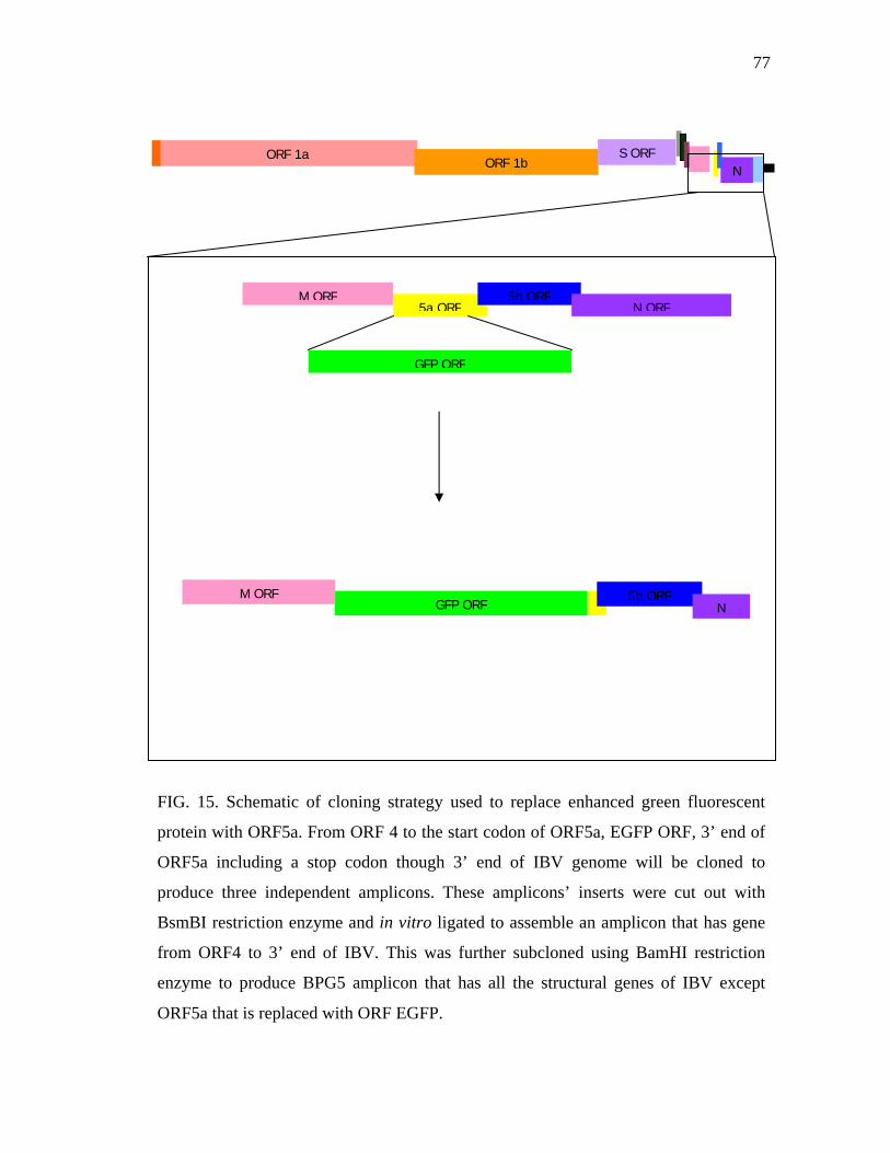
77
FIG. 15. Schematic of cloning strategy used to replace enhanced green fluorescent
protein with ORF5a. From ORF 4 to the start codon of ORF5a, EGFP ORF, 3’ end of
ORF5a including a stop codon though 3’ end of IBV genome will be cloned to
produce three independent amplicons. These amplicons’ inserts were cut out with
BsmBI restriction enzyme and in vitro ligated to assemble an amplicon that has gene
from ORF4 to 3’ end of IBV. This was further subcloned using BamHI restriction
enzyme to produce BPG5 amplicon that has all the structural genes of IBV except
ORF5a that is replaced with ORF EGFP.
ORF 1a ORF 1b
S ORF N
5a ORF5b ORF
GFP ORF
N ORFM ORF
GFP ORF5b ORF
N M ORF
78
ORF M and the intergenic region of ORF M and 5a including the start codon of ORF
5a, and the 3’ end of ORF 5a including stop codon through end of 3’ UTR of IBV were
PCR amplified using primer pairs listed on Table 3. EGFP was PCR amplified from
pLEGFP-N1 vector (Clontech, Palo Alto, CA) as a template using a primer pair that has
been designed to have BsmBI restriction enzyme recognition sequence at the each 3’ and
5’ ends. To replace EGFP with IBV 5a ORF, no see’m technology was employed (202).
Neighboring genes of the 5a ORF have been engineered to be compatible with EGFP
ORF using a BsmBI restriction enzyme as illustrated in Figure 16. For PCR
amplification upstream and downstream of ORF5a, the P5 amplicon described in chapter
II was used as a template.
For PCR amplification, the Expand High Fidelity PCR system (Roche,
Indianapolis, IN) was used throughout the study and primer pairs used for PCR cloning
though this study were listed in Table 3. For EGFP PCR amplification, 50 ng of
pLEGFP-N1 vector (Clontech laboratories Inc., Palo Alto, CA), 10 pM of each forward
and reverse primer (GFPSF and GFPER respectively), 1 ul of 10 mM deoxynucleotide
triphosphate (dNTP), 5 ul of 10X PCR buffer number 3 (Roche), 8 ul of 25 mM MgCl2,
and 0.75 ul of thermostable DNA polymerase mixture were added into 200 ul PCR tubes
and distilled water was added to bring the total reaction volume to 50 ul. Reaction
condition for the EGFP PCR amplification includes an initial denaturation at 94°C for 2
minutes followed by 30 cycles of denaturation at 94°C for 10 seconds, annealing at 55°C
for 30 seconds and elongation at 72°C for 30 seconds. The final step was an elongation
at 72°C for 7 minutes.
79
TABLE 3. Sequences of primer pairs used for construction of IBVG
Primer Nucleotide Sequence Location in IBV genome
GFPSF 5’ CGTCTCATGGTGAGCAAGGGCGAGGAG 3’
GFPER 5’ CGTCTCCGCTTACTTGTACAGCTCGTCC 3’
5ASR 5’ CGTCTCCACCATCGTCCGTATTTGTTAAGT 3’ 25574-25592
5AEF 5’ CGTCTCTAAGCGTATACGCCCACCCA 3’ 25658-25678
BMF 5’ ATGGCGGAAAATTGCACACTTGATTC 3’ 24509-24535
GNRT 5’ TTTTTTTTTTTTTTTTTTTGCTCTAACTCTATACTAGCC 3’ 27593-27613
80
5a ORF and its neighboring sequence CAAAAACTTAACAAATACGGACGATGAAA GCGTATACGCCCACCCAATC GTTTTTGAATTGTTTATGCCTGCTACTTT CGCATATGCGGGTGGGTTAG
GFP sequences including start and stop codon
ACCATGGTGAGCAAGGGCGAGGAGCTG CTCGGCATGGACGAGCTGTACAAGTAAAGC TGGTACCACTCGTTCCCGCTCCTCGAC GAGCCGTACCTGCTCGACATGTTCATTTCG
5ASR 5’ CAAAAACTTAACAAATACGGACGATGGTGGAGACG 3’ 3’ GTTTTTGAATTGTTTATGCCTGCTACCACCTCTGC 5’
GFPSF 5’ CGTCTCATGGTGAGCAAGGGCGAGGAGCTGTTCAC 3’ 3’ GCAGAGTACCACTCGTTCCCGCTCCTCGACAAGTG 5’
5a S and GFP S junction after ligation
CAAAAACTTAACAAATACGGACGA TGGTGAGCAAGGGCGAGGAGCTGTTCAC GTTTTTGAATTGTTTATGCCTGCTACCA CTCGTTCCCGCTCCTCGACAAGTG
GFPE R 5’ CTCGGCATGGACGAGCTGTACAAGTAAGCGGAGACG 3’ 3’ GAGCCGTACCTGCTCGACATGTTCATTCGCCTCTGC 5’
5a E F 5’ CGTCTCTAAGCGTATACGCCCACCCAATC 3’ 3’ GCAGAGATTCGCATATGCGGGTGGGTTAG 5’
GFP stop codon and ORF5a 3’ end junction sequence after ligation
CTCGGCATGGACGAGCTGTACAAGT AAGCGTATACGCCCACCCAATC GAGCCGTACCTGCTCGACATGTTCATTCG CATATGCGGGTGGGTTAG
FIG. 16. “No see’m technology” used to replace EGFP with ORF5a. Pink nucleotides
denote BsmBI restriction enzyme sequence inserted primer pairs. Red nucleotides denote
start codon or stop codon of EGFP and ORF5a. Underlined sequences are the primer used
to amplified each gene by PCR.
81
The 5ASR and BMF were used as reverse and forward primers, respectively, for
amplification upstream of ORF5a and ORF M. 50 ng of the template, amplicon P5, 10
pM of each forward and reverse primer, 1 ul of 10 mM of each deoxynucleotide
triphosphate (dNTP), 5 ul of 10X PCR buffer number 3 (Roche), 8 ul of 25 mM MgCl2,
and 0.75 ul of thermostable DNA polymerase mixture were placed in 200 ul PCR tubes
and distilled water was added to bring the total reaction volume to 50 ul. Reaction
conditions for the PCR amplification includes an initial denaturation at 94°C for 2
minutes followed by 30 cycles of denaturation at 94°C for 10 seconds, annealing at 55°C
for 60 seconds and elongation at 68°C for one minute. The final step was an elongation
at 68°C for 7 minutes.
The 5AEF and GNRT were used as forward and reverse primers respectively for
amplification downstream of ORF5a to the 3’ UTR of IBV. 50 ng of template, amplicon
P5, 10 pM of each forward and reverse primer, 1 ul of 10 mM deoxynucleotide
triphosphate (dNTP), 5 ul of 10X PCR buffer number 3 (Roche), 8 ul of 25 mM MgCl2,
and 0.75 ul of thermostable DNA polymerase mixture were added into 200 ul PCR tubes
and the total reaction volume was brought to 50 ul with distilled water. Reaction
conditions for the PCR amplification includes an initial denaturation at 94°C for 2
minutes followed by 30 cycles of denaturation at 94°C for 10 seconds, annealing at 55°C
for 30 seconds and elongation at 68°C for one minute 20 seconds. The final step was an
elongation at 68°C for 7 minutes.
Following amplification, 2 ul of each PCR product was run on a 1% agarose gel
in Tris-acetate-EDTA (TAE) buffer containing ethidium bromide (EtBr) and the results
82
were recorded using a FluoChem TM gel analysis system (Alpha Innotech, Corporation,
San Leandro, CA). Each PCR product was purified from a 1% agarose gel containing
crystal violet (2 ug/ ml). DNA bands were excised from the agarose gel and then purified
using a QIAquick Gel Extraction Kit (QIAGEN Inc., Valencia, CA) following the
manufacture’s directions. The concentration of gel purified PCR products were
measured with a SmartSpec 3000 spectrophotometer (BIO-RAD, Hercules, CA). Each
PCR product was ligated into a cloning vector. EGFP and upstream of ORF5a were
ligated into a pSTBlue-1 (Novagen, Darmstadt, Germany) according to manufacturer’s
directions. Briefly, equal-molar ratios of PCR product and pSTBlue vector were ligated
for 2 hours at 16°C and then transformed into chemically competent NovaBlue cells
(Novagen, Darmstadt, Germany). Final constructs of the EGFP ORF and the upstream
region of ORF5a were named as pSTBGFP and pSTBM, respectively. The region
beginning from downstream of ORF5a to 3’UTR of IBV was ligated into the pCR-XL-
TOPO cloning vector (Invitrogen, Carlsbad, CA) for 5 minutes at room temperature and
transformed into chemically competent Oneshot competent cells (Invitrogen). The final
construct was named pCRB3’. Transformed bacteria were plated on Luria-Bertani (LB)
medium agar plates containing kanamycin (5 ug/ml) and grown overnight at 37°C. Mini-
prep cultures were prepared from the transformed colonies to confirm the insert size and
direction using unique restriction enzyme recognition sequence in the inserts. The
construct was confirmed with nucleotide sequencing using the universal primers SP6 and
T7 primers synthesized in a commercial laboratory.
83
To assemble recombinant P5G amplicon, pSTBGFP, pSTBM and pCRB3’ were
digested with BsmBI. Appropriate inserts from each amplicon were gel purified as
described above. Because pCRB3’ has an internal BsmBI restriction recognition
sequence other then BsmBI sites at 3’ and 5’ ends inserted by PCR mutagenesis, two
DNA bands were prepared from pCRB3’ BsmBI digestion reactions. Also, pCRB3’ was
used as a cloning vector after the restriction enzyme digestion because it has a pCR-XL-
TOPO backbone. Equal-molar amounts of each insert were overnight ligated using T4
DNA ligase (3 units/ul, Promega) at 16°C and transformed into chemically competent
one shot competent cells (Invitrogen) by heat shock at 45°C for 30 seconds. Subcloned
recombinant P5G were confirmed by restriction enzyme digestion and sequencing.
Nucleotide Sequencing
Each amplicon DNA was prepared from transformed bacterial midi-cultures
grown overnight and DNA templates were prepared using QIAGEN mini prep kit
(QIAGEN). Sequencing was performed using the ABI prism BigDye Terminator Cycle
Sequencing Ready Reaction Kit (Applied Biosystems Inv., Foster City, CA). For a
standard sequencing reaction, 350-500 ng of plasmid DNA was mixed with 2 ul of
BigDye mixture (ABI) and 10 pM of primers. The remainder of the reaction mixture was
adjusted to 6 ul with distilled water. Sequencing reactions were performed using a
GeneAmp thermocycler (PerkinElmer, Boston, MA) using conditions that included pre-
denaturation at 96°C for 2 minutes and 30 cycles of 96°C for 30 seconds, 52°C for 15
84
seconds and 60°C for 4 minutes. Non-incorporated nucleotides were removed using
micro Bio-spin chromatography columns (BIO-RAD, Hercules, CA) and completely
dried using a vacuum drier. Completed sequencing reactions were sent to the Gene
Technology Laboratory in the Department of Biology in Texas A&M University.
Sequences were read using an ABI automatic sequencer 3100 (ABI).
Analysis of Sequence
Sequence data were analyzed using SDS biology workbench
(http://workbench.sdsc.edu) (San Diego Supercomputer Center, University of California,
San Diego, CA). LALIGN program was used for DNA and amino acid sequence
comparison. The TACG program was used to search for restriction enzyme digestion
profiles for cloning purposes. The SIXFRAME program was used to convert DNA
sequence into amino acid sequence.
Assembly of a Full-length IBV Infectious Construct
Inserted DNAs from each amplicon were excised with the appropriate restriction
enzymes. PI was digested with XhoI (Promega, Madison, WI) and then treated with calf
intestine phosphatase (CIP) (Promega) to inhibit self-ligation. CIP treated DNA was
phenol:chloroform extracted, ethanol precipitated and then digested with restriction
enzyme SapI (New England Biolabs Inc., Beverly, MA). P2–1, P2-2 and P3-1 amplicons
were double digested with restriction enzymes SapI and BsmBI. When the buffer
85
conditions were not compatible, after one restriction enzyme digestion, the reactions
were ethanol precipitated and then the reaction was reconstituted with the appropriate
buffer and in the appropriate volume before adding the second restriction enzyme.
Except for the BsmBI restriction enzyme digestion that needed to be incubated at 55°C,
the restriction enzymes were incubated at 37°C. P3-2 and P4 amplicons were single
digested with restriction enzyme BsmBI. The P5 amplicon was digested with restriction
enzyme EcoRI (Promega), then CIP treated and digested with BsmBI. Each DNA insert
was run on a TAE agarose gel (0.8 to 1%) containing 2.4 ug/ml crystal violet rather than
ethidium bromide to avoid UV-induced DNA damage that could impact subsequent
manipulations of DNA fragments. Target size inserts were excised from the agarose gel
and the DNA inserts were extracted using a QIAquick Gel Extraction kit (QIAGEN Inc,
Valencia, CA) following the manufacturer’s instruction.
A relatively high concentration of T4 DNA ligase (20 U/ul, Promega) was used
to assemble infectious cDNA of IBV. Ligation reactions were incubated at 16°C
overnight, before inactivating by incubating at 65°C for 10 minutes. An aliquot of the
ligation reactions were run on an agarose gel to confirm the ligation. The appropriately
sized larger ligation fragments were agarose gel purified and then ligated step-wise with
other fragments. The final ligation reaction of P1 through P3-1 and P3-2 though P5 were
pooled without further purification and then ligated overnight at 16°C.
86
RNA Preparation
Viral RNA was prepared from the supernatant of IBV infected Vero cells or
recombinant IBVG rescued from transfection. Briefly, Vero cells were infected with 0.1
multiplication of infection (m.o.i) and incubated in a CO2 incubator at 37°C until the
cells showed more than 80% cytopathogenic effect (CPE) such as syncytia formation.
The cells showing extensive CPE were frozen and thawed three times and then cell
debris were precipitated by centrifugation at 3,000 rpm using a table top centrifuge (CR
412; Jouan Inc. Winchester, VA). The supernatants were then ultracentrifuged at 35,000
rpm for 2 hours at 4°C using an L7-55 ultracentrifuge (Beckman, Palo alto, CA) with an
SW55Ti swing bucket rotor to pellet the virus. The viral pellet was re-suspended in
small volumes of TEN (10 mM Tris-Cl, pH 8.0, 1 mM ethylendiaminotetraacetate
(EDTA), pH 8.0 and 0.1 M NaCl) buffer.
TRIZOL reagent (Invitrogen, Carlsbad, CA) was used to extract the RNA from
the re-suspended viral pellet following the manufacturer’s instruction. Briefly, 1000 ul of
TRIZOL reagent were added to 200 ul of viral pellet suspension in TEN buffer before
mixing thoroughly by inverting the microfuge tube several times. The mixture was
incubated for 5 minutes at room temperate, 200 ul of chloroform were added and then
mixed thoroughly by inversion. This mixture was incubated at room temperature for 3
minutes. To separate the aqueous and the organic phases, the mixture was centrifuged at
12,000 rpm for 15 minutes at 4°C using a microcentrifuge (5417 R; Eppendorf,
Hamburg, Germany). The upper phase was then transferred to a new nuclease free
microfuge tube and a 0.5 volume of isopropanol was added to precipitate the RNA. After
87
a 10 minute incubation at room temperature, the RNA was pelleted by centrifugation at
12,000 rpm for 15 minutes at 4°C. Before suspending the RNA pellet with nuclease-free
water, the RNA pellet was washed with 70% ethanol, then briefly air-dried. The RNA
solution was quantified using a SmartSpecTM 3000 spectrophotometer (BIO-RAD,
Hercules, CA) and stored at –70°C until it was used for Northern blot analysis or RT-
PCR.
Northern Blot Analysis
Preparation of a random primed isotope labeled probe.
To prepare a random primed probe against GFP and the 3’ UTR, pGEGFP and
pGEBN-2 were used as templates for in vitro transcription reactions. The template DNA
was digested with PstI restriction enzyme and then phenol:chloroform extracted
followed by ethanol precipitation to remove contaminating RNase. One ug of template
was used for an in vitro transcription and labeling reaction using T7 RNA polymerase
(Promega). Briefly, 4 ul of 5X transcription buffer, 2 ul of 100 mM DTT, 1 ul of RNase
inhibitor (Promega, 30 units/ul), 4 ul of rNTP mix containing 2.5 mM rCTP, rATP, and
rGTP, 1.2 ul of 200 uM rUTP, 5 ul of [α32P] rUTP (Perkin-Elmer), 1.3 ul of T7 RNA
polymerase (20 units/ul) were used in a 20 ul reaction. The reaction was incubated at
37°C for one hour and then ethanol precipitated. The probe was resuspended in
deionized formamide and stored at –20°C. Radioisotope activity was measured with a
liquid scintillation system (Beckman Coulter Inc., Fullerton, CA).

88
RNA transfer
To transfer RNA to nylon membranes, 1 ul of each in vitro transcribed RNA was
run on a denaturing agarose gel containing formaldehyde following the standard
methods (153) with modifications. RNA was denatured with sample buffer containing
1/10 v/v of 10X formaldehyde gel–running buffer (0.2M 3-(N-norpholino)
propanesulfonic acid (MOPS) (pH 7.0), 80 mM sodium acetate and 10 mM EDTA (pH
8.0)) with formaldehyde, 1/2 v/v of formamide, 2 ug of ethidium bromide, and 1/10 v/v
of formaldehyde gel-loading buffer (50% glycerol, 0.25% bromophenol blue, 0.25%
xylene cyanol FF). RNA and sample buffer were mixed, denatured at 70°C for 5 minutes
and chilled on ice. A 1% denaturing agarose gel containing formaldehyde was prepared
with a 1/10 volume of 10 X formaldehyde gel running buffer and formaldehyde to give a
2.2 M final concentration. Each RNA sample was loaded on the agarose gel and run at
90 V in 1 X formaldehyde gel-running buffer. At one hour intervals, the buffer from
each reservoir was collected, mixed and returned for electrophoresis. When the xylene
cyanol dye front was at the bottom of the gel, electrophoresis was terminated. The gel
was washed several times with distilled water and then soaked in 0.05 N NaOH for 20
minutes and exposed to long wavelength UV light for 60 seconds. The gel was then
washed with distilled water and soaked for 20 minutes in 10 X sodium chloride sodium
citrate (SSC) buffer. RNA from the gel was transferred overnight to a nylon membrane
by capillary transfer in 10 X SSC. RNA transfer was confirmed by ethidium bromide
staining of the nylon membrane and the molecular weight markers were indicated with
pencil. The membrane was then fixed by UV cross-linking for 30 seconds.
89
Hybridization reaction
The nylon membrane was hybridized to random primed α-32P labeled probes
with an activity of 2 x 10⁷ cpm. Hybridization reactions were performed using an
autoblot hybridization oven (Bellco Glass Inc., Vineland, NJ). Hybridization conditions
included incubation at 55°C for 12 hours in hybridization buffer containing 50%
deionized formaldehyde, 2 X Denhardt’s reagent, 5 X SSC and 0.1% SDS. After
hybridization, excess probe was removed with 1 X SSC, 0.1% SDS, followed by three
washes for 20 minutes each at 68°C in 0.2 X SSC and 0.1% SDS. After hybridization,
the nylon paper was exposed to an image plate (IP) for 20 minutes to one hour,
depending on the signal intensity. The image was analyzed using a BAS-1800 II
phosphoimage reader (Fusifilm Electronic Imaging Ltd. Tokyo, Japan) and analyzed
using the Image gauge V3.12 and L processor V1.72 programs.
In vitro Transcription and Electroporation
Capped runoff T7 transcripts were synthesized in vitro from in vitro ligated IBV
cDNA using an mMessage mMachine kit (Ambion, Austin, TX.) as described by the
manufacturer with some modifications. Twenty ul of the IBV RNA transcription
reaction mixture was prepared with three ul of 30 mM GTP stock and incubated at 37C°
for 2 hrs.
To increase the efficiency of virus production, N transcript was also prepared for
co-transfecting with the entire IBV transcript. pGEMN-1 was used as a template for N
90
transcripts. The plasmid was digested with a PstI restriction enzyme (Promega) to
linearize the template and then phenol:chloroform extracted followed by ethanol
precipitation to remove any RNase contamination. The DNA was resuspended with
diethyl pyrocarbonate (DEPC) treated distilled water. One ug of the template was used
for an in vitro transcription reaction using mMESSAGE mMACHINE TM T7 Ultra kit
(Ambion, Austin, TX.). A poly(A) tail was added to the transcript with E. coli Poly(A)
polymerase. Both transcripts were used, with or without precipitation with 7.5 M lithium
chloride containing 50 mM EDTA.
BHK-21 cells were grown to subconfluence (about 70%) before electroporation.
BHK-21 cells were treated with trypsin to detach them from flasks. The cells were
washed twice with ice cold DEPC treated PBS and then resuspended in the cold PBS
with 2 x 10⁷ per ml infectious cDNA transcription reaction. N transcript was mixed with
the cells in a 1.5 ml microcentrifuge tube. The cells and RNA mixture were transferred
immediately to 2 mm gap electroporation cuvettes (electroporation cuvettes plus model
No. 620, BTX, Genetronics, Inc. San Diego, California) and then electroporated at 850
V, 25 mΩ at resistance 720 Ω using an electro-cell manipulator 600 (BTX, Genetronics,
Inc. San Diego, California) with three consecutive pulses. After electroporation, cells
were incubated at RT for 10 minutes and the transfectant was diluted 1 to 20 in complete
DMEM containing 10% FBS and resuspended with 1 x 10⁶ Vero cell per 100 mm petri-
dish.
91
Plaque Staining and Plaque Purification
Virus preparations were serially diluted 10-fold in DMEM lacking FBS. Then,
200 ul of each dilution was inoculated per well of six well cell culture plates that had
been seeded with monolayers of Vero cells. After a one hour incubation, residual virus
was removed by washing the cells twice with PBS before overlaying the cells with 0.8%
agarose containing 1 X DMEM, 1% FBS and antibiotics. The cells were incubated until
plaques were observed. For plaque staining, the agarose overlay was removed using a
spatula, the cell monolayers were air dried briefly and overlaid with 2 ml of crystal
violet staining solution per well. After 10 minutes, excessive crystal violet was removed
and the cells were washed several times with PBS to remove residual crystal violet. For
plaque purification, the agarose over the plaques were picked with a 1000 λ micropipette
and suspended in 1 ml DMEM without FBS. Before inoculating, the plaque suspension,
the viral suspension was incubated at 4°C overnight and inoculated to Vero cell in 6 well
plates described before.
Kinetics of Viral Replication
Vero cells were infected with wild type IBV Beaudette strain and the IBV
Beaudette strain derived from an infectious cDNA clone and plaque purified three times.
The Vero cells were seeded in 35 mm petri dishes at a concentration of 106 per dish. The
cells were infected with three plaque forming units per cell for an hour and then the
media were replaced with DMEM containing 1% FBS with antibiotics, and 5, 10, 12, 19,
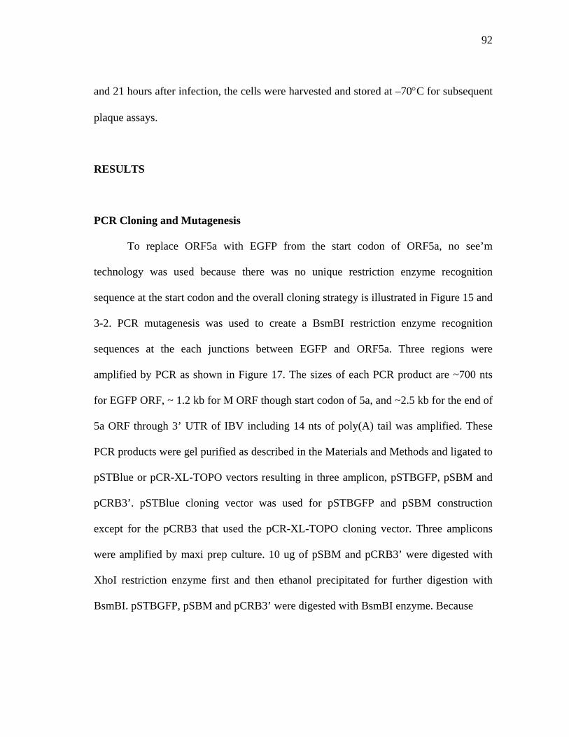
92
and 21 hours after infection, the cells were harvested and stored at –70°C for subsequent
plaque assays.
RESULTS
PCR Cloning and Mutagenesis
To replace ORF5a with EGFP from the start codon of ORF5a, no see’m
technology was used because there was no unique restriction enzyme recognition
sequence at the start codon and the overall cloning strategy is illustrated in Figure 15 and
3-2. PCR mutagenesis was used to create a BsmBI restriction enzyme recognition
sequences at the each junctions between EGFP and ORF5a. Three regions were
amplified by PCR as shown in Figure 17. The sizes of each PCR product are ~700 nts
for EGFP ORF, ~ 1.2 kb for M ORF though start codon of 5a, and ~2.5 kb for the end of
5a ORF through 3’ UTR of IBV including 14 nts of poly(A) tail was amplified. These
PCR products were gel purified as described in the Materials and Methods and ligated to
pSTBlue or pCR-XL-TOPO vectors resulting in three amplicon, pSTBGFP, pSBM and
pCRB3’. pSTBlue cloning vector was used for pSTBGFP and pSBM construction
except for the pCRB3 that used the pCR-XL-TOPO cloning vector. Three amplicons
were amplified by maxi prep culture. 10 ug of pSBM and pCRB3’ were digested with
XhoI restriction enzyme first and then ethanol precipitated for further digestion with
BsmBI. pSTBGFP, pSBM and pCRB3’ were digested with BsmBI enzyme. Because
93
3 kb
1.5 kb
0.5 kb
M 1 2 3
FIG. 17. Site-directed mutagenesis by PCR. To replace the enhanced green
fluorescent protein (EGFP) gene with ORF5a, BsmBI restriction enzyme recognition
sequences were created by PCR on the EGFP ORF and an ORF5a neighboring
sequence. These restriction enzyme sites will be further used to religate EGFP with
ORF5a neighboring gene.
M; 1 kb DNA ladder, lane 1; PCR product of EGFP, lane 2; PCR product from ORF
M to start codon of ORF5a, lane 3; PCR product from 3’ end of ORF5a to 3’ UTR
of IBV genome including the poly(A) tail.
94
pCRB3’ has an internal BsmBI recognition sequence, digestion of pCRB3’ with BsmBI
produces two insert fragments. Inserts were separated from the vector by electrophoresis
and purified from the gel after restriction enzyme digestion. These four fragments were
ligated in one reaction. Because each BsmBI overhang has a unique sequence,
theoretically the products can only be religated with the correct fragment. Furthermore,
only the final ligation product with all the fragments having a pCR-XL-TOPO backbone
can survive after transformation. Transformed plasmid constructs were verified by
restriction enzyme digestion and the correct construct was named as pIBVG11.
Because the pIBVG11 construct only has the M ORF though 3’ UTR of IBV,
further subcloning was performed using the pIBVG11 construct and amplicon P5 to
produce P5 containing every structural genes as well as EGFP. Because pIBVG11 and
P5 have the same orientation and M ORF has a unique BamHI restriction recognition
sequence, both constructs were digested with BamHI. Then pIBVG11 was treated with
phosphatase to prevent self-ligation. The larger fragment from P5 BamHI reaction
containing the S ORF and part of the M ORF was replaced with a smaller fragment of
pIBVG11 digested with BamHI. The ligation reaction was transformed into chemically
competent cells and the recombinant GFP5 amplicon with EGFP instead of 5a were
cloned. This GFP5 construct was sequenced using a primer upstream of 5a ORF to make
sure there were no mutations during cloning. Sequences of EGFP and the upstream
region of the ORF5a junction is shown in Figure 18. The EGFP ORF was replaced with
ORF 5a exactly as intended. Furthermore, sequences, upstream of 5a and downstream of
5a did not have any unintentional mutations. This shows that IBV infectious cDNA is
95
ORF5a and GFP start codon junction after ligation
CAAAAACTTAACAAATACGGACGA TGGTGAGCAAGGGCGAGGAGCTGTTCAC GTTTTTGAATTGTTTATGCCTGCTACCA CTCGTTCCCGCTCCTCGACAAGTG
FIG. 18. Sequence verification of EGFP ORF and ORF5a replacement. The start codon was denoted in a red box. B is the enlargement of part of A containing start codon. Prior to the start codon, sequences are identical as the intergenic sequences between ORF5a, the start codon is from EGFP.
A
B
96
very stable for further manipulation and subsequent serial cultures compared to the
MHV infectious cDNA.
Sequencing Analysis
After verifying sequences of the EGFP junction, it was assumed that there were
no mutations introduced by PCR procedure because the PCR product was comparatively
small and high fidelity thermostable polymerase developed for cloning of large PCR
product was used to amplify the regions. After in vitro transcription and transfection, an
infectious cDNA clone of IBV expressing EGFP could not be rescued. It was then
decided to sequence the entire GFP5 amplicons and three nucleotide changes in 3’ UTR
introduced by PCR were identified. The region with mutations was replaced with the
same region of amplicon P5 and used to produce the infectious cDNA clones of
recombinant IBV with the EGFP.
In vitro Assembly
Based on the experience acquired during development of the infectious cDNA
clone of IBV, I used an orderly in vitro ligation approach for construction of the cDNA
clone of IBV as described in chapter II. P1 and P2-1 were ligated and P2-2 and P3-1
were ligated then P3-2 and P4 were ligated together and GP5L, and P5S were ligated.
The larger ligation products from each of the four ligation reactions were separated by
electrophoreses on agarose gels. The ligation products of P1/P21-1 and P2-2/P3-1 were
ligated and ligation products of P3-2/P4 and P5 were ligated separately and without
97
further purification, these two ligation reactions were pooled and ligated overnight at
16°C.
In vitro Transcription and Electroporation
Approximately 0.2 pM of cDNA template was used for an in vitro transcription
reaction. To confirm that the in vitro transcript had a full-length RNA with the EGFP
gene instead of ORF5a, a Northern blot analysis was performed using two different
probes. One probe used was the same as that used for the 3’UTR of IBV for the
infectious cDNA construction. The other probe was specific for EGFP. Because it has
been shown that the N transcript increased the rescue of coronaviruses after transfection
(56, 184, 201), N ORF transcripts were used as described in chapter II.
In vitro transcripts of IBVG cDNA and N ORF were electroporated into BHK-21
cells. Because BHK-21 cells are not susceptible to IBV infection, Vero cells were
overlaid on the electroporated BHK-21 cells. Two days after transfection, apparent CPE
was noticed in the cells transfected with total cellular RNA from IBV infected cell and
three days after transfection, CPE was noticed in the cells transfected with recombinant
IBVG infectious cDNA. To confirm whether this recombinant virus expressed EGFP,
the cells were examined under the UV microscope. As shown in Figure 19, the
recombinant IBVG expressed EGFP that 5a was replaced with EGFP. RT-PCR was used
to confirm the size difference of ORF 5a region.
98
BA
FIG. 19. Recombinant infectious cDNA clone which 5a ORF was replaced with EGFP
ORF was viable in infected cells and expressed EGFP. Cells transfected with
recombinant infectious cDNA expressing EGFP. (A) Cells 3 days after transfection
with the recombinant EGFP IBV transcript as viewed under a phase contrast
microscope (B) Cells transfected with recombinant EGFP IBV transcript after 3 days
transfection under an UV microscope.
99
DISCUSSION
Infectious cDNA clones of RNA viruses are one of the most powerful tools to
study the function of viral proteins, especially during viral replication and viral
assembly. The availability of infectious cDNA clones for many small RNA viruses has
resulted in a tremendous development of RNA virus research. With the development of
molecular biological techniques, it has been possible to construct infectious cDNA
clones of coronaviruses overcoming the difficulties the large genome size and toxicity of
the coronavirus genome in bacterial hosts. Several groups used three different
approaches to construct infectious cDNA clones of coronaviruses, including MHV,
TGEV, HCoV and IBV. These infectious cDNA clones have been developed to express
foreign genes for potential gene expression system, gene transfer vector, and as vaccine.
Infectious cDNAs of coronaviruses can be used to study the molecular biology of the
viruses. Many studies have been done using defective interfering (DI) particle or
targeted recombination in MHV and FIPV before the development of infectious cDNA
clones. However, it is hard to conclude the result of mutants, especially deletion
mutation studies, because of the extremely high rate of recombination during viral
replication and transcription and the multiple crossovers during the recombination
process. Therefore, using infectious cDNA clones is a more reasonable approach
compared to using a targeted mutagenesis approach to study coronaviruses.
Coronaviruses have several small ORFs, some of these ORF are expressed in
virus infected cells. However, the exact functions of these genes are not well understood.
These small genes, referred as group specific genes, are not necessary for viral
100
replication in vitro in MHV, even though deletion mutant viruses of the region were
significantly attenuated. IBV has four small group specific antigens including 3a, 3b, 5a
and 5b.
Previously, we were successful in constructing an infectious cDNA clone of the
Vero cell adapted Beaudette strain of IBV. In this study, we wanted to use the infectious
cDNA clone to resolve the issue of whether a small group specific antigen is necessary
for the survival of IBV in vitro. One of the group specific genes, 5a ORF was replaced
with green fluorescent protein. The C-terminal region of 5a protein was kept for the
expression of 5b protein. After in vitro assembly and electroporation, a recombinant IBV
5a deletion mutant of IBV expressing EGFP was successfully rescued and the
fluorescence was observed. By sequence analysis and northern blot analysis, it was
confirmed that there was no unintentional mutations in the amplicons used to construct
the recombinant clone. Based on this study, 5a protein of IBV is not necessary for
survival of IBV in vitro, even though the function of the protein is not yet understood.
In our laboratory, group specific antigens of several different strains of IBV have
been sequenced and shown that the proteins are conserved among different strains,
especially putative 5a. Therefore, even though these proteins are not necessary for viral
survival in vitro, it is possible that these proteins are important for viral replication in the
natural host or maybe a protective protein against host defense mechanisms, such as
cytokine mimicry molecules or anti-apoptotic proteins.
In summary, in this study, a recombinant infectious cDNA clone of IBV
containing EGFP instead of 5a ORF was constructed. By sequence analysis of
101
amplicons, targeted mutagenesis was confirmed and the recombinant infectious cDNA
was viable implying that the 5a protein is not necessary for IBV amplification in Vero
cells. This also showed the possibility of using IBV as a gene transfer vector for poultry.
102
CHAPTER IV
RECOMBINANT SEMLIKI FOREST VIRUS REPLICON CAN BE PACKAGED
INTO IBV VIRIONS WITHOUT IBV CIS ACTING SIGNAL SEQUENCE
INTRODUCTION
Infectious bronchitis virus (IBV) has been a recognized problem in the poultry
industry since 1931 (158). IBV has been controlled by traditional attenuated live vaccine
strategies. The high mutation rate and the high frequency of recombination of IBV,
however, has made it necessary to seek alternative methods which can control the disease
more effectively and to lower the possibility of emerging recombinant IBV originating
from field and vaccine strains (reviewed in (39)).
IBV belongs to the order Nidovirales in the Coronaviridae family. Coronaviruses
cause clinical disease in a number of animals, including humans (34, 126). Most
coronaviruses cause disease only in the species from which they were isolated and, if
they replicate in cell culture, predominantly replicate only in cell lines derived from their
natural hosts (197). IBV has four known structural proteins, including the spike
glycoprotein (S) that is post-translationally cleaved into S1 and S2, the integral
membrane glycoprotein (M), the small membrane protein (E) and the nucleocapsid
protein (N) (94). S, M, and E proteins are envelope proteins that are embedded in the
host-derived bilipid layer of the viral envelope. The S protein is not required for virus
budding process but is responsible for viral attachment and entry, determining host
103
specificity and creating the binding site for cellular receptors. Receptors for MHV,
TGEV and HCoV have been identified (46, 54, 77, 136, 200). Determinants responsible
for the generation of neutralizing antibodies also lie on N terminal of the S protein,
especially S1 (40, 81, 84, 183).
Both M and E proteins localize in the cis-Golgi compartment and several studies
have suggested that the M and E proteins are critical for intracellular viral budding (47,
50, 98, 118). Expression of the E proteins of MHV and IBV, in the absence of other viral
proteins, produces vesicles that can be released from the cells (47, 120). However, co-
expression of M and E are necessary in TGEV and bovine coronavirus (BoCV) to make
virus like particles (VLP) (13, 17, 193). The N protein binds to the viral RNA genome,
producing the ribonucleoprotein complex (10, 43, 125, 130, 144). The N protein is the
only viral protein that is phosphorylated and the phosphorylation of the N protein has
been implicated as having a role in viral RNA and protein interaction.
Packaging mechanisms of several coronaviruses, such as MHV, BoCV and more
recently TGEV have been studied (44, 60, 124). MHV packages only viral genomic
RNA, whereas IBV and BoCV have been shown to package not only genomic RNA, but
also subgenomic RNAs (204). Using defective interfering (DI) RNA and deletion
mutagenesis, Makino et al. have located packaging signal sequences in MHV and shown
that M protein and packaging signal sequence interaction might mediate specific viral
genomic RNA packaging into MHV virions (124, 129). The BoCV packaging signal
sequence was identified based on a sequence homology search of BoCV genome with
the MHV packaging signal sequence (44). In the presence of the packaging signal
104
sequence, non-viral RNA can be packaged into BoCV or MHV virions, respectively.
However, it is interesting that one of the naturally occurring BoCV DIs, Drep can be
packaged into BoCV virions without the known packaging signal sequence (44).
Recently, the packaging signal sequence of TGEV has been identified by Escobars et al.
(60). They also showed that highly purified TGEV only packaged genomic RNA like
MHV and the packaging signal sequence is located within the 649nts at the 5’ UTR of
TGEV genomic RNA (60). In the study, it was suggested that not only the packaging
signal sequence itself, but also viral replication status, is important for efficient viral
packaging in TGEV.
Semliki forest virus (SFV), a prototype of the alphavirus genus and a member of
the Togaviridae family, has a single-strand, positive sense RNA genome (160). Several
members of this group, in particular Sindbis virus (SIN), SFV, and Venezuelan equine
encephalitis (VEE) virus, have provided valuable tools for understanding virus structure
and replication. The two alphaviruses most extensively studied in laboratories, SIN and
SFV, are generally considered avirulent for humans. The alphavirus genome is
approximately 11.7 kb and is encapsidated within an icosahedral capsid protein (177).
Cells infected with these viruses synthesize abundant amounts of viral genomic RNA
and subgenomic RNA. The genomic and subgenomic RNAs are 5' capped and 3'
polyadenylated (27). The 5’ two thirds of the genomic RNA encode the replicase
polyproteins, which are cleaved to produce nsP1, nsP2, nsP3 and nsP4 products. The
subgenomic RNA is identical in sequence to the 3' one-third of the genomic RNA and
encodes the structural proteins. These include the capsid protein, which interacts with
105
the genomic RNA to form the viral nucleocapsid and two membrane glycoproteins
(reviewed in (177)). Cytoplasmic replication of the genomic and subgenomic RNAs is
mediated by the replicase gene using a full-length negative sense RNA as a template.
The viral structural proteins are translated from the subgenomic mRNA as a polyprotein
that is processed into the individual virion components by a viral protease.
Specific genomic RNA packaging of alphaviruses has been shown to depend on
the packaging signal sequence that is present only on the genomic RNA. The signal
sequences of SIN, SFV and Ross river virus (RRV) have been identified and used to
make replicon vectors that can replicate and be packaged into virions produced by a
helper construct. The helper construct encodes viral structural genes under the control of
the subgenomic RNA promoter. Because the helper construct lacks the packaging signal
sequence, it results in aberrant self-packaging, in spite of its high concentration in the
cytoplasm during viral replication and transcription. The replicon, packaged into the
newly made virions, can infect cells and replicate, but the progeny RNAs cannot be
transmitted into new cells because they lack the viral structural proteins.
As an alternative to live attenuated vaccines, we were interested in developing a
new type of vaccine by combining the advantages of two different viruses, IBV and SFV.
In this study, as a prerequisite of this purpose, we decided to develop recombinant SFVs as
a tool for identifying the packaging signal sequence of IBV. The SFV replicon can be
replicated in the cytoplasm to produce high concentrations of this RNA. We hypothesized
that if a recombinant SFV has the packaging signal sequence of IBV, the replicon can be
packaged into an IBV virion. To our surprise, however, with or without any candidate
106
packaging signal sequence of IBV, rSFV can be packaged into an IBV virion suggesting
IBV virion can be a useful candidate for pseudotyping of SFV vectors for specific
targeting to respiratory system of poultry as a vaccine or for gene transfer.
MATERIALS AND METHODS
Viruses and Cells
A Vero cell culture adapted Beaudette strain of IBV was obtained from
American Type Culture Collection (ATCC, Manassas, VA) and then plaque purified
three times in the laboratory before use. The virus was propagated in the African green
monkey kidney Vero cell line obtained from the ViroMed Laboratory (Minnetonka,
MN) and maintained in Dubecco’s modified Eagle medium (DMEM) containing 5%
fetal bovine serum (FBS) supplemented with penicillin G (100 unit/ml) and streptomycin
(100 ug/ml).
RNA Preparation
Viral RNA was prepared from the supernatant of IBV infected Vero cells.
Briefly, Vero cells were infected with 0.1 multiplication of infection (m.o.i) and
incubated in a CO2 incubator at 37°C until the cells showed more than 80%
cytopathogenic effect (CPE), such as syncytia formation. The cells showing extensive
CPE were frozen and thawed three times and then cell debris was removed by
centrifugation at 3,000 rpm using a tabletop centrifuge (CR 412; Jouan Inc. Winchester,
107
VA). The supernatants were then ultracentrifuged at 35,000 rpm for 2 hours at 4°C using
an L7-55 ultracentrifuge (Beckman, Palo Alto, CA) with an SW55Ti swing bucket rotor
to pellet the virus. The viral pellet was re-suspended in small volumes of TEN (10 mM
Tris-Cl, pH 8.0, 1 mM ethylendiaminotetraacetate (EDTA), pH 8.0 and 0.1M NaCl)
buffer.
TRIZOL reagent (Invitrogen, Carlsbad, CA) was used to extract the RNA from
the re-suspended viral pellet following by the manufacturer’s instruction. Briefly, 1 ml
of TRIZOL reagent was added to 200 ul of viral pellet suspension in TEN buffer and
then mixed thoroughly by inverting the microfuge tube several times. The mixture was
incubated for 5 minutes at room temperature before adding 200 ul of chloroform and
mixing thoroughly. The mixture was then incubated at room temperature for 3 minutes.
To separate the aqueous and the organic phases, the mixture was centrifuged at 12,000
rpm for 15 minutes at 4°C using a microcentrifuge 5417 R (Eppendorf, Hamburg,
Germany). The upper (aqueous) phase was then transferred to a new nuclease free
microfuge tube and a 500 ul of isopropanol were added to precipitate the RNA. After a
10-minutes incubation at room temperature, the RNA was pelleted by centrifugation at
12,000 rpm for 15 minutes at 4°C. Before suspending the RNA pellet with nuclease free-
water, the RNA pellet was washed with 70% ethanol, and then briefly air-dried. The
RNA solution was quantified using a SmartSpec TM 3000 spectrophotometer (BIO-RAD,
Hercules, CA) and stored at –70°C until further use as a template for RT-PCR or as a
size marker for electrophoresis.
108
RT-PCR and PCR Amplification of Target Genes
The 3’UTR, 5’UTR, 1b region of DI 61(175) and bovine coronavirus packaging
signal sequence homologous region of IBV (44) were cloned by RT-PCR. Sequences
and locations of primers used in this study are listed in Table 4. The first cDNA reaction
was done using the SUPERSCRIPTTM First-Stranded Synthesis System for RT-PCR
(Invitrogen, Carlsbad, CA) according to manufacture’s directions. Briefly, 1-2 ug of
RNA in 8 ul of nuclease free water with 1 ul of 10 mM deoxynucleotide-triphosphate
(dNTP) and 10 pM of each reverse primer BE5R, IBVPR, and CPR were incubated in a
70°C water bath for 5 minutes. Reactions were chilled on ice for 5 minutes before
adding 2 ul of 10X first strand buffer, 4 ul of 25 mM MgCl2, 0.1 M dithiothreitol (DTT),
1 ul of RNase inhibitor, (50 units/ul, Invitrogen) and then incubated at 42°C for 50
minutes after adding 1 ul of Superscript reverse transcriptase (50 Units/ul). The reaction
was terminated by incubating at 75°C for 15 minutes before degrading RNA template
with 1 ul of E. coli RNase H (2 Units/ul), and incubating for 20 minute at 37°C.
Two ul of each completed reverse transcription reaction was used as a template
for PCR reaction. A 50 ul reaction included 6 ul of 25 mM MgCl2, 1 ul of 10 mM
deoxynucleotide triphosphate (dNTP), 5 ul of PCR buffer A (Promega, Madison, WI)
and 1 ul each of 10 pM of forward and reverse primer, and 0.5 ul of Taq polymerase (5
unit/ul, Promega) and distilled water was added to bring the total reaction volume to 50
ul. The BE5R and IBVLF oligos were used as forward and reverse primers, respectively
for 5’ UTR RT-PCR amplification. The IBVPR and IBVPF primer pair were used for
amplification of the region homologous to the bovine coronavirus packaging signal
109
TABLE 4. Primer pairs used for RT-PCR or PCR amplification and sequencing of recombinant SFV replicons
Name position Sequence polarity
IBVLF 1-26 5’ ACTTAAGATAGATATTAATATATATC 3’ +
BE5R 528-561 5’ GGGAGATACTCCCTGTTTTAG 3’ _
IBVPF 18821-18843 5’ AAATGGCCTAGTAGTTCTGTATG 3’ +
IBVPR 19115-19139 5’ ATACCTTTCTACAAAACTCTCCTC 3’ _
CPF 18200-18224 5’ CCTAAATTTGACCGCATTAGCTTCC 3’ +
CPR 18486-18510 5’ CAATATTGTCGATAGACTGGAGAGC 3’ _
EGFPF2 5’ GGATCCCAAGGGCGAGGAGCTGTTC 3’ +
EGFPR 5’ GGATCCTTACTTGTACAGCTCGTC C 3’ _
SFVR 7440-7466 5’ GGCGCCACCGGCGGCCGTAAAACGTC 3’ _
nsP4F 5761-5780 5’ CGGTGGTGGACAGGCTCACA 3’ +
nsP4R 6001-6200 5’ TCTGTCCAAGCAACTATCCG 3’ _
110
sequence. CPR and CPF primers were used to amplify the 1b region of CD-61. Reaction
conditions included an initial denaturation at 94°C for 5 minutes followed by 30 cycles
of denaturation at 94°C for 30 seconds, annealing at 55°C for 30 seconds and an
elongation at 72°C for 30 minutes. The final step was an elongation at 72°C for 10
minutes. 10 pM of each forward and reverse primer, GFPF2, and EGFPR with 50 ng of
pLEGFP-N1 vector as a template were used for EGFP PCR amplification. For the PCR
reaction condition, the same condition described previously was used except for the
primer sets. PCR amplification conditions included initial denaturation at 94°C for 5
minutes followed by 30 cycle of denaturation at 94°C for 30 seconds, annealing at 55°C
for 30 seconds and elongation at 72°C for 40 seconds. This was followed by 7 minutes
incubation at 72°C for the final elongation.
Cloning and Construction of Recombinant SFV Replicons
PCR products were run on 0.7% agarose gels in Tris-acetate-EDTA (TAE) buffer
containing ethidium bromide (EtBr). DNA bands of appropriate size were excised from
the gels on UV transilluminator and the PCR products from the gels were purified using
a QUIAquick Gel Extraction Kit (QIAGEN Inc. Valencia, CA) following the
manufacture’s directions. The concentration of gel purified PCR products was measured
with a SmartSpec 3000 spectrophotometer (BIO-RAD). Each PCR product was ligated
into a cloning vector, pSTBlue (Novagen, Darmstadt, Germany) using T4 DNA ligase
(Promega). Equal-molar ratios of insert and vector were incubated for 2 hours at 16°C
and then transformed into chemically competent NovaBlue cell by the heat shock
111
method. Transformed bacteria were plated on Luria-Bertani (LB) medium agar plates
containing 50 ug/ml ampicillin and grown overnight at 37°C. Each colony was grown
overnight in LB media for mini preparation of plasmids. Plasmid prepared by the alkali
lysis method and insert size and direction were confirmed by restriction enzyme
digestion and nucleotide sequencing. pGEBN-1 described in chapter II was used to
subclone the 3’UTR of IBV.
To subclone EGFP as a reporter gene into pSFV-1, pSFV-1 and pSTBEGFP
plasmids were digested with BamHI followed by CIP treatment of pSFV-1 to prevent
self-ligation. The small DNA fragment of the pSFTBEGFP BamHI digestion reaction
was separated from the vector by running in a 0.7% agarose gel and the insert was
purified with the gel extraction kit (QIAGEN) following manufacturer’s directions. An
equal-molar ratio of pSFV-1 and the EGFP insert were ligated overnight and
transformed into Oneshot chemically competent cells (Invitrogen, Carlsbad, CA). The
plasmids were prepared from the overnight mini culture of the colonies and plasmid
having correct orientation of the EGFP gene downstream of SFV subgenomic promoter
in the correct frame was confirmed by restriction enzyme digestion and nucleotide
sequencing.
Candidate packaging signal sequences were subcloned downstream of EGFP in
pSFGFP. pSFGFP was digested with SmaI restriction enzyme and then CIP treated to
inhibit self-ligation. Amplicons containing 5’ UTR, 3’ UTR, ORF1b region and the
BoCV packaging sequence homologous region were digested with EcoRI to excise the
inserts from the vectors. Inserts were gel purified by agarose gel electrophoresis and to
112
make the ends blunt, the inserts were treated with T4 DNA polymerase (Promega).
These prepared DNA inserts were phosphorylated using T4 DNA kinase (Promega) then
ligated to pSFEGFP vector prepared by SmaI digestion and subsequent phosphatase
treatment.
Nucleotide Sequencing
Each replicon DNA was prepared from transformed bacterial cultures grown
overnight and DNA templates were prepared using Mini Prep kits (QIAGEN Inc.,
Valencia, CA). Sequencing was performed using the ABI prism BigDye Terminator
Cycle Sequencing Ready Reaction Kit (Applied Biosystems, Foster City, CA). A
standard sequencing reaction included 350-500 ng of plasmid DNAs mixed with 2 ul of
Bigdye mixture (ABI) and 10 pM of primers. The remainder of the reaction mixture was
adjusted to 6 ul with distilled water. Sequencing reactions were performed using a
GeneAmp thermocycler (PerkinElmer) according to manufacturer’s suggestion including
a pre-denaturation at 96°C for 2 minutes and 30 cycles of 96°C for 30 seconds, 52°C for
15 seconds and 60°C for 4 minutes. Non-incorporated nucleotides were removed using
micro Bio-spin chromatography columns (BIO-RAD, Hercules, CA) and completely
dried using a vacuum drier. Completed sequencing reactions were sent to the Gene
Technology Laboratory in the Department of Biology at Texas A&M University.
Sequences were read using an ABI automatic sequencer 3100 (ABI, Foster City,
California).
113
Analysis of Sequence
Sequence data were analyzed using SDS Biology Workbench
(http://workbench.sdsc.edu) (San Diego Supercomputer Center, University of California,
San Diego, CA). The LALIGN program was used for DNA and amino acid sequence
comparison. The TACG program was used to search for restriction enzyme digestion
profiles for the purpose of subcloning. The SIXFRAME program was used to translate
the DNA sequence into the amino acid sequence. The MFOLD program
(http://bioweb.pasteur.fr/seqanal/interfaces/mfold-simple.html) was used to compare
RNA secondary structures.
In vitro transcription
Capped runoff SP6 transcripts were synthesized in vitro from each recombinant
SFV plasmids that were linearized by SpeI restriction digestion (Promega). Digested
plasmids were phenol:chloroform extracted and then ethanol precipitated. Precipitated
DNA was dissolved with diethypyrocarobonate (DEPC) treated water. 1.5 ug of template
was used for each transcription reaction. In vitro transcription was performed using SP6
RNA polymerase (Invitrogen, Carlsbad, CA) following the transcription reaction
directions described for the SFV-1 expression vector (Invitrogen). Briefly, 50 ul of
transcription reaction consisting of 5X SP6 Buffer (0.2M Tris-HCl pH 7.9, 30 mM
MgCl2, 10 mM spermidine- (HCl)3), 20 ul of 2 NTP mix containing 2.5 mM ATP, 2.5
mM CTP, 2.5 mM UTP, 1.25 mM GTP, 5 ul of 10 mM capping analog, 10 mM DTT, 2
ul of recombinant ribonuclease inhibitor (40 units/ul) and 38 units of SP6 RNA
114
polymerase and the remaining reaction was filled with DEPC treated water. The
transcription reaction was performed at 37°C for 2 hours and followed by DNase I
digestion to remove template DNA. The transcripts were precipitated with isopropanol
and washed with 70% ethanol. The transcripts were resuspended in DEPC treated water
and then the concentration of the transcripts was measured using a SmartSpec TM 3000
spectrophotometer (BIO-RAD, Hercules, CA) before storing at –70°C until used for
transfection.
Transfection and Infection
DMRIE-C reagent (Invitrogen) was used for RNA transfection following the
manufacturer’s instructions. Five ug of in vitro transcribed RNA was used for
transfection of cells in each 30 mm cell culture dish. Vero cells were seeded in six wells
plates the day before transfection. The Vero cells were washed with phosphate buffered
saline twice and 2 multiplicities of infection (m.o.i) of IBV Beaudette strain were added
for 1 hour at 37°C. After adsorption, the cells were washed twice with PBS and then
transfected with each replicon transcript. The infected cells were washed once with 2 ml
of serum free media OPTI-MEM (Invitrogen) per well. Five ug of each transcript was
suspended in 1 ml of OPTI-MEM containing 10 ul of DMRIE-C reagent. The reaction
was thoroughly mixed by briefly vortexing and then overlaid on the Vero cells. After
four hours incubation, medium was replaced with complete DMEM containing 10%
FBS and further incubated for 16 more hours.
115
RNaseA and Antibody Treatment
Hyper-immune serum from chicks infected with IBV Beaudette strain was kindly
provided by Dr. King (USDA, Southeast Poultry Research Laboratory, Athens, GA).
The neutralizing titer of the antibody was determined. The antibody was two fold
serially diluted with DMEM. One hundred plaque forming units (PFU) of IBV in 200 ul
of DMEM was prepared by diluting viral stock with DMEM. Serial two-fold diluents of
antibody and 100 PFU of IBV were incubated at 37°C for 30 minute and the mixture
was overlaid on six well plates seeded with monolayers of Vero cells. After five days
incubation, antibody titer was measured as the lowest dilution factor that completely
neutralized plaque formation.
Passage 0 (P0) supernatants were treated with RNase A or neutralizing antibody
before passage 1 (P1). A 500 ul aliquot of each passage 0 supernatant was incubated
with a 2 fold dilution of antibody or 20 ul of RNase A (10 mg/ml), for 30 minutes at
37°C. A control was similarly incubated without serum or RNaseA. After incubation, the
supernatants were inoculated onto fresh Vero cells seeded on 35 mm cell culture petri
dish as passage 1.
Northern Blot Analysis
Preparation of a random primed isotope labeled probe.
Three different probes were used in this study to differentiate coronavirus
subgenomic RNAs, SFV genomic RNA, GFP, genomic and subgenomic RNA of
116
recombinant SFV containing GFP. pGEBN-1 that is described in chapter II was used as
a probe specific to the genomic and subgenomic RNAs of IBV. pGEGFP described in
chapter III was used as a specific probe against GFP in recombinant SFV genomic RNA
and subgenomic RNA. Part of nsP4 of pSFV was PCR amplified and cloned into pGEM
vector and used as a specific probe against SFV genomic RNA. To prepare random
primed probes, each amplicon was digested with PstI restriction enzyme and then
phenol:chloroform extracted followed by ethanol precipitation to remove contaminating
RNase. One ug of template was used for an in vitro transcription and labeling using T7
RNA polymerase (Promega). Briefly, 4 ul of 5X transcription buffer, 2 ul of 100 mM
DTT, 1 ul of RNase inhibitor (Promega, 30 unit/ul), 4 ul of rNTP mix (2.5 mM rCTP,
rATP, and rGTP, 1.2 ul of 200 uM rUTP, 5 ul of [α-32P] rUTP) and 1.3 ul of T7 RNA
polymerase (20 units/ul) were used in a 20 ul reaction. The reaction was incubated at
37°C for 1 hour and then ethanol precipitated. The probe was resuspended in deionized
formamide and stored at –20°C. Radioisotope activity was measured with a liquid
scintillation system (Beckman coulter Inc., Fullerton, CA) before using for hybridization
reaction.
RNA transfer
To transfer RNAs to a nylon membrane for northern hybridization analysis, less
than 5 ug of each total cellular RNAs was prepared from each sample and
electrophoresed on a denaturing agarose gel containing formaldehyde following the
standard method (153) with the following modifications; RNAs were denatured with
117
sample buffer with 1/10 v/v formaldehyde gel–running buffer (0.2M 3-(N-norpholino)
propanesulfonic acid (MOPS) (pH 7.0), 80 mM sodium acetate and 10 mM EDTA (pH
8.0)), 1/5 v/v of formaldehyde, 1/2 v/v of deionized formamide, 2 ug of EtBr, and 1/10
v/v of formaldehyde gel-loading buffer (50% glycerol, 0.25% bromophenol blue, 0.25%
xylene cyanol FF) per RNA sample containing a total 15 ul of reaction volume. RNAs
and sample buffer were mixed and then denatured at 70°C for 5 minute and chilled on
ice. Then, a 1% denaturing agarose gel was prepared with a 1/10 volume of 10 X
formaldehyde gel running buffer and formaldehyde to give final concentration of 2.2 M
formaldehyde. Each RNA sample was loaded on the agarose gel and run at 90 V in 1 X
formaldehyde gel-running buffer. After one hour, the buffer from each reservoir was
collected, mixed and returned to continue the electrophoresis. When the xylene cyanol
dye front was at the bottom of the gel, electrophoresis was terminated. The gel was
washed several times with distilled water and then soaked in 0.05N NaOH for 20
minutes and exposed to long wavelength UV light for 60 seconds. The gel was then
washed with distilled water and soaked for 20 minutes in 10 X sodium chloride sodium
citrate (SSC) buffer. RNA from the gel was transferred to a nylon membrane by
capillary transfer in 10 X SSC overnight. RNA transfer was confirmed by EtBr staining
of the nylon membrane and the molecular weight controls were marked with a pencil.
The membrane was then fixed by UV cross-linking for 30 seconds with GS gene linker
UV chamber (BIO-RAD).
118
Hybridization reactions
Duplicate or triplicate nylon membranes with the same samples were prepared
and hybridized with different probes at the same time. The nylon membranes were
hybridized with a different specific probe having specific activity of 2 x 10⁷ cpm/ug that
was α-32P labeled. Hybridization reactions were performed using an autoblot
hybridization oven (Bellco Glass Inc., Vineland, NJ). Hybridization conditions included
incubation at 55°C for 12 hours in hybridization buffer containing 50% deionized
formaldehyde, 2 X Denhardt’s reagent, 5 X SSC and 0.1% SDS. After hybridization,
excess probe was removed by washing with buffer containing 1 X SSC, 0.1% SDS,
followed by three washes for 20 minutes each at 68°C in 0.2 X SSC and 0.1% SDS.
After hybridization, the nylon paper was exposed to an image plate (IP) for 20 minutes
to one hour depending on the signal intensity. The image was analyzed using a BAS-
1800 II phosphoimage reader (Fusifilm Electronic Imaging Ltd. Tokyo, Japan) and
analyzed using the Image gauge V3.12 and L processor V1.72 programs.
RESULTS
RT-PCR and Cloning
Based on a study which has shown that the IBV virion can package genomic
RNA, as well as subgenomic RNA (38), I hypothesized that there must be more than one
packaging signal sequence in IBV. To identify the packaging signal sequence, I selected
several regions as putative packaging signal sequences of IBV based on several studies.
119
The 5’ and 3’ untranslated region of the IBV have been shown to be important for viral
replication and transcription (48). Based on a study by Penzes et al, part of the ORF1b
region in DI, CD-61 was selected as a candidate for a packaging signal sequence of IBV
(140). CD-61 can be efficiently packaged into IBV virions. Another candidate signal
sequence was selected after a homologous sequence search with bovine coronavirus
signal sequences using the same approach as that used by Cologna et al. (44) to identify
the packaging signal sequence of BoCV. The result was that only one candidate gene in
IBV showed homology with BoCV and shared a similar location as the BoCV packaging
signal. Packaging signal sequences of MHV, BoCV and putative candidate packaging
signal sequence of IBV were compared in Figures 20 and 21. These four candidates
genes were RT-PCR amplified using total cellular RNA prepared from IBV infected
cells.
The MHV packaging signal sequence was identified by deletion mutation studies
using DIs of MHV (124). We considered using a DI system to find the packaging signal
sequence of IBV, but we rationalized that the interpretation of data in a DI system would
be complex, especially if there was more than one packaging signal sequence for IBV.
Therefore, we chose the more straightforward approach of constructing a system that can
replicate by itself, while containing only one cis signal sequence of IBV per construct.
We decided to use the SFV vector as the RNA expression system because it can replicate
by itself in the absence of the IBV replicase. Furthermore, SFV only replicates in the
cytoplasm so its recombinant RNA can be carried by the IBV virion in the presence of a
packaging signal sequence.
120
ALIGN calculates a global alignment of two sequences BCV-pack 290 nt vs. MHV-packaging 291 nt scoring matrix: DNA, gap penalties: -16/-4 73.3% identity; Global alignment score: 722 10 20 30 40 50 BCV AATGGCGTAGTGGTGGACAAGGTTGGAGACACAGATTGTGTGTTTTATTTTGCTGTGCG- ::::::::::: ::::: :: :::::::: : ::: : :::: ::::::::::: MHV AATGGCGTAGTTGTGGAGAAAGTTGGAGATTCTGATGTGGAATTTTGGTTTGCTGTGCGT 10 20 30 40 50 60 60 70 80 90 100 110 BCV AAAGAGGGTCAGGATGTCATCTTCAGCCAATTCGACAGCCTGAGAGTCAGCTCTAACCAG ::::: ::: : ::::: :::::::::: : ::::: : ::: : ::: : MHV AAAGACGGTGACGATGTTATCTTCAGCCGTACAGGGAGCCTTGAACCGAGCCATTACCGG 70 80 90 100 110 120 120 130 140 150 160 170 BCV AGCCCACAAGGTAATCTGGGGAGTAATGAACCCGGTAATGTCGGTGGTAATGATGCTCTG :::::::::::::::::::: ::::: ::: :: :: : :::::::: ::::: MHV AGCCCACAAGGTAATCTGGGTGGTAATCGCGTGGGTGATCTCAGCGGTAATGAAGCTCTA 130 140 150 160 170 180 180 190 200 210 220 230 BCV GCAACCTCCACTATCTTTACACAAAGCCGTGTTATTAGC-TCTTTTACATGTCGTACTGA :: :::::::::::: :::::: : :::::: : ::::: ::: ::: : :: MHV GCGCGTGGCACTATCTTTACTCAAAGCAGA-TTATTATCCTCTTTCACACCTCGATCAGA 190 200 210 220 230 240 250 260 270 280 290 BCV TATGGAAAAAGATTTTATAGCTTTAGATCAAGATGTGTTTATTCAGAAGTAT ::::: ::::::::::: : ::::::: : :::::::: ::: :: ::: MHV GATGGAGAAAGATTTTATGGATTTAGATGATGATGTGTTCATTGCAAAATAT 240 250 260 270 280 290
FIG. 20. Sequence alignment of MHV and BoCV packaging signal sequences.
121
ALIGN calculates a global alignment of two sequences version 2.0uPlease cite: Myers and Miller, CABIOS (1989) 4:11-17 BCV-pack 290 nt vs. ibv-pack 319 nt scoring matrix: DNA, gap penalties: -16/-4 53.9% identity; Global alignment score: 74 10 20 30 40 50 BCV -AATGGCGTAGTGGT---GGACAAGGTTGGAGACACAGATTGTGTGTTTTATTTTGCTG- :::::: :::: :: : : : : : :: : :::: :: :: : :::::: ibv AAATGGCCTAGTAGTTCTGTATGATGATAGATATGGTGATTACCAGTCTTTTCTTGCTGC 10 20 30 40 50 60 60 70 80 90 100 BCV TGCGAAAGAGGGTC-AGGATGT-CA-----TCTTCAGCCA-ATTCGACAGCCTGAGAGTC :: :: : : :: :: : : :: : : ::: : ::: :: : ::: ibv TGATAATGCTGTTCTAGTTTCTACACAGTGTTATAAGCGATATT---CATACGTAGAAAT 70 80 90 100 110 110 120 130 140 150 BCV AGC-TCTAACCAGAGCC--CACAA-GGTA----ATCTGGGGA-GTAATGAACCCGG---- : : ::::: : : :: :: :::: :: :: : : ::::: : ibv ACCATCTAATTTGCTCGTTCAGAATGGTATGCCATTAAAAGATGGAGCGAACCTGTATGT 120 130 140 150 160 170 160 170 180 190 200 BCV TAATGTCGGTGGTAATGATGCTCTGG-----CAACCTC-CACTATCTTTACACAAAGCCG : :: ::: ::::: ::: : : : :::: ::: :: :: :: : :: ibv TTATAAGCGTGTTAATGGTGCGTTTGTTACACTACCTAACACAATAAACACCCAGGGTCG 180 190 200 210 220 230 210 220 230 240 250 260 BCV T-GTTATTAGCTCTTTTACATGTCGTACTGATATGGAAAAAGATTTTATAGCTTTAGATC ::::: : ::::: : ::::: ::: :: :: :::::: : ::: : ibv AAGTTATGAAA-CTTTTGAACCTCGTAGTGACATTGAGCGTGATTTTCTCGCTATGTCAG 240 250 260 270 280 290 270 280 290 BCV AAGATGTGTTTATTCAGAAGTAT : :: ::: : : : :::: ibv AGGAGAGTTTTGTAGAAAGGTAT 300 310 FIG 21. Sequence alignment of BoCV packaging signal sequence and the putative
IBV homologue.
122
We decided to use enhanced green fluorescence protein (EGFP) as a reporter
gene in order to verify transfectant replication by UV microscopy before northern
analysis. The overall constructs of recombinant SFV replicons are illustrated in Figure
22. EGFP was PCR amplified using a primer pair containing a BamHI restriction
enzyme recognition sequence at both the 3’ and 5’ ends, because the pSFV-1 expression
vector has only one cloning site, a BamHI restriction enzyme recognition sequence that
contains an internal start codon to express the recombinant protein. The genuine start
codon of EGFP was removed and the 5’ sequence of EGFP was mutated by PCR
mutagenesis to use the SFV vector start codon. The EGFP PCR product was cloned into
a TA-PCR cloning vector, pSTBlue (Novagen). The EGFP gene was cut from the
cloning vector and then subcloned into SFV-1 digested with BamHI. The direction of
EGFP ORF in SFV-1 was verified by PCR amplification using primer sets EGFPF and
SFVR as forward and reverse primers, respectively. The recombinant SFV construct
with EGFP in the proper orientation was named pSFGFP. Before subcloning the
candidate packaging signal sequences into pSFGFP, expression of EGFP from the
construct was confirmed by transfection of the construct into BHK-21 and Vero cells.
The four RT-PCR amplified candidate IBV packaging signal sequences were cloned into
pSTBLUE (Novagen) for further amplification and subcloning into pSFGFP. Except for
a BamHI site that was used for the cloning of EGFP, pSFGFP has only one cloning site,
SmaI. pSFGFP was digested with SmaI and treated with phosphatase to inhibit self-
ligation of the vector. Each candidate sequence was digested from the cloning vector
using EcoRI and each end was blunted using T4 DNA polymerase. Each candidate
123
pSFV-1
pSFGFP pSFGPS pSFGF5UTR pSFGF3UTR pSFGFCPS
nsP1 nsP2 nsP3 nsP4
GFnsP1 nsP2 nsP3 nsP4
GFnsP1 nsP2 nsP3 nsP4
GFnsP1 nsP2 nsP3 nsP4
GFnsP1 nsP2 nsP3 nsP4
GFnsP1 nsP2 nsP3 nsP4
BamHI cloning site
FIG. 22. Schematics of pSFV-1 eukaryotic expression vector and recombinant SFV
replicons constructed using pSFV-1 vector. The reporter protein, EGFP was denoted as
green boxes. The 3’ and 5’ untranslated region of SFV were denoted with gray boxes.
Each candidate packaging sequence was marked with different colors. Small boxes in
front of EGFP represent subgenomic promoter sequence of SFV.
124
packaging signal sequence was cloned downstream of EGFP. The direction of each
candidate sequence in pSFGFP was analyzed by RT-PCR using the forward primer used
to clone each gene and SFVR as a reverse primer. Sequencing reactions and analysis
were performed to confirm the sequence and direction of each construct using SFVR.
Each construct was named as illustrated in Figure 22. The construct with a homologous
sequence of the BoCV packaging signal sequence was named pSFGPS and the construct
having a sequence 1b region of DI was named pSFGFCPS. The other two constructs
having 3’ and 5’ UTR sequences were referred to as pSFGF3’UTR and pSFGF5’UTR,
respectively.
Three different probes were used for northern blot analysis. To identify the
replication and transcription of IBV, a probe against the 3’UTR of IBV was used that is
described in chapter II. To identify the replication and transcription of each SFV
replicon, a probe against EGFP was used as described in chapter III. To differentiate the
genomic RNA from subgenomic RNA of each recombinant SFV replicon, a probe
against the nsP4 region of SFV was PCR amplified and cloned into a pGEM vector.
Each probe was prepared by in vitro transcription and labeled by random priming with
32P. Specific interaction of each probe with the target gene was examined by northern
blot analysis using in vitro transcribed recombinant SFV replicons and total cellular
RNA extracted from IBV infected Vero cells. The highly specific interaction of these
probes with the target genes was confirmed. The probe against nsP4 showed small
amount of non-specific interaction compared to the other two probes.
125
Recombinant Replicons can be Packaged into IBV Virions
Each replicon was in vitro transcribed using SP6 RNA polymerase following the
manufacture’s instructions, precipitated with isopropyl alcohol and resuspended in
DEPC treated water. Before transfection, the concentration of each replicon was
quantified with spectrophotometer at OD260 and denaturing gel electrophoresis was used
to confirm the size of each replicon. The overall experimental procedure is illustrated in
Figure 23. Vero cells were infected with IBV Beaudette helper virus at 2 PFU per cell in
order to provide virion structural proteins. One hour after infection, each replicon was
transfected with DMRIE-C reagent (Invitrogen). Four hours after transfection, the media
was replaced with complete DMEM media containing 10% FBS. Vero cells showed
extensive syncytia formation 16 hours after infection. The supernatants were harvested
and frozen before passaging to new Vero cells and the RNA from the cells was harvested
for northern blot analysis using TRIZOL reagent. Passage 0 cellular RNAs were used to
confirm the replication and transcription of replicons by northern blot analysis. To verify
whether the replicons could be packaged into IBV virions, we passaged the supernatant
into fresh susceptible cells instead of identifying the virion RNA from the supernatants
after transfection. If the replicon could be packaged into virions, expression of EGFP
could be detected using UV microscopy at Passage 1. IBV helper virus that supplied the
structural proteins of IBV was inoculated into Vero cells before the transfection of the
replicons. The total cellular RNA profile was analyzed by northern blot assays from
passage 0 and PASSAGE 1. In multiple experiments, we found that every replicon could
be passaged from passage 0 into passage 1
126
Infection of helper virus
- Northern blot analysis- Confirm expression of GFP
FIG. 23. Schematic of the overall experimental design.
In vitro transcription
of Recombinant SFV
replicons
Passage 1
Passage 0
Transfection of the transcripts
Total cellular RNA extraction
Construction of replicons
127
Figure 24. shows the northern blot analysis of the total cellular RNAs of passage 1
transfected with pSFGFP and pSFGPS. A probe against EGFP can detect the genomic
and subgenomic RNAs of replicons (Figure 24). Lanes 2 and 4 were used as controls
that didn’t have packaging signal sequences in the construct. pSFGPS can be passaged
into and replicates in passage 1 cells. These results show that not only the IBV virion can
package recombinant SFV replicon without any cis signal sequence of IBV, but also the
replicons can successfully infect and replicate in the infected cells.
Packaging of the Recombinant SFV Replicons into IBV Virion is Helper Virus
Dependent
To determine whether packaging is truly helper virus dependant, we repeated the
experiment with and without helper virus. As shown in Figure 25 in pictures B and D,
only with helper virus infection the recombinant SFV replicon was transferred into new
Vero cells and expressed EGFP in passage 1. To assure that this observation was not due
to contamination of in vitro transcripts from passage 0, we treated passage 0 supernatant
with RNaseA to remove possible residual RNA or RNA coating the outside of helper
virions. As shown in Figure 24, there was no difference between RNase treatment and a
control without RNase treatment. This suggests that the RNA is packaged inside of the
128
C
B A 1 2 3 4 5 6 7 8 1 2 3 4 5 6 7 8
FIG. 24. Recombinant SFV replicons can be packaged into IBV virions with or
without IBV cis sequences. Northern blot analysis of total cellular RNA of passage 1
Vero cells infected with helper virus and then transfected with pSFGFP and pSFPS. A.
Northern blot analysis using a probe specific to the IBV 3’ UTR to show each
subgenomic RNA of IBV. Arrows indicate each subgenomic RNA of IBV. B.
Northern blot analysis using a probe specific to the green fluorescent protein gene.
The arrow heads indicates the genomic size of recombinant SFV replicons and arrow
indicate subgenomic RNAs of replicons. C. Northern blot analysis using a probe
against nsP4 gene of SFV. Odd numbers indicate the replicons containing putative
packaging signal sequences and even numbers indicate the replicons without any IBV
sequences. Lane 1 to 2, control, lane 3-4, treated with RNaseA, lane 5-6, treated with
hyper immune serum against the IBV Beaudette strain from chickens, and lane 7-8,
129
A B
C D
FIG. 25. Without helper virus, recombinant SFV replicons cannot be passaged
from passage 0 to passage 1. A and B are P0 and C and D are P1. A and C are the
cells transfected with only pSFGFP constructs. B and D are the cells transfected
with pSFGFP construct and infected with IBV Beaudette virus one hour post
transfection.
130
virion or that RNA coating the outside of the virion can be protected from RNaseA
digestion by virion protein. To verify whether the replicons are inside of the virion, we
decided to remove the virion from the supernatant to see whether the replicon can be
passaged into passage 1 from passage 0 without virion. To remove the virion from the
supernatant, we utilized a neutralizing antibody against the helper virus to inhibit the
infection of the helper virus and inhibit the carry over of recombinant SFV interacting
with helper virion. To determine the amount of antibody that can completely neutralize
the virus in the supernatant, a neutralization assay was performed. Only a 2-fold dilution
of the antibody could completely neutralize the helper virus, therefore, this dilution was
used to neutralize the passage 0 supernatant. After antibody treatment, no replicon was
detected in passage 1 by Northern blot analysis nor was any helper virus replication as
observed in Figure 24.
Surprisingly, replication of every replicon was detected even though the controls
only have EGFP but no IBV sequences and no enhancement of packaging was observed
in other recombinant SFVs containing packaging candidate sequences. It is concluded
that packaging is more dependent on the concentration of transfectant than the presence
of IBV sequences. With RNase treatment, the pSFGFP construct could still be passaged
form passage 0 to passage 1. Based on this finding, we conclude that recombinant SFV
replicons can be packaged into IBV virion even though we don’t know the mechanism
of this packaging.
131
DISCUSSION
Viral RNA packaging mechanisms are comparatively well studied in plant
viruses and retroviruses. It has been shown that the primary interaction of protein and
RNA is important for viral RNA encapsidation and packaging into virions. In single
stranded RNA viruses with icosahedral virions, it has been proposed that binding of
structural proteins to a specific RNA structure forms an assembly initiation complex,
which nucleates the subsequent addition of capsid protein subunits to complete a mature
virion (80). For Tobacco mosaic virus (TMV), viral RNA interacts with the capsid
protein and this interaction results in a polymer of helical ribonucelocapid complexes
depending on the pH or other environmental factors and that the encapsidation process
has been shown in vitro (29). For retroviruses, one protein, gag, is sufficient for virus
particle assembly and is capable of specifically packaging the genomic RNA into
particles (127). Without the packaging signal sequence that is referred as ψ, viral
genomic packaging is significantly decreased. However, virions still contain non-viral
RNA inside. Murjaux et al. (127) have shown that in Moloney murine leukemia virus
(MMLV), viral RNA confers the stability of the virion. This study also reported an
interesting observation. When the gag protein is expressed with SFV, the virus like
particles package the SFV replicon inside of the virion. These study showed the
possibility that RNA plays role as a structural component of virus and not only as a
packaging signal sequence but also other unknown packaging. However, other unknown
packaging mechanisms are important for viral RNA packaging.
132
Coronaviruses are largest known RNA virus containing from 27.6 to 31 kb of
genomic RNA. It has been reported that TGEV have icosahedral cores inside of the viral
envelope but this kind of internal core structure has not been shown in IBV (143).
Because MHV produces subgenomic RNAs inside of infected cells and because of its
specificity of genomic RNA packaging, it has been speculated that the packaging signal
sequence of MHV might be located only in the genomic RNA region and not in
subgenomic RNA.
However, it has been reported that IBV, BoCV and TGEV package some
subgenomic RNA into their virion, in addition to genomic RNA (38, 204). The MHV
packaging signal sequence was identified by studying DI by deletion mutation (124). It
has been shown that the presence of the packaging signal sequence can confer the
packaging of heterologous sequences into MHV virions (63).
The bovine coronavirus packaging signal sequence was identified by using
sequence homologue searches (44). Cologna et al. found one region showing high
sequence homology (~70%) with the MHV packaging signal sequence and went on to
show that the presence of the packaging signal sequence conferred the specific
packaging of foreign genes into BoCV virions (44, 63). One interesting point is that
every DI that has the MHV packaging signal sequence can be packaged into MHV
virions, but one of BoCV DI, Drep, that does not have the known BoCV packaging
signal sequence, can still be packaged into BoCV virions (44).
We are interested in developing an efficient vaccine against IBV. One of the
problems with IBV live vaccine development is its high rate of recombination and
133
frequent mutations caused by the lack of proofreading of the RdRp. Attenuated live
vaccines, therefore, sometimes evolve into a new recombinant variant increasing the
viral gene pool in field strains. To reduce the possible homologous recombination
between vaccine strains and field strains, we proposed to express IBV proteins using
other viral vectors in order to induce the cellular and humoral immunity against IBV.
One of the candidates was the SFV vector. SFV eukaryotic expression vector has been
known to be an efficient eukaryotic expression vector because of its promiscuous cell
tropism and high expression of recombinant proteins. To engineer the SFV for an IBV
vaccine, we wanted to use the IBV virion as a transfer vehicle for specifically targeting
to the organs affected by IBV. To package the recombinant SFV into an IBV virion,
identification of the packaging mechanism or packaging signal sequence is a
prerequisite. Location of the packaging signal sequence of IBV was a focus of this work.
Because previous reports indicated that the IBV virion packages not only genomic RNA
but also packages varying amounts of subgenomic RNA depending on the virus strain,
we proposed that IBV may have a different packaging mechanism from MHV. IBV
might have more than one packaging signal sequence to allow packaging of both
subgenomic and genomic RNA. We identified four different candidate packaging signal
sequences. These sequences are located in the 3’ and 5’ untranslated regions, the ORF1b
region in one DI of IBV that is efficient packaged into the virion and the one located by
a sequence homology search with BoCV packaging sequence.
As explained previously, the packaging signal sequence of IBV could not be
identified using a recombinant SFV replicon. From our study, however, an interesting
134
finding is that rSFV replicons can be packaged into IBV virions in a concentration
dependent manner. The exact mechanism of rSFV replicon packaging into the IBV
virion is unknown. There is no significant sequence homology between the IBV and
SFV genomes, but the putative packaging signal sequence may share a secondary
structure. It is a possibility that, like retroviruses, coronaviruses need RNA to stabilize
the RNP structure in order to facilitate the viral assembly process. This possibility is
supported by the observation that N proteins interact with viral RNA producing RNP and
that the protein has a high affinity to RNA. In MHV, Makino et al. (128) proposed that
contrary to icosahedral virus packaging mechanisms, which utilize capsid protein
interaction with viral RNA, integral membrane protein and packaging signal sequence
interaction confer the specificity of genomic RNA packaging in MHV. M proteins have
also been shown to interact with viral RNA.
It is possible that one of the candidates we have identified is the packaging signal
sequence even though significant differences in packaging efficiency of each
recombinant replicon was not seen. In retroviruses, it has been shown that not only the
packaging signal sequence itself, but also neighboring sequences significantly increase
the packaging efficiency. It is also possible that the putative packaging signal sequence
in SFV may have changed its secondary structure, such that the SFV RNA buries the
packaging sequence. Because every candidate sequence was located down stream of
EGFP, the location of the packaging signal may be important for efficient packaging and
does not solely rely on the secondary structure of the packaging signal sequence. It is
135
also possible that IBV may need the cooperation of more than one packaging signal
sequences.
Alternatively, in addition to using packaging signal sequence for packaging,
other packaging mechanisms have been reported. It is noteworthy that Cavangh’s group
tried to identify the cis signal sequence necessary for viral RNA packaging using
deletion mutations with IBV (48). Although this approach has not identified definitive
sequences, it has been successful in understanding the cis signal sequences necessary for
viral replication and transcription.
Because of the availability of infectious cDNA clones of coronaviruses, it will be
interesting to see whether the recombinant infectious cDNA having deletions in known
packaging signal sequences of coronaviruses such as MHV, BoCV and TGEV can be
packaged into virions. The infectious cDNA clone could further elucidate the packaging
mechanisms of IBV.
136
CHAPTER V
CONCLUSION
Recently, there has been an escalation of interest in coronaviruses because of
recent outbreak of severe acute respiratory syndrome (SARS) caused by a coronavirus
now referred to as SCoV. However, coronavirus infection is a well known problem
causing severe illness in infected animals from laboratory animals to agricultural animals
including swine, cattle and poultry subsequently causing severe economic losses in
agricultural industry. Avian infectious bronchitis is one of the oldest known diseases
caused by the coronavirus, infectious bronchitis virus.
A major focus of my efforts has been to develop an effective vaccine against
IBV. Knowing the mechanism of viral pathogenesis is the prerequisite of developing
effective methods to control viral infection. Infectious cDNA clones are the most useful
tools to study the viral replication and pathogenesis. Several groups have been successful
in constructing infectious cDNA clones of coronaviruses in the past three years. Three
different approaches have been used for construction of infectious cDNA clones of
coronaviruses. To overcome the cloning size limitation of plasmid vector, two vector
systems were used which included bacterial artificial chromosome (BAC) for the
construction of infectious cDNA of transmissible gastroenteritis virus (TGEV) and
vaccinia virus for the infectious cDNA clones of infectious bronchitis virus (IBV) and
human coronavirus (HCoV) (2, 31, 185). The third approach is an in vitro assembly
137
strategy. Instead of cloning the entire viral genome into a cloning vector, in vitro
assembled DNA templates are used to transcribe full-length RNA transcripts using T7
RNA polymerase (201, 202). By using this approach, infectious cDNA clones of MHV
and TGEV have been constructed.
Although, IBV infectious cDNA clones have been constructed using vaccinia
virus as a cloning vector (31), the vaccinia system is time consuming because of its
selection process for the recombinant vaccinia virus containing infectious cDNA clones
of coronaviruses, and it is cumbersome to manipulate the recombinant vaccinia vector
for producing mutations in coronavirus genes inside of vaccinia virus. Casai et al. (30)
mentioned that they were not able to rescue infectious cDNA clones of IBV using the in
vitro assembly technique. However, in our study we showed that construction of an
infectious cDNA clone of IBV using an in vitro assembly strategy is possible.
Several subsequent publications suggest that not only the infectious cDNA of
coronaviruses can be used to study the molecular biology of the virus itself, but also can
be used as a gene transfer vector or expression vector. One recent report also exploited
the coronavirus to demonstrate that coronaviruses can be valuable candidates for a
multigene transfer or making expression vectors because of their ability to produce
multiple subgenomic RNA. Using more than two different promoters, proteins can be
co-expressed without the interruption from other transcription unit that is inherent in
some other expression vectors (187).
To show the potential for using infectious cDNA clones of IBV as a gene transfer
vector, I constructed a recombinant cDNA clone of IBV with the enhanced green
138
fluorescent protein (EGFP) gene. Compared to other coronaviruses, the IBV genome is
compact and several ORFs overlap. However, IBV has several small ORFs known as
group specific proteins whose functions are not well understood. Based on work with
MHV, it is possible that these gene products are not necessary for viral survival in vitro.
In this study, we replaced ORF5a with EGFP and the recombinant virus was viable and
expressed EGFP in the virus infected cells. This showed the possibility that other group
specific proteins may not be necessary for the viral survival at least in vitro.
One of the problems with IBV live vaccine development is its high rate of
recombination and frequent mutations, likely caused by the lack of the proofreading
ability of the RdRp. Attenuated live vaccines sometimes evolve into a new recombinant
variants, increasing the viral gene pool in field situations. To reduce the possible
homologous recombination between vaccine strains and field strains, we proposed using
other viral vectors to express the IBV S protein that can induce both cellular and
humoral immunity against IBV. One of the candidates was SFV vector. SFV eukaryotic
expression vector is an efficient eukaryotic expression vector because of its promiscuous
cell tropism and high expression of recombinant proteins. To engineer SFV for an IBV
vaccine, we wanted to use the IBV virion as a transfer vehicle that can be specifically
targeted to the organs affected by IBV. To package the recombinant SFV into an IBV
virion, identification of the packaging mechanism or packaging signal sequence is a
prerequisite. Location of the packaging signal sequence of IBV was a focus of the work.
Because previous reports indicated that the IBV virion packages not only genomic RNA
but also packages varying amounts of subgenomic RNA depending on the virus strain,
139
we proposed that IBV may have a different packaging mechanism from MHV. IBV
might have more than one packaging signal sequence that could be located in both
subgenomic and genomic RNA. We identified four different candidate packaging signal
sequences. These sequences are located in the 3’ and 5’ untranslated regions, the ORF1b
region in one DI of IBV that is efficiently packaged into the virion and the one located
by a sequence homology search with the BoCV packaging sequence.
As explained previously, the packaging signal sequence of IBV could not be
identified using a recombinant SFV replicon, however, an interesting finding of our
studies is that rSFV replicons can be packaged into IBV virions in a concentration
dependent manner. The exact mechanism of rSFV replicon packaging into the IBV
virion is unknown. There is no significant sequence homology between the IBV and
SFV genomes but the putative packaging signal sequence may share a secondary
structure. It is a possibility that, like retroviruses, coronaviruses need RNA to stabilize
the RNP structure facilitating the viral assembly process. This possibility is supported by
the observation that N proteins interact with viral RNA producing RNP and that the
protein has a high affinity for RNA. In MHV, Makino et al. proposed that contrary to
icosahedral virus packaging mechanisms, which utilize capsid protein interaction with
viral RNA, integral membrane protein and the packaging signal sequence interaction
confer the specificity of genomic RNA packaging in MHV. M proteins have also been
shown to interact with viral RNA.
Because we now have infectious cDNA clones of coronaviruses, it will be
interesting to see whether the recombinant infectious cDNA having deletions in known
140
packaging signal sequences of coronaviruses such as MHV, BoCV and TGEV can be
packaged into virions. In addition, the infectious cDNA clone constructed in our
laboratory could be a better tool for the study of the packaging mechanisms of IBV.
In summary, an infectious cDNA clone originating from the Vero cell adapted
Beaudette strain of IBV was constructed using the in vitro assembly technique. This
cDNA clone was used to construct a recombinant infectious cDNA clone of IBV
expressing EGFP. ORF5a was replaced with EGFP and this also showed that ORF5a is
not necessary for the viral survival. The recombinant SFV vector system was used to
identify the packaging signal sequence of IBV using four candidate IBV cis acting signal
sequences. A specific packaging signal sequence was not found in any of the candidate
signal sequences, however, it has been shown that SFV can be packaged into IBV virion
without any IBV cis acting signal sequence showing the possibility of using SFV for the
construction of chimeric SFV-IBV vector for vaccine development in the absence of
IBV specific packaging signal sequences.
141
REFERENCES
1. Albassam, M. A., R. W. Winterfield, and H. L. Thacker. 1986. Comparison of
the nephropathogenicity of four strains of infectious bronchitis virus. Avian Dis.
30:468-476.
2. Almazan, F., J. M. Gonzalez, Z. Penzes, A. Izeta, E. Calvo, J. Plana-Duran,
and L. Enjuanes. 2000. Engineering the largest RNA virus genome as an
infectious bacterial artificial chromosome. Proc. Natl. Acad. Sci. USA 97:5516-
5521.
3. Alonso, S., I. Sola, J. P. Teifke, I. Reimann, A. Izeta, M. Balasch, J. Plana-
Duran, R. J. Moormann, and L. Enjuanes. 2002. In vitro and in vivo expression
of foreign genes by transmissible gastroenteritis coronavirus-derived minigenomes.
J. Gen. Virol. 83:567-579.
4. Alonso-Caplen, F. V., Y. Matsuoka, G. E. Wilcox, and R. W. Compans. 1984.
Replication and morphogenesis of avian coronavirus in Vero cells and their
inhibition by monensin. Virus Res. 1:153-167.
5. Armstrong, J., H. Niemann, S. Smeekens, P. Rottier, and G. Warren. 1984.
Sequence and topology of a model intracellular membrane protein, E1
glycoprotein, from a coronavirus. Nature 308:751-752.
6. Avellaneda, G. E., P. Villegas, M. W. Jackwood, and D. J. King. 1994. In vivo
evaluation of the pathogenicity of field isolates of infectious bronchitis virus. Avian
Dis. 38:589-597.
7. Baker, S. C., and M. M. Lai. 1990. An in vitro system for the leader-primed
transcription of coronavirus mRNAs. EMBO J. 9:4173-4179.
8. Banner, L. R., J. G. Keck, and M. M. Lai. 1990. A clustering of RNA
recombination sites adjacent to a hypervariable region of the peplomer gene of
murine coronavirus. Virology 175:548-555.
142
9. Baric, R. S., K. Fu, M. C. Schaad, and S. A. Stohlman. 1990. Establishing a
genetic recombination map for murine coronavirus strain A59 complementation
groups. Virology 177:646-656.
10. Baric, R. S., G. W. Nelson, J. O. Fleming, R. J. Deans, J. G. Keck, N. Casteel,
and S. A. Stohlman. 1988. Interactions between coronavirus nucleocapsid protein
and viral RNAs: implications for viral transcription. J. Virol. 62:4280-4287.
11. Baric, R. S., S. A. Stohlman, M. K. Razavi, and M. M. Lai. 1985.
Characterization of leader-related small RNAs in coronavirus-infected cells: further
evidence for leader-primed mechanism of transcription. Virus Res. 3:19-33.
12. Baric, R. S., and B. Yount. 2000. Subgenomic negative-strand RNA function
during mouse hepatitis virus infection. J. Virol. 74:4039-4046.
13. Baudoux, P., C. Carrat, L. Besnardeau, B. Charley, and H. Laude. 1998.
Coronavirus pseudoparticles formed with recombinant M and E proteins induce
alpha interferon synthesis by leukocytes. J. Virol. 72:8636-8643.
14. Beaudette F.R., H. C. B. 1937. Cultivation of the virus of infectious bronchitis. J
Am. Vet. Med. Assoc. 90:51-60.
15. Becker, W. B., K. McIntosh, J. H. Dees, and R. M. Chanock. 1967.
Morphogenesis of avian infectious bronchitis virus and a related human virus
(strain 229E). J. Virol. 1:1019-1027.
16. Binns, M. M., M. E. Boursnell, D. Cavanagh, D. J. Pappin, and T. D. Brown.
1985. Cloning and sequencing of the gene encoding the spike protein of the
coronavirus IBV. J. Gen. Virol. 66 :719-726.
17. Bos, E. C., W. Luytjes, H. V. van der Meulen, H. K. Koerten, and W. J. Spaan.
1996. The production of recombinant infectious DI-particles of a murine
coronavirus in the absence of helper virus. Virology 218:52-60.
18. Bosch, B. J., R. Van Der Zee, C. A. De Haan, and P. J. Rottier. 2003. The
coronavirus spike protein is a class I virus fusion protein: structural and functional
characterization of the fusion core complex. J. Virol. 77:8801-8811.
143
19. Bost, A. G., R. H. Carnahan, X. T. Lu, and M. R. Denison. 2000. Four proteins
processed from the replicase gene polyprotein of mouse hepatitis virus colocalize in
the cell periphery and adjacent to sites of virion assembly. J. Virol. 74:3379-3387.
20. Bost, A. G., E. Prentice, and M. R. Denison. 2001. Mouse hepatitis virus
replicase protein complexes are translocated to sites of M protein accumulation in
the ERGIC at late times of infection. Virology 285:21-29.
21. Boursnell, M. E., T. D. Brown, and M. M. Binns. 1984. Sequence of the
membrane protein gene from avian coronavirus IBV. Virus Res. 1:303-313.
22. Boursnell, M. E., T. D. Brown, I. J. Foulds, P. F. Green, F. M. Tomley, and M.
M. Binns. 1987. Completion of the sequence of the genome of the coronavirus
avian infectious bronchitis virus. J. Gen. Virol. 68 :57-77.
23. Boyer, J. C., and A. L. Haenni. 1994. Infectious transcripts and cDNA clones of
RNA viruses. Virology 198:415-426.
24. Brayton, P. R., M. M. Lai, C. D. Patton, and S. A. Stohlman. 1982.
Characterization of two RNA polymerase activities induced by mouse hepatitis
virus. J. Virol. 42:847-853.
25. Brierley, I., M. E. Boursnell, M. M. Binns, B. Bilimoria, V. C. Blok, T. D.
Brown, and S. C. Inglis. 1987. An efficient ribosomal frame-shifting signal in the
polymerase-encoding region of the coronavirus IBV. EMBO J. 6:3779-3385.
26. Brierley, I., P. Digard, and S. C. Inglis. 1989. Characterization of an efficient
coronavirus ribosomal frameshifting signal: requirement for an RNA pseudoknot.
Cell 57:537-547.
27. Brzeski, H., and S. I. Kennedy. 1978. Synthesis of alphavirus-specified RNA. J.
Virol. 25:630-640.
28. Budzilowicz, C. J., S. P. Wilczynski, and S. R. Weiss. 1985. Three intergenic
regions of coronavirus mouse hepatitis virus strain A59 genome RNA contain a
common nucleotide sequence that is homologous to the 3' end of the viral mRNA
leader sequence. J. Virol. 53:834-840.
144
29. Butler, P. J., and A. Klug. 1971. Assembly of the particle of tobacco mosaic virus
from RNA and disks of protein. Nat. New. Biol. 229:47-50.
30. Casais, R., B. Dove, D. Cavanagh, and P. Britton. 2003. Recombinant avian
infectious bronchitis virus expressing a heterologous spike gene demonstrates that
the spike protein is a determinant of cell tropism. J. Virol. 77:9084-9089.
31. Casais, R., V. Thiel, S. G. Siddell, D. Cavanagh, and P. Britton. 2001. Reverse
genetics system for the avian coronavirus infectious bronchitis virus. J. Virol.
75:12359-12369.
32. Cavanagh, D. 1983. Coronavirus IBV glycopolypeptides: size of their polypeptide
moieties and nature of their oligosaccharides. J. Gen. Virol. 64:1187-1191.
33. Cavanagh, D. 1983. Coronavirus IBV: structural characterization of the spike
protein. J. Gen. Virol. 64 :2577-2583.
34. Cavanagh, D. 1997. Nidoviales: a new order comprising Coronaviridae and
Arteriviridae. Arch. Virol. 142:629-633.
35. Cavanagh, D., D. A. Brian, L. Enjuanes, K. V. Holmes, M. M. Lai, H. Laude,
S. G. Siddell, W. Spaan, F. Taguchi, and P. J. Talbot. 1990. Recommendations
of the Coronavirus Study Group for the nomenclature of the structural proteins,
mRNAs, and genes of coronaviruses. Virology 176:306-307.
36. Cavanagh, D., P. J. Davis, J. H. Darbyshire, and R. W. Peters. 1986.
Coronavirus IBV: virus retaining spike glycopolypeptide S2 but not S1 is unable to
induce virus-neutralizing or haemagglutination-inhibiting antibody, or induce
chicken tracheal protection. J. Gen. Virol. 67 :1435-1442.
37. Cavanagh, D., P. J. Davis, D. J. Pappin, M. M. Binns, M. E. Boursnell, and T.
D. Brown. 1986. Coronavirus IBV: partial amino terminal sequencing of spike
polypeptide S2 identifies the sequence Arg-Arg-Phe-Arg-Arg at the cleavage site of
the spike precursor propolypeptide of IBV strains Beaudette and M41. Virus Res.
4:133-143.
38. Cavanagh, D., K. Shaw, and X. Zhao. 1993. Analysis of messenger RNA within
virions of IBV. Adv. Exp. Med. Biol. 342:123-128.
145
39. Cavanagh, D. N., S. 1997. Diseases of Poultry. Iowa State University Press, Ames,
IA.
40. Chang, S. H., J. L. Bae, T. J. Kang, J. Kim, G. H. Chung, C. W. Lim, H.
Laude, M. S. Yang, and Y. S. Jang. 2002. Identification of the epitope region
capable of inducing neutralizing antibodies against the porcine epidemic diarrhea
virus. Mol. Cells 14:295-299.
41. Chew, P. H., P. S. Wakenell, and T. B. Farver. 1997. Pathogenicity of attenuated
infectious bronchitis viruses for oviducts of chickens exposed in ovo. Avian Dis.
41:598-603.
42. Collins, A. R., R. L. Knobler, H. Powell, and M. J. Buchmeier. 1982.
Monoclonal antibodies to murine hepatitis virus-4 (strain JHM) define the viral
glycoprotein responsible for attachment and cell-cell fusion. Virology 119:358-
371.
43. Collisson, E. W., A. K. Williams, S. I. Chung, and M. Zhou. 1995. Interactions
between the IBV nucleocapsid protein and RNA sequences specific for the 3' end
of the genome. Adv. Exp. Med. Biol. 380:523-528.
44. Cologna, R., and B. G. Hogue. 2000. Identification of a bovine coronavirus
packaging signal. J. Virol. 74:580-583.
45. Compton, S. R., D. B. Rogers, K. V. Holmes, D. Fertsch, J. Remenick, and J. J.
McGowan. 1987. In vitro replication of mouse hepatitis virus strain A59. J. Virol.
61:1814-1820.
46. Compton, S. R., C. B. Stephensen, S. W. Snyder, D. G. Weismiller, and K. V.
Holmes. 1992. Coronavirus species specificity: murine coronavirus binds to a
mouse-specific epitope on its carcinoembryonic antigen-related receptor
glycoprotein. J. Virol. 66:7420-7428.
47. Corse, E., and C. E. Machamer. 2000. Infectious bronchitis virus E protein is
targeted to the Golgi complex and directs release of virus-like particles. J. Virol.
74:4319-4326.
146
48. Dalton, K., R. Casais, K. Shaw, K. Stirrups, S. Evans, P. Britton, T. D. Brown,
and D. Cavanagh. 2001. cis-acting sequences required for coronavirus infectious
bronchitis virus defective-RNA replication and packaging. J. Virol. 75:125-133.
49. Davies, H. A., R. R. Dourmashkin, and M. R. Macnaughton. 1981.
Ribonucleoprotein of avian infectious bronchitis virus. J. Gen. Virol. 53:67-74.
50. de Haan, C. A., L. Kuo, P. S. Masters, H. Vennema, and P. J. Rottier. 1998.
Coronavirus particle assembly: primary structure requirements of the membrane
protein. J. Virol. 72:6838-6850.
51. de Haan, C. A., P. S. Masters, X. Shen, S. Weiss, and P. J. Rottier. 2002. The
group-specific murine coronavirus genes are not essential, but their deletion, by
reverse genetics, is attenuating in the natural host. Virology 296:177-189.
52. de Haan, C. A., M. Smeets, F. Vernooij, H. Vennema, and P. J. Rottier. 1999.
Mapping of the coronavirus membrane protein domains involved in interaction
with the spike protein. J. Virol. 73:7441-7452.
53. de Haan, C. A., H. Vennema, and P. J. Rottier. 2000. Assembly of the
coronavirus envelope: homotypic interactions between the M proteins. J. Virol.
74:4967-4978.
54. Delmas, B., J. Gelfi, R. L'Haridon, L. K. Vogel, H. Sjostrom, O. Noren, and H.
Laude. 1992. Aminopeptidase N is a major receptor for the entero-pathogenic
coronavirus TGEV. Nature 357:417-420.
55. Delmas, B., and H. Laude. 1990. Assembly of coronavirus spike protein into
trimers and its role in epitope expression. J. Virol. 64:5367-5375.
56. Denison, M. R., W. J. Spaan, Y. van der Meer, C. A. Gibson, A. C. Sims, E.
Prentice, and X. T. Lu. 1999. The putative helicase of the coronavirus mouse
hepatitis virus is processed from the replicase gene polyprotein and localizes in
complexes that are active in viral RNA synthesis. J. Virol. 73:6862-6871.
57. Dougherty, W. G., and B. L. Semler. 1993. Expression of virus-encoded
proteinases: functional and structural similarities with cellular enzymes. Microbiol.
Rev. 57:781-822.
147
58. Dveksler, G. S., C. W. Dieffenbach, C. B. Cardellichio, K. McCuaig, M. N.
Pensiero, G. S. Jiang, N. Beauchemin, and K. V. Holmes. 1993. Several
members of the mouse carcinoembryonic antigen-related glycoprotein family are
functional receptors for the coronavirus mouse hepatitis virus-A59. J. Virol. 67:1-8.
59. Dveksler, G. S., M. N. Pensiero, C. B. Cardellichio, R. K. Williams, G. S.
Jiang, K. V. Holmes, and C. W. Dieffenbach. 1991. Cloning of the mouse
hepatitis virus (MHV) receptor: expression in human and hamster cell lines confers
susceptibility to MHV. J. Virol. 65:6881-6891.
60. Escors, D., A. Izeta, C. Capiscol, and L. Enjuanes. 2003. Transmissible
gastroenteritis coronavirus packaging signal is located at the 5' end of the virus
genome. J. Virol. 77:7890-7902.
61. Evans, M. R., and R. W. Simpson. 1980. The coronavirus avian infectious
bronchitis virus requires the cell nucleus and host transcriptional factors. Virology
105:582-591.
62. Flanagan, E. B., J. M. Zamparo, L. A. Ball, L. L. Rodriguez, and G. W. Wertz.
2001. Rearrangement of the genes of vesicular stomatitis virus eliminates clinical
disease in the natural host: new strategy for vaccine development. J. Virol.
75:6107-6114.
63. Fosmire, J. A., K. Hwang, and S. Makino. 1992. Identification and
characterization of a coronavirus packaging signal. J. Virol. 66:3522-3530.
64. Gallagher, T. M., C. Escarmis, and M. J. Buchmeier. 1991. Alteration of the pH
dependence of coronavirus-induced cell fusion: effect of mutations in the spike
glycoprotein. J. Virol. 65:1916-1928.
65. Gelb, J., Jr., J. B. Wolff, and C. A. Moran. 1991. Variant serotypes of infectious
bronchitis virus isolated from commercial layer and broiler chickens. Avian Dis.
35:82-87.
66. Godeke, G. J., C. A. de Haan, J. W. Rossen, H. Vennema, and P. J. Rottier.
2000. Assembly of spikes into coronavirus particles is mediated by the carboxy-
terminal domain of the spike protein. J. Virol. 74:1566-1571.
148
67. Gonzalez, J. M., Z. Penzes, F. Almazan, E. Calvo, and L. Enjuanes. 2002.
Stabilization of a full-length infectious cDNA clone of transmissible gastroenteritis
coronavirus by insertion of an intron. J. Virol. 76:4655-4661.
68. Gorbalenya, A. E., E. V. Koonin, A. P. Donchenko, and V. M. Blinov. 1989.
Coronavirus genome: prediction of putative functional domains in the non-
structural polyprotein by comparative amino acid sequence analysis. Nucleic Acids
Res. 17:4847-4861.
69. Grotzinger, C., G. Heusipp, J. Ziebuhr, U. Harms, J. Suss, and S. G. Siddell.
1996. Characterization of a 105-kDa polypeptide encoded in gene 1 of the human
coronavirus HCV 229E. Virology 222:227-235.
70. Guan, Y., B. J. Zheng, Y. Q. He, X. L. Liu, Z. X. Zhuang, C. L. Cheung, S. W.
Luo, P. H. Li, L. J. Zhang, Y. J. Guan, K. M. Butt, K. L. Wong, K. W. Chan,
W. Lim, K. F. Shortridge, K. Y. Yuen, J. S. Peiris, and L. L. Poon. 2003.
Isolation and characterization of viruses related to the SARS coronavirus from
animals in southern china. Science. 302:276-278
71. Haijema, B. J., H. Volders, and P. J. Rottier. 2003. Switching species tropism:
an effective way to manipulate the feline coronavirus genome. J. Virol. 77:4528-
4538.
72. Hatta, M., G. Neumann, and Y. Kawaoka. 2001. Reverse genetics approach
towards understanding pathogenesis of H5N1 Hong Kong influenza A virus
infection. Philos. Trans R. Soc. Lond. B Biol. Sci. 356:1841-1843.
73. Hegyi, A., and J. Ziebuhr. 2002. Conservation of substrate specificities among
coronavirus main proteases. J. Gen. Virol. 83:595-599.
74. Heusipp, G., C. Grotzinger, J. Herold, S. G. Siddell, and J. Ziebuhr. 1997.
Identification and subcellular localization of a 41 kDa, polyprotein 1ab processing
product in human coronavirus 229E-infected cells. J. Gen. Virol. 78 :2789-2794.
75. Hoffmann, E., K. Mahmood, C. F. Yang, R. G. Webster, H. B. Greenberg, and
G. Kemble. 2002. Rescue of influenza B virus from eight plasmids. Proc. Natl.
Acad. Sci. USA 99:11411-11416.
149
76. Hofstad, M. S., and H. W. Yoder, Jr. 1966. Avian infectious bronchitis--virus
distribution in tissues of chicks. Avian Dis. 10:230-239.
77. Holmes, K. V., J. F. Boyle, D. G. Weismiller, S. R. Compton, R. K. Williams,
C. B. Stephensen, and M. F. Frana. 1987. Identification of a receptor for mouse
hepatitis virus. Adv. Exp. Med. Biol. 218:197-202.
78. Horzinek, M. C., H. Lutz, and N. C. Pedersen. 1982. Antigenic relationships
among homologous structural polypeptides of porcine, feline, and canine
coronaviruses. Infect. Immun. 37:1148-1155.
79. Jacobs, L., W. J. Spaan, M. C. Horzinek, and B. A. van der Zeijst. 1981.
Synthesis of subgenomic mRNA's of mouse hepatitis virus is initiated
independently: evidence from UV transcription mapping. J. Virol. 39:401-406.
80. James M. Fox, J. E. J. a. M. J. Y. 1994. RNA/protein interactions in icosahedral
virus assembly. Sem. Virol. 5:51-60.
81. Jimenez, G., I. Correa, M. P. Melgosa, M. J. Bullido, and L. Enjuanes. 1986.
Critical epitopes in transmissible gastroenteritis virus neutralization. J. Virol.
60:131-139.
82. Keck, J. G., G. K. Matsushima, S. Makino, J. O. Fleming, D. M. Vannier, S. A.
Stohlman, and M. M. Lai. 1988. In vivo RNA-RNA recombination of coronavirus
in mouse brain. J. Virol. 62:1810-1813.
83. Krijnse-Locker, J., M. Ericsson, P. J. Rottier, and G. Griffiths. 1994.
Characterization of the budding compartment of mouse hepatitis virus: evidence
that transport from the RER to the Golgi complex requires only one vesicular
transport step. J. Cell Biol. 124:55-70.
84. Kubo, H., S. Takase-Yoden, and F. Taguchi. 1993. Neutralization and fusion
inhibition activities of monoclonal antibodies specific for the S1 subunit of the
spike protein of neurovirulent murine coronavirus JHMV c1-2 variant. J. Gen.
Virol. 74 :1421-1425.
85. Kuo, L., and P. S. Masters. 2003. The small envelope protein e is not essential for
murine coronavirus replication. J. Virol. 77:4597-4608.
150
86. Kusters, J. G., E. J. Jager, H. G. Niesters, and B. A. van der Zeijst. 1990.
Sequence evidence for RNA recombination in field isolates of avian coronavirus
infectious bronchitis virus. Vaccine 8:605-608.
87. Ladman, B. S., C. R. Pope, A. F. Ziegler, T. Swieczkowski, C. J. Callahan, S.
Davison, and J. Gelb, Jr. 2002. Protection of chickens after live and inactivated
virus vaccination against challenge with nephropathogenic infectious bronchitis
virus PA/Wolgemuth/98. Avian Dis. 46:938-944.
88. Lai, M. M. 1990. Coronavirus: organization, replication and expression of genome.
Annu. Rev. Microbiol. 44:303-333.
89. Lai, M. M., R. S. Baric, P. R. Brayton, and S. A. Stohlman. 1984.
Characterization of leader RNA sequences on the virion and mRNAs of mouse
hepatitis virus, a cytoplasmic RNA virus. Proc. Natl. Acad. Sci. USA 81:3626-30.
90. Lai, M. M., and D. Cavanagh. 1997. The molecular biology of coronaviruses.
Adv. Virus Res. 48:1-100.
91. Lai, M. M., C. D. Patton, R. S. Baric, and S. A. Stohlman. 1983. Presence of
leader sequences in the mRNA of mouse hepatitis virus. J. Virol. 46:1027-1033.
92. Lai, M. M., C. D. Patton, and S. A. Stohlman. 1982. Further characterization of
mRNA's of mouse hepatitis virus: presence of common 5'-end nucleotides. J. Virol.
41:557-565.
93. Lai, M. M., C. D. Patton, and S. A. Stohlman. 1982. Replication of mouse
hepatitis virus: negative-stranded RNA and replicative form RNA are of genome
length. J. Virol. 44:487-492.
94. Lanser, J. A., and C. R. Howard. 1980. The polypeptides of infectious bronchitis
virus (IBV-41 strain). J. Gen. Virol. 46:349-361.
95. Le, S. Y., N. Sonenberg, and J. V. Maizel, Jr. 1994. Distinct structural elements
and internal entry of ribosomes in mRNA3 encoded by infectious bronchitis virus.
Virology 198:405-411.
151
96. Liao, C. L., and M. M. Lai. 1992. RNA recombination in a coronavirus:
recombination between viral genomic RNA and transfected RNA fragments. J.
Virol. 66:6117-6124.
97. Lim, K. P., and D. X. Liu. 1998. Characterization of the two overlapping papain-
like proteinase domains encoded in gene 1 of the coronavirus infectious bronchitis
virus and determination of the C-terminal cleavage site of an 87-kDa protein.
Virology 245:303-312.
98. Lim, K. P., and D. X. Liu. 2001. The missing link in coronavirus assembly.
Retention of the avian coronavirus infectious bronchitis virus envelope protein in
the pre-Golgi compartments and physical interaction between the envelope and
membrane proteins. J. Biol. Chem. 276:17515-17523.
99. Lim, K. P., L. F. Ng, and D. X. Liu. 2000. Identification of a novel cleavage
activity of the first papain-like proteinase domain encoded by open reading frame
1a of the coronavirus Avian infectious bronchitis virus and characterization of the
cleavage products. J. Virol. 74:1674-1685.
100. Liu, D. X., I. Brierley, and T. D. Brown. 1995. Identification of a trypsin-like
serine proteinase domain encoded by ORF 1a of the coronavirus IBV. Adv. Exp.
Med. Biol. 380:405-411.
101. Liu, D. X., I. Brierley, K. W. Tibbles, and T. D. Brown. 1994. A 100-kilodalton
polypeptide encoded by open reading frame (ORF) 1b of the coronavirus infectious
bronchitis virus is processed by ORF 1a products. J. Virol. 68:5772-5780.
102. Liu, D. X., and T. D. Brown. 1995. Characterization and mutational analysis of an
ORF 1a-encoding proteinase domain responsible for proteolytic processing of the
infectious bronchitis virus 1a/1b polyprotein. Virology 209:420-427.
103. Liu, D. X., D. Cavanagh, P. Green, and S. C. Inglis. 1991. A polycistronic
mRNA specified by the coronavirus infectious bronchitis virus. Virology 184:531-
544.
104. Liu, D. X., and S. C. Inglis. 1991. Association of the infectious bronchitis virus 3c
protein with the virion envelope. Virology 185:911-917.
152
105. Liu, D. X., and S. C. Inglis. 1992. Identification of two new polypeptides encoded
by mRNA5 of the coronavirus infectious bronchitis virus. Virology 186:342-347.
106. Liu, D. X., and S. C. Inglis. 1992. Internal entry of ribosomes on a tricistronic
mRNA encoded by infectious bronchitis virus. J. Virol. 66:6143-6154.
107. Liu, D. X., S. Shen, H. Y. Xu, and S. F. Wang. 1998. Proteolytic mapping of the
coronavirus infectious bronchitis virus 1b polyprotein: evidence for the presence of
four cleavage sites of the 3C-like proteinase and identification of two novel
cleavage products. Virology 246:288-297.
108. Locker, J. K., G. Griffiths, M. C. Horzinek, and P. J. Rottier. 1992. O-
glycosylation of the coronavirus M protein. Differential localization of
sialyltransferases in N- and O-linked glycosylation. J. Biol. Chem. 267:14094-
14101.
109. Locker, J. K., J. Klumperman, V. Oorschot, M. C. Horzinek, H. J. Geuze, and
P. J. Rottier. 1994. The cytoplasmic tail of mouse hepatitis virus M protein is
essential but not sufficient for its retention in the Golgi complex. J. Biol. Chem.
269:28263-28269.
110. Locker, J. K., D. J. Opstelten, M. Ericsson, M. C. Horzinek, and P. J. Rottier.
1995. Oligomerization of a trans-Golgi/trans-Golgi network retained protein occurs
in the Golgi complex and may be part of its retention. J. Biol. Chem. 270:8815-
8821.
111. Locker, J. K., J. K. Rose, M. C. Horzinek, and P. J. Rottier. 1992. Membrane
assembly of the triple-spanning coronavirus M protein. Individual transmembrane
domains show preferred orientation. J. Biol. Chem. 267:21911-21918.
112. Lomniczi, B. 1977. Biological properties of avian coronavirus RNA. J. Gen. Virol.
36:531-533.
113. Lomniczi, B., and I. Kennedy. 1977. Genome of infectious bronchitis virus. J.
Virol. 24:99-107.
153
114. Lu, X. T., A. C. Sims, and M. R. Denison. 1998. Mouse hepatitis virus 3C-like
protease cleaves a 22-kilodalton protein from the open reading frame 1a
polyprotein in virus-infected cells and in vitro. J. Virol. 72:2265-2271.
115. Lu, Y., X. Lu, and M. R. Denison. 1995. Identification and characterization of a
serine-like proteinase of the murine coronavirus MHV-A59. J. Virol. 69:3554-
3559.
116. Luytjes, W., P. J. Bredenbeek, A. F. Noten, M. C. Horzinek, and W. J. Spaan.
1988. Sequence of mouse hepatitis virus A59 mRNA 2: indications for RNA
recombination between coronaviruses and influenza C virus. Virology 166:415-
422.
117. Luytjes, W., L. S. Sturman, P. J. Bredenbeek, J. Charite, B. A. van der Zeijst,
M. C. Horzinek, and W. J. Spaan. 1987. Primary structure of the glycoprotein E2
of coronavirus MHV-A59 and identification of the trypsin cleavage site. Virology
161:479-487.
118. Machamer, C. E., S. A. Mentone, J. K. Rose, and M. G. Farquhar. 1990. The
E1 glycoprotein of an avian coronavirus is targeted to the cis Golgi complex. Proc.
Natl. Acad. Sci. USA 87:6944-6948.
119. Macnaughton, M. R., and H. A. Davies. 1980. Two particle types of avian
infectious bronchitis virus. J. Gen. Virol. 47:365-372.
120. Maeda, J., A. Maeda, and S. Makino. 1999. Release of coronavirus E protein in
membrane vesicles from virus-infected cells and E protein-expressing cells.
Virology 263:265-272.
121. Maeda, J., J. F. Repass, A. Maeda, and S. Makino. 2001. Membrane topology of
coronavirus E protein. Virology 281:163-169.
122. Mahy, B. W., S. Siddell, H. Wege, and V. ter Meulen. 1983. RNA-dependent
RNA polymerase activity in murine coronavirus-infected cells. J. Gen. Virol. 64
:103-111.
123. Makino, S., J. G. Keck, S. A. Stohlman, and M. M. Lai. 1986. High-frequency
RNA recombination of murine coronaviruses. J. Virol. 57:729-737.
154
124. Makino, S., K. Yokomori, and M. M. Lai. 1990. Analysis of efficiently packaged
defective interfering RNAs of murine coronavirus: localization of a possible RNA-
packaging signal. J. Virol. 64:6045-6053.
125. Masters, P. S. 1992. Localization of an RNA-binding domain in the nucleocapsid
protein of the coronavirus mouse hepatitis virus. Arch. Virol. 125:141-160.
126. Mcintosh, K. 1974. Coronaviruses: a comparative review. current topics in
microbiology and immunology 63:85-129.
127. Muriaux, D., J. Mirro, D. Harvin, and A. Rein. 2001. RNA is a structural
element in retrovirus particles. Proc. Natl. Acad. Sci. USA 98:5246-5251.
128. Narayanan, K., C. J. Chen, J. Maeda, and S. Makino. 2003. Nucleocapsid-
independent specific viral RNA packaging via viral envelope protein and viral
RNA signal. J. Virol. 77:2922-2927.
129. Narayanan, K., and S. Makino. 2001. Cooperation of an RNA packaging signal
and a viral envelope protein in coronavirus RNA packaging. J. Virol. 75:9059-
9067.
130. Nelson, G. W., and S. A. Stohlman. 1993. Localization of the RNA-binding
domain of mouse hepatitis virus nucleocapsid protein. J. Gen. Virol. 74 :1975-
1979.
131. Neumann, G., and Y. Kawaoka. 2002. Generation of influenza A virus from
cloned cDNAs--historical perspective and outlook for the new millennium. Rev.
Med. Virol. 12:13-30.
132. Neumann, G., and Y. Kawaoka. 2002. Synthesis of influenza virus: new impetus
from an old enzyme, RNA polymerase I. Virus Res. 82:153-158.
133. Ng, L. F., and D. X. Liu. 2002. Membrane association and dimerization of a
cysteine-rich, 16-kilodalton polypeptide released from the C-terminal region of the
coronavirus infectious bronchitis virus 1a polyprotein. J. Virol. 76:6257-6267.
134. Niemann, H., B. Boschek, D. Evans, M. Rosing, T. Tamura, and H. D. Klenk.
1982. Post-translational glycosylation of coronavirus glycoprotein E1: inhibition by
monensin. EMBO J. 1:1499-1504.
155
135. Niemann, H., and H. D. Klenk. 1981. Coronavirus glycoprotein E1, a new type of
viral glycoprotein. J. Mol. Biol. 153:993-1010.
136. Oleszak, E. L., S. Perlman, and J. L. Leibowitz. 1992. MHV S peplomer protein
expressed by a recombinant vaccinia virus vector exhibits IgG Fc-receptor activity.
Virology 186:122-132.
137. Opstelten, D. J., M. J. Raamsman, K. Wolfs, M. C. Horzinek, and P. J. Rottier.
1995. Envelope glycoprotein interactions in coronavirus assembly. J. Cell. Biol.
131:339-349.
138. Patterson, S., and R. W. Bingham. 1976. Electron microscope observations on
the entry of avian infectious bronchitis virus into susceptible cells. Arch. Virol.
52:191-200.
139. Pekosz, A., B. He, and R. A. Lamb. 1999. Reverse genetics of negative-strand
RNA viruses: closing the circle. Proc. Natl. Acad. Sci. USA 96:8804-8806.
140. Penzes, Z., K. Tibbles, K. Shaw, P. Britton, T. D. Brown, and D. Cavanagh.
1994. Characterization of a replicating and packaged defective RNA of avian
coronavirus infectious bronchitis virus. Virology 203:286-293.
141. Rice, C. M., R. Levis, J. H. Strauss, and H. V. Huang. 1987. Production of
infectious RNA transcripts from Sindbis virus cDNA clones: mapping of lethal
mutations, rescue of a temperature-sensitive marker, and in vitro mutagenesis to
generate defined mutants. J. Virol. 61:3809-3819.
142. Risco, C., I. M. Anton, L. Enjuanes, and J. L. Carrascosa. 1996. The
transmissible gastroenteritis coronavirus contains a spherical core shell consisting
of M and N proteins. J. Virol. 70:4773-4777.
143. Risco, C., M. Muntion, L. Enjuanes, and J. L. Carrascosa. 1998. Two types of
virus-related particles are found during transmissible gastroenteritis virus
morphogenesis. J. Virol. 72:4022-4031.
144. Robbins, S. G., M. F. Frana, J. J. McGowan, J. F. Boyle, and K. V. Holmes.
1986. RNA-binding proteins of coronavirus MHV: detection of monomeric and
156
multimeric N protein with an RNA overlay-protein blot assay. Virology 150:402-
410.
145. Rossen, J. W., C. P. Bekker, G. J. Strous, M. C. Horzinek, G. S. Dveksler, K.
V. Holmes, and P. J. Rottier. 1996. A murine and a porcine coronavirus are
released from opposite surfaces of the same epithelial cells. Virology 224:345-351.
146. Rossen, J. W., C. P. Bekker, W. F. Voorhout, G. J. Strous, A. van der Ende,
and P. J. Rottier. 1994. Entry and release of transmissible gastroenteritis
coronavirus are restricted to apical surfaces of polarized epithelial cells. J. Virol.
68:7966-7973.
147. Rossen, J. W., G. J. Strous, M. C. Horzinek, and P. J. Rottier. 1997. Mouse
hepatitis virus strain A59 is released from opposite sides of different epithelial cell
types. J. Gen. Virol. 78 :61-69.
148. Rottier, P., J. Armstrong, and D. I. Meyer. 1985. Signal recognition particle-
dependent insertion of coronavirus E1, an intracellular membrane glycoprotein. J.
Biol. Chem. 260:4648-4652.
149. Rottier, P. J., and J. K. Rose. 1987. Coronavirus E1 glycoprotein expressed from
cloned cDNA localizes in the Golgi region. J. Virol. 61:2042-2045.
150. Rottier, P. J., W. J. Spaan, M. C. Horzinek, and B. A. van der Zeijst. 1981.
Translation of three mouse hepatitis virus strain A59 subgenomic RNAs in
Xenopus laevis oocytes. J. Virol. 38:20-26.
151. Rottier, P. J., G. W. Welling, S. Welling-Wester, H. G. Niesters, J. A. Lenstra,
and B. A. Van der Zeijst. 1986. Predicted membrane topology of the coronavirus
protein E1. Biochemistry 25:1335-1339.
152. Rottier, P. J. M. 1995. The coronavirus. Plenum Press, New York.
153. Sambrook, J., E.F. Fritsch, and T. Maniatis. 1989. Molecular Cloning: a
Laboratory Manual, 2nd ed. Cold Spring Harbor Laboratory Press, Plainview, NY.
154. Sawicki, S. G., and D. L. Sawicki. 1986. Coronavirus minus-strand RNA
synthesis and effect of cycloheximide on coronavirus RNA synthesis. J. Virol.
57:328-334.
157
155. Sawicki, S. G., and D. L. Sawicki. 1990. Coronavirus transcription: subgenomic
mouse hepatitis virus replicative intermediates function in RNA synthesis. J. Virol.
64:1050-1056.
156. Sawicki, S. G., and D. L. Sawicki. 1998. A new model for coronavirus
transcription. Adv. Exp. Med. Biol. 440:215-219.
157. Schaad, M. C., and R. S. Baric. 1994. Genetics of mouse hepatitis virus
transcription: evidence that subgenomic negative strands are functional templates.
J. Virol. 68:8169-8179.
158. Schalk, A. F., and M.C. Hawn. 1931. An apparently new respiratory disease of
baby chicks. J. Am. Vet. Med. Assoc. 78:413-422.
159. Schiller, J. J., A. Kanjanahaluethai, and S. C. Baker. 1998. Processing of the
coronavirus MHV-JHM polymerase polyprotein: identification of precursors and
proteolytic products spanning 400 kilodaltons of ORF1a. Virology 242:288-302.
160. Schlesinger, S. S. a. M. J. 2001. Togaviridae: The viruses and their replication, p.
895-916. In A. P. M. H. Bernard N. Field (ed.), Fields-Virology, vol. 1. Lippincott
Williams and Wilkins Publisher, Philadelphia.
161. Schochetman, G., R. H. Stevens, and R. W. Simpson. 1977. Presence of
infectious polyadenylated RNA in coronavirus avian bronchitis virus. Virology
77:772-782.
162. Sethna, P. B., M. A. Hofmann, and D. A. Brian. 1991. Minus-strand copies of
replicating coronavirus mRNAs contain antileaders. J. Viro.l 65:320-325.
163. Shen, S., Z. L. Wen, and D. X. Liu. 2003. Emergence of a coronavirus infectious
bronchitis virus mutant with a truncated 3b gene: functional characterization of the
3b protein in pathogenesis and replication. Virology 311:16-27.
164. Shi, S. T., J. J. Schiller, A. Kanjanahaluethai, S. C. Baker, J. W. Oh, and M.
M. Lai. 1999. Colocalization and membrane association of murine hepatitis virus
gene 1 products and De novo-synthesized viral RNA in infected cells. J. Virol.
73:5957-5969.
158
165. Siddell, S., H. Wege, and V. Ter Meulen. 1983. The biology of coronaviruses. J.
Gen. Virol. 64 :761-776.
166. Sims, A. C., J. Ostermann, and M. R. Denison. 2000. Mouse hepatitis virus
replicase proteins associate with two distinct populations of intracellular
membranes. J. Virol. 74:5647-5654.
167. Snijder, E. J., P. J. Bredenbeek, J. C. Dobbe, V. Thiel, J. Ziebuhr, L. L. Poon,
Y. Guan, M. Rozanov, W. J. Spaan, and A. E. Gorbalenya. 2003. Unique and
conserved features of genome and proteome of SARS-coronavirus, an early split-
off from the coronavirus group 2 lineage. J. Mol. Biol. 331:991-1004.
168. Snijder, E. J., and M. C. Horzinek. 1993. Toroviruses: replication, evolution and
comparison with other members of the coronavirus-like superfamily. J. Gen. Virol.
74 ( Pt 11):2305-2316.
169. Sola, I., S. Alonso, S. Zuniga, M. Balasch, J. Plana-Duran, and L. Enjuanes.
2003. Engineering the transmissible gastroenteritis virus genome as an expression
vector inducing lactogenic immunity. J. Virol. 77:4357-4369.
170. Spaan, W., D. Cavanagh, and M. C. Horzinek. 1988. Coronaviruses: structure
and genome expression. J. Gen. Virol. 69 ( Pt 12):2939-2952.
171. Spaan, W. J., P. J. Rottier, M. C. Horzinek, and B. A. van der Zeijst. 1982.
Sequence relationships between the genome and the intracellular RNA species 1, 3,
6, and 7 of mouse hepatitis virus strain A59. J. Virol. 42:432-439.
172. Stern, D. F., and S. I. Kennedy. 1980. Coronavirus multiplication strategy. I.
Identification and characterization of virus-specified RNA. J. Virol. 34:665-674.
173. Stern, D. F., and S. I. Kennedy. 1980. Coronavirus multiplication strategy. II.
Mapping the avian infectious bronchitis virus intracellular RNA species to the
genome. J. Virol. 36:440-449.
174. Stern, D. F., and B. M. Sefton. 1982. Synthesis of coronavirus mRNAs: kinetics
of inactivation of infectious bronchitis virus RNA synthesis by UV light. J. Virol.
42:755-759.
159
175. Stirrups, K., K. Shaw, S. Evans, K. Dalton, R. Casais, D. Cavanagh, and P.
Britton. 2000. Expression of reporter genes from the defective RNA CD-61 of the
coronavirus infectious bronchitis virus. J. Gen. Virol. 81:1687-1698.
176. Stohlman, S. A., R. S. Baric, G. N. Nelson, L. H. Soe, L. M. Welter, and R. J.
Deans. 1988. Specific interaction between coronavirus leader RNA and
nucleocapsid protein. J. Virol. 62:4288-4295.
177. Strauss, J. H., and E. G. Strauss. 1994. The alphaviruses: gene expression,
replication, and evolution. Microbiol. Rev. 58:491-562.
178. Sturman, L. S., and K. V. Holmes. 1977. Characterization of coronavirus II.
Glycoproteins of the viral envelope: tryptic peptide analysis. Virology 77:650-60.
179. Sturman, L. S., K. V. Holmes, and J. Behnke. 1980. Isolation of coronavirus
envelope glycoproteins and interaction with the viral nucleocapsid. J. Virol.
33:449-462.
180. Sturman, L. S., C. S. Ricard, and K. V. Holmes. 1990. Conformational change of
the coronavirus peplomer glycoprotein at pH 8.0 and 37 degrees C correlates with
virus aggregation and virus-induced cell fusion. J. Virol. 64:3042-3050.
181. Sturman, L. S., C. S. Ricard, and K. V. Holmes. 1985. Proteolytic cleavage of
the E2 glycoprotein of murine coronavirus: activation of cell-fusing activity of
virions by trypsin and separation of two different 90K cleavage fragments. J. Virol.
56:904-911.
182. Swift, A. M., and C. E. Machamer. 1991. A Golgi retention signal in a
membrane-spanning domain of coronavirus E1 protein. J. Cell. Biol. 115:19-30.
183. Taguchi, F., H. Kubo, H. Suzuki, and Y. K. Yamada. 1995. Localization of
neutralizing epitopes and receptor-binding site in murine coronavirus spike protein.
Adv. Exp. Med. Biol. 380:359-365.
184. Tahara, S. M., T. A. Dietlin, C. C. Bergmann, G. W. Nelson, S. Kyuwa, R. P.
Anthony, and S. A. Stohlman. 1994. Coronavirus translational regulation: leader
affects mRNA efficiency. Virology 202:621-630.
160
185. Thiel, V., J. Herold, B. Schelle, and S. G. Siddell. 2001. Infectious RNA
transcribed in vitro from a cDNA copy of the human coronavirus genome cloned in
vaccinia virus. J. Gen. Virol. 82:1273-1281.
186. Thiel, V., K. A. Ivanov, A. Putics, T. Hertzig, B. Schelle, S. Bayer, B.
Weissbrich, E. J. Snijder, H. Rabenau, H. W. Doerr, A. E. Gorbalenya, and J.
Ziebuhr. 2003. Mechanisms and enzymes involved in SARS coronavirus genome
expression. J. Gen. Virol. 84:2305-2315.
187. Thiel, V., N. Karl, B. Schelle, P. Disterer, I. Klagge, and S. G. Siddell. 2003.
Multigene RNA vector based on coronavirus transcription. J. Virol. 77:9790-9798.
188. Thiel, V., and S. G. Siddell. 1994. Internal ribosome entry in the coding region of
murine hepatitis virus mRNA 5. J. Gen.Virol. 75 :3041-3046.
189. Tooze, J., S. Tooze, and G. Warren. 1984. Replication of coronavirus MHV-A59
in sac- cells: determination of the first site of budding of progeny virions. Eur. J.
Cell Biol. 33:281-293.
190. Tooze, J., S. A. Tooze, and S. D. Fuller. 1987. Sorting of progeny coronavirus
from condensed secretory proteins at the exit from the trans-Golgi network of
AtT20 cells. J. Cell Biol. 105:1215-1226.
191. Tooze, S. A., J. Tooze, and G. Warren. 1988. Site of addition of N-acetyl-
galactosamine to the E1 glycoprotein of mouse hepatitis virus-A59. J. Cell Biol.
106:1475-1487.
192. van der Meer, Y., E. J. Snijder, J. C. Dobbe, S. Schleich, M. R. Denison, W. J.
Spaan, and J. K. Locker. 1999. Localization of mouse hepatitis virus
nonstructural proteins and RNA synthesis indicates a role for late endosomes in
viral replication. J. Virol. 73:7641-7657.
193. Vennema, H., G. J. Godeke, J. W. Rossen, W. F. Voorhout, M. C. Horzinek, D.
J. Opstelten, and P. J. Rottier. 1996. Nucleocapsid-independent assembly of
coronavirus-like particles by co-expression of viral envelope protein genes. EMBO
J. 15:2020-2028.
161
194. Wang, G., C. Deering, M. Macke, J. Shao, R. Burns, D. M. Blau, K. V.
Holmes, B. L. Davidson, S. Perlman, and P. B. McCray, Jr. 2000. Human
coronavirus 229E infects polarized airway epithelia from the apical surface. J.
Virol. 74:9234-9239.
195. Wang, L., Y. Xu, and E. W. Collisson. 1997. Experimental confirmation of
recombination upstream of the S1 hypervariable region of infectious bronchitis
virus. Virus Res. 49:139-145.
196. Wege, H., A. Muller, and V. ter Meulen. 1978. Genomic RNA of the murine
coronavirus JHM. J. Gen. Virol. 41:217-227.
197. Wege, H., S. Siddell, and V. ter Meulen. 1982. The biology and pathogenesis of
coronaviruses. Curr. Top. Microbiol. Immunol. 99:165-200.
198. Wilbur, S. M., G. W. Nelson, M. M. Lai, M. McMillan, and S. A. Stohlman.
1986. Phosphorylation of the mouse hepatitis virus nucleocapsid protein. Biochem.
Biophys. Res. Commun. 141:7-12.
199. Williams, A. K., L. Wang, L. W. Sneed, and E. W. Collisson. 1992.
Comparative analyses of the nucleocapsid genes of several strains of infectious
bronchitis virus and other coronaviruses. Virus Res. 25:213-222.
200. Williams, R. K., G. S. Jiang, and K. V. Holmes. 1991. Receptor for mouse
hepatitis virus is a member of the carcinoembryonic antigen family of
glycoproteins. Proc. Natl. Acad. Sci. USA 88:5533-5536.
201. Yount, B., K. M. Curtis, and R. S. Baric. 2000. Strategy for systematic assembly
of large RNA and DNA genomes: transmissible gastroenteritis virus model. J.
Virol. 74:10600-10611.
202. Yount, B., M. R. Denison, S. R. Weiss, and R. S. Baric. 2002. Systematic
assembly of a full-length infectious cDNA of mouse hepatitis virus strain A59. J.
Virol. 76:11065-10678.
203. Yu, X., W. Bi, S. R. Weiss, and J. L. Leibowitz. 1994. Mouse hepatitis virus gene
5b protein is a new virion envelope protein. Virology 202:1018-1023.
162
204. Zhao, X., K. Shaw, and D. Cavanagh. 1993. Presence of subgenomic mRNAs in
virions of coronavirus IBV. Virology 196:172-178.
205. Zhou, M., A. K. Williams, S. I. Chung, L. Wang, and E. W. Collisson. 1996.
The infectious bronchitis virus nucleocapsid protein binds RNA sequences in the 3'
terminus of the genome. Virology 217:191-199.
206. Ziebuhr, J., J. Herold, and S. G. Siddell. 1995. Characterization of a human
coronavirus (strain 229E) 3C-like proteinase activity. J. Virol. 69:4331-4338.
207. Ziebuhr, J., E. J. Snijder, and A. E. Gorbalenya. 2000. Virus-encoded
proteinases and proteolytic processing in the Nidovirales. J. Gen. Virol. 81:853-
879.
208. Ziegler, A. F., B. S. Ladman, P. A. Dunn, A. Schneider, S. Davison, P. G.
Miller, H. Lu, D. Weinstock, M. Salem, R. J. Eckroade, and J. Gelb, Jr. 2002.
Nephropathogenic infectious bronchitis in Pennsylvania chickens 1997-2000.
Avian Dis. 46:847-858.
163
VITA
SOONJEON YOUN
Degree Institution Date Major
Ph.D. Texas A&M University
College Station, Texas
Veterinary Microbiology
D.V.M. Konkuk University
Seoul, Korea
December, 1996
M.S. Konkuk University
Seoul, Korea
December, 1996 Public Health
B.S. Konkuk University
Seoul, Korea
December, 1994 Veterinary Medicine
Professional Experience
1998-present Ph.D. graduate student in Veterinary Pathobiology, Texas A&M
University
Ph.D. graduate student in Veterinary Medical School, Konkuk
University
1997-1998
Lecturer, Samyook Univeristy
1996-1997 Veterinarian, Daehan Inti. Inc.
1995-1996 Research Associate, National Veterinary Research & Quarantine
Service (NVRQS)
1994-1995 Research Associate, Korean Food and Drug Administration (KFDA)


![Expression multifunctional · We report the isolation of a cDNA clone encoding a GA 20-oxidase[gibberellin, 2-oxoglutarate:oxygen oxidoreductase (20-hydroxylating, oxidizing) EC 1.14.11.-]](https://img.pdfslide.us/doc/110x75/61025b607f589d169e1821be/expression-multifunctional-we-report-the-isolation-of-a-cdna-clone-encoding-a-ga.jpg)