Embed Size (px)
Citation preview
Aberrant right subclavian artery Rev Arg de Anat Clin; 2013, 5 (2): 80-87__________________________________________________________________________________________
Todos los derechos reservados. Reg. Nº: 5104953 www.anatclinar.com.ar80
Review
IN THE GROSS ANATOMY LABORATORY: A REVIEW OF THE EMBRYOLOGY AND MOLECULAR GENETICS OF THE ABERRANT
RIGHT SUBCLAVIAN ARTERY
Todd M. Chappell, Prakash N. Panchani, James Barksdale, Kenneth H. Astrin, Anthony C. DiLandro, Anthony V. D’Antoni*
New York College of Podiatric Medicine, New York, NY, USA
RESUMEN
La disección del cadáver embalsamado de una mujer de 66-años por los estudiantes de medicina de primer año de anatomía general, reveló la presencia de una arteria subclavia derecha aberrante (ASDA) de trayecto retroesofágico. La prevalencia de una ASDA en la población normal es del 0.2-2.0%. Se ha reportado que la ASDA tiene una asociación con varias deformidades congénitas, tales como el síndrome de Down, Kommerell divertículo, y varias otras anomalías. No es común asociar síntomas clínicos con la ASDA, sin embargo, el síntoma más común es la disfagia lusoria. Hemos descubierto que la ASD se originó desde la porción más distal del arco aórtico en una posición retroesofágica. Medidas pertinentes de las arterias se grabaron y un análisis para obtener información clínica, genética, y embriológica acerca de la ASDA se realizó. Como en la mayoría de los casos, el curso de la ASDA fue entre el esófago y la columna vertebral. Se ha demostrado que una región en el cromosoma humano 22 (22q11) está involucrada en el desarrollo normal de los vasos del arco aórtico. Este artículo ilustra cómo el descubrimiento de una variante a través de la disección da pie a estudiantes de medicina a aprender y repasar la literatura, sobre la embriología y la genética molecular, sobre anomalías del arco aórtico y sus correlaciones clínicas.
Palabras clave: síndromes del arco aórtico, embriología, genética, arterias subclavias.
ABSTRACT
Dissection of a 66-year-old female embalmed cadaver by medical students in a first-year gross anatomy
course revealed the presence of an aberrant (retroesophageal) right subclavian artery (ARSA). The prevalence of an ARSA is between 0.2-2.0% in the normal population. ARSA has been reported to have an association with various congenital deformities, such as Down syndrome, Kommerell diverticulum, and various other anomalies. Clinical symptoms are usually not associated with ARSAs but when present, the most common symptom is dysphagia lusoria. We discovered that the RSA originated from the most distal portion of the aortic arch in a retroesophageal position. Relevant measurements of the vessel were recorded and a review was conducted to obtain clinical, genetic, and embryological information about the ARSA. As in the majority of cases, the course of the ARSA was between the esophagus and the vertebral column. A region on human chromosome 22 (22q11) has been shown to be involved in normal development of the aortic arch vessels. This paper illustrates how discovery of a variant via dissection prompted medical students to learn and review the literature of aortic arch anomalies and their clinical correlations.
Key words: aortic arch syndromes, embryology, genetics, subclavian arteries.
* Correspondence to: Anthony V. D’Antoni, DC, PhD. Division of Pre-clinical Sciences, New York College of Podiatric Medicine, 53 East 124th Street, New York, NY, 10035, USA. [email protected]
Received: 16 February, 2013. Revised: 25 February, 2013. Accepted: 10 April, 2013.
Aberrant right subclavian artery Rev Arg de Anat Clin; 2013, 5 (2): 80-87__________________________________________________________________________________________
Todos los derechos reservados. Reg. Nº: 5104953 www.anatclinar.com.ar81
INTRODUCTION
Dissection of a 66 year-old formalin-fixed female cadaver by medical students during a first-year human gross anatomy course revealed an aberrant (retroesophageal) right subclavian artery (ARSA) that was the fourth and most distal branch of the arch of the aorta. In this paper we describe the clinical relevancy of this variant from an embryologic perspective and review its underlying molecular genetics.The presence of an ARSA was first described in 1735 by Hunauld from autopsy studies (Hunauld, 1735). Bayford (1794) correlated symptoms associated with an ARSA and labeled the clinical syndrome dysphagia lusoria. Presence of an ARSA is now considered to be the most common congenital intrathoracic anomaly of the arch of the aorta with a prevalence that ranges from 0.2 – 2.0% (Brauner et al, 2011; Chaoui et al, 2005; Coppi et al, 2012; Felson et al, 1950; Kopp et al, 2007; Ramaswamy et al, 2008; Saito et al, 2005; Stone et al, 2011; Thorpe et al, 2011). The proximal part of the right subclavian artery (RSA) normally develops from the right fourth pharyngeal arch artery, whereas the distal part develops from the right seventh intersegmental artery and right dorsal aorta (Brauner et al, 2011; Chaoui et al, 2005; Kopp et al, 2007; Moore and Persaud, 2008; Stone et al, 2011; Thorpe et al, 2011). Involution of the right fourth pharyngeal arch artery and the cranial segment of the right dorsal aorta results in the formation of an ARSA from the right seventh intersegmental artery and distal segment of the right dorsal aorta (Brauner et al, 2011; Chaoui et al, 2005; Kopp et al, 2007; Moore and Persaud, 2008; Stone et al, 2011; Thorpe et al, 2011). In approximately 80% of cases, an ARSA courses between the esophagus and vertebral column (Brauner et al, 2011; Stone et al, 2011; Thorpe et al, 2011). In the remaining 20% of cases the ARSA is positioned posteriorly (15%) or anteriorly (5%) to the trachea (Brauner et al, 2011; Stone et al, 2011; Thorpe et al, 2011). ARSAs are associated with various congenital anomalies that include Down syndrome, septal defects, Kommerell diverticulum (KD) and other anomalies of the aortic arch (Chaoui et al, 2005; Chaoui et al, 2008; Ramaswamy et al, 2008; Stone et al, 2011; Thorpe et al, 2011). Chaoui et al (2005) reported that 19-36% of Down syndrome fetuses had a retroesophageal RSA compared to an incidence of less than one percent in the normal population. KD is an enlargement of the aberrant artery at its origin from the aortic arch (Stone et al, 2011; Thorpe et al, 2011) and 29-60% of individuals with an
ARSA also have a KD (Stone et al, 2011; Thorpe et al, 2011). The diagnosis of an ARSA has increased due to improved imaging techniques, however, 60-80% of patients are asymptomatic (Brauner et al, 2011; Chaoui et al, 2008; Moore and Persaud, 2008; Stone et al, 2011). Clinically, this variant has been reported to cause dysphagia lusoria and arteria lusoria (Simek et al, 2008; Thorpe et al, 2011) with dysphagia being the most common presenting symptom (Kopp et al, 2007; Stone et al, 2011). Other reported but uncommon pathologies associated with the presence of an ARSA include progressive dyspnea, upper extremity claudication, aneurysm, and acute myocardial infarction (Alirhayim et al, 2013; Biasutto et al, 2013; Coppi et al, 2012; Kopp et al, 2007; Stone et al, 2011). Treatments include open surgical technique, intraluminal endo-vascular approach, carotid-subclavian bypass, and a hybrid technique combining a surgical endovascular approach with RSA transposition and thoracic aortic stent graft implantation (Kopp et al, 2007; Shennib and Diethrich, 2008; Stone et al, 2011; Thorpe et al, 2011).
MATERIALS AND METHODS
We describe a 66 year-old, formalin-fixed female cadaver whose ARSA arose from the most distal portion of the aortic arch and passed posterior to the esophagus. Located in the superior mediastinum, the arch of the aorta is a direct continuation of the ascending aorta (Standring, 2008a). Developmentally, three branches usually (65%) originate from the arch of the aorta: the brachiocephalic trunk, left common carotid artery, and left subclavian artery (Standring, 2008a). The brachiocephalic trunk usually gives rise to the right common carotid and right subclavian arteries (Standring, 2008a).During the first-year gross anatomy course, we dissected and removed the anterior thoracic wall using Grant’s method (Tank, 2013). We transected the lungs and heart and then carefully dissected the aortic arch and great vessels. We discovered that the RSA did not arise from the brachiocephalic trunk (Standring, 2008b), but rather, originated from the most distal portion of the aortic arch (Chaoui et al., 2008) in a retroesophageal position (Figure 1).A PubMed search was conducted in the English language from 1950 to present along with a hand search to obtain information about the embryologic development, molecular genetics, resultant pathology, and current advances in the treatment of patients diagnosed with ARSAs.
Aberrant right subclavian artery Rev Arg de Anat Clin; 2013, 5 (2): 80-87__________________________________________________________________________________________
Todos los derechos reservados. Reg. Nº: 5104953 www.anatclinar.com.ar82
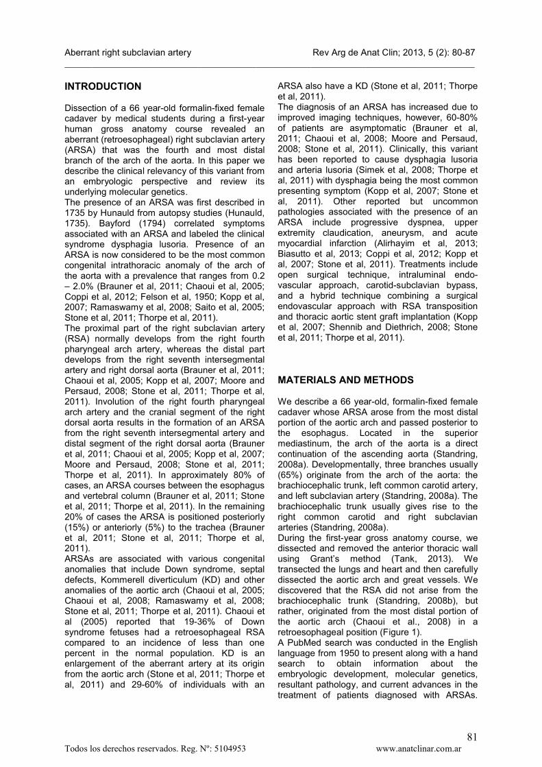
Figure 1. A: Anterior view of the arch of the aorta and great vessels. The trachea has been moved to the right to demonstrate the underlying esophagus. Note the position of the aberrant right subclavian artery (ARSA) posterior to the trachea and esophagus. In some cases, this arrangement has been reported to be the cause of dysphagia (Saito et al, 2005). RCCA, right common carotid artery; LCCA, left common carotid artery; LSA, left subclavian artery.
Aberrant right subclavian artery Rev Arg de Anat Clin; 2013, 5 (2): 80-87__________________________________________________________________________________________
Todos los derechos reservados. Reg. Nº: 5104953 www.anatclinar.com.ar83
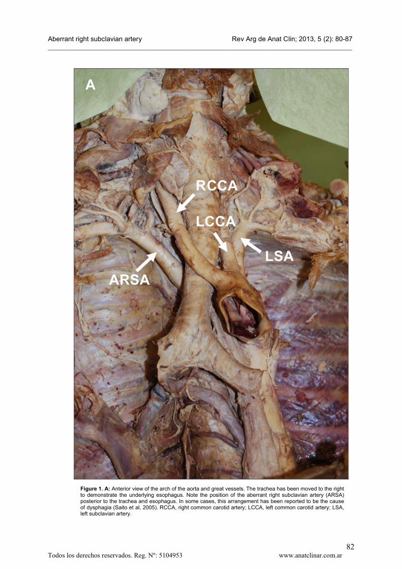
Figure 1. B: Close-up of the anatomy demonstrated in A with the posterior aspect of the arch of the aorta exposed. The origin (●) of the four vessels is clearly seen in this view as well as the location of the esophagus (ESO). From proximal to distal, they are the RCCA (diameter 9 mm), LCCA (diameter 9 mm), LSA (diameter 10 mm), and ARSA (diameter 12 mm).
RESULTS
Most of the literature surrounding the ARSA was prompted by a unique case, including incidental
findings of asymptomatic patients due to improved diagnostic imaging modalities. Most cases of an ARSA are asymptomatic, however, in those cases where symptoms do exist, the
Aberrant right subclavian artery Rev Arg de Anat Clin; 2013, 5 (2): 80-87__________________________________________________________________________________________
Todos los derechos reservados. Reg. Nº: 5104953 www.anatclinar.com.ar84
overwhelming majority present with progressive dysphagia (Brauner et al, 2011; Chaoui et al, 2008; Kopp et al, 2007; Moore and Persaud, 2008; Stone et al, 2011). It is common for the presence of a KD to be associated with an ARSA, 29-60% of cases (Stone et al, 2011; Thorpe et al, 2011); however, this was not the case in our report. The most common course of an ARSA is between the esophagus and
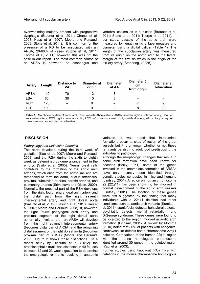
vertebral column as in our case (Brauner et al, 2011; Stone et al, 2011; Thorpe et al, 2011). In our study, vessels of the aortic arch were measured for length using a tape measure and diameter using a digital caliper (Table 1). The length of the subclavian artery was measured from its origin on the aortic arch to the lateral margin of the first rib which is the origin of the axillary artery (Standring, 2008b).
Table 1. Morphometric data of aortic arch blood vessels. Abbreviations: ARSA, aberrant right subclavian artery; LSA, left subclavian artery; RCC, right common carotid; LCC, left common carotid; VA, vertebral artery; AA, axillary artery. All measurements are reported in millimeters.
DISCUSSION
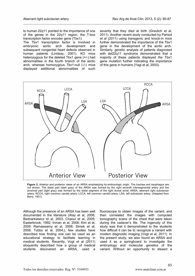
Embryology and Molecular GeneticsThe aorta develops during the third week of gestation (Kau et al, 2007; Moore and Persaud, 2008) and the RSA during the sixth to eighth week as determined by gene arrangement in the embryo (Saito et al, 2005). Neural crest cells contribute to the formation of the aortic arch arteries, which arise from the aortic sac and are remodeled to form the aorta, ductus arteriosus, proximal subclavian arteries, carotid arteries, and pulmonary arteries (Srivastava and Olson, 2000). Normally, the proximal part of the RSA develops from the right fourth pharyngeal arch artery and the distal part from the right seventh intersegmental artery and right dorsal aorta (Biasutto et al, 2012; Biasutto et al, 2013; Kau et al, 2007; Moore and Persaud, 2008). If, however, the right fourth pharyngeal arch artery and proximal segment of the right dorsal aorta abnormally involute, then an ARSA will develop from the right seventh intersegmental artery (becomes distal part of ARSA) and the remaining distal segment of the right dorsal aorta (becomes proximal part of ARSA) (Moore and Persaud, 2008). Figure 2 shows these distinctions. In a recent study by Biasutto et al (2012) the brachiocephalic trunk was dissected in 40 fetuses between 12 and 23 weeks gestation to determine the embryologic remnants resulting in anatomic
variation. It was noted that intraluminal formations occur at sites of fusion of the great vessels but it is unknown whether or not these remnants persist into adulthood predisposing the individual to pathology.Although the morphologic changes that result in aortic arch formation have been known for decades (Barry, 1951), some of the genes involved in the anomalous formation of ARSAs have only recently been identified through genetic studies conducted in mice and humans(Lindsay, 2001). A region on human chromosome 22 (22q11) has been shown to be involved in normal development of the aortic arch vessels (Lindsay, 2001). The location of these genes were first suggested by the finding that many individuals with a 22q11 deletion had other conditions such as aortic arch variants (Sureka et al, 2011), craniofacial defects, behavioral defects, psychiatric defects, mental retardation, and DiGeorge syndrome. These genes were found to be localized to the region involved in aortic arch formation (Lindsay, 2001). A review by Momma (2010) noted that 80% of patients with congenital cardiovascular defects had a chromosome 22q11 deletion. Comparison of the human 22q11 region with the murine homologous chromosome identified around 30 genes in the deleted region (Yagi et al, 2003).Further studies using knockout (KO) mice with deletions in the mouse chromosome homologous
Artery LengthDistance to
VADiameter at
originDiameter
at AA
Diameter 5 cm
from origin
Diameter at bifurcation
ARSA 110 70 12 8 – –
LSA 60 30 10 8 – –
RCC 120 – 9 – 7 9
LCC 100 – 9 – 8 8
Aberrant right subclavian artery Rev Arg de Anat Clin; 2013, 5 (2): 80-87__________________________________________________________________________________________
Todos los derechos reservados. Reg. Nº: 5104953 www.anatclinar.com.ar85
to human 22q11 pointed to the importance of one of the genes in the 22q11 region, the T-box transcription factor encoder gene (Tbx1).The Tbx1 transcription factor is involved in embryonic aortic arch development and subsequent congenital heart defects observed in human patients (Lindsay, 2001). KO mice heterozygous for the deleted Tbx1 gene (+/-) had abnormalities in the fourth branch of the aortic arch, whereas homozygous Tbx1-null (-/-) micedisplayed additional abnormalities of such
severity that they died at birth (Greulich et al, 2011). Another recent study conducted by Parisot et al (2011) using transgenic and knock-in mice further demonstrated the importance of the Tbx1 gene in the development of the aortic arch. Similarly, genetic analysis of patients diagnosed with del22q11 syndrome demonstrated that a majority of these patients displayed the Tbx1 gene mutation further indicating the importance of this gene in humans (Yagi et al, 2003).
Figure 2. Anterior and posterior views of an ARSA emphasizing its embryologic origin. The trachea and esophagus are not shown. The distal part (dark gray) of the ARSA was formed by the right seventh intersegmental artery and the proximal part (light gray) was formed by the distal segment of the right dorsal aorta. ARSA, aberrant right subclavian artery; RCCA, right common carotid artery; LCCA, left common carotid artery; LSA, left subclavian artery. (Adapted from Barry, 1951).
Although the presence of an ARSA has been well documented in the literature (Atay et al, 2006; Bednarkiewicz et al, 2003; Chaoui et al, 2005; Easterbrook, 1992; Inman et al, 2008; Kim et al, 2009; Ramaswamy et al, 2008; Simek et al, 2008; Tubbs et al, 2004;), few studies have described how finding one can be used as an educational strategy to facilitate learning in medical students. Recently, Vogt et al (2011) eloquently described how a group of medical students discovered an ARSA, used a
fluoroscope to obtain images of the variant, and then correlated the images with computed tomography scans of the chest that were taken during the cadaver’s life. The ingenuity of this study was that it demonstrated to the students how difficult it can be to recognize a variant with modern diagnostic imaging (Vogt et al, 2011). In the present study, we also found an ARSA and used it as a springboard to investigate the embryology and molecular genetics of the variant. Without an opportunity to dissect a
Aberrant right subclavian artery Rev Arg de Anat Clin; 2013, 5 (2): 80-87__________________________________________________________________________________________
Todos los derechos reservados. Reg. Nº: 5104953 www.anatclinar.com.ar86
human body, we would have never learned about this variation in so much depth, nor would we have had the opportunity to experience the process of anatomical research. Our study and that of Vogt et al (2011) provide novel examples of how valuable clinically-relevant cadaveric dissection is in medical education.
REFERENCES
Alirhayim Z, Qureshi W, Shafiq A, Hassan S.2013. Aortic arch variant presenting as an acute ST elevation myocardial infarction. BMJ case reports 2013.
Atay Y, Engin C, Posacioglu H, Ozyurek R, Ozcan C, Yagdi T, Ayik F, Alayunt EA. 2006. Surgical approaches to the aberrant right subclavian artery. Tex Heart Inst J 33: 477-481.
Barry A. 1951. The aortic arch derivatives in the human adult. Anat Rec 111: 221-238.
Bayford D. 1794. An account of a singular case of obstructed deglutition. Memoirs Med Soc London: 275-286.
Bednarkiewicz M, Bruschweiler I, Christenson JT. 2003. Undiagnosed aberrant right subclavian artery: Pitfall in aortic arch surgery. Cardiovascular surgery (London, England) 11:61-63.
Biasutto SN, Ceccón AF, Bortolín PA, de la Rosa M. 2012. Clinically important formations in the interior surface of the brachiocephalic trunk. Rev Arg de Anat Clin 4: 57-64.
Biasutto SN, Cecenarro RR, Ceccón GF, Álvarez ML, de la Rosa M, Bortolín PA. 2013. Rev Arg de Anat Clin 5: 21-28.
Brauner E, Lapidot M, Kremer R, Best LA, Kluger Y. 2011. Aberrant right subclavian artery-suggested mechanism for esophageal foreign body impaction: Case report. World J Emerg Surg 6: 12.
Chaoui R, Heling KS, Sarioglu N, Schwabe M, Dankof A, Bollmann R. 2005. Aberrant right subclavian artery as a new cardiac sign in second- and third-trimester fetuses with Down syndrome. Am J Obstet Gynecol 192: 257-263.
Chaoui R, Rake A, Heling KS. 2008. Aortic arch with four vessels: aberrant right subclavian artery. Ultrasound Obstet Gynecol 31: 115-117.
Coppi G, Tshomba Y, Psacharopulo D, Marone EM, Chiesa R. 2012. Aberrant right subclavian artery in blunt aortic injury: implication for treatment and review of the literature. Annals of vascular surgery 26: 861 e861-866.
Easterbrook JS. 1992. Identification of aberrant right subclavian artery on MR images of the cervical spine. J Magn Reson Imaging 2: 507-509.
Felson F, Cohen S, Courter SR, McGuire J. 1950. Anomalous right subclavian artery. Radiology 54: 340-349.
Greulich F, Rudat C, Kispert A. 2011. Mechanisms of T-box gene function in the developing heart. Cardiovascular research.
Hunauld P. 1735. Examen de quelques parties d'un singe. Hist Acad Roy Sci 135;2: 516-523.
Inman JC, Kim P, McHugh R. 2008. Retro-esophageal subclavian artery - esophageal fistula: A rare complication of a salivary bypass tube. Head & neck 30: 1120-1123.
Kau T, Sinzig M, Gasser J, Lesnik G, Rabitsch E, Celedin S, Eicher W, Illiasch H, Hausegger KA. 2007. Aortic development and anomalies. Semin Intervent Radiol 24: 141-152.
Kim Y-D, Yeo H-T, Cho Y-D. 2009. Anomalous variations of the origin and course of vertebral arteries in patients with retroesophageal right subclavian artery. J Korean Neurosurg Soc 45:297-299.
Kopp R, Wizgall I, Kreuzer E, Meimarakis G, Weidenhagen R, Kuhnl A, Conrad C, Jauch KW, Lauterjung L. 2007. Surgical and endovascular treatment of symptomatic aberrant right subclavian artery (arteria lusoria). Vascular 15: 84-91.
Lindsay EA. 2001. Chromosomal microdeletions: dissecting del22q11 syndrome. Nat Rev Genet 2: 858-868.
Momma K. 2010. Cardiovascular anomalies associated with chromosome 22q11.2 deletion syndrome. Am J Cardiol 105: 1617-1624.
Moore KL, Persaud TVN. 2008. The Developing Human: Clinically Oriented Embryology. 8th Ed. Philadelphia: Saunders Elsevier.
Parisot P, Mesbah K, Theveniau-Ruissy M, Kelly RG. 2011. Tbx1, subpulmonary myocardium and conotruncal congenital heart defects. Birth defects research 91: 477-484.
Ramaswamy P, Lytrivi ID, Thanjan MT, Nguyen T, Srivastava S, Sharma S, Ko HH, Parness IA, Lai WW. 2008. Frequency of aberrant subclavian artery, arch laterality, andassociated intracardiac anomalies detected by echocardiography. Am J Cardiol 101: 677-682.
Saito T, Tamatsukuri Y, Hitosugi T, Miyakawa K, Shimizu T, Oi Y, Yoshimoto M, Yamamoto Y, Spanel-Browski K, Steinke H. 2005. Three cases of retroesophageal right subclavian artery. Journal of Nippon Medical School = Nihon Ika Daigaku zasshi 72: 375-382.
Shennib H, Diethrich EB. 2008. Novel approaches for the treatment of the aberrant
Aberrant right subclavian artery Rev Arg de Anat Clin; 2013, 5 (2): 80-87__________________________________________________________________________________________
Todos los derechos reservados. Reg. Nº: 5104953 www.anatclinar.com.ar87
right subclavian artery and its aneurysms. Journal of vascular surgery 47: 1066-1070.
Simek M, Nemec P, Studentova H, Novotny J. 2008. Distal aortic arch aneurysm with a retroesophageal right subclavian artery (arteria lusoria). Eur J Cardiothorac Surg 34: 670.
Srivastava D, Olson EN. 2000. A genetic blueprint for cardiac development. Nature 407:221-226.
Standring S. 2008a. Heart and great vessels. In: Standring S, editor. Gray's Anatomy. 40th Ed. Edinburgh: Churchill Livingston Elsevier. p 959-987.
Standring S. 2008b. Pectoral girdle, shoulder region and axilla. In: Standring S, editor. Gray's Anatomy. 40th Ed. Edinburgh: Churchill Livingston Elsevier. p 791-822.
Stone WM, Ricotta JJ, 2nd, Fowl RJ, Garg N, Bower TC, Money SR. 2011. Contemporary management of aberrant right subclavian arteries. Annals of vascular surgery 25: 508-514.
Sureka AO, Peng LF, Reinhartz O, Reddy VM, Hanley FL. 2011. Anatomy in Patients with 22q11 Deletion and Pulmonary Atresia with Ventricular Septal Defect and Major
Aortopulmonary Collaterals. Surgical Science 2: 294-296.
Tank PW. 2013. Grant's Dissector. 15th Ed. Baltimore: Lippincott Williams & Wilkins.
Thorpe SW, Hohl JB, Gilbert S, Tannoury CA, Lee JY. 2011. Aberrant right subclavian artery encountered during debridement of T2 osteomyelitis and associated phlegmon. The spine journal : official journal of the North American Spine Society 11: e6-10.
Tubbs RS, Oakes WJ, Salter EG, Zehren SJ. 2004. Retroesophageal right subclavian artery with persistent ductus arteriosus. Anatomical Science International 79: 98-100.
Vogt KM, Kauh CY, Holder DM, DePhilip RM. 2011. Fluoroscopic angiography in the gross anatomy dissection laboratory: visualizing the aortic arch and its branches in a cadaver. Clinical Anatomy 24: 253-257.
Yagi H, Furutani Y, Hamada H, Sasaki T, Asakawa S, Minoshima S, Ichida F, Joo K, Kimura M, Imamura S, Kamatani N, Momma K, Takao A, Nakazawa M, Shimizu N, Matsuoka R. 2003. Role of TBX1 in human del22q11.2 syndrome. Lancet 362: 1366-1373.


![THE URINARY SYSTEM MODULE - kaukau.edu.sa/files/140/subjects/9941_urinary_module_-_january_2009[1].pdf · 4 Gross anatomy of upper and lower urinary tract Anatomy 5 Histology / Embryology](https://img.pdfslide.us/doc/110x75/60c9e788a5727742cd1eb962/the-urinary-system-module-1pdf-4-gross-anatomy-of-upper-and-lower-urinary.jpg)