Embed Size (px)
Citation preview
Gross anatomy, histology and developmental anatomy of
pituitary glandDr. Laxman Khanal
Asst. Professor, Department of Anatomy
BPKIHS, Dharan
17-11-2016

Q. ‘Pituitary’ is named so because it was though thata. Its size is like that of peab. It secret mucus like secretion which released from nosec. It is located in sella tursicad. It is master endocrine gland
Q. What structure lies lateral to the pituitary?a. Optic chiasma b. Diaphragm sellaec. Cavernous sinus d. sphenoid bone
Q. Hormone ‘Vasopressin’ is synthesized in.a. Posterior pituitary b. Anterior pituitaryb. Hypothalamus d. Thalamus

Introduction
• The pituitary gland is a pea-shaped structure measuring about 0.5inch in diameter that lies in the hypophysial fossa of the sphenoidbone and attaches to the hypothalamus by a stalk, the infundibulum.
• For long time pituitary gland was regarded as master endocrine glanddue to its control over other gland, but we now know that pituitaryitself has master that is hypothalamus.
• Pituitary gland is also called as ‘hypophysis cerebri’.
(Hypo=under, physis= growth, cerebri=cerebrum)

• Lies in hypophyseal fossa of sphenoid bone• Covered by dural fold (diaphragm sellae)• Above connected with hypothalamus by infundibulum.
3rd ventricle
Hypothalamus
Pituitary stalk
Pituitary gland CS
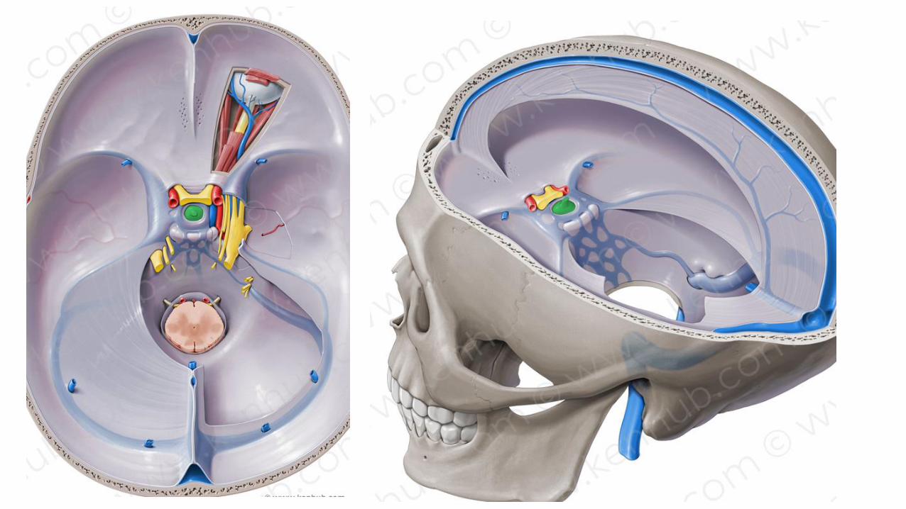


3rd ventricle
Post lobe
Ant lobe
Infundibulum Neurohypophysis
1. Pars posterior2. Infundibulum3. Median eminence
Cleft of pituitary
Intermediate lobe

3rd ventricle
Neurohypophysis
1. Pars posterior2. Infundibulum3. Median eminence
Adenophypophysis
1. Pars tuberalis2. Pars intermedia3. Pars anterior

3rd ventricle
OC

Prosencephalon (Forebrain)
1. Diencephalon 2. Telencephalon
Primitive oral cavity(stomodeum)
Floor of diencephalon
Roof of stomodeum
Rathke’s pouch
Neuroectodermaldiverticulum
Infundibulum
Neurohypophysis
AdenohypophysisAnt pituitaryIntermediate pituitary

Development of pituitary gland

CraniopharyngiomaRemnants of Rathke’s pocuh

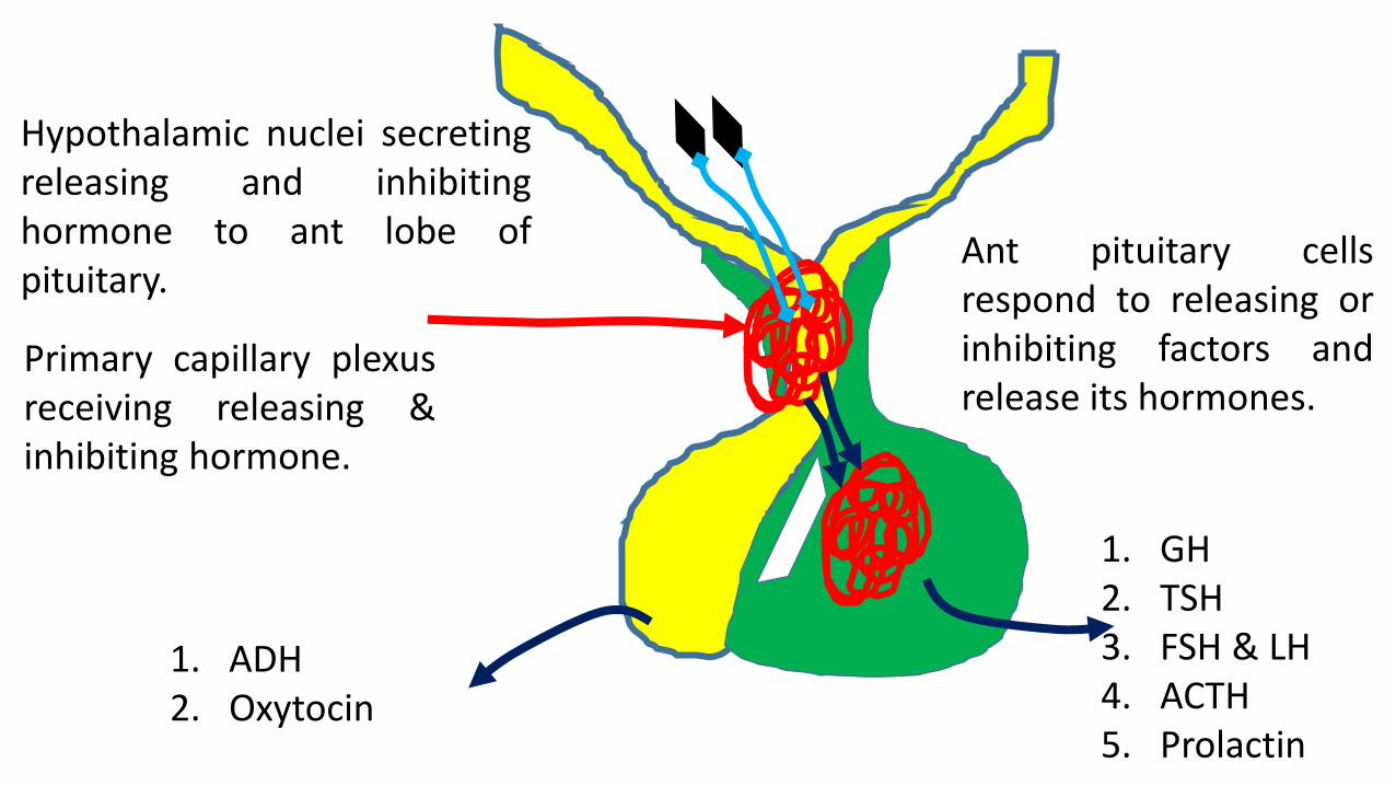
Primary capillary plexus
Secondary capillary plexus
Hypophysial Portal vein
Hypophysial vein
Superior and inferior hypophysial artery
Capillary plexus of posterior lobe
Hypophysial vein
Trabecular artery

1. GH2. TSH3. FSH & LH4. ACTH5. Prolactin
1. ADH2. Oxytocin
Hypothalamic nuclei secretingreleasing and inhibitinghormone to ant lobe ofpituitary.
Primary capillary plexusreceiving releasing &inhibiting hormone.
Ant pituitary cellsrespond to releasing orinhibiting factors andrelease its hormones.

1. GH2. TSH3. FSH & LH4. ACTH5. Prolactin
1. ADH2. Oxytocin
Hypothalamo-hypophysial tract (HHT)
PVSO
Herring bodies

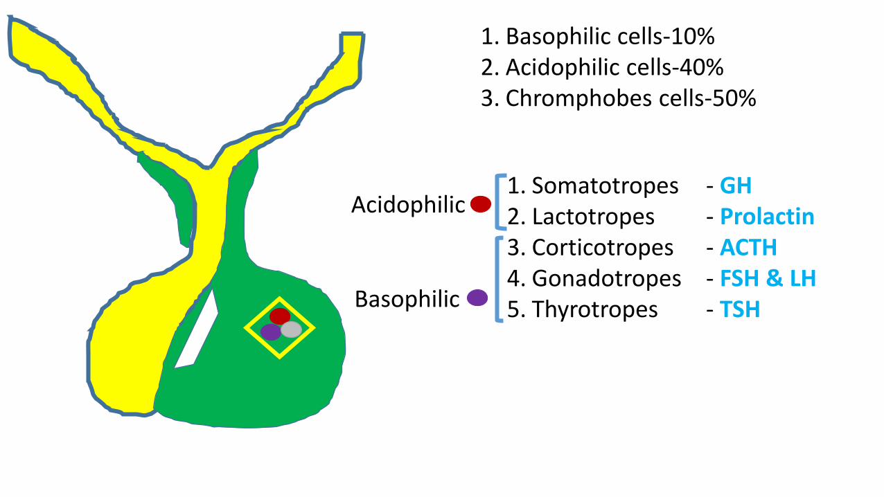
1. Basophilic cells-10%2. Acidophilic cells-40%3. Chromphobes cells-50%
1. Somatotropes - GH2. Lactotropes - Prolactin3. Corticotropes - ACTH4. Gonadotropes - FSH & LH5. Thyrotropes - TSH
Acidophilic
Basophilic
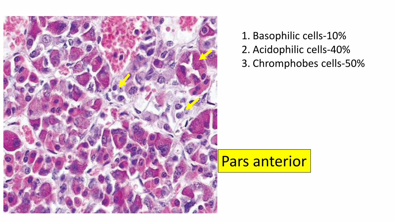
1. Basophilic cells-10%2. Acidophilic cells-40%3. Chromphobes cells-50%
Pars anterior
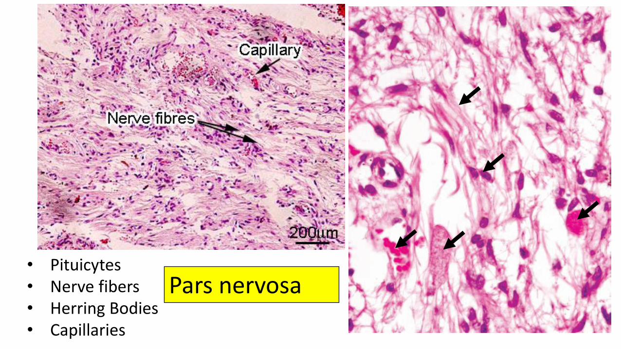
• Pituicytes• Nerve fibers • Herring Bodies• Capillaries
Pars nervosa

ADH stored in posterior pituitaryV2 R
V2 R
More water retain in body.Volume of urine decreases.
HHT system
Diabetes insipidus
Central DI
Nephrogenic DI No ADH
ADH synthesized in hypothalamus

Cushing disease
Pituitary adenoma of ant lobe
High ACTH secretion
High cortisol from adrenal cortex
• High BP• High blood glucose level• Moon face• Fatigueness
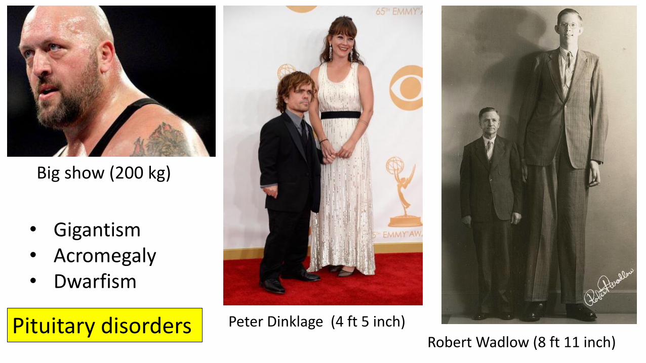
Robert Wadlow (8 ft 11 inch)
Peter Dinklage (4 ft 5 inch)
Big show (200 kg)
• Gigantism • Acromegaly• Dwarfism
Pituitary disorders