Embed Size (px)
Citation preview

Knee Anatomy
Ernest F. Talarico, Jr., Ph.D.
Associate Director of Medical Education
Associate Professor and Course Director, Human Gross Anatomy & Embryology
Coordinator, Anatomical Education Program
Indiana University School of Medicine-Northwest
AY14-15
Knee Joint
• The most complex joint in the body. Femur round, tibia flat.
• Comprised of 3 bones.– Femur– Tibia– patella

Femur
• Medial and Lateral Condyles- distal ends of the femur.
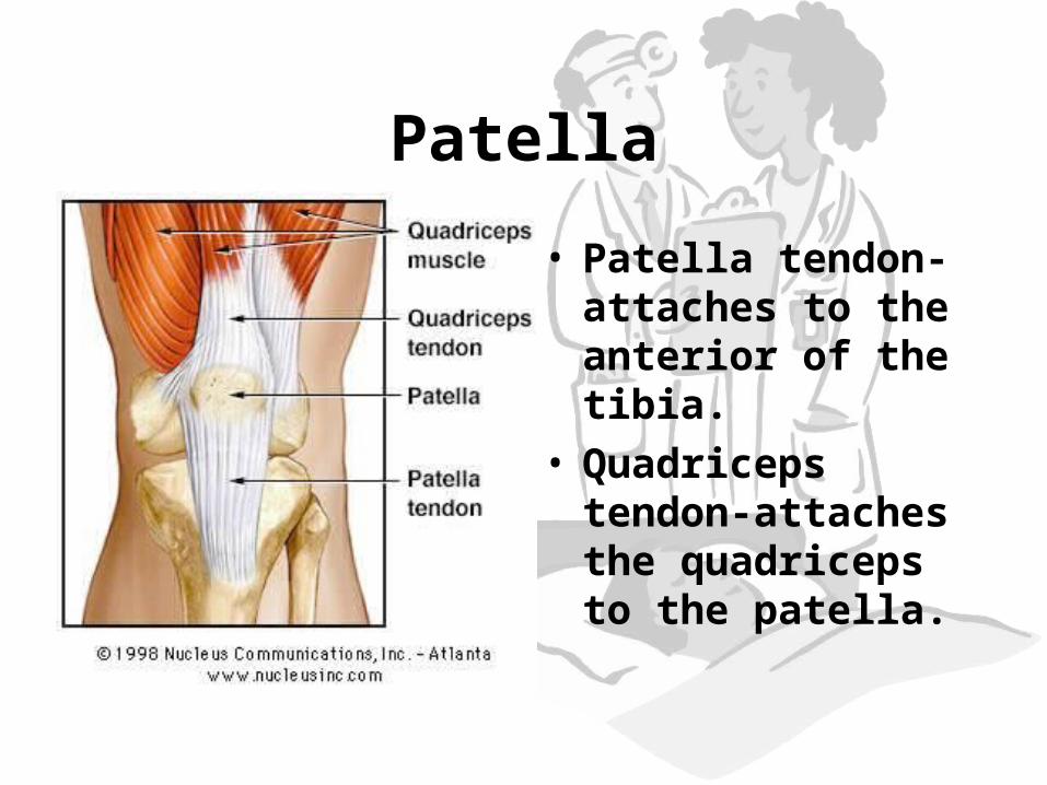
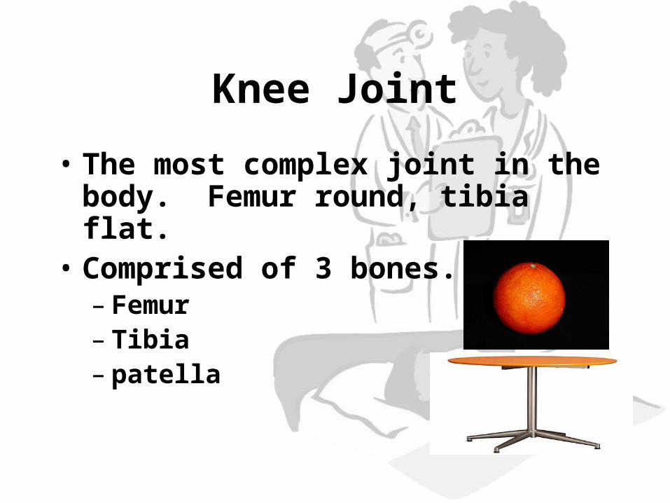
Patella
• Patella tendon- attaches to the anterior of the tibia.
• Quadriceps tendon-attaches the quadriceps to the patella.

Cruciate Ligaments• Major
stabilizing ligaments in the knee
• Anterior Cruciate Ligament (ACL)-prevents the tibia from sliding out in front of the femur
• Injuries caused by hyperflexion
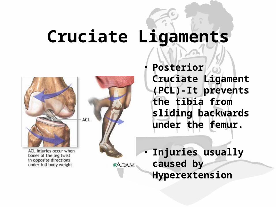
Cruciate Ligaments
• Posterior Cruciate Ligament (PCL)-It prevents the tibia from sliding backwards under the femur.
• Injuries usually
caused by Hyperextension
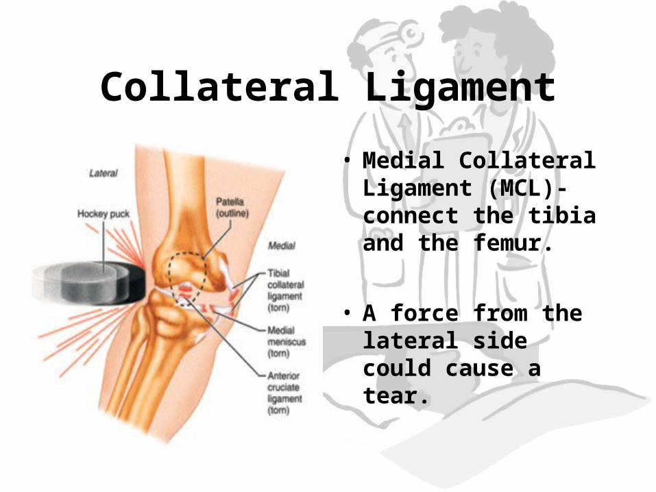
Collateral Ligament
• Medial Collateral Ligament (MCL)- connect the tibia and the femur.
• A force from the lateral side could cause a tear.

Collateral Ligament
• Lateral Collateral Ligament (LCL)- connect the fibula to the femur.
• A force from the medial side can cause a tear of the LCL


Cartilage
• Articulate Cartilage-covers the moving parts of the knee.
• Chronic damage to articulate cartilage leads to arthritis.

Cartilage
• Meniscus- half moon shaped cartilage lying between the knee joint.

Meniscus Tear
• Surgery
• Chronic Arthritis

Knee Injuries
• ACL Replacement surgery.