Embed Size (px)
Citation preview
Impact of Obesity on Nuclear Medicine Imaging
Mohammad A. Ghanem, Nafeesa A. Kazim, and Abdelhamid H. Elgazzar
Department of Nuclear Medicine, Mubarak Al Kabeer Hospital, Jabriya, Kuwait; and Department of Nuclear Medicine, Facultyof Medicine, Kuwait University, Safat, Kuwait
Obesity, with its alarming increase among adults and children,represents a significant health problem with serious medical,social, psychologic, and economic reverberations. The burden ofthis problem significantly affects the medical care system,including medical imaging. The effect of obesity on nuclearmedicine imaging spans many aspects, from preimaging patientpreparation to radiotracer administration, image acquisition, andimage interpretation. The acquired images may be suboptimalbecause of artifacts due to soft-tissue attenuation and incom-plete whole-body coverage, and quantification may be subopti-mal, especially for PET. Other difficulties include mechanicalproblems such as the weight limit of the imaging table and thebore size of the PET or SPECT/CT scanner and the need to alterthe timing, duration, or protocol of many imaging procedures.These issues are discussed in this review, which clarifies theimpact of this epidemic health problem on nuclear medicineservices and proposes possible solutions to overcome obesity-related difficulties encountered in nuclear medicine practice.
Key Words: obesity imaging; nuclear medicine; imaging
J Nucl Med Technol 2011; 39:40–50DOI: 10.2967/jnmt.110.078881
Obesity, a complex condition representing a significanthealth problem with serious medical, social, and psycho-logic reverberations, affects virtually all ages and socio-economic groups. It has reached an epidemic figureglobally, with more than 1 billion overweight adults, at least300 million of whom are clinically obese (1–9). Forming alarge economic burden, obesity has been estimated to ac-count for 2%–7% of the total health care cost in severaldeveloped countries (10).Additionally, obese and overweight children are a growing
concern. This disorder has doubled in children 10–17 y oldover the last decade, making it the most prevalent childhoodnutritional disorder in many parts of the world (11).Obese individuals encounter more health problems through-
out the course of their lives. Because of the alarming rate at
which the number of obese patients, both adults and children,is increasing, a bigger burden is being placed on the medicalcare system as it deals with obesity-related problems (12).One such problem is radiologic images that are difficult toperform and interpret (13). This review discusses these diffi-culties and suggests ways to overcome them.
The effects of obesity on imaging can be classified intopreimaging, imaging, and postimaging categories.
PREIMAGING CONSIDERATIONS
Obesity affects several preimaging aspects, includingcalculation of radiopharmaceutical activity, injection of theradiopharmaceutical, and preparation of the patient.
Administered Activity
The standard activity of radiopharmaceuticals in adults isbased on the ideal standard weight of a patient, 70 kg (14).Parameters such as pregnancy and sometimes renal func-tion are considered limiting factors in the radiation dosedelivered to the patient. Obtaining acceptable image qualityin an obese patient frequently requires use of an activityhigher than used in a patient of ideal weight. However, theradiation dose to the patient recommended by the Interna-tional Commission on Radiological Protection limits theincrease in activity, and hence the dose may not be highenough to produce an adequate study for proper interpreta-tion due to a reduced signal-to-noise ratio and increasedscatter in the acquired image. In PET, for example, anincrease in the injected dose according to patient weightcan be used to overcome poor image quality due to scatter,but the dose cannot exceed 925 MBq (25 mCi) of 18F-FDG(15). A common procedure used in myocardial perfusionimaging is to calculate the activity on the basis of patientweight and adjust upward for heavier patients by using afixed formula dose such as 11.47 MBq (0.31 mCi)/kg for99mTc agents or 1.48 MBq (0.04 mCi)/kg for 201Tl. Anotheroption to overcome this limitation and improve image qual-ity is to lengthen the acquisition time or use a multidetectorsystem for higher statistical counts (14,16).
Injection
Subcutaneous adipose tissue in obese patients can act as abarrier to visualization and palpation of the underlyingvascular structure and make intravenous access cumbersome.Such cases should be dealt with by more experienced nuclear
Received May 11, 2011; revision accepted Oct. 29, 2010.For correspondence or reprints contact: Abdelhamid Elgazzar, Department
of Nuclear Medicine, Kuwait University, P.O. Box 24923, Safat, Kuwait,13110.E-mail: [email protected] ª 2011 by the Society of Nuclear Medicine, Inc.
40 JOURNAL OF NUCLEAR MEDICINE TECHNOLOGY • Vol. 39 • No. 1 • March 2011
by on July 10, 2018. For personal use only. tech.snmjournals.org Downloaded from

medicine technologists, an intravenous team, or even ananesthesiologist.Some hospitals have sophisticated settings in which
ultrasonography-guided peripheral intravenous access canbe used (17,18). Costantino el al (19), in studying a sub-group of 60 patients with difficult intravenous access, con-cluded that ultrasonography-guided peripheral intravenousaccess is more successful than traditional blind techniques,requires less time, decreases the number of percutaneouspunctures, and improves patient satisfaction.
Patient Preparation
In addition to the well-known aspects of patient prepa-ration before a nuclear medicine study, such as goodhydration, fasting, body hair shaving for cardiac studies,and specific drug cessation, some special considerationsshould be kept in mind in obese or overweight patients,such as the amount of hydration (when required in somestudies) and longer acquisition times. Greater fluid intakeshould be encouraged because, among other factors,adequate hydration is dependent on the patient’s weight.The patient should be told how long an acquisition isexpected, since this will encourage greater cooperation bythe patient during positioning and acquisition.
IMAGING CONSIDERATIONS
Patient and Staff Safety
Obese patients generally need more staff assistance.Patient transfer from stretcher or wheelchair to the imagingtable may be cumbersome, and the patient may fall and beinjured if not carefully assisted. Any accessory gadgetssuch as mobile steps and walkers available in the imagingroom should be used to prevent possible injury to thepatient. Furthermore, the members of the staff may be givenspecial training on how to use an assistive device, such as aHoyer lift, to reduce their chance of being injured whileattempting to lift or transfer obese patients. If patienttransfer is still a problem, it is recommended that a mobileg-camera, if available, be wheeled to the patient’s stretcher.
Camera Setup and Limitations
Most g-camera tables have a weight limit of 180 kg (400lb). It is unsafe for overweight patients to be positioned on ag-camera table if the maximum bearing weight is exceeded.A mobile g-camera, if available, can be wheeled to thepatient’s bed or stretcher for certain studies such as lungventilation–perfusion scans. Imaging equipment that is usedfor heavy patients may show increased wear and tear; poten-tial stress to the table motors requires particular attention.
FIGURE 1. Whole-body PET/CT scan of 48-y-old woman (body mass index, 39; blood glucose level, 101 mg/dL, or 5.6 mmol/L) withhistory of breast cancer whose body contour exceeds PET and CT field of view, causing cropping and reconstruction artifact at left-breast region.
IMPACT OF OBESITY ON NUCLEAR MEDICINE • Ghanem et al. 41
by on July 10, 2018. For personal use only. tech.snmjournals.org Downloaded from
When multihead g-cameras are used, the position of thedetectors is usually adjusted automatically; however, in thecase of an obese patient, the detector position may beadjusted manually to accommodate the contour of thepatient. A g-camera integrated with a CT scanner has anentrance gantry that does not exceed 80 cm, creating diffi-culty in accommodating some obese patients. Artifacts suchas cropping and beam hardening can occur when thepatient’s girth exceeds the field of view, leading to a lowersensitivity for detection of peripheral lesions (Fig. 1) (20).Even large-field-of-view cameras may not accommodatethe body of the patient in whole-body imaging. Parts ofthe body may not be included, and additional images shouldbe obtained. If the patient’s body is larger than the borediameter of the imaging system, the patient cannot undergothe scan, even though body weight may not exceed the limitspecified by the manufacturer. New-generation scannershave promising potential advantages for the imaging ofheavy patients, such as an increased table weight limitand an increased gantry bore diameter (21).
Positioning
Nuclear medicine studies generally take longer thanradiologic studies. This increases the chance of patientmovement during acquisition, since remaining still for long
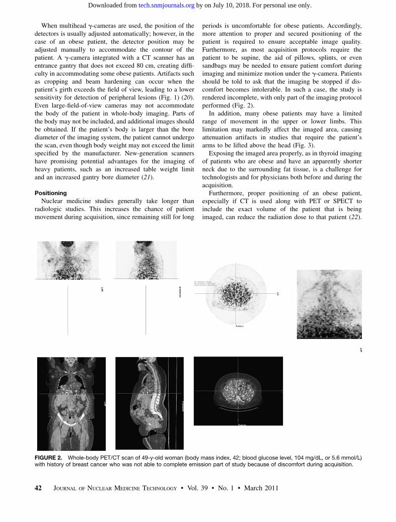
periods is uncomfortable for obese patients. Accordingly,more attention to proper and secured positioning of thepatient is required to ensure acceptable image quality.Furthermore, as most acquisition protocols require thepatient to be supine, the aid of pillows, splints, or evensandbags may be needed to ensure patient comfort duringimaging and minimize motion under the g-camera. Patientsshould be told to ask that the imaging be stopped if dis-comfort becomes intolerable. In such a case, the study isrendered incomplete, with only part of the imaging protocolperformed (Fig. 2).
In addition, many obese patients may have a limitedrange of movement in the upper or lower limbs. Thislimitation may markedly affect the imaged area, causingattenuation artifacts in studies that require the patient’sarms to be lifted above the head (Fig. 3).
Exposing the imaged area properly, as in thyroid imagingof patients who are obese and have an apparently shorterneck due to the surrounding fat tissue, is a challenge fortechnologists and for physicians both before and during theacquisition.
Furthermore, proper positioning of an obese patient,especially if CT is used along with PET or SPECT toinclude the exact volume of the patient that is beingimaged, can reduce the radiation dose to that patient (22).
FIGURE 2. Whole-body PET/CT scan of 49-y-old woman (body mass index, 42; blood glucose level, 104 mg/dL, or 5.6 mmol/L)with history of breast cancer who was not able to complete emission part of study because of discomfort during acquisition.
42 JOURNAL OF NUCLEAR MEDICINE TECHNOLOGY • Vol. 39 • No. 1 • March 2011
by on July 10, 2018. For personal use only. tech.snmjournals.org Downloaded from

Difficulty in positioning after all efforts may lead to sub-optimal imaging, longer procedures, and increased radia-tion dose to the patient and technologist.Including the entire body within the acquired images can
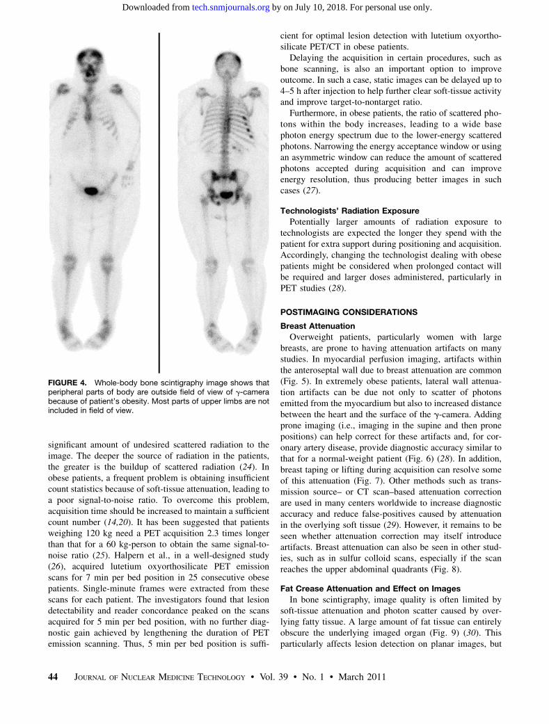
be troublesome in obese patients. This issue is a problem inwhole-body bone scans or PET scans, as parts of the bodyperiphery may not be included in the imaged area (Fig. 4).The standard acquisition protocol may need to be
modified in obese patients. For instance, a combined supine
and prone acquisition may be required to maintain thespecificity of gated myocardial perfusion imaging in thediagnosis of coronary artery disease (23).
Acquisition
The number of photons reaching the g-detector dependsgreatly on the amount of attenuating tissue and the geom-etry of the patient’s body. Compton scattering attenuatesprimary photons reaching the g-camera and contributes a
FIGURE 3. Lung perfusion images of overweight female patient who was unable to lift arms over head during acquisition, causingattenuation defects (arrows) on lateral, right anterior oblique, and left anterior oblique images.
IMPACT OF OBESITY ON NUCLEAR MEDICINE • Ghanem et al. 43
by on July 10, 2018. For personal use only. tech.snmjournals.org Downloaded from
significant amount of undesired scattered radiation to theimage. The deeper the source of radiation in the patients,the greater is the buildup of scattered radiation (24). Inobese patients, a frequent problem is obtaining insufficientcount statistics because of soft-tissue attenuation, leading toa poor signal-to-noise ratio. To overcome this problem,acquisition time should be increased to maintain a sufficientcount number (14,20). It has been suggested that patientsweighing 120 kg need a PET acquisition 2.3 times longerthan that for a 60 kg-person to obtain the same signal-to-noise ratio (25). Halpern et al., in a well-designed study(26), acquired lutetium oxyorthosilicate PET emissionscans for 7 min per bed position in 25 consecutive obesepatients. Single-minute frames were extracted from thesescans for each patient. The investigators found that lesiondetectability and reader concordance peaked on the scansacquired for 5 min per bed position, with no further diag-nostic gain achieved by lengthening the duration of PETemission scanning. Thus, 5 min per bed position is suffi-
cient for optimal lesion detection with lutetium oxyortho-silicate PET/CT in obese patients.
Delaying the acquisition in certain procedures, such asbone scanning, is also an important option to improveoutcome. In such a case, static images can be delayed up to4–5 h after injection to help further clear soft-tissue activityand improve target-to-nontarget ratio.
Furthermore, in obese patients, the ratio of scattered pho-tons within the body increases, leading to a wide basephoton energy spectrum due to the lower-energy scatteredphotons. Narrowing the energy acceptance window or usingan asymmetric window can reduce the amount of scatteredphotons accepted during acquisition and can improveenergy resolution, thus producing better images in suchcases (27).
Technologists’ Radiation Exposure
Potentially larger amounts of radiation exposure totechnologists are expected the longer they spend with thepatient for extra support during positioning and acquisition.Accordingly, changing the technologist dealing with obesepatients might be considered when prolonged contact willbe required and larger doses administered, particularly inPET studies (28).
POSTIMAGING CONSIDERATIONS
Breast Attenuation
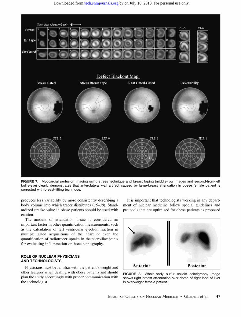
Overweight patients, particularly women with largebreasts, are prone to having attenuation artifacts on manystudies. In myocardial perfusion imaging, artifacts withinthe anteroseptal wall due to breast attenuation are common(Fig. 5). In extremely obese patients, lateral wall attenua-tion artifacts can be due not only to scatter of photonsemitted from the myocardium but also to increased distancebetween the heart and the surface of the g-camera. Addingprone imaging (i.e., imaging in the supine and then pronepositions) can help correct for these artifacts and, for cor-onary artery disease, provide diagnostic accuracy similar tothat for a normal-weight patient (Fig. 6) (28). In addition,breast taping or lifting during acquisition can resolve someof this attenuation (Fig. 7). Other methods such as trans-mission source– or CT scan–based attenuation correctionare used in many centers worldwide to increase diagnosticaccuracy and reduce false-positives caused by attenuationin the overlying soft tissue (29). However, it remains to beseen whether attenuation correction may itself introduceartifacts. Breast attenuation can also be seen in other stud-ies, such as in sulfur colloid scans, especially if the scanreaches the upper abdominal quadrants (Fig. 8).
Fat Crease Attenuation and Effect on Images
In bone scintigraphy, image quality is often limited bysoft-tissue attenuation and photon scatter caused by over-lying fatty tissue. A large amount of fat tissue can entirelyobscure the underlying imaged organ (Fig. 9) (30). Thisparticularly affects lesion detection on planar images, but
FIGURE 4. Whole-body bone scintigraphy image shows thatperipheral parts of body are outside field of view of g-camerabecause of patient’s obesity. Most parts of upper limbs are notincluded in field of view.
44 JOURNAL OF NUCLEAR MEDICINE TECHNOLOGY • Vol. 39 • No. 1 • March 2011
by on July 10, 2018. For personal use only. tech.snmjournals.org Downloaded from

FIGURE 5. Multislice views (A) and raw images (B) show large-breast-attenuation artifact (arrows) involving anterolateralmyocardial wall of obese female patient.
IMPACT OF OBESITY ON NUCLEAR MEDICINE • Ghanem et al. 45
by on July 10, 2018. For personal use only. tech.snmjournals.org Downloaded from

the problem can sometimes be resolved by performingSPECT of the region of interest. Different techniques forattenuation correction have been proposed for better imagequality and enable better quantification (31).Similarly, in a study on sentinel lymph node biopsy in
842 breast cancer patients, unsuccessful mapping wassignificantly higher in patients with a body mass indexgreater than 30 than in those less than 30 (14/113 [12.4%]vs. 8/425 [1.9%], respectively; P , 0.001), among otherfactors (32).The addition of SPECT/CT to the acquisition protocol
used for lymphoscintigraphy in overweight and obesepatients improves nodal identification and avoids false-positive interpretations of sites of nonnodal uptake (33).Another example of the effect of fat crease attenuation on
image quality during whole-body bone or gallium scans isseen in Figure 10. The folded skin creates thicker soft tissueoverlying the imaged area of interest, causing linear abnor-mally increased uptake at the edge of the fat crease. If at thelevel of a vertebral body, this artifact could be mistaken for
a compression fracture (34). This can also affect bone den-sitometry studies, causing falsely higher values at the levelof the crease. The amount of soft tissue in obese patientshas been observed to cause diffusely increased skull activ-ity on bone scans due to disparate attenuation of overlyingsoft tissues (35).
Steatopygia
A high degree of fat accumulation in the buttocks cancause marked attenuation and a decrease in counts in thelumbar spine and pelvic region, obscuring or mimickingdisease (Fig. 11). SPECT acquisitions can help in such sit-uations to confirm or rule out any disease.
Quantitation Difficulties
Standardized uptake value takes into account the differ-ences between normalizing for body weight, for lean bodymass, and for surface area. Obese or overweight patientsusually have an overestimated standardized uptake valuewhen calculated using patient weight. Although not popu-lar, using lean body mass or body surface area usually
FIGURE 6. Myocardial perfusion imaging using supine and prone stress technique clearly demonstrates that inferior wall artifact(arrows) caused by diaphragmatic attenuation in obese male patient is corrected on prone images (middle row).
46 JOURNAL OF NUCLEAR MEDICINE TECHNOLOGY • Vol. 39 • No. 1 • March 2011
by on July 10, 2018. For personal use only. tech.snmjournals.org Downloaded from
produces less variability by more consistently describing abody volume into which tracer distributes (36–38). Stand-ardized uptake value in obese patients should be used withcaution.The amount of attenuation tissue is considered an
important factor in other quantification measurements, suchas the calculation of left ventricular ejection fraction inmultiple gated acquisitions of the heart or even thequantification of radiotracer uptake in the sacroiliac jointsfor evaluating inflammation on bone scintigraphy.
ROLE OF NUCLEAR PHYSICIANSAND TECHNOLOGISTS
Physicians must be familiar with the patient’s weight andother features when dealing with obese patients and shouldplan the study accordingly with proper communication withthe technologist.
It is important that technologists working in any depart-ment of nuclear medicine follow special guidelines andprotocols that are optimized for obese patients as proposed
FIGURE 7. Myocardial perfusion imaging using stress technique and breast taping (middle-row images and second-from-leftbull’s-eye) clearly demonstrates that anterolateral wall artifact caused by large-breast attenuation in obese female patient iscorrected with breast-lifting technique.
FIGURE 8. Whole-body sulfur colloid scintigraphy imageshows right-breast attenuation over dome of right lobe of liverin overweight female patient.
IMPACT OF OBESITY ON NUCLEAR MEDICINE • Ghanem et al. 47
by on July 10, 2018. For personal use only. tech.snmjournals.org Downloaded from

in Table 1. Knowing the camera weight limit and borewidth is important to avoid overloading the mobile tableor jamming the patient into the CT or PET opening. Askingpatients about their weight before they make the appoint-ment will save the staff a lot of time and prevent unneces-sary cancellations. In addition, choosing the right dose andcamera setting with the proper energy windowing is essen-
tial to get an acceptable image quality. If patient manipu-lation is required before and after placement under theg-camera, an extra hand is probably advisable to avoidtechnologist injury or prolonged radiation exposure (18).Proper communication with the referring physician andnuclear medicine staff is mandatory to ensure a safe andsmooth transfer of the patient.
CONCLUSION
No less than other medical services, nuclear medicineservices are affected by obesity in important and variableways. If nuclear medicine personnel make proper arrange-ments, problems arising during patient transportation,preparation, and imaging can be limited. Special protocolsshould be established to deal with overweight and obesepatients, depending on the prevalence of this problem in theserviced area. Such preparations can help correct for theartifacts arising from obesity and in the interpretation ofimages and can improve imaging outcomes in thesepatients.
FIGURE 9. Photon attenuation caused by massive abdominalfat, which renders interpretation of lumbar spine and pelvisdifficult on anterior view of this male subject.
FIGURE 10. Fat tissue over lower abdomen and buttocks ongallium scan causes soft-tissue attenuation of detectedphotons on anterior and posterior views.
FIGURE 11. Whole-body bone scan of obese patientillustrates effect of body build on images. Attenuation is seenin region of lower lumbar spine and pelvis, in addition to edgeartifact caused by fat crease in mid posterior lumbar spine and,to lesser extent, in anterior ribs.
48 JOURNAL OF NUCLEAR MEDICINE TECHNOLOGY • Vol. 39 • No. 1 • March 2011
by on July 10, 2018. For personal use only. tech.snmjournals.org Downloaded from

REFERENCES
1. Gu D, He J, Duan X, et al. Body weight and mortality among men and women in
China. JAMA. 2006;295:776–783.
2. Al-Isa AN. Body mass index, overweight and obesity among Kuwaiti intermedi-
ate school adolescents aged 10–14 years. Eur J Clin Nutr. 2004;58:1273–1277.
3. Obesity: Preventing and Managing the Global Epidemic—Report of a WHO Con-
sultation on Obesity. Geneva, Switzerland: World Health Organization; 1998.
4. Barth N, Ziegler A, Himmelmann GW, et al. Significant weight gains in a
clinical sample of obese children and adolescents between 1985 and 1995. Int
J Obes Relat Metab Disord. 1997;21:122–126.
5. Troiano RP, Flegal KM. Overweight children and adolescents: description, epi-
demiology, and demographics. Pediatrics. 1998;101:497–504.
6. Kopelman PG. Obesity as a medical problem. Nature. 2000;404:635–643.
7. Cernerud L. Height and body mass index of seven-year-old Stockholm school-
children from 1940 to 1990. Acta Paediatr. 1993;82:304–305.
8. Popkin BM, Doak CM. The obesity epidemic is a worldwide phenomenon. Nutr
Rev. 1998;56:106–114.
9. Freedman DS, Srinivasan SR, Valdez RA, et al. Secular increases in relative
weight and adiposity among children over two decades: the Bogalusa Heart
Study. Pediatrics. 1997;99:420–426.
10. Obesity: Preventing and Managing the Global Epidemic—Report of a WHO
Consultation. Geneva, Switzerland: World Health Organization, 2000. WHO
Technical Report Series, no. 894.
11. Sherina MS, Rozali A. Childhood obesity: contributing factors, consequences
and intervention. Mal J Nutr. 2004;10:13–22.
12. Wang G, Dietz WH. Economic burden of obesity in youths aged 6 to 17 years:
1979–1999. Pediatrics. 2002;109:E81.
13. Uppot RN, Sahani DV, Hahn PF, Kalra MK, Saini SS, Mueller PR. Effect of
obesity on image quality: fifteen-year longitudinal study for evaluation of dic-
tated radiology reports. Radiology. 2006;240:435–439.
14. Hansen CL, Goldstein RA, Berman DS, et al. Myocardial perfusion and function
single photon emission computed tomography. J Nucl Cardiol. 2006;13:e97–e120.
15. Everaert H, Vanhove C, Lahoutte T, et al. Optimal dose of 18F-FDG required for
whole-body PET using an LSO PET camera. Eur J Nucl Med Mol Imaging.
2003;30:1615–1619.
16. Karesh SM. Principles of radiopharmacy. In: Henkin RE, Bova D, Dillehay GL,et al.,
eds. Nuclear Medicine. 2nd ed. Philadelphia, PA: Mosby/Elsevier; 2006:342–343.
17. Keyes LE, Frazee BW, Snoey ER, Simon BC, Christy D. Ultrasound-guided
brachial and basilic vein cannulation in emergency department patients with
difficult intravenous access. Ann Emerg Med. 1999;34:711–714.
18. Botkin CD, Osman MM. Prevalence, challenges, and solutions for 18F-FDG PET
studies of obese patients: a technologist’s perspective. J Nucl Med Technol.
2007;35:80–83.
19. Costantino TG, Parikh AK, Satz WA, Fojtik JP. Ultrasonography-guided periph-
eral intravenous access versus traditional approaches in patients with difficult
intravenous access. Ann Emerg Med. 2005;46:456–461.
20. Uppot RN, Sahani DV, Hahn PF, Gervais D, Mueller PR. Impact of obesity on
medical imaging and image-guided intervention. AJR. 2007;188:433–440.
21. Surli S, Karp J, Werner M, et al. Imaging performance of an LYSO-based TOF
PET scanner [abstract]. J Nucl Med. 2006;47(suppl):54P.
22. Li J, Udayasankar UK, Toth TL, Seamans J, Small WC, Kalra MK. Automatic
patient centering for MDCT: effect on radiation dose. AJR. 2007;188:
547–552.
23. Nishina H, Slomka PJ, Abidov A, et al. Combined supine and prone quanti-
tative myocardial perfusion SPECT: method development and clinical valida-
tion in patients with no known coronary artery disease. J Nucl Med.
2006;47:51–58.
24. Barnes WE. In vivo quantification of activity by planar imaging. In: Henkin RE,
Bova D, Dillehay GL, et al., eds. Nuclear Medicine. 2nd ed. Philadelphia, PA:
Mosby/Elsevier; 2006:179–180.
TABLE 1Main Effects of Obesity on Nuclear Medicine Imaging and Possible Remedies
Obesity-related difficulty Impact on imaging Possible remedy
Body configuration Attenuation Proper preparation such as adequate hydration
(higher amount)
Increase of injected activityDelayed acquisition in some studies
Increased time of acquisition
Access to veins Possible infiltration Use of more experienced nuclear medicine technologist
Patient discomfort Use of intravenous teamHelp from an anesthesiologist
Use of ultrasound-guided peripheral intravenous
access injection method
Patient mobility Possible fall injuries Use of greater cautionUse of accessory gadgets
Imaging of patient on stretcher (mobile camera
may be needed)Adequate communication with patient
Positioning for acquisition Difficulty in achieving proper positions Attention to proper and secured positioning
Use of pillow, splints, or other means to ensure
patient’s comfort and thus minimize motionBody contouring Variable distances from camera at different
parts of body
Manual adjustment of camera head to obtain
adequate images
Patient girth Compton scatter Use of narrower or asymmetric energy window
Insufficient count statistics Increase of acquisition timeBeam-hardening artifact on CT. Use of caution during reading and quantification
Reduced sensitivity to peripheral lesions Comparison with non–attenuation-corrected images
Acquisition of additional spot image studiesDiaphragmatic attenuation
(cardiac)
Masking of underlying organ activity
(inferior wall artifact)
Use of prone acquisition in myocardial perfusion
Use of attenuation correction
Breast attenuation (cardiac) Masking of underlying organ activity
(anteroseptal-lateral wall defect)
Breast lifting and binding
Use of attenuation correctionFat crease and steatopygia Edge effect/attenuation
Masking of bone details and creation
of false findings
Manipulation of crease and reimaging, or
addition of SPECT
Addition of extra views or SPECT
IMPACT OF OBESITY ON NUCLEAR MEDICINE • Ghanem et al. 49
by on July 10, 2018. For personal use only. tech.snmjournals.org Downloaded from

25. Watson CC, Casey ME, Bendriem B, et al. Optimizing injected dose in clinical
PET by accurately modeling the counting rate response functions specific to
individual patient scans. J Nucl Med. 2005;46:1825–1834.
26. Halpern BS, Dahlbom M, Auerbach MA, et al. Optimizing imaging protocols for
overweight and obese patients: a lutetium orthosilicate PET/CT study. J Nucl
Med. 2005;46:603–607.
27. Kojima A, Matsumoto M, Takahashi M, Uehara S. Effect of energy resolution on
scatter fraction in scintigraphic imaging: Monte Carlo study.Med Phys. 1993;20:
1107–1113.
28. Berman DS, Kang X, Nishina H, et al. Diagnostic accuracy of gated Tc-99m
sestamibi stress myocardial perfusion SPECT with combined supine and prone
acquisitions to detect coronary artery disease in obese and nonobese patients.
J Nucl Cardiol. 2006;13:191–201.
29. Downer J. Hybrid SPECT�CT: making a difference.Medical Solutions: Changing the
Way Healthcare Is Delivered. June 2006: 79–83. Availableat: http://www.medical.
siemens.com/siemens/en_US/rg_marcom_FBAs/files/brochures/magazin_
2006/solution_internet_june_06_eng.pdf. Accessed December 20, 2010.
30. Weiner GM, Jenicke L, Mueller V, Bohuslavizki KH. . Artifacts and non-osseous
uptake in bone scintigraphy: imaging reports of 20 cases. Radiol Oncol. 2001;35:
185–191.
31. Case JA, Licho R, King MA, Weaver JP. Bone SPECT of the spine: a comparison
of attenuation correction techniques. J Nucl Med. 1999;40:604–613.
32. Goyal A, Newcombe RG, Chhabra A, Mansel RE; ALMANAC Trialists Group.
Factors affecting failed localisation and false-negative rates of sentinel node
biopsy in breast cancer: results of the ALMANAC validation phase. Breast
Cancer Res Treat. 2006;99:203–208.
33. Lerman H, Lievshitz G, Zak O, Metser U, Schneebaum S, Even-Sapir E. Im-
proved sentinel node identification by SPECT/CT in overweight patients with
breast cancer. J Nucl Med. 2007;48:201–206.
34. Elgazzar AH. Basic sciences of bone and joint disease. In: Elgazzar AH.
Orthopedic Nuclear Medicine. New York, NY: Springer; 2004:26.
35. Lorberboym M, Macadziob S, Nikolov G, Kim CK. The hot skull sign on bone
scans of obese patients resulting from disparate soft tissue attenuation. Clin Nucl
Med. 2005;30:680–681.
36. Basu S, Zaidi H, Houseni M, et al. Novel quantitative techniques for assessing
regional and global function and structure based on modern imaging modalities:
implications for normal variation, aging and diseased states. Semin Nucl Med.
2007;37:223–239.
37. Schomburg A, Bender H, Reichel C, et al. Standardized uptake values of fluo-
rine-18 fluorodeoxyglucose: the value of different normalization procedures. Eur
J Nucl Med. 1996;23:571–574.
38. Thie JA. Understanding the standardized uptake value, its methods, and impli-
cations for usage. J Nucl Med. 2004;45:1431–1434.
50 JOURNAL OF NUCLEAR MEDICINE TECHNOLOGY • Vol. 39 • No. 1 • March 2011
by on July 10, 2018. For personal use only. tech.snmjournals.org Downloaded from

Doi: 10.2967/jnmt.110.078881Published online: February 14, 2011.
2011;39:40-50.J. Nucl. Med. Technol. Mohammad A. Ghanem, Nafeesa A. Kazim and Abdelhamid H. Elgazzar Impact of Obesity on Nuclear Medicine Imaging
http://tech.snmjournals.org/content/39/1/40This article and updated information are available at:
http://tech.snmjournals.org/site/subscriptions/online.xhtml
Information about subscriptions to JNMT can be found at:
http://tech.snmjournals.org/site/misc/permission.xhtmlInformation about reproducing figures, tables, or other portions of this article can be found online at:
(Print ISSN: 0091-4916, Online ISSN: 1535-5675)1850 Samuel Morse Drive, Reston, VA 20190.SNMMI | Society of Nuclear Medicine and Molecular Imaging
is published quarterly.Journal of Nuclear Medicine Technology
© Copyright 2011 SNMMI; all rights reserved.
by on July 10, 2018. For personal use only. tech.snmjournals.org Downloaded from