Embed Size (px)
Citation preview

IFSSH Scientific Committee on Anatomy and Biomechanics
Chair: Eduardo R. Zancolli (Argentina)
Committee: Carlos Zaidenberg (Argentina)
Diego Piazza (Argentina)
Hernan Iriarte (Argentina)
Report submitted February 2013

Anatomy of the Hand
Knowledge of anatomy of the hand is the basis for understanding pathology and for
precise application of surgical techniques.
This committee has selected to write a report on the TFCC anatomy but believing also
that it will be extremely useful for hand surgeons to have a brief actualization of the
most significant new papers on hand anatomy.
Since the last IFSSH´s Committees Reports (November 2010), a lot of papers on hand
anatomy have been published, many confirming what is already known but some adding
new knowledge on the already know structures. This last group mainly refers to
anatomical variations that need to be known in order to diminish surgical surprises and
to imagine how to deal with them.
We are conscious that due to the huge amount of bibliographical sources we might have
missed some important papers and we apologize for it.
Committee Report on Anatomy of the TFCC
The Triangular Fibrocartilage was initially described by Weitbrecht (1742) as
intermedia triangularis cartilage. In modern literature were Palmer and Werner
(1981) that introduced the term Triangular-Fibro Cartilage-Complex (TFCC) to describe
these structures.
Anatomical description
The TFCC is a structure running from the medial border of the radius to the styloid
area of the ulna and with a distal volar expansion to the carpus. This fibrocartilage-
ligament complex stabilizes the distal radio-ulnar and ulno-carpal joints, distributing
load between the carpus and ulna, and allowing smooth wrist flexion-extension, radial-
ulnar deviation and forearm pronation-supination.
Other structures also intervene in stability: musculotendinous structures, the bony
anatomy and the interosseous membrane.
The TFCC different components are:
Articular disc
Dorsal and palmar distal radio-ulnar ligaments
Meniscus homologue
Capsule
Extensor carpi ulnaris subsheath
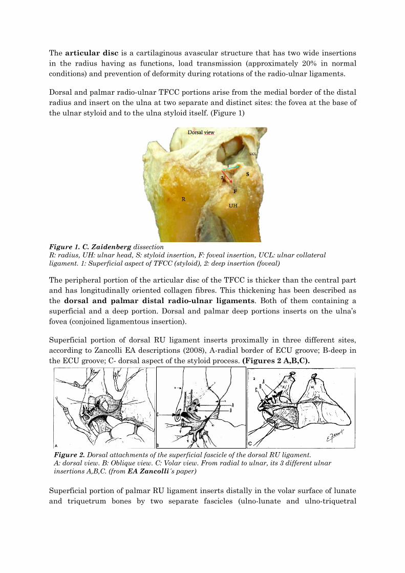
The articular disc is a cartilaginous avascular structure that has two wide insertions
in the radius having as functions, load transmission (approximately 20% in normal
conditions) and prevention of deformity during rotations of the radio-ulnar ligaments.
Dorsal and palmar radio-ulnar TFCC portions arise from the medial border of the distal
radius and insert on the ulna at two separate and distinct sites: the fovea at the base of
the ulnar styloid and to the ulna styloid itself. (Figure 1)
Figure 1. C. Zaidenberg dissection
R: radius, UH: ulnar head, S: styloid insertion, F: foveal insertion, UCL: ulnar collateral
ligament. 1: Superficial aspect of TFCC (styloid), 2: deep insertion (foveal)
The peripheral portion of the articular disc of the TFCC is thicker than the central part
and has longitudinally oriented collagen fibres. This thickening has been described as
the dorsal and palmar distal radio-ulnar ligaments. Both of them containing a
superficial and a deep portion. Dorsal and palmar deep portions inserts on the ulna’s
fovea (conjoined ligamentous insertion).
Superficial portion of dorsal RU ligament inserts proximally in three different sites,
according to Zancolli EA descriptions (2008), A-radial border of ECU groove; B-deep in
the ECU groove; C- dorsal aspect of the styloid process. (Figures 2 A,B,C).
Figure 2. Dorsal attachments of the superficial fascicle of the dorsal RU ligament.
A: dorsal view. B: Oblique view. C: Volar view. From radial to ulnar, its 3 different ulnar
insertions A,B,C. (from EA Zancolli´s paper)
Superficial portion of palmar RU ligament inserts distally in the volar surface of lunate
and triquetrum bones by two separate fascicles (ulno-lunate and ulno-triquetral
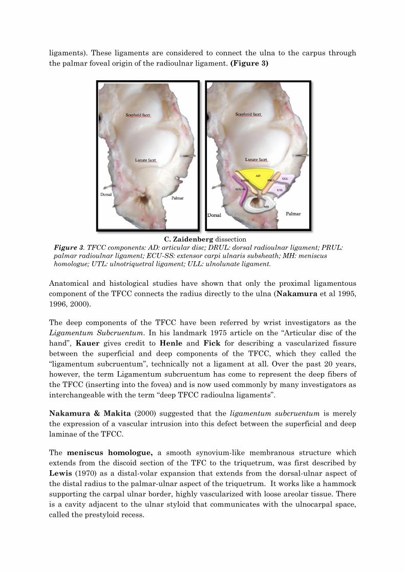
ligaments). These ligaments are considered to connect the ulna to the carpus through
the palmar foveal origin of the radioulnar ligament. (Figure 3)
C. Zaidenberg dissection
Figure 3. TFCC components: AD: articular disc; DRUL: dorsal radioulnar ligament; PRUL:
palmar radioulnar ligament; ECU-SS: extensor carpi ulnaris subsheath; MH: meniscus
homologue; UTL: ulnotriquetral ligament; ULL: ulnolunate ligament.
Anatomical and histological studies have shown that only the proximal ligamentous
component of the TFCC connects the radius directly to the ulna (Nakamura et al 1995,
1996, 2000).
The deep components of the TFCC have been referred by wrist investigators as the
Ligamentum Subcruentum. In his landmark 1975 article on the “Articular disc of the
hand”, Kauer gives credit to Henle and Fick for describing a vascularized fissure
between the superficial and deep components of the TFCC, which they called the
“ligamentum subcruentum”, technically not a ligament at all. Over the past 20 years,
however, the term Ligamentum subcruentum has come to represent the deep fibers of
the TFCC (inserting into the fovea) and is now used commonly by many investigators as
interchangeable with the term “deep TFCC radioulna ligaments”.
Nakamura & Makita (2000) suggested that the ligamentum subcruentum is merely
the expression of a vascular intrusion into this defect between the superficial and deep
laminae of the TFCC.
The meniscus homologue, a smooth synovium-like membranous structure which
extends from the discoid section of the TFC to the triquetrum, was first described by
Lewis (1970) as a distal-volar expansion that extends from the dorsal-ulnar aspect of
the distal radius to the palmar-ulnar aspect of the triquetrum. It works like a hammock
supporting the carpal ulnar border, highly vascularized with loose areolar tissue. There
is a cavity adjacent to the ulnar styloid that communicates with the ulnocarpal space,
called the prestyloid recess.
Garcia Elias, based on histological studies, considered the tissue which continued from
the TFCC to the carpal bone as a meniscus homologue which is difficult to separate from
the TFCC.
Others authors (Ishii, Palmer & Werner 1998) redefined in three configurations of the
meniscus homologue and the prestyloid recess, based on how the prestyloid recess
communicates with the ulnocarpal space: 1- narrow opening type; 2- wide opening type;
3-no opening type.
Different studies have shown that there are variations in the attachment of the TFCC to
the triquetrum. Hogikyan & Louis subdivided the patterns of its attachment to the
triquetrum into four types: a small, thin structure and focal attachment (group 1: 28%);
a small, thick structure and focal attachment (group 2: 39%); a thick structure and
broad attachment to between one-third and one-quarter of the triquetrum (group 3:
28%); and a broad attachment covering the entire triquetrum (group 4: 5%).
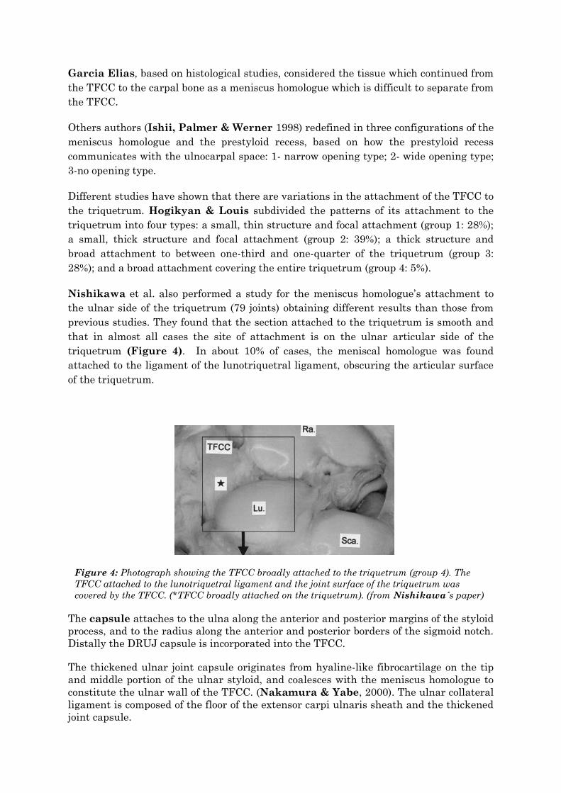
Nishikawa et al. also performed a study for the meniscus homologue’s attachment to
the ulnar side of the triquetrum (79 joints) obtaining different results than those from
previous studies. They found that the section attached to the triquetrum is smooth and
that in almost all cases the site of attachment is on the ulnar articular side of the
triquetrum (Figure 4). In about 10% of cases, the meniscal homologue was found
attached to the ligament of the lunotriquetral ligament, obscuring the articular surface
of the triquetrum.
Figure 4: Photograph showing the TFCC broadly attached to the triquetrum (group 4). The
TFCC attached to the lunotriquetral ligament and the joint surface of the triquetrum was
covered by the TFCC. (*TFCC broadly attached on the triquetrum). (from Nishikawa´s paper)
The capsule attaches to the ulna along the anterior and posterior margins of the styloid
process, and to the radius along the anterior and posterior borders of the sigmoid notch.
Distally the DRUJ capsule is incorporated into the TFCC.
The thickened ulnar joint capsule originates from hyaline-like fibrocartilage on the tip
and middle portion of the ulnar styloid, and coalesces with the meniscus homologue to
constitute the ulnar wall of the TFCC. (Nakamura & Yabe, 2000). The ulnar collateral
ligament is composed of the floor of the extensor carpi ulnaris sheath and the thickened
joint capsule.
The Extensor Carpi Ulnaris (ECU) tendon courses through the sixth dorsal
compartment of the wrist, passing dorsal on the lower end of the ulna through a small
fibro-osseous tunnel. The tendon is held tightly in the ulnar groove by a thin
subsheath, a proper relatively rigid retinaculum, attached on the margins of the ulnar
groove and ensuring its stability during pronation-supination. It is a pulley described by
Bourgery et al as “petit arcade fibrose” which prevents ECU tendon subluxation. The
ECU retinaculum is separate from the dorsal or extensor retinaculum and covered by
expansions of the extensor retinaculum, which plays no stabilizing role with regard to
the ECU tendon.
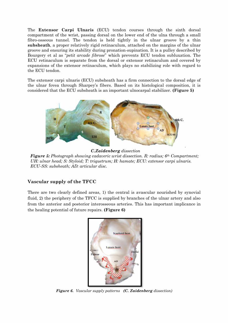
The extensor carpi ulnaris (ECU) subsheath has a firm connection to the dorsal edge of
the ulnar fovea through Sharpey’s fibers. Based on its histological composition, it is
considered that the ECU subsheath is an important ulnocarpal stabilizer. (Figure 5)
.
C.Zaidenberg dissection
Figure 5: Photograph showing cadaveric wrist dissection. R: radius; 6th Compartment;
UH: ulnar head; S: Styloid; T: triquetrum; H: hamate; ECU: extensor carpi ulnaris.
ECU-SS: subsheath; AD: articular disc.
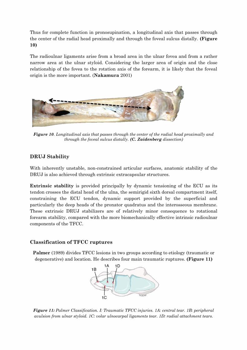
Vascular supply of the TFCC
There are two clearly defined areas, 1) the central is avascular nourished by synovial
fluid, 2) the periphery of the TFCC is supplied by branches of the ulnar artery and also
from the anterior and posterior interosseous arteries. This has important implicance in
the healing potential of future repairs. (Figure 6)
Figure 6. Vascular supply patterns (C. Zaidenberg dissection)

Innervation of the TFCC
Gupta et al (2001) studied the innervation of the TFCC. Central and radial aspects of
the TFCC do not have any nerve fascicles or fibers present.
The volar portion of the TFCC is innervated by a branch of the ulnar nerve and the
dorsal sensory branch of the ulnar nerve.
The ulnar and dorsal aspects of the TFCC are more variable in their patterns of
innervation. Branches of the ulnar nerve and the dorsal sensory branch of the ulnar
nerve innervate the ulnar aspect of the complex. Branches of the posterior interosseous
nerve and the dorsal sensory branch of the ulnar nerve innervate the dorsal aspect of
the TFCC. (Figure 7)
Figure 7. Low-power view of preserved and sectioned TFCC showing sampled regions including
central/radial (c), palmar (p), ulnar (u), and dorsal (d). (from Gupta´s paper)
Cavalcante ML, Rodrigues CJ, R. Mattar Jr (2004) went futher and studied
mechanoreceptors and nerve endings. The free nerve endings, (Figure 8) responsible
for sensing pain, predominate in the ulnar and dorsal areas. The Vater-Pacini
corpuscles predominate in the radial and dorsal area, promoting perception of the onset
or cessation of movement and mechanical stress change. The Golgi-Mazzoni corpuscles
were more frequent in the ulnar and ventral areas, linking these areas to function of
slow adaptation and sensation of extreme movements. The propioceptive function
receptors were found in all areas of TFCC because Ruffini corpuscles have homogeneous
distribution in its fibrocartilaginous tissue.

Figure 8. Free nerve endings (arrows) in the fibrocartilaginous tissue at the peripheral area of
the TFCC. (from R. Matta Jr´s paper)
Biomechanics in short
The distal radioulnar joint (DRUJ) has been defined as a diarthrodial trochoid
articulation formed by the head of the ulna and the shallow sigmoid cavity of the lower
end of the radius. The curvatures of the two articulating surfaces are not equal. The
radius of the ulna is about two thirds the length of the sigmoid notch concavity. This
results in a relatively unstable articulation with reduced area of contact between the
two bones. To overcome this, different stabilizing structures exist: (a) the TFCC,
composed of the discus articularis, the palmar and dorsal radioulnar ligaments, the
ulnocarpal ligaments, and the ECU sheath; (b) the pronator quadratus muscle; and (c)
the interosseous membrane.
The concave radius-of-curvature of the sigmoid notch is greater than that of the ulna
head (Figure 9).
Full congruity of two articulating surfaces is therefore not possible. This incongruity of
articular surfaces creates a geometrically non-constrained articulation at the DRUJ,
subject to translational dorsal and palmar instability. In the extremes of forearm
rotation, <10% of the ulnar head may be in contact with the sigmoid notch.
Figure 9. Different radius-of-curvature between distal radius and ulna
(C.Zaidenberg dissection)
Thus for complete function in pronosupination, a longitudinal axis that passes through
the center of the radial head proximally and through the foveal sulcus distally. (Figure
10)
The radioulnar ligaments arise from a broad area in the ulnar fovea and from a rather
narrow area at the ulnar styloid. Considering the larger area of origin and the close
relationship of the fovea to the rotation axis of the forearm, it is likely that the foveal
origin is the more important. (Nakamura 2001)
Figure 10. Longitudinal axis that passes through the center of the radial head proximally and
through the foveal sulcus distally. (C. Zaidenberg dissection)
DRUJ Stability
With inherently unstable, non-constrained articular surfaces, anatomic stability of the
DRUJ is also achieved through extrinsic extracapsular structures.
Extrinsic stability is provided principally by dynamic tensioning of the ECU as its
tendon crosses the distal head of the ulna, the semirigid sixth dorsal compartment itself,
constraining the ECU tendon, dynamic support provided by the superficial and
particularly the deep heads of the pronator quadratus and the interosseous membrane.
These extrinsic DRUJ stabilizers are of relatively minor consequence to rotational
forearm stability, compared with the more biomechanically effective intrinsic radioulnar
components of the TFCC.
Classification of TFCC ruptures
Palmer (1989) divides TFCC lesions in two groups according to etiology (traumatic or
degenerative) and location. He describes four main traumatic ruptures. (Figure 11)
Figure 11: Palmer Classification. I: Traumatic TFCC injuries. 1A: central tear. 1B: peripheral
avulsion from ulnar styloid. 1C: volar ulnocarpal ligaments tear. 1D: radial attachment tears.
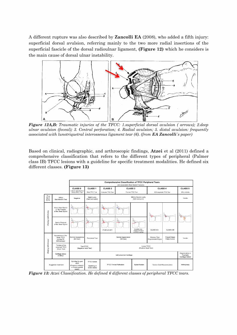
A different rupture was also described by Zancolli EA (2008), who added a fifth injury:
superficial dorsal avulsion, referring mainly to the two more radial insertions of the
superficial fascicle of the dorsal radioulnar ligament, (Figure 12) which he considers is
the main cause of dorsal ulnar instability.
Figure 12A,B: Traumatic injuries of the TFCC: 1.superficial dorsal avulsion ( arrows); 2.deep
ulnar avulsion (foveal); 3. Central perforation; 4. Radial avulsion; 5. distal avulsion: frequently
associated with lunotriquetral interosseous ligament tear (6). (from EA Zancolli´s paper)
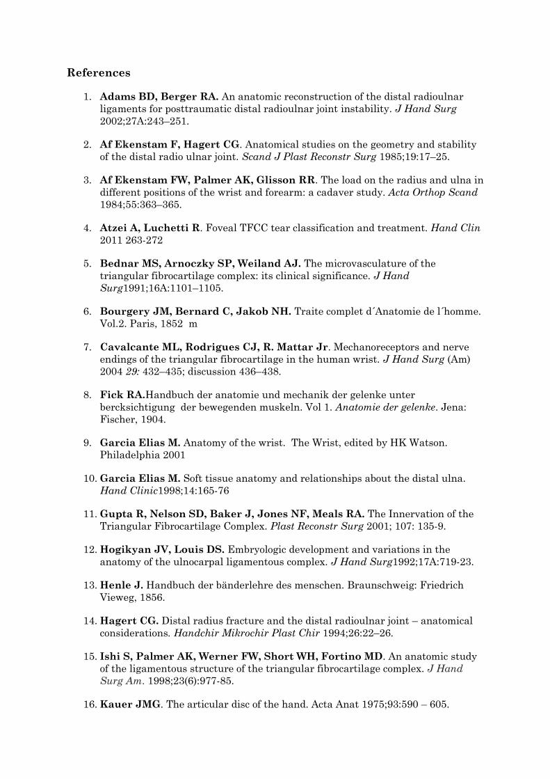
Based on clinical, radiographic, and arthroscopic findings, Atzei et al (2011) defined a
comprehensive classification that refers to the different types of peripheral (Palmer
class IB) TFCC lesions with a guideline for specific treatment modalities. He defined six
different classes. (Figure 13)
Figure 13: Atzei Classification. He defined 6 different classes of peripheral TFCC tears.
References
1. Adams BD, Berger RA. An anatomic reconstruction of the distal radioulnar
ligaments for posttraumatic distal radioulnar joint instability. J Hand Surg
2002;27A:243–251.
2. Af Ekenstam F, Hagert CG. Anatomical studies on the geometry and stability
of the distal radio ulnar joint. Scand J Plast Reconstr Surg 1985;19:17–25.
3. Af Ekenstam FW, Palmer AK, Glisson RR. The load on the radius and ulna in
different positions of the wrist and forearm: a cadaver study. Acta Orthop Scand
1984;55:363–365.
4. Atzei A, Luchetti R. Foveal TFCC tear classification and treatment. Hand Clin
2011 263-272
5. Bednar MS, Arnoczky SP, Weiland AJ. The microvasculature of the
triangular fibrocartilage complex: its clinical significance. J Hand
Surg1991;16A:1101–1105.
6. Bourgery JM, Bernard C, Jakob NH. Traite complet d´Anatomie de l´homme.
Vol.2. Paris, 1852 m
7. Cavalcante ML, Rodrigues CJ, R. Mattar Jr. Mechanoreceptors and nerve
endings of the triangular fibrocartilage in the human wrist. J Hand Surg (Am)
2004 29: 432–435; discussion 436–438.
8. Fick RA.Handbuch der anatomie und mechanik der gelenke unter
bercksichtigung der bewegenden muskeln. Vol 1. Anatomie der gelenke. Jena:
Fischer, 1904.
9. Garcia Elias M. Anatomy of the wrist. The Wrist, edited by HK Watson.
Philadelphia 2001
10. Garcia Elias M. Soft tissue anatomy and relationships about the distal ulna.
Hand Clinic1998;14:165-76
11. Gupta R, Nelson SD, Baker J, Jones NF, Meals RA. The Innervation of the
Triangular Fibrocartilage Complex. Plast Reconstr Surg 2001; 107: 135-9.
12. Hogikyan JV, Louis DS. Embryologic development and variations in the
anatomy of the ulnocarpal ligamentous complex. J Hand Surg1992;17A:719-23.
13. Henle J. Handbuch der bänderlehre des menschen. Braunschweig: Friedrich
Vieweg, 1856.
14. Hagert CG. Distal radius fracture and the distal radioulnar joint – anatomical
considerations. Handchir Mikrochir Plast Chir 1994;26:22–26.
15. Ishi S, Palmer AK, Werner FW, Short WH, Fortino MD. An anatomic study
of the ligamentous structure of the triangular fibrocartilage complex. J Hand
Surg Am. 1998;23(6):977-85.
16. Kauer JMG. The articular disc of the hand. Acta Anat 1975;93:590 – 605.
17. Kleinman WB, GrahamTJ. Distal ulnar injury and dysfunction. In: Peimer CA,
ed. Surgery of the hand and upper extremity. New York: McGraw-Hill, 1996:667–
709.
18. Kleinman WB, Graham TJ. The distal radioulnar joint capsule: clinical anatomy
and role in posttraumatic limitation of forearm rotation. J Hand Surg 1998;
23A:588–599.
19. Lewis OJ, Hamshere RJ, Bucknill TM. The anatomy of the wrist joint. J Anat
1970;106:539-552.
20. Lewis OJ, Hamshere RJ, Bucknill TM. The anatomy of the wrist joint. J Anat
1969;106:539-52.
21. Nakamura T, Yabe Y, Horiuchi Y. Functional Anatomy of the Triangular
Fibrocartilage Complex. Journal of Hand Surgery 1996. 21B. 5: 581-586.
22. Ono H, Gilula LA, Marzke MW, Obermann WR. Bicompartmentalization of
the radiocarpal joint. J Hand Surg1996;21A:788-93.
23. Palmer AK, Werner FW. The triangular fibrocartilage complex of the wrist:
anatomy and function. J Hand Surg1981;6:153-62.
24. Palmer AK, Werner FW. Biomechanics of the distal radioulnar joint. Clin
Orthop Relat Res 1984;187:26–35.
25. Palmer AK. Triangular fibrocartilage complex lesions: A classification. J Hand
Surg(Am.) 14: 594, 1989.
26. Pogue DJ, Viegas SF, Patterson RM, Peterson PD, Jenkins DK, Sweo TD,
Hokanson JA. Effects of distal radius fracture malunion on wrist joint
mechanics. J Hand Surg 1990; 15A:721–727.
27. Schuind F, An KN, Berglund L, Rey R, Cooney WP, Linscheid RL, Chao
EYS. The distal radioulnar ligaments: a biomechanical study. J Hand Surg
1991;16A:1106 –1114.
28. Schuind F, An KN, Berglund L, Rey R, Cooney WP, Linscheid RL, Chao
EYS. The distal radioulnar ligaments: a biomechanical study. J Hand Surg
1991;16A:1106 –1114.
29. Stuard P, Berger R, Linscheid R, An K. Dorso Palmar Stability of the Distal
Radio Ulnar Joint. J Hand Surg 2000, 25A : 689-6
30. Spinner M, Kaplan EB. Extensor carpi ulnaris: its relationship to stability of
the distal radioulnar joint. Clin Orthop Relat Res 1970;68:124–128.
31. Thiru-Pathi RG, Ferlic DC, Clayton ML, McClure DC. Arterial anatomy of
the triangular fibrocartilage of the wrist and its clinical significance. J Hand
Surg 1986;11A:258– 263.
32. Viegas SF, Pogue DJ, Patterson RM, Peterson PD. Effects of radioulnar
instability on the radiocarpal joint: a biomechanical study. J Hand Surg
1990;15A:728–732.
33. Weitbrecht J. Syndesmologia sive Historia Ligamentarum corporis Humani,
quam secundum observations Anatomicas Concinnavit, et Figuris and Objecta
Recentia Adumbratis Illustravit. Academy of Sciences, Petropoli, 1742.
34. Zancolli EA. Etiopatogenia y tratamiento de la inestabilidad dorsal del extremo
distal del cúbito consecutiva a la rotura traumática del fibrocartílago triangular.
Rev Asoc Argent OrtopTraumatol2008; 73(3):2-23.
Latest Papers on Hand Anatomy
I. Ligaments
The insertion points of the thumb`s MP joint collateral ligaments has been described
with some precision. The ulnar collateral ligament (UCL) has a metacarpal origin 4.2
mm from the dorsal surface and 5.3 mm from the articular surface. The center of the
phalangeal insertion of the UCL was 2.8 mm from the volar surface and 3.4 mm from
the articular surface. The volar aspect of the phalangeal insertion extended up to 0.7
mm from the volar edge of the phalanx.
The radial collateral ligament (RCL) inserts at the metacarpal, having its center at
3.5 mm from the dorsal surface and 3.3 mm from the articular surface, the dorsal aspect
being 1.5 mm from the dorsal edge of the metacarpal. The RCL`s center at phalangeal
insertion was 2.8 mm from the volar surface and 2.6 mm from the articular surface,
being its volar aspect 0.5 mm from the volar edge of the phalanx. This data is relevant
for successful repair and reconstruction.
- Carlson MG, Warner KK, Meyers KN, Hearns KA, Kok PL. Anatomy of the Thumb
Metacarpophalangeal Ulnar and Radial Collateral Ligaments. J Hand Surg Am.
2012 Oct; 37(10): 2021-6.
II. Nerves
The TMC joint has been described as innervated by the radial nerve (main innervation),
the lateral antebrachial nerve innervation and the median nerve. Even though
denervation’s based on these structures not always lead to good results. A new study on
19 cadaveric specimens shows that 58% had superficial radial nerve, 47% had median
nerve innervation from the motor branch and 47% had ulnar nerve innervation from the
motor branch. This paper supposed to speak for the first time that ulnar innervation
may also be present for the TMC joint.
- Miki RA, Kam CC, Gennis ER, Barkin JA, Riel RU, Robinson PG, Owens PW. Ulnar
nerve component to innervation of thumb carpometacarpal joint. Iowa Orthop
J. 2011; 31:225-30.
Deep palmar communications between the ulnar and median nerves have continued to
be studied. (50 hands, 25 cadavers). In 16% of the hands communicating branches were
found
- Marios Loukas, Sharath S Bellary, R Shane Tubbs, Mohammadali M Shoja, Aaron A
Cohen Gadol. Deep palmar communications between the ulnar and median
nerves. Clin Anat. 2011 Mar; 24 (2): 197-201.
Another paper also describes that a connecting third common palmar digital branch of
the median nerve with the fourth common palmar and proper palmar digital branches of
the median nerve presented a plexiform nature.
- Sirasanagandla SR, Patil J, Potu BK, Nayak BS, Shetty SD, Bhat KM. A rare
anatomical variation of the Berrettini anastomosis and third common palmar
digital branch of the median nerve. Anat Sci Int. 2013 Jan 17.
The median nerve branches for the pronator teres have been studied in one paper. All
specimens (20 upper limbs) showed to have a branch from the median nerve long enough
to reach the radial nerve in the cubital fossa in potential for neurotization cases.
- Tubbs RS, Beckman JM, Loukas M, Shoja MM, Cohen-Gadol AA. Median nerve
branches to the pronator teres: cadaveric study with potential use in
neurotization procedures to the radial nerve at the elbow. J Neurosurg. 2011
Jan; 114(1): 253-5.
The sublime bridge is the tendinous arch connecting the radial and humeral heads of
the flexorum digitorum superficialis muscle. Located at the mean distance of 8.1 mm
from the medial epicondyle, it was found to be tendinous in 75% and muscular in 25% of
the specimens. As known, it is a potential factor for median nerve compression at the
proximal forearm.
- Tubbs RS, Marshall T, Loukas M, Shoja MM, Cohen-Gadol AA. The sublime bridge:
anatomy and implications in median nerve entrapment. J Neurosurg. 2010 Jul;
113(1): 110-2.

III. Muscles
The flexor carpi radialis brevis muscle is a muscular variant that can be present as
much as 3.95 % in cadaveric studies. On volar approaches for distal radius fractures it
may be found as a separate tendon running between the FCR and the radial vessels
(inserting distally at the FCR tunnel) and superficial to the pronator quadratus.
- Ho SY, Yeo CJ, Sebastin SJ, Tan TC, Lim AY. The flexor carpi radialis brevis
muscle - an anomaly in forearm musculature: a review article. Hand Surg. 2011;
16(3): 245-9.
The Palmaris Profundus variant when present (incidence 1/530 limbs) may prohibit
endoscopic carpal tunnel release. It was found inserting onto the undersurface of the
transverse carpal ligament.
- McClelland WB Jr, Means KR Jr. Palmaris profundus tendon prohibiting
endoscopic carpal tunnel release: case report. J Hand Surg Am. 2012 Apr; 37(4):
695-8.
IV. Tendons
A new study of the flexor tendon sheaths shows high incidence of variations (33% in 12
cadavers), which have communication between the radial and ulnar bursae. This might
explain variations to the classical presentation of spread of infection through the digital
flexor sheaths.
- Fussey JM, Chin KF, Gogi N, Gella S, Deshmukh SC. An anatomic study of flexor
tendon sheaths: a cadaveric study. J Hand Surg Eur Vol. 2009 Dec; 34(6): 762-5.
The extensor pollicis brevis (EPB) tendon has been determined to run through a
separate sheath in the first dorsal compartment in 28% (50 wrists, 25 cadavers)
- Mirzanli C, Ozturk K, Esenyel CZ, Ayanoglu S, Imren Y, Aliustaoglu S. Accuracy of
intrasheath injection techniques for de Quervain's disease: a cadaveric study.
J Hand Surg Eur Vol. 2012 Feb; 37(2): 155-60.
Accessory abductor pollicis longus tendons have been studied once more (78
cadaveric upper limbs) with a presence of 85%. This paper speaks of the potentiality of
the tendons as a graft source for TMC osteoarthritis.
- Bravo E, Barco R, Bullón A. Anatomic study of the abductor pollicis longus: a
source for grafting material of the hand. Clin Orthop Relat Res. 2010 May; 468(5):
1305-9.
The sheaths and tendons of the first dorsal compartment were also studied in 124
cadavers. A unique compartment was found in 63.4%. In 32.1% two complete or partial
separate compartments were observed, while 4.5% specimens showed no extensor
pollicis brevis in the first dorsal compartment.
- Motoura H, Shiozaki K, Kawasaki K. Anatomical variations in the tendon sheath
of the first compartment. Anat Sci Int. 2010 Sep; 85(3): 145-51.
The accessory tendon slip from the extensor carpi ulnaris (ECU) has also been
studied in 54 specimens with an incidence of 5.6 %. Originating from the ECU, they
ended in the extensor apparatus of the fifth finger, running ulnar side of extensor
digiti minimi tendon.
The mean width was 1.4 +/- 0.01 mm. This slip must be considered in cases of ECU
tenosynovitis and MRI images of longitudinal split of ECU.
- Pınar Y, Gövsa F, Bilge O, Celik S. Accessory tendon slip arising from the
extensor carpi ulnaris and its importance for wrist pain. Acta Orthop Traumatol
Turc. 2012; 46(2): 132-5.
V. Myotomes
In brachial plexus dissections (38 arms, 19 cadavers), branches from the lateral cord to
the ulnar nerve or medial cord have been identified in 13.1%. Flexor carpi ulnaris
(FCU) in electrodiagnostic studies (in cases of C6, C7 and C8 radiculopathies) showed
abnormal findings in 46.2% of C7 radiculopathies, 76.5% in C8 radiculopathies and 0%
in C6 radiculopathies.
This study shows that the FCU can also be affected in C7 neuropathies (not only in C8
cases as classically mentioned).

- Pyun SB, Kang S, Kwon HK. Anatomical and electrophysiological myotomes
corresponding to the flexor carpi ulnaris muscle. J Korean Med Sci. 2010 Mar;
25(3): 454-7.
VI. Vascular
The persistent median artery has been addressed in three papers. In one of them
giving an incidence of 4%. It´s relations, superficial to the third common digital nerve
and the extraligamentous recurrent thenar motor branch of the median nerve have been
determined.
- Eid N, Ito Y, Shibata MA, Otsuki Y. Persistent median artery: Cadaveric study
and review of the literature. Clin Anat. 2011 Jan 12.
The other addresses the palmar type of the persistent median artery (PMA) with an
incidence of 15.4% (42 cadavers, 84 limbs). In 11.9% of the 15.4 % the PMA took part in
the formation of the superficial palmar arch.
- Nayak SR, Krishnamurthy A, Kumar SM, Prabhu LV, Potu BK, D'Costa S, Ranade
AV. Palmar type of median artery as a source of superficial palmar arch: a
cadaveric study with its clinical significance. Hand (N Y). 2010 Mar; 5(1): 31-6.
Another study on 60 upper limbs demonstrated a 6.6 % persistent median artery.
- Singla RK, Kaur N, Dhiraj GS. Prevalence of the persistant median artery. J Clin
Diagn Res. 2012 Nov; 6(9): 1454-7.
A study about the arteries of the thumb (30 hands) showed that the princeps pollicis
artery was present in all specimens and was the origin of the radial and ulnar digital
arteries in 73.3 %. The dorsal ulnar artery was present in all cases and also originated
in the princeps pollicis artery in 73.3%. The dorsal radial artery was present only in
66.7% of dissections as a direct branch of the radial artery. Several anastomoses were
found between the radial and ulnar digital arteries and between dorsal and palmar
systems.
- Ramírez AR, Gonzalez SM. Arteries of the thumb: description of anatomical
variations and review of the literature. Plast Reconstr Surg. 2012 Mar; 129(3):
468e-476e.