Embed Size (px)
Citation preview

1
Clumping Factor A interaction with complement factor I increases C3b cleavage on the
bacterial surface of Staphylococcus aureus, and decreases complement-mediated
phagocytosis.
Pamela S. Hair, Charlene G. Echague, Amber M. Sholl, Justin A. Watkins, Joan A. 5
Geoghegan, Timothy J. Foster, Kenji M. Cunnion*
P. S. Hair, C. G. Echague, and J. A. Watkins, Department of Pediatrics, Eastern Virginia
Medical School, Norfolk, VA, USA.
J.A. Geoghegan and T. J. Foster, Department of Microbiology, Trinity College, The 10
University of Dublin, Dublin 2, Ireland.
K. M. Cunnion, Department of Pediatrics, Eastern Virginia Medical School, Children’s
Specialty Group, and The Children’s Hospital of The King’s Daughters, Norfolk, VA,
USA.
15
Running title: S. aureus ClfA and factor I.
20
K. M. Cunnion, M.D., M.P.H.
I.D. Division
Children's Hospital of The King's Daughters
Copyright © 2010, American Society for Microbiology and/or the Listed Authors/Institutions. All Rights Reserved.Infect. Immun. doi:10.1128/IAI.01065-09 IAI Accepts, published online ahead of print on 25 January 2010
at TRIN
ITY CO
LLEGE on February 1, 2010
iai.asm.org
Dow
nloaded from

2
601 Children's Lane
Norfolk, VA 23507
(757) 668-7238 ph
(757) 668-8275 fax
at TRIN
ITY CO
LLEGE on February 1, 2010
iai.asm.org
Dow
nloaded from

3
Abstract.
The human complement system is important in the immunological control of
Staphylococcus aureus infection. We have previously shown that the S. aureus surface
protein clumping factor A (ClfA), when expressed in recombinant form, bound the
complement control protein factor I and increased factor I cleavage of C3b to iC3b. In 5
the present study, we show that, when compared to the wild type, isogenic ClfA-deficient
S. aureus mutants when incubated in serum bound less factor I, generated less iC3b on
the bacterial surface, and bound less C3-fragments. It has been shown previously that
two amino acids in ClfA (P336 and Y338) were essential for fibrinogen binding. However
S. aureus expressing ClfA P336A Y338S were less virulent than ClfA-deficient strains in 10
animal models. This suggested that ClfA contributed to S. aureus virulence by a
mechanism different to fibrinogen binding. In the present study, we have shown that S.
aureus expressing ClfA P336A Y338S was more susceptible to complement-mediated
phagocytosis compared to the ClfA-null mutant or wild-type. Unlike ClfA, ClfA P336A
Y338S did not enhance factor I cleavage of C3b to iC3b and inhibited the cofactor 15
function of factor H. Fibrinogen enhanced factor I binding to ClfA and the S. aureus
surface. Twenty clinical S. aureus strains all expressed ClfA and bound factor I. High
factor I-binding by clinical strains correlated with poor phagocytosis. In summary, these
results suggest the interaction of ClfA with factor I contributes to S. aureus virulence by a
complement-mediated mechanism.20
at TRIN
ITY CO
LLEGE on February 1, 2010
iai.asm.org
Dow
nloaded from

4
Introduction.
Staphylococcus aureus is a significant cause of morbidity and mortality; methicillin-
resistant S. aureus (MRSA) caused an estimated 18,650 deaths in the United States in
2005 (19). Antibiotic resistance continues to increase among S. aureus isolates including
community-associated MRSA (7, 30), healthcare-associated MRSA (12), and S. aureus 5
with reduced susceptibility to vancomycin (20). Understanding how this organism avoids
host immune defenses is crucial for the development of new strategies to prevent and
treat infections.
Complement is a major component of innate immunity and plays a vital role in the 10
control of many bacterial pathogens (28) including S. aureus (15, 21, 33). Indeed this
organism secretes several small soluble proteins that interfere with normal complement
host defense mechanisms, including SCIN and Efb (15, 32). We have previously shown
that the human complement regulator factor I is captured on the S. aureus cell surface
where it is activated and cleaves the crucial opsonin C3b (22) to iC3b (3). This results in 15
decreased phagocytosis by human neutrophils (2). We subsequently showed that the A
domain of clumping factor A (ClfA), an important surface-located fibrinogen binding
protein, bound factor I and acted as a cofactor to trigger cleavage of C3b to iC3b (13).
The binding to fibrinogen by ClfA involves the C-terminus of the -chain binding to a 20
trench located between subdomains N2 and N3 by “dock-lock-latch” mechanism (18).
Residues Pro336 and Tyr338 are located in the trench and are crucial for ligand binding -
a P336S Y338A mutant (ClfAPYII) is completely defective in fibrinogen binding (23).
at TRIN
ITY CO
LLEGE on February 1, 2010
iai.asm.org
Dow
nloaded from

5
Clumping factor A is covalently anchored to the cell wall of S. aureus and promotes
adhesion of the bacterium to fibrin clots and to thrombi created on heart valves in a rat
model of endocarditis (25). In addition, ClfA is required for survival of bacteria
following injection into the blood stream of mice (16). This was attributed to the ability 5
of the protein to promote bacterial resistance to phagocytosis by neutrophils. It was
proposed that binding to fibrinogen prevented the deposition or recognition of opsonins.
However, phagocytosis experiments performed in the absence of fibrinogen demonstrated
that the expression of ClfA still contributed an anti-phagocytic, suggesting the existence
of another mechanism (14). 10
In mouse models of S. aureus bacteremia and septic arthritis, bacteria expressing the non-
fibrinogen binding mutant of ClfA were less virulent than a null mutant that was devoid
of the surface protein (17). It was difficult to explain these effects by the loss of
fibrinogen binding alone. In the present study, we have analyzed the interaction of ClfA 15
with factor I on the bacterial cell surface and their roles in triggering cleavage of C3b to
iC3b. In doing so, we have provided a novel explanation for the role of ClfA in
disrupting opsonophagocytosis.
20
at TRIN
ITY CO
LLEGE on February 1, 2010
iai.asm.org
Dow
nloaded from

6
Materials and Methods.
Bacteria and growth conditions. The S. aureus strains Newman or Reynolds were used
in all experiments. Bacteria were grown in Columbia 2% NaCl broth at 37ºC to mid-
logarithmic phase, unless otherwise noted. Two ClfA-deficient strains that are isogenic
mutants of strain Newman (9) were tested. ClfA-(2) denotes the isogenic mutant with the 5
genotype clfA2::Tn917 (21). ClfA-(5) denotes the isogenic mutant with a frame-shift
mutation in clfA5 (11). The ClfAPYII-expressing strain that expresses a non-fibrinogen
binding variant of ClfA (ClfA P336A Y338S) (17). Newman srtA::Tcr is a sortase A-
deficient mutant (27).
S. aureus strain Reynolds in mid-logarithmic phase produces undetectable 10
amounts of capsule by rocket immunoelectophoresis (4). To evaluate the role of capsule,
a capsule-deficient isogenic mutant of Reynolds strain, JL022 (29), was tested.
Clinical S. aureus strains were obtained as discarded de-identified isolates from the
Clinical Microbiology Laboratory of The Children’s Hospital of The King’s Daughters
(Eastern Virginia Medical School IRB protocol #06-04-WC-0040). Twenty isolates were 15
tested, 5 CA-MRSA invasive isolates, 5 CA-MRSA non-invasive isolates, 5 methicillin-
susceptible S. aureus (MSSA) invasive isolates, and 5 MSSA non-invasive isolates. CA-
MRSA isolates were defined as MRSA susceptible to clindamycin, while hospital-
associated MRSA were defined as resistant to clindamycin. Invasive isolates were
recovered from blood, bone, or joint cultures. Non-invasive isolates were recovered from 20
nasopharyngeal cultures, superficial wounds, or colonized tracheostomies of patients
without systemic or deep tissue S. aureus infection.
at TRIN
ITY CO
LLEGE on February 1, 2010
iai.asm.org
Dow
nloaded from

7
Recombinant ClfA. Recombinant ClfA (rClfA) was expressed from pQE30 in E. coli.
It comprised the full A region (residues 40-599) with an N-terminal His-tag (26).
Recombinant ClfAPYII (rClfAPYII, P336S Y338A) is a variant that does not bind
fibrinogen. It was also expressed in E. coli with a His-tag (17). Recombinant protein
expression and purification were performed as previously described (26). 5
Buffers. S. aureus was incubated in serum diluted in 60% DGVBS++ buffer (60%
Veronal buffered saline [VBS] with 3% dextrose, 0.1% gelatin, 0.15 mM CaCl2, and
1.0mM MgCl2), unless otherwise stated. Complement activation was inhibited with
EDTA-GVBS- - (VBS with 0.1% gelatin and 0.01 M EDTA). 10
Serum, hirudin plasma, and factor I. Normal human serum (NHS) was obtained from
the blood of healthy human volunteers in accordance with an Institutional Review Board
approved protocol (Eastern Virginia Medical School IRB 02-06-EX-0216). The serum
from 5 persons was pooled, aliquoted, and frozen at -80ºC, as previously described (4). 15
Heat-inactivated serum was generated by warming NHS to 56ºC for 30 minutes. Hirudin
plasma was generated from the blood of human volunteers, as previously described (31).
Hirudin plasma was used because it does not alter the function of the complement system
unlike other methods of generating plasma like EDTA and heparin. Purified factor I was
purchased commercially (CompTech, Tyler TX) and tested for purity and functionality 20
(3). C3-depleted serum was purchased commercially (CompTech).
at TRIN
ITY CO
LLEGE on February 1, 2010
iai.asm.org
Dow
nloaded from

8
Cell wall preparations. Cell wall preparations were generated as previously described
(13). Briefly, S. aureus cell walls were digested with lysostaphin in a 30% raffinose
buffer, to stabilize the bacterial protoplasts, containing protease inhibitors and DNAse.
The protoplasts were sedimented and the supernatant was recovered as the cell wall
preparation. Detection of ClfA in cell wall preparations was performed by Western blot 5
analysis as previously described (13).
Serum factor I binding to S. aureus. After washing, mid-logarithmic phase S. aureus
were adjusted to a concentration of 1 × 10⁹ bacteria per mL. Staphylococcal suspension
(0.125mL) was added to 0.1 mL 60% DGVBS buffer and serum to achieve the described 10
final serum concentration and incubated for 5 minutes at 37° C. Bacteria were washed
twice in GVBS- - buffer and then boiled in 30µL of 2% SDS (0.05 M Tris) buffer for 5
minutes to remove surface bound proteins. Staphylococci were then sedimented and the
supernatant recovered for analysis by factor I ELISA (below). Calculation of factor I
expressed as molecules/CFU: 15
÷
÷=
mole
molecules106
mole
g88
CFU10
0.24mL
g
ng10
mL
ngX
CFU
molecules Ifactor 23
9
9
Factor I detection and quantitation. Factor I quantitation was performed by ELISA as
previously described (13). Briefly, ELISA plates were coated with goat anti-human
factor I antibody (CompTech, Tyler, TX). Plates were blocked with 3% BSA in 20
PBS/Tween. Test samples were then incubated for 1 hour at room temperature. Factor I
was detected with mouse monoclonal anti-human factor I antibody (Quidel, San Diego,
at TRIN
ITY CO
LLEGE on February 1, 2010
iai.asm.org
Dow
nloaded from

9
CA) followed by goat anti-mouse horseradish peroxidase-linked antibody. Western blot
detection of factor I was performed as previously described (13). Briefly, the membrane
was probed with a monoclonal anti-factor I antibody, a polyclonal goat anti-mouse-HRP
antibody and then developed by ECL. A modified ELISA was used to quantitate factor I
from NHS binding to rClfA. Wells of an ELISA plate were coated with 20 µg/ml of 5
either rClfA or rClfAPYII in carbonate buffer. Wells were washed, blocked, and then
incubated with NHS in 3% BSA for 1 hour. Washed wells were then incubated with an
anti-factor I monoclonal antibody followed by an anti-mouse HRP antibody as previously
described.
10
Generation of iC3b in the presence of recombinant ClfA and ClfAPYII. In 100 µl of
60% DGVBS++, purified C3b (1 µg) and purified factor I (0.1 µg) were combined with
rClfA or rClfAPYII (20, 100, or 200 µg) at 37°C for 16 hr. A control containing C3b,
factor I, and factor H was used for these experiments. The samples were measured for
iC3b generation by ELISA, as described elsewhere (2). 15
Bacterial opsonization. S. aureus isolates were grown as described, washed with
GVBS++ and diluted to a concentration of 1 × 10⁹ CFU based on optical density at 600
nm. An aliquot of 1 × 10⁸ CFU was incubated in 1% NHS in a total volume of 0.5 mL of
GVBS++ for 5 min. at 37° C. The bacteria were then washed twice with GVBS- -. 20
Bacteria were also opsonized with C3b using purified complement components to
activate the classical pathway as previously described (3). Briefly, antibody-sensitized S.
aureus were successively incubated with purified complement components C1, C4, C2,
at TRIN
ITY CO
LLEGE on February 1, 2010
iai.asm.org
Dow
nloaded from

10
and C3 to generate the classical pathway C3-convertase and bind C3b to the bacterial
surface in the absence of other serum proteins. C3b-coated staphylococci were then
incubated with factor I (4µg/mL) or factor H (4µg/mL) and factor I in GVBS- -.
C3-fragment detection and quantitation. Deposited C3-fragments were stripped from 5
bacteria with 25 mM methylamine as previously described (4). Measurement of iC3b
was performed by ELISA using polyclonal anti-C3 antibody for capture and monoclonal
anti-iC3b for detection, as previously described (2). Total C3-fragments were measured
by ELISA using different polyclonal anti-C3 antibodies for capture and detection, as
previously described (2). C3-fragments were also analyzed by Western blot probing with 10
polyclonal anti-C3 antibody that recognizes the peptide chains of both C3b and iC3b, as
previously described (5). Images were captured digitally using Versadoc (BioRad).
Phagocytosis. Human neutrophils were prepared from heparinized human blood from
healthy human volunteers by Hypaque-ficoll step gradient centrifugation, dextran 15
sedimentation, and hypotonic lysis. S. aureus isolates were grown, washed, and diluted
as described above. An aliquot of 1 × 10⁸ S. aureus was incubated in 1% NHS or 1%
heat-inactived serum in a total volume of 0.5 mL of GVBS++ for 30 min. at 37°C. These
were not washed, but instead an aliquot of 2 × 10⁷ opsonized S. aureus was directly
incubated with 1 × 10⁶ neutrophils and tumbled for 45 min. at 37°C. A 100 µL aliquot of 20
the mixture was removed, stained with acridine orange (0.01% final) and then quenched
with crystal violet (0.03% final) as previously described (2).
at TRIN
ITY CO
LLEGE on February 1, 2010
iai.asm.org
Dow
nloaded from
11
Western blot detection of ClfA. The PVDF membrane was blocked in 3% BSA/TBST,
which was also used for antibody diluents. The primary probe was a chicken anti-ClfA
antibody (1:5000), the secondary probe was a goat anti-chicken HRP antibody (1:5000)
(Genway Biotech Inc., San Diego, CA) and then the blot was developed by ECL.
5
Recombinant ClfA binding to serum factor I. Wells of an ELISA plate were coated
with a goat anti-factor I antibody at a 1:1000 dilution in carbonate buffer for 4 hours,
washed with PBS (0.5% Tween), as done between each of the following steps, and
blocked with 3% BSA/PBST overnight at 4oC. Plates were then incubated with 10%
NHS for 1 hour at room temperature to capture the serum factor I onto the plate. 10
Titrating amounts of rClfA or rClfAPYII were then applied to the plate starting at 200
µg/ml diluted in 60% DGVBS++ for 3 hours at 37oC. Bound rClfA was detected using a
chicken anti-ClfA (1:1000 in 3% BSA/PBST for 1 hour at room temperature) and a goat
anti-chicken HRP (1:1000 in 3% BSA/PBST for 1 hour at room temperature). Plates
were developed with TMB, stopped with 1 N H2SO4 and read at 450 nm. 15
Statistical analysis. The values of replicate experiments were averaged, from which
means and standard errors were calculated (Microsoft Excel XP). Statistical comparisons
were made with a Student t test with p values of < 0.05 considered significant (GraphPad
InStat). 20
at TRIN
ITY CO
LLEGE on February 1, 2010
iai.asm.org
Dow
nloaded from
12
Results.
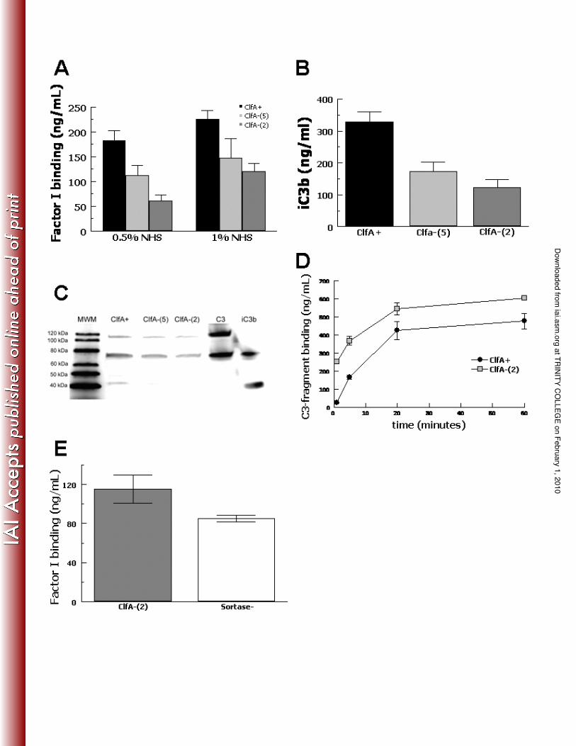
Factor I binding and C3b cleavage by S .aureus. Previously we have shown that the
soluble A domain of ClfA can bind complement factor I and act as a cofactor (13). In
order to analyze the ability of ClfA expressed on the surface of S .aureus to promote 5
binding of factor I, the wild type ClfA+ strain Newman was compared with two null
mutants lacking ClfA. Bacteria were incubated in human serum and factor I bound to the
cells was stripped off by boiling cells in 2% SDS and measured by ELISA (Fig. 1A). In
0.5% serum, the ClfA+ strain bound 112% more factor I than the mutants (mean value
for the two null mutants; P = 0.012) whereas in 1% serum, the wild-type bound 69% 10
more factor I than the mutants (P = 0.012). These results show that expression of ClfA
results in higher factor I binding compared to cells lacking the protein, but that some
factor I still bound to the ClfA- mutants.
In order to evaluate the effect of ClfA on cleavage of C3b to iC3b on the S.
aureus cell surface, the wild-type and ClfA-defective mutants were incubated in 1% NHS 15
for 5 minutes and the bound iC3b was stripped off using methylamine and measured by
ELISA (Fig. 1B). The level of iC3b on the ClfA+ strain was 125% higher than the
mutants (mean value for the two mutants; P = 0.005). This showed that expression of
ClfA increased the generation of iC3b, but that some C3 degradation occurred in the
absence of ClfA. A decrease in iC3b was also detected by Western blotting analysis of 20
C3 fragments solubilized from the cell surface (Fig. 1C).
Cleavage of C3b to iC3b on the S. aureus surface should decrease the number of
alternative pathway C3-convertases on the bacterial surface and inhibit the deposition of
at TRIN
ITY CO
LLEGE on February 1, 2010
iai.asm.org
Dow
nloaded from
13
additional C3b. Therefore, we tested the total amount of C3-fragments deposited by
serum on the surface of the wild-type compared with a ClfA-null strain over 60 minutes
(Fig. 1D). The differences were most striking at 1 minute with the ClfA-null strain
binding 9-fold more C3-fragments compared with the wild-type (P < 0.001). The
difference in C3-fragment binding narrowed over time, but remained significant. Thus, 5
the presence of ClfA appears to correlate with an overall decrease in the deposition of
C3-fragment opsonins.
In order to evaluate whether the residual factor I binding to the ClfA-null strains
could be non-specific, we tested a sortase-null mutant (27) of S. aureus strain Newman
(Fig. 1E). The sortase-null mutant is unable to anchor any LPXTG-motif cell wall 10
associated proteins (24). No significant difference in factor I binding was evident
between the ClfA-null strain [ClfA-(2)] and the sortase-null (Sortase-) strain (P = 0.17),
suggesting that the non-ClfA-dependent factor I-binding is unrelated to covalently
anchored surface proteins.
15
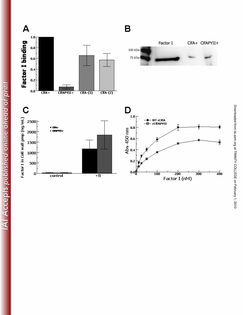
C3b cleavage and ClfAPYII. ClfAPYII is a ClfA mutant with substitutions at
positions 336 and 338 (ClfA P336A Y338S) that does not bind fibrinogen (6), the
primary function described for ClfA. In order to test the role of ClfAPYII in factor I
cleavage of C3b to iC3b on the S. aureus surface, a mutant of Newman expressing
ClfAPYII was incubated in 1% normal human serum, stripped of C3-fragments and iC3b 20
was measured by ELISA (Fig. 2A). The ClfAPYII expressing strain generated 57% less
iC3b compared with wild-type (P = 0.02, based on absolute values). This suggested that
at TRIN
ITY CO
LLEGE on February 1, 2010
iai.asm.org
Dow
nloaded from

14
the presence of ClfAPYII altered the ability of factor I in serum to cleave C3b to iC3b on
the bacterial surface.
In order to further elucidate how the expression of ClfAPYII affects factor I
cleavage of C3b to iC3b on the S. aureus surface, experiments were conducted using
purified complement components. S. aureus strains expressing ClfAPYII or ClfA (wild-5
type) were incubated with purified components to activate the classical pathway and bind
C3b to the bacterial surface (3). The C3b-coated bacteria were then incubated with
purified factor I or both purified factor I and purified factor H, stripped of C3-fragments
using methylamine and iC3b and total C3-fragments measured by ELISA (Fig. 2B). The
ClfA-expressing (wild-type) strain incubated with factor I alone showed increased C3b 10
cleavage by iC3b/C3 ratio (P = 0.05) compared with control. However, the ClfAPYII-
expressing strain incubated with factor I alone showed no increase in C3b cleavage above
control, suggesting that ClfAPYII could not act as a cofactor for factor I on the S. aureus
surface. Curiously, when the C3b-coated ClfAPYII-expressing strain was exposed to
both factor I and a strong cofactor, factor H, C3b cleavage was similar to control. This 15
suggests that ClfAPYII interaction with factor I on the S. aureus surface may inhibit the
normal cofactor activity of factor H.
We then tested whether recombinant ClfAPYII would show differences in
cofactor activity for factor I compared with recombinant ClfA. Recombinant ClfA and
ClfAPYII were incubated with purified C3b and purified factor I and iC3b generation 20
was measured by ELISA (Fig. 2C). In the presence of 200 µg of recombinant ClfAPYII,
factor I produced 57% less iC3b compared with the same amount of recombinant ClfA (P
at TRIN
ITY CO
LLEGE on February 1, 2010
iai.asm.org
Dow
nloaded from

15
= 0.01). These findings again suggest that ClfAPYII has diminished cofactor activity for
factor I compared with wild-type ClfA.
Factor I binding to S. aureus expressing ClfAPYII and recombinant ClfAPYII. In
order to test factor I binding on the bacterial surface, serum factor I binding to the S. 5
aureus expressing ClfAPYII was compared with the wild-type and ClfA-null strains.
The bacteria were incubated in serum, stripped of surface proteins by boiling in 2% SDS
and factor I was measured by ELISA (Fig. 3A). Minimal factor I could be detected in the
stripped surface protein supernatant for the ClfAPYII-expressing strain and was 86%
decreased compared with the ClfA-deficient strains (P < 0.03). This suggested that either 10
the factor I was not binding to the bacterial surface of the ClfAPYII-expressing strain or
that factor I could not be stripped from the ClfAPYII strain.
In order to test these possibilities, we incubated both strains in purified factor I
and then solubilized cell-wall proteins with lysostaphin (13). The cell-wall preparations
were analyzed by Western blotting and probed for factor I with anti-factor I antibody 15
(Fig. 3B). The factor I content of the cell wall preparations was also measured by ELISA
(Fig. 3C). The Western blot analysis showed that factor I was present in the cell wall
preparation of both organisms. The ELISA measured similar amounts of factor I in the
cell wall preparations of the ClfAPYII-expressing S. aureus compared with the wild-type.
This demonstrated that similar amounts factor I bound to the cell wall of the ClfAPYII-20
expressing strain, but could not be removed from the cell wall by boiling in 2% SDS,
which readily removed factor I from the surface of the wild-type strain. These results
at TRIN
ITY CO
LLEGE on February 1, 2010
iai.asm.org
Dow
nloaded from

16
suggested that ClfAPYII bound factor I, but the binding properties were altered compared
to wild-type ClfA.
In order to evaluate further the binding of factor I to ClfAPYII, we then tested the
binding of serum factor I to recombinant ClfA (rClfA) or recombinant ClfAPYII
(rClfAPYII) in an ELISA-type assay (Fig. 3D). Both rClfA and rClfAPYII bound serum 5
factor I with similar half-maximal binding values of 50nM showing that both forms bind
serum factor I effectively.
Phagocytosis of S. aureus expressing ClfAPYII. In order to understand better the
impact of ClfA on complement-mediated phagocytosis, ClfAPYII-expressing S. aureus 10
was compared with the wild-type and ClfA-null strains. The bacteria were incubated
with 1% normal human serum (NHS) or heated serum without complement activity and
then added to purified human neutrophils in a 20:1 ratio. Bacteria were stained with
acridine orange and extracellular bacteria were quenched with crystal violet. With heat
inactivated serum minimal phagocytosis occurred for each strain, whether evaluated as 15
the absolute number of bacteria phagocytized per 100 neutrophils (Fig. 4A) or as the
percent of neutrophils phagocytizing bacteria (Fig. 4B). In normal human serum,
neutrophils phagocytized 3-fold more ClfAPYII-expressing S. aureus compared with
wild-type (P < 0.01) and phagocytized 2-fold more ClfAPYII-expressing S. aureus
compared with the ClfA-null strain (P = 0.01). More neutrophils ingested ClfAPYII-20
expressing bacteria than either the wild-type (P = 0.02) or the ClfA-null strain (P = 0.03).
Although a trend towards increased phagocytosis was noticed for the ClfA-null strain
compared with wild-type, this did not reach statistical significance. These findings show
at TRIN
ITY CO
LLEGE on February 1, 2010
iai.asm.org
Dow
nloaded from
17
that ClfAPYII-expressing S. aureus are much more susceptible to complement-mediated
phagocytosis than the wild-type or the ClfA-null mutant suggesting that the substitutions
at positions 336 and 338 of ClfA are important in modulating S. aureus susceptibility to
phagocytosis.
5
Effect of complement activation on factor I binding. In order to determine if
complement activation and C3b deposition had any effect on the binding of factor I to the
S. aureus surface, the ClfA+ wild type strain Newman was incubated in normal human
serum or in serum where complement was inactivated by heat treatment or with EDTA-
GVBS-- buffer. Complement proteins were stripped and factor I was measured by ELISA 10
(Fig. 5A). No difference in the level of factor I was detected when comparing samples
incubated in NHS or complement-inactivated sera (P > 0.11).
In order to test if C3 or C3b was necessary for factor I binding to ClfA,
recombinant ClfA was incubated with normal human serum or C3-depleted serum and
factor I binding was measured by ELISA (Fig. 5B). Serum factor I binding to rClfA was 15
not significantly different in the presence or the absence of C3 or C3b at 25% serum (P =
0.07), but in 50% serum, rClfA bound 16% less factor I in the presence of C3 compared
with C3-deficient serum (P = 0.02). These findings suggest that C3 is not required for
serum factor I binding to ClfA.
20
Effect of capsule on factor I binding to S .aureus. In order to determine if expression
of capsular polysaccharide interfered with factor I binding to S. aureus, the well
characterized capsule type 5 expressing strain Reynolds and an isogenic capsule deficient
at TRIN
ITY CO
LLEGE on February 1, 2010
iai.asm.org
Dow
nloaded from

18
mutant were compared. Bacteria were grown in conditions where capsule was expressed
strongly (stationary phase) or poorly (mid logarithmic phase) (4). Similar levels of factor
I bound to the two strains under both growth conditions (Fig. 6A, P = 0.17) suggesting
that neither capsule expression nor the phase of growth significantly affected factor I
binding. Control experiments showed that ClfA was expressed by bacteria in both phases 5
(Fig. 6B).
Factor I binding to clinical isolates. To evaluate if factor I could bind to clinical
isolates of S. aureus, five strains from each of four categories (invasive CA-MRSA, non-
invasive CA-MRSA, invasive MSSA, non-invasive MSSA) were incubated with serum, 10
surface proteins were stripped and factor I measured by ELISA (Fig. 7A). All 20 strains
bound factor I. A three-fold difference was found comparing the strains which bound the
most factor I compared with those which bound the least. We calculated that
approximately 60,000 molecules of factor I were bound on average to the S. aureus
isolates. These experiments demonstrate that all clinical strains of S. aureus bind factor I. 15
Control experiments showed that ClfA was expressed by all clinical strains; a
representative blot is shown in figure 7B.
Clinical strains that bound the most factor I would be expected to cleave more
effectively C3b to iC3b and limit opsonic C3-fragment deposition on the S. aureus
surface by the alternative pathway, as shown in figure 1D. Neutrophil phagocytosis 20
experiments were performed with the two clinical strains that bound the most factor I and
the two clinical strains which bound the least factor I (Fig. 7C). A 1.6-fold increase in
phagocytosis efficiency was found for clinical isolates that poorly bound factor I
at TRIN
ITY CO
LLEGE on February 1, 2010
iai.asm.org
Dow
nloaded from

19
compared with strains that bound the most factor I (P = 0.003). These findings support
the likely clinical relevance of an association between factor I binding and virulence.
Effect of fibrinogen on factor I binding to ClfA. In order to test whether the presence
of fibrinogen interferes with factor I binding to ClfA, we compared factor I binding in 5
serum, where there is minimal fibrinogen, to plasma which has a physiological
concentration of fibrinogen (Fig 8A). Use of the anti-coagulant hirudin does not affect
activation of complement as occurs in plasma stabilized with EDTA or heparin (1).
Factor I binding to wild-type (ClfA+) S. aureus was increased in both plasma
preparations compared with serum (P 0.01). There was no difference in factor I 10
binding to the ClfA-null strain [ClfA-(2)] in the plasma compared to serum (P 0.1).
These results suggest that the presence of fibrinogen increases factor I binding to wild-
type S. aureus.
In order to test whether factor I bound to the S. aureus continued to cleave C3b to
iC3b in the presence of a physiological concentration of fibrinogen (2-4 mg/mL), we 15
compared the generation of iC3b on the surface of bacteria incubated in NHS or hirudin
plasma (Fig. 8B). For the wild-type strain (ClfA+), there was no difference in iC3b
generated on the bacterial surface in NHS compared to hirudin plasma (P = 0.63). This
suggests that factor I functions normally on the S. aureus surface in the presence of
physiologic fibrinogen. 20
The influence of fibrinogen was also tested using purified factor I and purified
fibrinogen in a solid phase binding assay with immobilized rClfA (Fig. 8C). Purified
fibrinogen increased purified factor I binding to rClfA in a dose-dependent manner. This
at TRIN
ITY CO
LLEGE on February 1, 2010
iai.asm.org
Dow
nloaded from

20
result is consistent with the results of the experiments shown in figure 5A where factor I
binding to S. aureus was increased in plasma, suggesting that fibrinogen increases the
association of factor I with ClfA.
We then tested if fibrinogen in plasma was associated detectably with factor I in a
solid phase assay where factor I was captured from plasma with an immobilized 5
monoclonal anti-factor I antibody (Fig. 8D). Factor I was readily captured from plasma,
but no bound plasma fibrinogen was detected when probed with an anti-fibrinogen
antibody. We also investigated whether fixing fibrinogen to a surface would cause
binding by factor I (Fig. 8E). Here an immobilized anti-fibrinogen antibody was used to
capture fibrinogen from plasma and bound plasma factor I measured with an anti-factor I 10
antibody. No plasma factor I bound to the solid-phased plasma fibrinogen.
In order to clarify whether fibrinogen is required for ClfA binding to factor I, we
performed a solid phase assay to capture factor I from serum followed by incubation with
increasing concentrations of rClfA or rClfAPYII (Fig. 8F). Wells were coated with anti-
factor I antibody to capture factor I from serum. After washing, rClfA or rClfAPYII were 15
added and binding was detected with anti-ClfA antibody. The rClfA or rClfAPYII bound
to serum factor I dose-dependently with similar affinities suggesting that ClfA binding to
serum factor I does not require fibrinogen.
at TRIN
ITY CO
LLEGE on February 1, 2010
iai.asm.org
Dow
nloaded from

21
Discussion.
The studies described in this paper show that ClfA expression on the surface of S.
aureus is associated with increased binding by factor I from serum, increased cleavage of
surface-bound C3b to iC3b, and decreased C3-fragment binding to the S. aureus surface.
We have previously shown that factor I-mediated cleavage of C3b to iC3b on the S. 5
aureus surface is associated with decreased phagocytosis by human neutrophils (2).
Taken together, these findings suggest that ClfA binding of serum factor I and the
resultant C3b cleavage to iC3b on the S. aureus surface is a plausible mechanism
contributing to S. aureus evasion of complement host defenses.
Previous studies have shown that ClfA P336A Y338S does not bind fibrinogen 10
(23) and that in mouse models of S. aureus bacteremia and septic arthritis a consistent
virulence pattern is found with the ClfAPYII mutant being less virulent that the ClfA null
mutant (17). It is quite striking that complement-mediated phagocytosis of these strains
shows a similar progression in that ClfAPYII-expressing S. aureus are more readily
phagocytized than the ClfA-null or the wild-type strain. These differences in 15
complement-mediated phagocytosis can be at least partly explained by the failure of
ClfAPYII to act as a cofactor for factor I-mediated cleavage of C3b to iC3b. ClfAPYII
also appears to inhibit the cofactor activity of factor H. Thus, ClfA has an important
interaction with the complement system that likely contributes to the differences in
virulence found in vivo that could not be attributed to ClfA interaction with fibrinogen. 20
These findings support the hypothesis that factor I binding to ClfA results in C3b
cleavage to iC3b on the S. aureus surface and contributes to immune evasion and
virulence.
at TRIN
ITY CO
LLEGE on February 1, 2010
iai.asm.org
Dow
nloaded from

22
The presence of fibrinogen at physiological concentrations increases factor I
binding to the S. aureus surface, increases factor I binding to ClfA, and does not
adversely affect the cleavage of C3b on the bacterial surface. Factor I in serum does not
appear to require fibrinogen for binding to ClfA or ClfAPYII (the latter being a molecule
that cannot bind fibrinogen). 5
Previous studies have suggested that upon binding the factor H-C3b complex,
factor I undergoes a conformational change that enables it to cleave C3b (8, 10). We
speculate that factor I may undergo a similar conformational change upon binding the
fibrinogen-ClfA complex that enables it to cleave C3b. We also propose that the
‘activating’ conformational change for factor I does not occur upon binding to ClfAPYII. 10
Based on these data, we propose the model illustrated in figure 9. The apo form
of ClfA is shown with the latching peptide emanating from the C-terminus of the N3
domain unbound to the N2 domain. When the D domain of fibrinogen contacts ClfA and
the gamma-chain peptide inserts into the ligand-binding trench, a conformational change
occurs which results in the fibrinogen peptide being locked in place by the latching 15
peptide undergoing beta-strand complementation with two beta strands in N2. Factor I
binds the fibrinogen-ClfA complex and undergoes a conformational change into an
‘active’ form that can cleave C3b to iC3b. ClfAPYII, in contrast, is unable to bind
fibrinogen and remains in the apo form with the latching peptide free. Factor I is also
able to bind ClfAPYII, but does not undergo a conformational change to an ‘active’ form. 20
Thus, factor I complexed with ClfAPYII is unable to cleave C3b to iC3b. The stronger
binding of factor I to ClfAPYII could be due to local changes in conformation due to the
amino acid substitutions.
at TRIN
ITY CO
LLEGE on February 1, 2010
iai.asm.org
Dow
nloaded from

23
All 20 clinical isolates expressed ClfA and bound factor I. Clinical isolates with
increased factor I binding were poorly phagocytized relative to isolates with decreased
factor I binding. These data support the likely physiological relevance of ClfA-mediated
factor I cleavage of C3b.
Future studies will continue to investigate the binding and functional interaction 5
between factor I, fibrinogen, ClfA, and ClfAPYII in order to further elucidate this
relationship.
at TRIN
ITY CO
LLEGE on February 1, 2010
iai.asm.org
Dow
nloaded from
24
Acknowledgements.
This work was supported by the Children’s Hospital of the King’s Daughters Research
Endowment. TJF acknowledges the support of a Science Foundation Ireland Principal
Investigator Programme Grant.
5
Strain JL022 was kindly provided by Dr. Jean Lee, Channing Laboratory, Harvard
Medical School.
at TRIN
ITY CO
LLEGE on February 1, 2010
iai.asm.org
Dow
nloaded from
25
References.
1. Bexborn, F., A. E. Engberg, K. Sandholm, T. E. Mollnes, J. Hong, and K. Nilsson Ekdahl. 2008. Hirudin versus heparin for use in whole blood in vitro biocompatibility models. Journal of biomedical materials research.
2. Cunnion, K. M., E. S. Buescher, and P. S. Hair. 2005. Serum complement 5 factor I decreases Staphylococcus aureus phagocytosis. The Journal of laboratory and clinical medicine 146:279-286.
3. Cunnion, K. M., P. S. Hair, and E. S. Buescher. 2004. Cleavage of complement C3b to iC3b on the surface of Staphylococcus aureus is mediated by serum complement factor I. Infection and immunity 72:2858-2863. 10
4. Cunnion, K. M., J. C. Lee, and M. M. Frank. 2001. Capsule production and growth phase influence binding of complement to Staphylococcus aureus. Infection and immunity 69:6796-6803.
5. Cunnion, K. M., H. M. Zhang, and M. M. Frank. 2003. Availability of complement bound to Staphylococcus aureus to interact with membrane 15 complement receptors influences efficiency of phagocytosis. Infection and immunity 71:656-662.
6. Deivanayagam, C. C., E. R. Wann, W. Chen, M. Carson, K. R. Rajashankar, M. Hook, and S. V. Narayana. 2002. A novel variant of the immunoglobulin fold in surface adhesins of Staphylococcus aureus: crystal structure of the 20 fibrinogen-binding MSCRAMM, clumping factor A. The EMBO journal 21:6660-6672.
7. Deurenberg, R. H., and E. E. Stobberingh. 2008. The evolution of Staphylococcus aureus. Infect Genet Evol.
8. DiScipio, R. G. 1992. Ultrastructures and interactions of complement factors H 25 and I. J Immunol 149:2592-2599.
9. Duthie, E. S., and L. L. Lorenz. 1952. Staphylococcal coagulase; mode of action and antigenicity. Journal of general microbiology 6:95-107.
10. Ekdahl, K. N., U. R. Nilsson, and B. Nilsson. 1990. Inhibition of factor I by diisopropylfluorophosphate. Evidence of conformational changes in factor I 30 induced by C3b and additional studies on the specificity of factor I. J Immunol 144:4269-4274.
11. Fitzgerald, J. R., A. Loughman, F. Keane, M. Brennan, M. Knobel, J. Higgins, L. Visai, P. Speziale, D. Cox, and T. J. Foster. 2006. Fibronectin-binding proteins of Staphylococcus aureus mediate activation of human platelets 35 via fibrinogen and fibronectin bridges to integrin GPIIb/IIIa and IgG binding to the FcgammaRIIa receptor. Molecular microbiology 59:212-230.
12. Goetghebeur, M., P. A. Landry, D. Han, and C. Vicente. 2007. Methicillin-resistant Staphylococcus aureus: A public health issue with economic consequences. The Canadian journal of infectious diseases & medical 40 microbiology = Journal canadien des maladies infectieuses et de la microbiologie medicale / AMMI Canada 18:27-34.
13. Hair, P. S., M. D. Ward, O. J. Semmes, T. J. Foster, and K. M. Cunnion. 2008. Staphylococcus aureus clumping factor A binds to complement regulator
at TRIN
ITY CO
LLEGE on February 1, 2010
iai.asm.org
Dow
nloaded from

26
factor I and increases factor I cleavage of C3b. The Journal of infectious diseases 198:125-133.
14. Higgins, J., A. Loughman, K. P. van Kessel, J. A. van Strijp, and T. J. Foster. 2006. Clumping factor A of Staphylococcus aureus inhibits phagocytosis by human polymorphonuclear leucocytes. FEMS microbiology letters 258:290-296. 5
15. Jongerius, I., J. Kohl, M. K. Pandey, M. Ruyken, K. P. van Kessel, J. A. van Strijp, and S. H. Rooijakkers. 2007. Staphylococcal complement evasion by various convertase-blocking molecules. The Journal of experimental medicine 204:2461-2471.
16. Josefsson, E., O. Hartford, L. O'Brien, J. M. Patti, and T. Foster. 2001. 10 Protection against experimental Staphylococcus aureus arthritis by vaccination with clumping factor A, a novel virulence determinant. The Journal of infectious diseases 184:1572-1580.
17. Josefsson, E., J. Higgins, T. J. Foster, and A. Tarkowski. 2008. Fibrinogen binding sites P336 and Y338 of clumping factor A are crucial for Staphylococcus 15 aureus virulence. PLoS ONE 3:e2206.
18. Keane, F. M., A. Loughman, V. Valtulina, M. Brennan, P. Speziale, and T. J. Foster. 2007. Fibrinogen and elastin bind to the same region within the A domain of fibronectin binding protein A, an MSCRAMM of Staphylococcus aureus. Molecular microbiology 63:711-723. 20
19. Klevens, R. M., M. A. Morrison, J. Nadle, S. Petit, K. Gershman, S. Ray, L. H. Harrison, R. Lynfield, G. Dumyati, J. M. Townes, A. S. Craig, E. R. Zell, G. E. Fosheim, L. K. McDougal, R. B. Carey, and S. K. Fridkin. 2007. Invasive methicillin-resistant Staphylococcus aureus infections in the United States. Jama 298:1763-1771. 25
20. Kosowska-Shick, K., L. M. Ednie, P. McGhee, K. Smith, C. D. Todd, A. Wehler, and P. C. Appelbaum. 2008. Incidence and Characteristics of Vancomycin Non-Susceptible Strains of Methicillin-Resistant Staphylococcus Aureus at Hershey Medical Center. Antimicrobial agents and chemotherapy.
21. Lambris, J. D., D. Ricklin, and B. V. Geisbrecht. 2008. Complement evasion 30 by human pathogens. Nature reviews 6:132-142.
22. Lambris, J. D., A. Sahu, and R. A. Wetsel. 1998. The chemistry and biology of C3, C4, and C5., p. 83 -118. In J. E. Volanakis and M. M. Frank (ed.), The human complement system in health and disease. Marcel Dekker, New York.
23. Loughman, A., J. R. Fitzgerald, M. P. Brennan, J. Higgins, R. Downer, D. 35 Cox, and T. J. Foster. 2005. Roles for fibrinogen, immunoglobulin and complement in platelet activation promoted by Staphylococcus aureus clumping factor A. Molecular microbiology 57:804-818.
24. Mazmanian, S. K., H. Ton-That, and O. Schneewind. 2001. Sortase-catalysed anchoring of surface proteins to the cell wall of Staphylococcus aureus. Molecular 40 microbiology 40:1049-1057.
25. Moreillon, P., J. M. Entenza, P. Francioli, D. McDevitt, T. J. Foster, P. Francois, and P. Vaudaux. 1995. Role of Staphylococcus aureus coagulase and clumping factor in pathogenesis of experimental endocarditis. Infection and immunity 63:4738-4743. 45
at TRIN
ITY CO
LLEGE on February 1, 2010
iai.asm.org
Dow
nloaded from
27
26. O'Connell, D. P., T. Nanavaty, D. McDevitt, S. Gurusiddappa, M. Hook, and T. J. Foster. 1998. The fibrinogen-binding MSCRAMM (clumping factor) of Staphylococcus aureus has a Ca2+-dependent inhibitory site. The Journal of biological chemistry 273:6821-6829.
27. O'Neill, E., C. Pozzi, P. Houston, H. Humphreys, D. A. Robinson, A. 5 Loughman, T. J. Foster, and J. P. O'Gara. 2008. A novel Staphylococcus aureus biofilm phenotype mediated by the fibronectin-binding proteins, FnBPA and FnBPB. Journal of bacteriology 190:3835-3850.
28. Petry, F., and M. Loos. 1998. Bacteria and Complement, p. 375 - 392. In J. E. Volanakis and M. M. Frank (ed.), The human complement system in health and 10 disease. Marcel Dekker, New York.
29. Portoles, M., K. B. Kiser, N. Bhasin, K. H. Chan, and J. C. Lee. 2001. Staphylococcus aureus Cap5O has UDP-ManNAc dehydrogenase activity and is essential for capsule expression. Infection and immunity 69:917-923.
30. Stranden, A. M., R. Frei, H. Adler, U. Fluckiger, and A. F. Widmer. 2008. 15 Emergence of SCCmec Type IV as the Most Common Type of Methicillin-Resistant Staphylococcus aureus in a University Hospital. Infection.
31. Tjernberg, J., K. N. Ekdahl, J. D. Lambris, O. Korsgren, and B. Nilsson. 2008. Acute antibody-mediated complement activation mediates lysis of pancreatic islets cells and may cause tissue loss in clinical islet transplantation. 20 Transplantation 85:1193-1199.
32. van Wamel, W. J., S. H. Rooijakkers, M. Ruyken, K. P. van Kessel, and J. A. van Strijp. 2006. The innate immune modulators staphylococcal complement inhibitor and chemotaxis inhibitory protein of Staphylococcus aureus are located on beta-hemolysin-converting bacteriophages. Journal of bacteriology 188:1310-25 1315.
33. Watts, A., D. Ke, Q. Wang, A. Pillay, A. Nicholson-Weller, and J. C. Lee. 2005. Staphylococcus aureus strains that express serotype 5 or serotype 8 capsular polysaccharides differ in virulence. Infection and immunity 73:3502-3511.
30
at TRIN
ITY CO
LLEGE on February 1, 2010
iai.asm.org
Dow
nloaded from

28
Figure Legends.
FIG. 1. Factor I binding and C3b cleavage to iC3b on wild-type (ClfA+) and ClfA-
deficient [ClfA-(5) and ClfA-(2)] S. aureus strains. Bacteria were incubated in 1%
normal human serum (NHS) for 5 minutes, washed and boiled in 2% SDS to remove
bound factor I, which was measured by ELISA (A). Data are mean ± SE for 5 5
independent experiments. Bacteria were incubated in 1% NHS for 5 minutes, washed,
stripped of C3-fragments with methylamine and assayed for iC3b by ELISA (B). Data
are mean ± SE for 4 independent experiments. C3-fragments stripped from the surface of
serum-incubated S. aureus strains were analyzed by Western blot and probed for C3-
fragments (C). Total C3-fragment binding to S. aureus expressing wild-type ClfA 10
(ClfA+) or ClfA-null [ClfA-(2)] incubated in 1% NHS for increasing lengths of time and
assayed by ELISA (D). Data are mean ± SE for 3 independent experiments. Factor I
binding was tested for a ClfA-deficient strain [ClfA-(2)] and a sortase-deficient isogenic
mutant (Sortase-) (E). Bacteria were incubated in 1% plasma for 5 minutes, washed,
stripped and measured for bound factor I by ELISA. Data are mean ± SE for 3 15
independent experiments.
FIG. 2. C3b cleavage to iC3b for S. aureus expressing ClfAPYII and recombinant
ClfAPYII. S. aureus strains expressing the wild-type ClfA (ClfA+), the mutant
ClfAPYII (ClfAPYII+), or ClfA-null [(ClfA-(5) and ClfA-(2)] were incubated in 1% 20
NHS for 5 minutes, washed, stripped of C3-fragments with methylamine and measured
for iC3b by ELISA (A). Data are normalized to wild-type and are mean ± SE for 3
independent experiments. S. aureus expressing wild-type ClfA (ClfA+) or ClfAPY
at TRIN
ITY CO
LLEGE on February 1, 2010
iai.asm.org
Dow
nloaded from

29
(ClfAPYII+) were coated with C3b by complement activation with purified components,
incubated with purified factor (fI), purified factors I and H (+fI +fH), or buffer alone
(control), stripped of bound C3-fragments and analyzed by ELISA (B). Factor H (fH)
was used as a control cofactor. Data are mean ± SE for 3 independent experiments.
Recombinant ClfA (rClfA) and recombinant ClfAPYII (rClfAPYII) were incubated with 5
purified C3b and purified factor I and assayed for iC3b by ELISA (C). Data are mean ±
SE for 3 independent experiments.
FIG. 3. Factor I binding to S. aureus expressing ClfAPYII and recombinant ClfAPYII.
S. aureus strains expressing the wild-type ClfA (ClfA+), the mutant ClfAPYII 10
(ClfAPYII+), or ClfA-null [ClfA-(5) and ClfA-(2)] were incubated in 1% NHS, washed,
and boiled in 2%SDS to remove bound factor I, which was measured by ELISA (A).
Data are normalized to wild-type and are mean ± SE for 5 independent experiments. S.
aureus strains expressing the wild-type ClfA (ClfA+) or the mutant ClfAPYII
(ClfAPYII+) were incubated with purified factor I, washed and cell-wall preparations 15
were analyzed by Western blot probing for factor I (B). The cell-wall preparations of S.
aureus incubated with purified factor I (+fI) or buffer (control) were also measured for
factor I by ELISA (C). Data are the means ± SEM of 4 independent experiments. Serum
factor I binding to recombinant ClfA (rClfA) or recombinant ClfAPYII (rClfAPYII),
measured by ELISA (D). Data are the means ± SEM of 5 independent experiments. 20
FIG. 4. Phagocytosis efficiency for neutrophil engulfment of isogenic S. aureus strains
incubated with normal human serum (NHS) or heated serum (heated serum) without
at TRIN
ITY CO
LLEGE on February 1, 2010
iai.asm.org
Dow
nloaded from
30
complement activity. Wild-type (ClfA+), ClfA-null [ClfA-(2)], or ClfAPYII-expressing
(ClfAPYII+) strains were incubated with 1% serum for 30 minutes, to which were added
purified human neutrophils for 45 minutes, then stained with acridine orange, quenched
with crystal violet and assayed by fluorescence microscopy. The numbers of bacteria
phagocytized per 100 neutrophils were measured (A). The percent of neutrophils 5
phagocytizing bacteria were measured (B). Data are the means ± SEM of 4 independent
experiments.
FIG. 5. Factor I binding to ClfA-expressing S. aureus and recombinant ClfA in the
absence of complement activation or C3. S. aureus expressing ClfA (wild-type) were 10
incubated in 10% normal human serum (NHS), heat-inactivated sera (Heat sera) or sera
in EDTA-GVBS- - buffer (EDTAsera), washed and boiled in 2% SDS to remove factor
I, which was measured by ELISA (A). Data are the mean ± SE for 5 independent
experiments. Serum factor I binding to recombinant ClfA was tested for NHS, heat-
inactivated sera or C3-depleted sera (C3depl sera) and measured by ELISA (B). Data are 15
the means ± SEM of 6 independent experiments.
FIG. 6. Serum factor I binding to S. aureus in the presence of capsule. Isogenic ClfA-
sufficient S. aureus strains that expressed capsule (capsule+) or was capsule-deficient
(capsule-def) were grown to mid logarithmic (log) or stationary phase, incubated in 10% 20
normal human serum, washed, and boiled in 2% SDS to remove bound factor I, which
was measured by ELISA (A). Data are the means ± SEM of 4 independent experiments.
at TRIN
ITY CO
LLEGE on February 1, 2010
iai.asm.org
Dow
nloaded from

31
ClfA expression for each strain and each growth condition was assayed by Western
blotting of cell wall preparations (B).
FIG. 7. Clinical S. aureus isolates binding serum factor I, ClfA expression, and
phagocytosis efficiency. Five separate isolates were tested for each category: invasive 5
CA-MRSA (MRSA-Inv), non-invasive CA-MRSA (MRSA-Non), invasive MSSA
(MSSA-Inv), and non-invasive MSSA (MSSA-Non) (A). A laboratory control strain
(Reynolds) was also tested. Bacteria were incubated in 10% normal human serum,
washed and boiled in 2% SDS to remove bound factor I, which was measured by factor I
ELISA. Data are the mean ± SE of 5 independent experiments. ClfA expression of S. 10
aureus clinical isolates was assayed by Western blotting of cell wall preparations (B).
Phagocytosis efficiency for the two clinical isolates that bound the least factor I (M25 and
S68) and the two clinical isolates that bound the most factor I (M32 and M2) (C).
Isolates were incubated with 1% serum for 30 minutes, to which were added purified
human neutrophils for 45 minutes, then stained with acridine orange, quenched with 15
crystal violet and assayed by fluorescence microscopy. Data are the mean ± SE of 3
independent experiments.
Figure 8. Factor I binding and function in the presence of fibrinogen. Factor I binding to
S. aureus wild-type (ClfA+) or ClfA-null strain [ClfA-(2)] incubated in 1% serum 20
(NHS), EDTA-plasma, or hirudin-plasma, measured by ELISA (A). Data are the mean ±
SE of 7 independent experiments. Bound iC3b to S. aureus wild-type (ClfA+) or
ClfAPYII-expressing strain (ClfAPYII+) incubated in 1% serum or hirudin-plasma,
at TRIN
ITY CO
LLEGE on February 1, 2010
iai.asm.org
Dow
nloaded from

32
measured by ELISA (B). Data are the mean ± SE of 3 independent experiments.
Purified factor I (10 ng/mL) binding to recombinant ClfA (rClfA) in an ELISA in the
presence of increasing purified fibrinogen (C). Data are the mean ± SE of 6 independent
experiments. Factor I was captured from increasing concentrations of plasma and probed
for associated plasma fibrinogen by ELISA (D). Monoclonal anti-factor I was used to 5
capture plasma factor I (control fI) and bound plasma fibrinogen (pfibrinogen/captured
fI) was detected with anti-fibrinogen antibody. Data are the mean ± SE of 3 independent
experiments. Fibrinogen was captured from increasing concentrations of plasma and
probed from associated plasma factor I by ELISA (E). Anti-fibrinogen antibody was
used to capture plasma fibrinogen and bound plasma factor I (pfactor I/captured fg) was 10
detected with anti-factor I antibody. Data are the mean ± SE of 3 independent
experiments. Serum factor I was captured with anti-factor I antibody and assayed for
bound rClfA or rClfAPYII by ELISA (F). Increasing concentrations of rClfA or
rClfAPYII were added and detected with anti-ClfA antibody. Data are the mean ± SE of
4 independent experiments. 15
Figure 9. A scheme to the associations of factor I and fibrinogen with ClfA and
ClfAPYII. Fibrinogen (Fg) binds to ClfA and enhances the binding of factor I which is
then in an ‘active’ conformation and able to cleave C3b to iC3b. ClfAPYII, which is
unable to bind fibrinogen, is also able to bind factor I, but the factor I is not in an ‘active’ 20
conformation and unable to cleave C3b to iC3b.
at TRIN
ITY CO
LLEGE on February 1, 2010
iai.asm.org
Dow
nloaded from