Embed Size (px)
Citation preview

Lipid A Structure is critical for establishing dysbiotic dental plaque communities in the な rabbit ligature model に
ぬ Camille Zenobia
1, Hatice Hasturk
2, Daniel Nguyen
2, Thomas E. Van Dyke
2, Alpdogan ね
Kantarci2*
, Richard P. Darveau3*#
の 1. University of Washington 1959 NE Pacific Street, Seattle WA 98195, Microbiology は
Department. Current address University of Pennsylvania 240 S 40th Street, Philadelphia, ば PA 19104 ぱ
2. Forsyth Institute, Department of Applied Oral Sciences, Center for Periodontology, ひ Cambridge, MA, 02142 など
3. University of Washington 1959 NE Pacific Street, Seattle WA 98195, Department of なな Periodontics [email protected] なに *-
Contributed equally なぬ
なね Abstract なの Periodontitis is a disease of polymicrobial etiology characterized by inflammation, degradation なは of host tissue and bone that irreversibly destroys the supporting apparatus of teeth. P. gingivalis なば contains lipid A with structural heterogeneity that has been postulated to contribute to the なぱ initiation of dysbiosis in oral communities by modulating the host response thereby creating a なひ permissive environment for its growth. In this paper, two P. gingivalis lipid A phosphatase にど mutants, which contain different “locked” lipid A structures that induce different host cellular にな responses were examined for their ability to induce dysbiosis and periodontitis in rabbits. LPS にに preparations obtained from these strains were also examined. After repeated applications of all にぬ strains and their respective LPS preparations, P. gingivalis wild type, but not the lipid A mutants にね had a significant impact on both the oral commensal microbial load and composition. In contrast, にの in rabbits exposed to the mutant strains or the LPS preparations, the microbial load did not には increase yet significant changes in the oral microbial composition were observed. All strains and にば their respective LPS preparations induced periodontitis. Therefore, the ability to alter the lipid A にぱ composition in response to environmental conditions by lipid A phosphatases is required for both にひ colonization of the rabbit and increases in the microbial load. Furthermore, the data demonstrate ぬど that multiple dysbiotic oral microbial communities can elicit periodontitis. ぬな ぬに ぬぬ Introduction ぬね
Porphyromonas gingivalis (P. gingivalis) is a gram negative, anaerobic bacterium that is ぬの associated with periodontitis. Periodontitis is a complex disease that is characterized by a shift ぬは
IAI Accepts, published online ahead of print on 25 November 2013Infect. Immun. doi:10.1128/IAI.01136-13Copyright © 2013, American Society for Microbiology. All Rights Reserved.
on July 9, 2018 by guesthttp://iai.asm
.org/D
ownloaded from

from aerobic Gram-positive bacteria to anaerobic Gram-negative bacteria. The complex gram ぬば negative biofilm induces a significant increase in inflammation and eventually a complex ぬぱ immune lesion that results in degradation of host tissues, alveolar bone reduction, chronic ぬひ inflammatory disease and ultimately tooth loss. Over 600 bacterial taxa have been identified in ねど the oral cavity; yet, only a relatively small percentage has been associated with disease (1). P. ねな gingivalis has been shown to cause disease in several different animal models (2). The ねに mechanism by which P. gingivalis transforms healthy microbial/ host homeostasis to destructive ねぬ periodontitis is not clear. ねね
To date, two in vivo animal models have implicated commensal bacteria as having a role ねの in P. gingivalis induced periodontal disease. The first study, utilizing a rabbit model of ねは periodontal disease showed that inoculation of P. gingivalis via ligature leads to an outgrowth of ねば oral bacteria as well as a shift in commensal profile and bone loss (3). There was no direct ねぱ implication that commensal bacteria contributed to disease; however, it was clear that ねひ introduction of P. gingivalis caused the outgrowth of the oral bacterial community. More のど recently, P. gingivalis infection by oral gavage in mice was followed by an increase of oral のな bacteria that resulted in bone loss while P. gingivalis infected germ-free mice lacking oral のに bacteria were protected from disease (4). This latter study clearly demonstrated that oral のぬ commensal bacteria were required for disease in the mouse gavage model and that P. gingivalis のね orchestrated the shift from a healthy bacterial community to a periopathogenic and dysbiotic のの community. Due to its significant contribution to the remodeling of commensal bacterial のは community resulting in disease, P. gingivalis was termed a keystone pathogen (5-8). P. のば gingivalis contains several virulence factors that may contribute to its ability to modulate the oral のぱ microbial composition (9). One of the virulence factors, the lipopolysaccharide (LPS), has been のひ
on July 9, 2018 by guesthttp://iai.asm
.org/D
ownloaded from

proposed to contribute changes to the oral microbial community (6). For example, P. gingivalis はど can alter its lipid A phosphate composition in response to different environmental conditions はな resulting in lipid A structures that are either agonists or antagonists for inflammatory activation はに at TLR4 (10-12). Alterations in the host environment by modulation of host TLR4 activity can はぬ have global effects on the microbial community by enhancing or suppressing the growth of はね different members of the oral community (7). はの
Here we show that two P. gingivalis mutants that are unable to modulate their lipid A はは structural composition display significantly different phenotypes with respect to interactions with はば innate host components TLR4 and antimicrobial peptides. One mutant expresses a lipid A that is はぱ a TLR4 agonist whereas the other mutant expresses a lipid A that is capable of antagonizing はひ TLR4. One hypothesis we tested is that the antagonist structure disrupts host cell functions and ばど causes overgrowth of the oral microbiota. Therefore, the two mutants were examined for their ばな ability to induce periodontitis. LPS isolated from these two mutant strains was also examined. It ばに was found that in the rabbit model of periodontal disease the wild type but not the mutant strains ばぬ of P. gingivalis was able to colonize the rabbit periodontium, significantly increase the oral ばね commensal bacterial load, and result in periodontitis. Conversely, although the mutant strains ばの did not significantly colonize rabbit periodontal tissue they as well as the isolated LPS ばは preparations were able to create dysbiotic oral communities that were also associated with ばば periodontitis. These data demonstrate that in the rabbit ligature model of periodontitis P. ばぱ gingivalis lipid A phosphatases are required for colonization and that multiple different oral ばひ dysbiotic microbial communities can disrupt host homeostasis and result in disease. ぱど
ぱな Materials and Methods ぱに
on July 9, 2018 by guesthttp://iai.asm
.org/D
ownloaded from
Bacterial growth conditions P. gingivalis strain ATCC 33277 was obtained from ぱぬ our stock collection. Bacteria were grown in TYHK medium consisting of trypticase soy broth ぱね (30 g/ l (Becton Dickinson, Sparks, MD), yeast extract 5 g/l (Becton Dickinson, Sparks, MD), ぱの and vitamin K3 (menadione) (Sigma-Aldrich, St. Louis, MO). The basal TYHK medium was ぱは sterilized by autoclaving, followed by the addition of filter-sterilized hemin (Sigma-Aldrich, St. ぱば Louis, MO) to the desired final concentration of either (1 たg/ ml) or (10 たg/ml) as indicated in ぱぱ the text and figure legends. Cultures were grown in an anaerobic growth chamber (5% H2, 5% ぱひ CO2, 90% N2) and maintained at 37°C on TYHK-agar plates. ひど
Gene Deletions in P. gingivalis 33277 The genomic nucleotide sequences encoding the ひな putative lipid A 1-phosphatase, PG1773, and the putative lipid A 4'-phosphatase, PG1587, were ひに obtained from searches of the annotated P. gingivalis W83 genome at The Comprehensive ひぬ Microbial Resource (http://cmr.jcvi.org/tigr-scripts/CMR/CmrHomePage.cgi). Gene deletions ひね were created by introducing either a tetracycline resistance cassette (tetQ) in place of the coding ひの region for PG1773 or an erythromycin resistance cassette (ermF/AM) in place of the coding ひは region for PG1587. Polymerase chain reaction (PCR) amplification of genomic DNA from P. ひば gingivalis A7436 was performed using primer sets designed against the W83 sequence to ひぱ amplify 1000 base-pairs upstream and 1000 base-pairs downstream from the regions adjacent to ひひ the PG1773 and PG1587 coding regions respectively. The amplified 5' and 3' flanking regions などど for PG1773 and PG1587 respectively, were co-ligated with the tetQ and ermF/AM cassettes などな respectively into pcDNA3.1(−) to generate the gene disruption plasmids, p1773 などに 5'flank:tetQ:3'flank and p1587 5'flank:erm:3'flank. P. gingivalis 33277 deficient in either などぬ PG1587 (1587KO) or PG1773 (1773KO) was generated by introducing either p1587 などね 5'flank:erm:3'flank or p1773 5'flank:tetQ:3'flank into P. gingivalis 33277 by electroporation in a などの
on July 9, 2018 by guesthttp://iai.asm
.org/D
ownloaded from

GenePulser Xcell (BioRad, Hercules, CA). Bacteria were plated on TYHK/agar plates containing などは the appropriate selective medium, which included either erythromycin (5 たg ml−1) or などば tetracycline (1 たg ml−1) and incubated anaerobically. One week later, colonies were selected for などぱ characterization. Loss of the PG1587 and PG1773 coding sequences were confirmed in all clones などひ by PCR analyses using primers designed to detect the coding sequences. (11) ななど
Isolation of LPS and Lipid A Bacteria were cultured for 48 hours in TYHK medium ななな containing hemin at a concentration of either (1 たg/ml) or (10 たg/ml). LPS was isolated using a ななに modified version of the Tri-reagent protocol for LPS isolation as previously described (10). To ななぬ generate lipid A, dried LPS samples were resuspended in 10 mM sodium acetate [pH 4.5] ななね containing 1% sodium dodecyl sulfate (w/v). The solution was heated 100°C for 1 hour followed ななの by lyophilization overnight. The resulting lipid A pellets washed once in ice-cold 95% ethanol ななは containing 0.02 N HCl, three times in 95% ethanol, followed by a final extraction with 1160 たl ななば of chloroform-methanol-water (1:1:0.9, v:v:v) to remove residual carbohydrate contaminants. ななぱ The chloroform layer containing the lipid A was dried and used for MALDI-TOF MS or ななひ MALDI-TOF/TOF tandem MS analyses. なにど
MALDI-TOF MS Analyses For MALDI-TOF MS analyses, lipid A samples were なにな dissolved in 10 たl of a mixture of 5-chloro-2-mercaptobenzothiazole (20 mg/ml) in なにに chloroform/methanol 1:1 (v/v) and 0.5 たl of each sample was analyzed in both positive and なにぬ negative ion modes on an AutoFlex Analyzer (Bruker Daltonics). Data were acquired with a 50 なにね Hz repletion rate and up to 3000 shots were accumulated for each spectrum. Instrument なにの calibration and all other tuning parameters were optimized using HP Calmix (Sigma-Aldrich, St. なには Louis, MO). Data was acquired and processed using flexAnalysis software (Bruker なにば Daltonics).(11) なにぱ
on July 9, 2018 by guesthttp://iai.asm
.org/D
ownloaded from

HEK293 TLR4 Activation Assays HEK293 cells were plated in 96-well plates at a なにひ density of 4 × 10
4 cells per well and transfected the following day with plasmids bearing firefly なぬど
luciferase, Renilla luciferase, human TLR4 and MD-2 by a standard calcium phosphate なぬな precipitation method as described previously (13) The test wells were stimulated in triplicate for なぬに 4 hours at 37°C with the indicated doses of LPS or intact bacteria that had been suspended by なぬぬ vortexing in DMEM containing 10% human serum. Following stimulation, the HEK293 cells なぬね were rinsed with phosphate-buffered saline and lysed with 50 たl of passive lysis buffer (Promega, なぬの Madison, WI). Luciferase activity was measured using the Dual Luciferase Assay Reporter なぬは System (Promega, Madison, WI). Data are expressed as fold increase of NF-せB-activity, which なぬば represents the ratio of NF-せB-dependent fire-fly luciferase activity to く-Actin promoter-なぬぱ dependent Renilla luciferase activity.(11) なぬひ
Polymyxin B Sensitivity Assays Overnight cultures of wild type P. gingivalis A7436 and なねど its isogenic putative phosphatase mutants were grown in THYK media containing hemin (1 なねな たg/ml) or (10 たg/ml) as indicated above. Liquid cultures that had been grown for 24 hours in an なねに anaerobic growth chamber were diluted to a starting optical density (OD600) of 1.0, which なねぬ represents approximately 1 × 10
9 CFU/ml for all of the strains examined (data not shown). なねね
Subsequently, 10−3
to 10−8
dilutions of each strain were plated on TYHK-agar plates containing なねの PMB (0, 5, and 200 たg/ml). After 8–10 days of incubation in an anaerobic chamber, the resulting なねは colonies were counted to determine the viability at the different PMB treatments. The results for なねば each strain were plotted as percent survival, which was derived from the ratio of the number of なねぱ colonies detected on the experimental plates containing polymyxin B (5 or 200 たg/ml) to the なねひ number of colonies detected on the control plates that did not contain polymyxin B. (11) なのど
on July 9, 2018 by guesthttp://iai.asm
.org/D
ownloaded from
P. gingivalis Wild Type (WT) P. gingivalis (strain A7436; (14)) was grown using なのな standard procedures as described previously (15), (16), (17), (18). Briefly, bacteria were なのに cultured on agar plates containing trypticase soy agar supplemented with 0.5% (w/v) yeast なのぬ extract (Invitrogen Life Technologies), 5% defibrinated sheep red blood cells, 5 Ǵg hemin, and なのね 1 Ǵg/ml vitamin K (Sigma-Aldrich, St. Louis, MO). Plates were incubated for 3 days at 37°C in なのの an anaerobic chamber maintained by hydrogen gas mixture (hydrogen/nitrogen) that is circulated なのは through a heated palladium catalyst. Colonies were randomly selected and anaerobically cultured なのば overnight at 37°C in Wilkins-Chalgren Anaerobe Broth.(3) なのぱ
Slurry preparations Bacterial numbers with P. gingivalis strains and mutants were なのひ spectrophotometrically determined at 600 nm, adjusted to 10
9 CFU (0.8 OD) while 35ng of LPS なはど
was used for LPS preparations. Bacterial strains and LPS preparations were mixed with なはな carboxymethylcellulose (CMC) to form a thick slurry. なはに
Animal Model なはぬ The study was approved by the Forsyth Institute Institutional Animal Care and Use なはね
Committee (Forsyth IACUC). Specific pathogen-free New Zealand White rabbits (24 males, なはの 3.5– 4.0 kg) were purchased from Covance Research Products, Inc. (Denver, PA), equilibrated なはは for 7 days prior to experiments. The rabbits were, kept in individual cages, received water ad なはば libitum, and fed standard rabbit chow (Purina LabDiet 5321, Purina Mills, LLC, St. Louis, なはぱ MO) throughout the experiment. Periodontitis was induced and established in all animals using a なはひ previously established protocol (3). A 3-0 silk suture (ligature) was placed around the second なばど premolar teeth of both mandibular quadrants under general anesthesia using 40 mg/kg ketamine, なばな Ketaset (Fort Dodge Animal Health) and 5 mg/kg xylazine, Anased (Lloyd Laboratories, Inc.) なばに IM injections. The CMC slurry containing P. gingivalis, phosphatase mutants or purified LPS なばぬ
on July 9, 2018 by guesthttp://iai.asm
.org/D
ownloaded from
(35ng) was topically applied to the ligatures three times a week (Monday, Wednesday, and なばね Friday) over a 6-week period to induce periodontal disease. The sutures were checked at every なばの application, and lost or loose sutures were replaced (3). At the end of the 6-week period, animals なばは were euthanized using an overdose of sodium pentobarbital (120 mg/kg of Euthasol – Virbac なばば Animal Health) according to the approved protocol by the Forsyth IACUC. Upon harvest, each なばぱ animal was numbered arbitrarily so that examiner was blinded during all subsequent なばひ measurements and analyses. Due to the large number of animals required for this study, なぱど historical data was used for the ligated control. This control has been tested many times in our なぱな laboratory with consistent results (3). なぱに
Morphometric analysis Immediately after euthanasia, the mandibles were dissected free なぱぬ of muscle and soft tissue, keeping the attached gingiva intact. Hemi-mandibles were obtained by なぱね splitting each mandible from the midline between the central incisors. Left hemi-mandible was なぱの processed for morphometric analysis of bone loss, while the other half was utilized for なぱは histological evaluation. For morphometric analysis, the hemi-mandible was defleshed by なぱば immersion in 10% hydrogen peroxide followed by careful soft tissue removal, washed with なぱぱ distilled water, air- dried and stained with 1% methylene blue for visual distinction between the なぱひ tooth and bone. The bone level around the second premolar was measured directly by a 0.5 mm なひど calibrated periodontal probe. Measurements were made at three points each, at buccal and なひな lingual sides, for crestal bone level. The mean crestal bone level around the tooth was calculated なひに for statistical analysis. Similarly, for the proximal (intrabony) bone level, measurements were なひぬ made at mesial and distal aspects of the tooth. The measurements were taken from both the なひね buccal and lingual side on both proximal aspects of the second premolar and the mean proximal なひの bone level was calculated (3). The tip of the tooth at the measured site was used as the reference なひは
on July 9, 2018 by guesthttp://iai.asm
.org/D
ownloaded from
point for these measurements. なひば Qualitative histological evaluations Half of the mandible was immersed in 10 volumes なひぱ
of 10% EDTA with continuous agitation. The solution was replaced every 24 hours for 4 weeks. なひひ Demineralization was confirmed by serial radiographs. After series of hydration and drying, the にどど specimens were embedded in paraffin. About 30 sections (5 たm) were cut for each paraffin block. にどな Selected section for each specimen were either stained with hematoxylin-eosin (H&E) for にどに descriptive histology and histomorphometry or with tartrate-resistant acid phosphatase (TRAP) にどぬ to examine osteoclastic activity using light microscopy (3). にどね
Quantitative histomorphometry To quantify the changes in bone, the mean value (±SD) にどの of the linear distance and the area of bone loss were calculated for each group. Previously にどは developed measurement technique (3) was used to calculate the bone changes at three different にどば sections of the root using the ProImage software. The linear measurements were made at three にどぱ levels each corresponding to one third of the root and alveolar bone interface: crestal, mid, and にどひ apical. Linear distance is reported as the distance from the base of the epithelium to the alveolar になど crest border at the three chosen levels, the apical, middle, and the coronal third of the root and is になな expressed as the difference between ligated and unligated sites. Osteoclast counts were になに performed to evaluate the osteoclastogenic activity in TRAP-stained sections by calculating the になぬ osteoclasts in affected areas. The total number of osteoclasts at the surface of the bone was になね compared between the groups. になの
Microbial sampling Microbial dental plaque was sampled at baseline, at 3 and 6 weeks になは using paper points. The area was isolated to prevent saliva contamination, and 30 s samples were になば collected using sterile paper points according to previously reported methods (3). Each sample になぱ
on July 9, 2018 by guesthttp://iai.asm
.org/D
ownloaded from

was placed in an individual Eppendorf tube containing 0.15 ml TE (10 mM Tris-HCl, 1 mM になひ EDTA (pH 7.6)) and 0.5 M NaOH was added for long term stabilization and stored at -80°C ににど until analysis. Twenty eight species representing periodontal organisms including P. gingivalis, ににな Aggregatibacter actinomycetemcomitans, Actinomyces odontolyticus, Actinomy ces viscosus II, ににに Actinomyces israelii, Peptostreptococcus micros, Prevotella intermedia, Prevotella nigrescens, ににぬ Capnocytophaga curva, Capnocytophaga rectus, Streptococcus oralis, Streptococcus ににね intermedius, Treponema denticola, Eikenella corrodens, Fusobacterium nucleatum subsp. ににの vincenti, Escherichia coli, Campylobacter concisus, Capnocytophaga sputigena, Prevotella にには bivia, Selenomonad noxia, Veillonella parvula, Capnocytophaga ochracea, Filifactor alocis, ににば Actinomyces naeslundii, Lactobacillus acidophilus, Eubacterium saphenum and Streptococcus ににぱ sanguis were investigated in each plaque sample using the checkerboard DNA-DNA ににひ hybridization technique (3). Evaluation of the chemiluminescent signals is performed by にぬど radiographic detection, comparing the obtained signals with the signals generated by pooled にぬな standard comparisons of 0, 10
3 bacteria and 10
6 bacteria of each of the 28 species. にぬに
Statistical analyses Mean values for histomorphometric measurements were used to にぬぬ determine the changes in bone level. In addition, TRAP positive cell counts were calculated to にぬね detect the osteoclastogenesis. Data were analyzed by two-tailed unpaired t tests (GraphPad にぬの Prism) where indicated. P < 0.05 was considered indicative of statistical significance. にぬは にぬば Results にぬぱ
Phenotypic characterization of the P. gingivalis lipid A mutants employed in this study にぬひ Two phosphatase mutants that result in P. gingivalis bacteria that have different lipid A profiles にねど were created to test lipid A phenotypes in the rabbit model of periodontal disease. The initial にねな
on July 9, 2018 by guesthttp://iai.asm
.org/D
ownloaded from
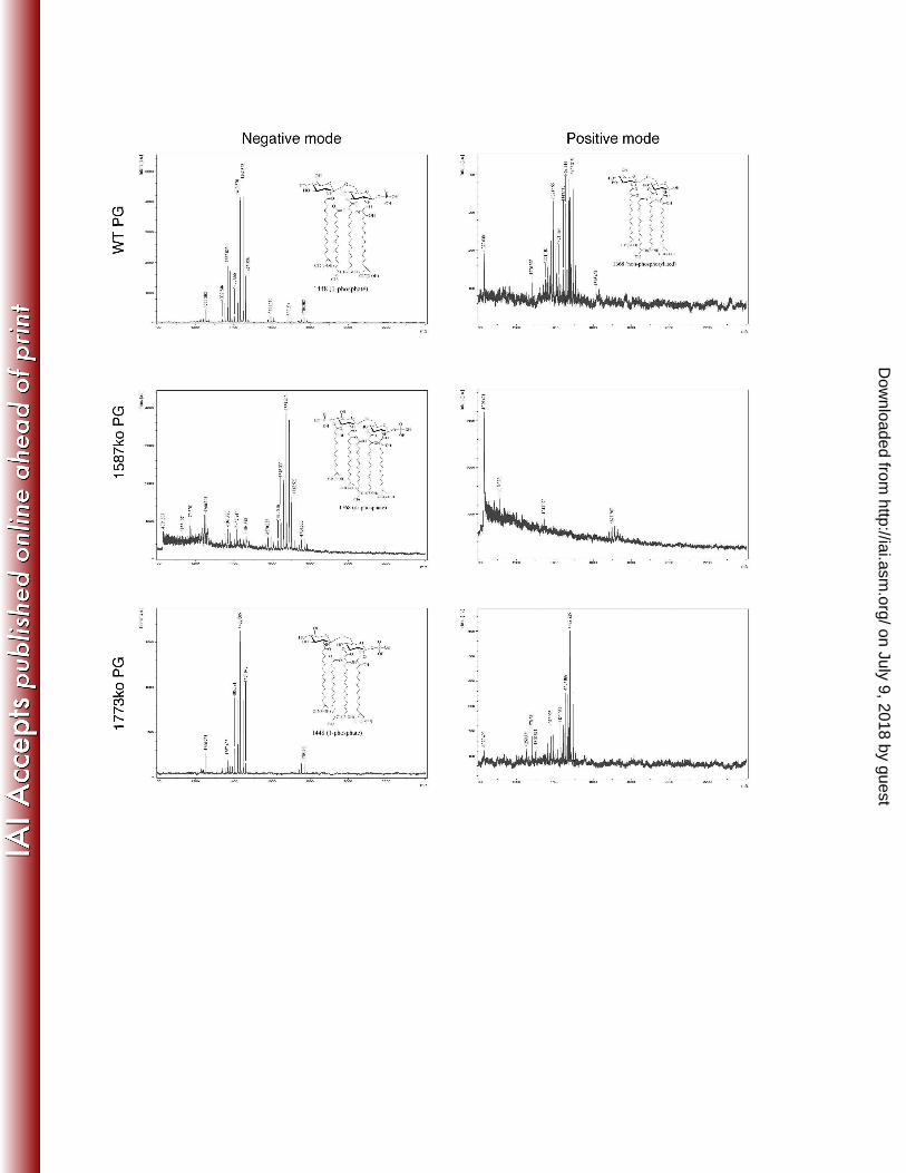
identification and characterization of P. gingivalis lipid A mutants that are unable to modify their にねに lipid A structural composition and display significantly different lipid A structural profiles was にねぬ performed in strain 33277 (11). However, since previous work in the rabbit ligature model was にねね performed with P. gingivalis strain A7436, in order to prevent potential undefined strain にねの variability effects in the rabbit model, the lipid A mutants were constructed in strain A7436. The にねは mutant strains, designated PG1587 and PG1773, contain deletion mutations in the lipid A 4’ にねば (PG1587) and 1 (PG1773) phosphatase genes, respectively (11). Characterization of the lipid A にねぱ structural composition confirmed that similar to strain 33277, strain A7436 containing these にねひ mutations displayed an altered lipid A structural composition in that PG1587 accumulated the di-にのど phosphate lipid A structural peak designated m/z 1770, whereas strain PG1773 accumulated にのな peak m/z 1449 (Fig. 1). This data confirms that the genes PG1587 and PG1773 result in the にのに same alterations in the lipid A structural profile in both P. gingivalis strains 33277 and A7436. にのぬ
Next, the mutant strains and their respective LPS preparations were examined for their にのね TLR4 and TLR2 responses. Similar to the results previously shown in strain 33277 (11) TLR4 にのの displayed differing responses to PG1587 and PG1773 in A7436. PG1587 bacteria as well as にのは their isolated LPS demonstrated strong TLR4 agonist activity (Fig. 2A). Likewise, LPS obtained にのば from PG1773 but not PG1587 displayed TLR4 antagonism (Fig. 2B). Interestingly, and different にのぱ from that observed in strain 33277 both PG1587 and PG1773 strain in A7436 displayed にのひ increased TLR2 activation compared to WT (Fig. 2B). We recently reported contaminating にはど lipoproteins in the P. gingivalis LPS preparations that could explain this TLR2 activity. ゅぱょ にはな
Finally, it has been previously reported that PG1587 and PG1773 in strain 33277 display にはに significantly different susceptibilities to polymyxin B, a cationic antimicrobial peptide antibiotic にはぬ (11). Examination of these mutations in A7436 yielded similar results in that PG1587 was にはね
on July 9, 2018 by guesthttp://iai.asm
.org/D
ownloaded from

exquisitely susceptible to polymyxin B whereas the wild type strain and PG1773 were にはの completely resistant (Fig. 2D). These data confirm that the lipid A phosphatase mutations にはは previously described in strain 33277 display a nearly identical phenotypes when contrasted in にはば A7436. にはぱ
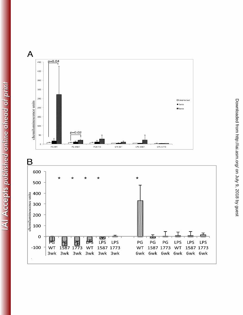
Porphyromonas gingivalis requires lipid A phosphatase modulation to colonize and にはひ cause overgrowth of commensal bacteria We used a rabbit model of periodontal disease to にばど examine P. gingivalis colonization and changes to total bacteria. Samples of plaque from the にばな ligature area were taken at three time-points including baseline (time zero), three weeks and six にばに weeks (Fig. 3). Chemiluminesence units from DNA-checkerboard were plotted for plaque にばぬ samples that were collected from each treatment group shown in Figure 4. Remarkably, P. にばね gingivalis wild type was the only group to cause robust P.gingivalis colonization (Fig 4A). にばの PG1587 also displayed a significant increase in P. gingivalis colonization at 6 weeks, although にばは much lower than wild type, (Fig. 4A). It is possible that the low chemiluminesence from the にばば baseline-uninfected rabbits is indicative of an endogenous strain of P. gingivalis that may have にばぱ increased over the time-course of infection with the PG1587. The test group LPS1587 shows a にばひ similar increase and since no bacteria were added this increase, although not significant, strongly にぱど implicates an endogenous strain that could be affected by the mutant strain or its LPS. にぱな
Next the relative biomass of the dental plaque was examined for the different にぱに experimental groups (Fig. 4B). Biomass was determined by the total chemiluminesence activity にぱぬ found in the dental plaque samples and it was found that plaque samples from each experimental にぱね group (except plaque LPS 1773ko group) showed a drastic reduction in total bacteria at three にぱの weeks from baseline-uninfected (Fig 4B). At 6 weeks, the colonization of wild type was also the にぱは only bacterial strain to cause significant increase in commensal biomass (Fig 4B). This result にぱば
on July 9, 2018 by guesthttp://iai.asm
.org/D
ownloaded from
was not found in any other experimental group demonstrating that both lipid A phosphate にぱぱ mutants were unable to both significantly colonize the rabbit periodontium or significantly にぱひ increase the total dental plaque biomass. にひど
Phosphatase mutants exert distinct affects on oral bacterial communities compared to にひな wild type while LPS preparations yield a less complex but discrete bacterial profile To examine にひに major increases and/or decreases to specific bacteria, the changes in abundance of bacterial にひぬ species that experienced a significant change (p ≥ 0.01 Mann Whitney Test) after six weeks of にひね applications were plotted in Table 1. Although wild type P. gingivalis was the only group to にひの generate an increase in oral bacteria at six weeks, all experimental groups had significant effects にひは on composition of oral bacterial community. The increase of total oral bacteria is markedly にひば increased and significant without the inclusion of P. gingivalis WT. Of the three species of にひぱ Campylobacter that were identified at baseline; two species (C. concisus and C. curva) showed にひひ significant reductions in all experimental groups. The changes to oral bacteria induced by wild ぬどど type P. gingivalis affected 16 species of the panel of 28 (59% G- and 41% G+). P. gingivalis ぬどな wild type altered the abundance of many more bacterial species than any of the other mutant ぬどに strains and LPS preparations. Conversely, the phosphatase mutants induced unique profiles ぬどぬ predominately in the Gram-negative communities. The changes to commensal bacteria by LPS ぬどね preparations were very few and virtually identical indicating a phenotype common to all LPS ぬどの types that caused the discrete changes to the bacterial communities. ぬどは
In order to assess the changes to Gram-negative and Gram-positive communities; the ぬどば relative abundance was plotted in a percentage plot (Fig 5A). To better visualize prominent ぬどぱ Gram-negative bacteria, P. gingivalis, C. curva and C. sputigena were separated by color (or ぬどひ pattern). The first major change observed was the reduction in the Gram-negative community at ぬなど
on July 9, 2018 by guesthttp://iai.asm
.org/D
ownloaded from

three weeks in all treatment groups, which was principally due to the marked decline of C. curva. ぬなな For example, the gram negative community, which includes C. curva makes up about 50% at ぬなに baseline in the animals received P. gingivalis WT, however, after three weeks of P. gingivalis ぬなぬ WT application resulted in a reduction of 30% in the Gram (-) bacteria and it seemed to be due to ぬなね the disappearance of C. curva. At the end of six weeks, P. gingivalis wild type became the most ぬなの predominant strain in the Gram-negative bacteria; this can be attributed largely to the ぬなは colonization of P. gingivalis. ぬなば
Low abundance of C. sputigena was seen at baseline, and at three weeks; however at six ぬなぱ weeks, the LPS preparations showed a remarkable increase in C. sputigena while the wild type ぬなひ and the mutant PG1587 showed decreases. The LPS groups showed no significant change in % ぬにど Gram-negative bacteria from baseline to 6 weeks. In these groups, the dramatic loss of C. curva ぬにな was replaced by the increase in C. sputigena masking overall changes to Gram-negative bacteria ぬにに while the phosphatase mutant bacteria and wild type had reduced amounts of C. sputigena in ぬにぬ addition to the reduction of C. curva. ぬにね
To examine the impact of bacterial strains and LPS preparations on C. sputigena, ぬにの chemiluminesence units were plotted in Fig 5B. All three LPS treatment groups yielded ぬには remarkable increases in C. sputigena while the bacterial treatment groups showed marked ぬにば reductions (Fig 5B). ぬにぱ
Clinical parameters of disease show similar bone loss for all groups In order to ぬにひ evaluate amount of periodontal disease caused by P. gingivalis or LPS preparations, clinical ぬぬど disease parameters including crestal bone level (distance between crest of the bone and tip of the ぬぬな tooth), intrabony defect depth (distance between the crestal bone and the base of the bone defect-ぬぬに vertical bone loss), were evaluated. Histological bone levels and osteoclast activity were assessed ぬぬぬ
on July 9, 2018 by guesthttp://iai.asm
.org/D
ownloaded from

by histological staining of tissue samples. Osteoclasts were identified by positive staining for ぬぬね their tartrate resistant acid phosphatases (TRAP). Strikingly, all experimental groups showed ぬぬの similar levels of clinical crestal and histological bone loss. The only difference was in the ぬぬは intrabony defect assessment; the rabbits receiving PGWT, PG1773 and LPS1773 had significant ぬぬば bone loss over ligature alone (historical data, (3)) while other differences were not statistically ぬぬぱ significant. It is interesting that this difference was common to all strains that had predominantly ぬぬひ the 1-phosphorylated, tetra-acylated lipid A structure capable of inhibiting host responses. ぬねど However, all other assessments of disease showed similar bone loss and osteoclast activity ぬねな between treatment groups. Together, these findings indicate that regardless of etiology, changes ぬねに to lipid A or bacterial communities, all the tested bacterial strains and LPS preparations cause ぬねぬ similar levels of periodontal disease. ぬねね ぬねの Discussion ぬねは
P. gingivalis has a number of virulence factors that contribute to disease such as ぬねば proteases (gingipains), lipopeptides, fimbriae, hemagluttinins and lipopolysaccharide. Although ぬねぱ some of these factors have been explored (9), the function of P. gingivalis’ unique heterogeneous ぬねひ LPS in disease in vivo has not been examined until now. In the current study, the use of P. ぬのど gingivalis lipid A phosphatase mutants that are unable to remodel their lipid A structural ぬのな composition do not colonize well or induce overgrowth of the commensal oral microbiota. ぬのに However, the P. gingivalis mutants and their respective isolated LPS preparations induced ぬのぬ significant changes in the qualitative composition of the oral bacterial communities and caused ぬのね similar disease profiles. This observation supports the idea that multiple different microbial ぬのの compositions result in dysbiosis and disease (7). ぬのは
on July 9, 2018 by guesthttp://iai.asm
.org/D
ownloaded from
Modifications to lipid A resulted in phenotypic changes to P. gingivalis. We have ぬのば previously reported that P. gingivalis grown in high temperature or lacking the 1587 phosphatase ぬのぱ can result in bacteria susceptible to antimicrobial peptides (AMPs) (11, 12). A potential ぬのひ explanation for this antimicrobial peptide sensitivity may be revealed by a recent study of ぬはど Helicobacter pylori lipid A phosphatases that showed the location of the phosphate group ぬはな determines susceptibility to polymyxin B (19). Considering this study, the dominant lipid A ぬはに structure possessing the phosphate at the 1 position in the PG1773 and P. gingivalis wild type ぬはぬ could confer resistance to polymyxin B whereas the lipid A with the 4’-phosphate is susceptible. ぬはね Since PG1587 was susceptible to polymyxin B, it was postulated that this mutant could be ぬはの sensitive to anti-microbial peptides in vivo. Indeed, the PG1587 was unable to colonize. However, ぬはは despite the antimicrobial peptide resistance of PG1773, it was also unable to colonize. It is likely ぬはば that the phosphatase mutants could not colonize due to the inability to modify the lipid A moiety ぬはぱ but for reasons not associated with antimicrobial peptide sensitivity. It has been shown in two ぬはひ other studies that alteration of phosphate position can affect colonization ability. Utilizing ぬばど bacterial mutants with changes to lipid A phosphate position in either Salmonella enterica ぬばな serovar Typhimurium or Helicobacter pylori resulted in a reduction or inability to colonize in a ぬばに mouse model of disease (20), (19). The phosphate position on lipid A appears to be a ぬばぬ determining factor for virulence; however, other pleiotropic effects from phosphate mutations ぬばね have not thoroughly been examined. For example, we have demonstrated here that TLR2 ぬばの activities are different between whole bacteria wild type and phosphatase mutants; presently, ぬばは there is no explanation for this and therefore more investigation is required. Regardless of ぬばば phosphatase phenotype or ability to colonize, all bacteria treatments caused changes to bacterial ぬばぱ communities. ぬばひ
on July 9, 2018 by guesthttp://iai.asm
.org/D
ownloaded from

DNA checkerboard hybridization was utilized to examine bacterial communities that ぬぱど have been shown to change with the application of P. gingivalis in a rabbit model of periodontal ぬぱな disease (3). Although the DNA checkerboard hybridization has limitations, such as high DNA ぬぱに requirement and species specific probes, this particular method allows for the analysis of a large ぬぱぬ panel of known bacterial species. The checkerboard assay has been compared to the 16S rDNA-ぬぱね based PCR method and found to be comparable in sensitivity for prevalent species although the ぬぱの sensitivity diminished with species at lower concentrations (21). The results from the ぬぱは checkerboard analysis revealed changes to bacterial communities were very different between ぬぱば the PGWT, PG1587 and PG1773. PGWT showed the most complex changes to oral microbiota. ぬぱぱ However, the bacterial changes seen from treatment with PG1587 and PG1773 were distinctly ぬぱひ different from each other, but common to PGWT. For example, significant increases to bacteria ぬひど associated with periodontal disease in humans were common to PGWT and PG1587 while ぬひな significant rise of unclassified bacteria were shared between PGWT and PG1773. It is interesting ぬひに to note that the increase of bacteria associated with PG1587 and PTWT were predominately part ぬひぬ of the complexes of bacteria associated with periodontal disease originally described by ぬひね Socransky (22) although it is not known if similar complexes exist in the rabbit periodontium. ぬひの The PG1587 and PGWT also showed a decrease in two bacterial species that are described in the ぬひは ‘green complex’ of bacteria associated with healthy flora. PG1773 did not have any increases in ぬひば bacteria associated with periodontal disease, save one of the ‘yellow complex’. Perhaps a shared ぬひぱ phenotype common to PGWT and PG1587 produced a suitable environment for the disease-ぬひひ associated bacterial complexes. For example, the agonist lipid A from PG1587, also produced by ねどど PGWT when environmental conditions allow (10, 11) could elicit an inflammatory host response ねどな favorable to the disease-associated bacteria. Alternately, the antagonist lipid A common to ねどに
on July 9, 2018 by guesthttp://iai.asm
.org/D
ownloaded from
PGWT and PG1773 could inhibit the host response and allow an outgrowth of certain ねどぬ commensal bacteria. Interestingly, PGWT, PG1773 and LPS1773 treatment groups caused ねどね similar intrabony defects again indicating a shared phenotype such as the antagonist lipid A ねどの structure; it is possible that this particular lipid A structure may be directly involved in disease ねどは progression since there are similarities between these treatment groups’ disease profiles but not ねどば bacterial profiles. However, the abundance and species of oral bacteria that resulted from each ねどぱ treatment were distinctly different from each other yet all resulted in disease. ねどひ
Different LPS preparations do not appear to cause any specific alterations rather; similar ねなど changes are made to bacterial profiles indicating a general environmental alteration. The striking ねなな feature common to all purified LPS was the robust TLR2 activity (8); this alteration of host-ねなに environment could be the cause of the similar bacterial profiles observed. Further, the LPS ねなぬ preparations appear to alter the environment in a way that is beneficial to C. sputigena, which is ねなね not seen in the whole bacteria preparations. ねなの
One common trend to all treatment groups except P. gingivalis WT was the ねなは disappearance of two species of Campylobacter, C. concisus and C. curva. The presence of P. ねなば gingivalis wild type did not reduce the C. concisus species as seen in all other treatment groups. ねなぱ This suggests that the environment created by the modulation of lipid A in wild type P. ねなひ gingivalis may be required for C. concisus to maintain its niche; this niche may change to allow ねにど some bacteria to bind where others may lose their binding sites. ねにな
The present study demonstrates that lipid A phosphatases are required for P. gingivalis ねにに colonization and commensal overgrowth. Overgrowth of commensal bacteria due to P. ねにぬ gingivalis colonization has recently been shown to be responsible for periodontitis using a mouse ねにね model of periodontal disease by gavage, (4). In this study, since all bacterial strains and their ねにの
on July 9, 2018 by guesthttp://iai.asm
.org/D
ownloaded from
respective LPS preparations caused microbial dysbiosis that resulted in very similar disease, it is ねには likely that disease in this model occurred through different etiologies that all resulted in a ねにば disruption of the normal oral flora. We could not distinguish those microbial changes which ねにぱ occurred from an altered local inflammatory environment from those that may have induced the ねにひ inflammatory response. Rather, this study demonstrates that multiple different bacterial ねぬど communities are associated with periodontal disease. ねぬな Acknowledgments ねぬに Supported in part by USPHS grants DE012768 (RPD); DE020906 (AK); DE19938; DE15566 ねぬぬ (TVD), and DE18917 to HH from the National Institute of Dental and Craniofacial Research ねぬね ねぬの
on July 9, 2018 by guesthttp://iai.asm
.org/D
ownloaded from
References ねぬは ねぬば な┻ Dewhirst FE, Chen T, Izard J, Paster BJ, Tanner AC, Yu WH, Lakshmanan A, ねぬぱ Wade WG. にどなど┻ The human oral microbiome┻ Journal of bacteriology 192:のどどに┽ねぬひ のどなば┻ ねねど に┻ Oz HS, Puleo DA. にどなな┻ Animal models for periodontal disease┻ Journal of ねねな biomedicine ┃ biotechnology 2011:ばのねぱのば┻ ねねに ぬ┻ Hasturk H, Kantarci A, Goguet-Surmenian E, Blackwood A, Andry C, Serhan CN, ねねぬ Van Dyke TE. にどどば┻ Resolvin Eな regulates inflammation at the cellular and tissue ねねね level and restores tissue homeostasis in vivo┻ Journal of immunology 179:ばどにな┽ねねの ばどにひ┻ ねねは ね┻ Hajishengallis G, Liang S, Payne MA, Hashim A, Jotwani R, Eskan MA, McIntosh ねねば ML, Alsam A, Kirkwood KL, Lambris JD, Darveau RP, Curtis MA. にどなな┻ Low┽ねねぱ abundance biofilm species orchestrates inflammatory periodontal disease through ねねひ the commensal microbiota and complement┻ Cell (ost Microbe 10:ねひば┽のどは┻ ねのど の┻ Hajishengallis G, Darveau RP, Curtis MA. にどなに┻ The keystone┽pathogen ねのな hypothesis┻ Nature reviews┻ Microbiology 10:ばなば┽ばにの┻ ねのに は┻ Darveau RP. にどどひ┻ The oral microbial consortiumfs interaction with the ねのぬ periodontal innate defense system┻ DNA Cell Biol 28:ぬぱひ┽ぬひの┻ ねのね ば┻ Darveau RP. にどなど┻ Periodontitis┺ a polymicrobial disruption of host homeostasis┻ ねのの Nature reviews┻ Microbiology 8:ねぱな┽ねひど┻ ねのは ぱ┻ Jain S, Coats SR, Chang AM, Darveau RP. にどなぬ┻ A Novel Class of Lipoprotein ねのば Lipase┽Sensitive Molecules Mediates Toll┽Like Receptor に Activation by ねのぱ Porphyromonas gingivalis┻ )nfection and immunity 81:なにばば┽なにぱは┻ ねのひ ひ┻ Bostanci N, Belibasakis GN. にどなに┻ Porphyromonas gingivalis┺ an invasive and ねはど evasive opportunistic oral pathogen┻ FEMS microbiology letters 333:な┽ひ┻ ねはな など┻ Al-Qutub MN, Braham PH, Karimi-Naser LM, Liu X, Genco CA, Darveau RP. にどどは┻ ねはに (emin┽dependent modulation of the lipid A structure of Porphyromonas gingivalis ねはぬ lipopolysaccharide┻ )nfection and immunity 74:ねねばね┽ねねぱの┻ ねはね なな┻ Coats SR, Jones JW, Do CT, Braham PH, Bainbridge BW, To TT, Goodlett DR, ねはの Ernst RK, Darveau RP. にどどひ┻ (uman Toll┽like receptor ね responses to P┻ gingivalis ねはは are regulated by lipid A な┽ and ねf┽phosphatase activities┻ Cell Microbiol 11:なのぱば┽ねはば なのひひ┻ ねはぱ なに┻ Curtis MA, Percival RS, Devine D, Darveau RP, Coats SR, Rangarajan M, Tarelli ねはひ E, Marsh PD. にどなな┻ Temperature┽dependent modulation of Porphyromonas ねばど gingivalis lipid A structure and interaction with the innate host defenses┻ )nfection ねばな and immunity 79:ななぱば┽ななひぬ┻ ねばに なぬ┻ Coats SR, Pham TT, Bainbridge BW, Reife RA, Darveau RP. にどどの┻ MD┽に mediates ねばぬ the ability of tetra┽acylated and penta┽acylated lipopolysaccharides to antagonize ねばね Escherichia coli lipopolysaccharide at the TLRね signaling complex┻ Journal of ねばの immunology 175:ねねひど┽ねねひぱ┻ ねばは なね┻ Shapira L, Champagne C, Van Dyke TE, Amar S. なひひぱ┻ Strain┽dependent activation ねばば of monocytes and inflammatory macrophages by lipopolysaccharide of ねばぱ Porphyromonas gingivalis┻ )nfection and immunity 66:にばぬは┽にばねに┻ ねばひ なの┻ Olsen I, Socransky SS. なひぱな┻ Ultrasonic dispersion of pure cultures of plaque ねぱど bacteria and plaque┻ Scand J Dent Res 89:ぬどば┽ぬなに┻ ねぱな
on July 9, 2018 by guesthttp://iai.asm
.org/D
ownloaded from
なは┻ Olsen I, Socransky SS. なひぱな┻ Comparison of three anaerobic culture techniques ねぱに amd media for viable recovery of subgingival plaque bacteria┻ Scand J Dent Res ねぱぬ 89:なはの┽なばね┻ ねぱね なば┻ Doan N, Contreras A, Flynn J, Morrison J, Slots J. なひひひ┻ Proficiencies of three ねぱの anaerobic culture systems for recovering periodontal pathogenic bacteria┻ Journal of ねぱは clinical microbiology 37:なばな┽なばね┻ ねぱば なぱ┻ Socransky SS, Haffajee AD. なひひね┻ Evidence of bacterial etiology┺ a historical ねぱぱ perspective┻ Periodontology にどどど 5:ば┽にの┻ ねぱひ なひ┻ Cullen TW, Giles DK, Wolf LN, Ecobichon C, Boneca IG, Trent MS. にどなな┻ ねひど (elicobacter pylori versus the host┺ remodeling of the bacterial outer membrane is ねひな required for survival in the gastric mucosa┻ PLoS pathogens 7:eなどどにねのね┻ ねひに にど┻ Kong Q, Six DA, Liu Q, Gu L, Wang S, Alamuri P, Raetz CR, Curtiss R, 3rd. にどなに┻ ねひぬ Phosphate groups of lipid A are essential for Salmonella enterica serovar ねひね Typhimurium virulence and affect innate and adaptive immunity┻ )nfection and ねひの immunity 80:ぬになの┽ぬににね┻ ねひは にな┻ Siqueira JF, Rocas IN, De Uzeda M, Colombo AP, Santos KR. にどどに┻ Comparison of ねひば なはS rDNA┽based PCR and checkerboard DNA┽DNA hybridisation for detection of ねひぱ selected endodontic pathogens┻ Journal of medical microbiology 51:などひど┽などひは┻ ねひひ にに┻ Socransky SS, Haffajee AD, Cugini MA, Smith C, Kent RL, Jr. なひひぱ┻ Microbial のどど complexes in subgingival plaque┻ J┻ Clin┻ Periodontol┻ 25:なぬね┽なねね┻ のどな のどに のどぬ
on July 9, 2018 by guesthttp://iai.asm
.org/D
ownloaded from
FIGURE LEGENDS のどね のどの のどは Figure 1 のどば のどぱ Figure 1 P. gingivalis expresses both lipid A 4ガ- and 1-phosphatase activities. Lipid A isolated のどひ from wild-type P. gingivalis A7436 or mutant bacteria bearing deletions in PG1587 and PG1773 のなど gene loci were examined by MALDI-TOF MS to elucidate the position of the lipid A phosphates. のなな Representative structures of lipid A corresponding to peak are shown. Lipid A samples were のなに examined in the negative ion mode or the positive ion mode for each sample. のなぬ のなね
on July 9, 2018 by guesthttp://iai.asm
.org/D
ownloaded from

のなの Figure 2 のなは のなば Figure 2 Lipid A phosphatases allow P. gingivalis to evade host innate immune differences. のなぱ Phosphatase mutants have different phenotypic host innate effects through toll like receptors and のなひ antimicrobial susceptibility. HEK293 cells expressing either human TLR4 and MD-2 (A, C) or のにど TLR2 and TLR1 (B) were exposed to the indicated doses of LPS or whole bacteria for 4 h. The のにな fold activation of NF-kB over the media control was determined by measuring inducible Firefly のにに luciferase activity. Positive control for TLR4 is E. coli and TLR2 is PamC3K4. The results のにぬ shown are means ± SD of triplicate samples from one of three independent experiments. のにね Asterisks indicate statistically significant differences (P < 0.001; two-tailed unpaired t-tests) in のにの the potency of TLR4 antagonism by PGWT or PG1773. (D) Lipid A phosphatases confer P. のには gingivalis resistance to killing by antimicrobial cationic peptides. The indicated strains of P. のにば gingivalis were plated on TYHK-agar plates containing polymyxin B (PMB) (0, 5 and 200 mg のにぱ ml-1) and measured by spectrophotometry OD600. のにひ のぬど
on July 9, 2018 by guesthttp://iai.asm
.org/D
ownloaded from
のぬな Figure 3 のぬに のぬぬ Figure 3 Timeline of experimental design. 3-0 silk ligatures were tied to second premolars in のぬね mandibular quadrants at baseline in all groups, and P. gingivalis, phosphatase mutants or purified のぬの LPS was applied in methylcellulose slurry three times per week (M-W-F) for 6 wk. Plaque のぬは samples were taken for microbial analysis at 0, 3 and 6 weeks during treatment. At 6 wk, all のぬば were sacrificed, and the extent of disease was determined. のぬぱ のぬひ のねど
on July 9, 2018 by guesthttp://iai.asm
.org/D
ownloaded from

のねな Figure 4 のねに のねぬ Figure 4 Porphyromonas gingivalis phosphate modulation is required for colonization and のねね commensal overgrowth. Chemiluminescence units of DNA-DNA checkerboard hybridization のねの assay specific to (A) P. gingivalis or (B) the summation of all bacteria analyzed (see Materials のねは and Methods) (P < 0.01; Mann-Whitney Test) のねば のねぱ
on July 9, 2018 by guesthttp://iai.asm
.org/D
ownloaded from

のねひ Table 1 ののど ののな ののに Table 1 Significant changes to chemiluminescence signal from specific oral bacteria at 6 weeks ののぬ compared to uninfected controls (P < 0.01; Mann-Whitney Test). Bacterial complexes coded ののね with regard to (Socransky and Haffajee ref); *Red complex, #Orange complex, Yellow ののの complex, Purple complex, Green complex. ののは ののば
on July 9, 2018 by guesthttp://iai.asm
.org/D
ownloaded from

ののぱ Figure 5 ののひ のはど Figure 5 Distinct changes to bacterial communities result from application of P. gingivalis, のはな phosphatase mutants or LPS. Relative biomass of Gram-negative bacteria including P. のはに gingivalis, C. curva and C. sputegina and Gram-positive bacteria profiles (A) and のはぬ chemiluminescence units of C. sputegina plotted for each treatment group over time. Statistics のはね were scored using Mann-Whitney Test のはの のはは
on July 9, 2018 by guesthttp://iai.asm
.org/D
ownloaded from

のはば Figure 6 のはぱ のはひ Figure 6 Analyses of periodontal disease from P. gingivalis, phosphatase or LPS-treated rabbit のばど lesions. Alveolar bone loss for all animals was directly measured on defleshed jaws (see のばな Materials and Methods) for characteristics of human periodontitis including soft (A, intrabony のばに defect) and hard tissue (B, crestal bone loss) destruction. Rabbit mandibles were harvested and のばぬ prepared for histologic analysis (see Materials and Methods). Histologic analysis and のばね quantification of histomorphometric changes (C). Osteoclastogenesis in the alveolar bone was のばの assessed by TRAP staining (D) (see Materials and Methods). Asterisks indicate statistically のばは significant differences (P < 0.01; Mann-Whitney Test) のばば のばぱ
on July 9, 2018 by guesthttp://iai.asm
.org/D
ownloaded from