Embed Size (px)
Citation preview
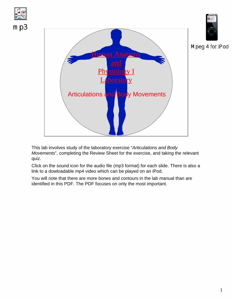
1
Human Anatomy and
Physiology ILaboratory
Articulations and Body Movements
This lab involves study of the laboratory exercise “Articulations and Body Movements”, completing the Review Sheet for the exercise, and taking the relevant quiz. Click on the sound icon for the audio file (mp3 format) for each slide. There is also a link to a dowloadable mp4 video which can be played on an iPod.You will note that there are more bones and contours in the lab manual than are identified in this PDF. The PDF focuses on only the most important.
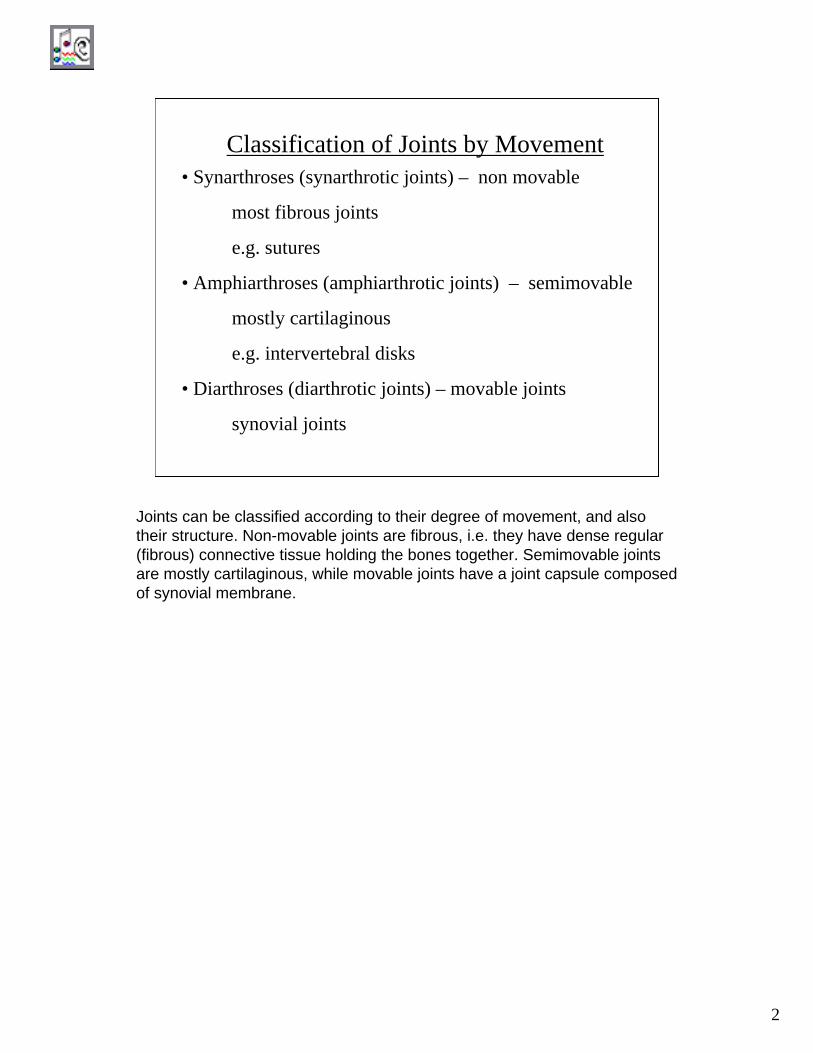
2
Classification of Joints by Movement• Synarthroses (synarthrotic joints) – non movable
most fibrous joints
e.g. sutures
• Amphiarthroses (amphiarthrotic joints) – semimovable
mostly cartilaginous
e.g. intervertebral disks
• Diarthroses (diarthrotic joints) – movable joints
synovial joints
Joints can be classified according to their degree of movement, and also their structure. Non-movable joints are fibrous, i.e. they have dense regular (fibrous) connective tissue holding the bones together. Semimovable joints are mostly cartilaginous, while movable joints have a joint capsule composed of synovial membrane.
3
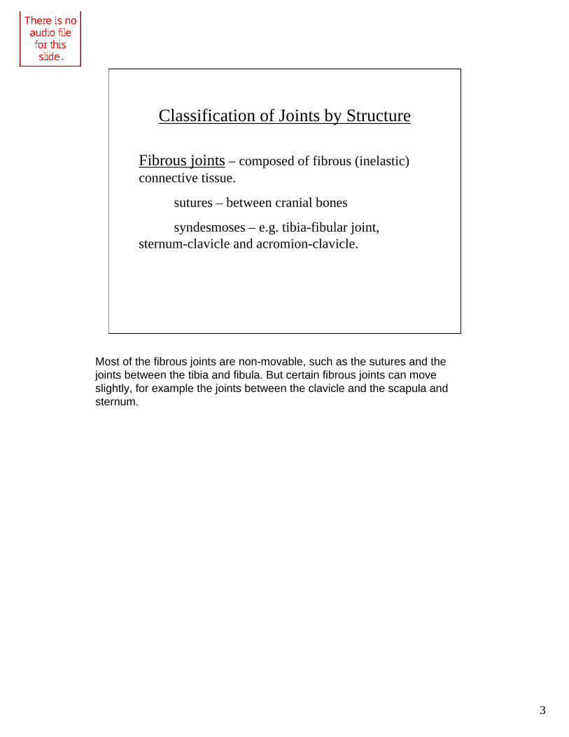
Classification of Joints by Structure
Fibrous joints – composed of fibrous (inelastic) connective tissue.
sutures – between cranial bones
syndesmoses – e.g. tibia-fibular joint, sternum-clavicle and acromion-clavicle.
Most of the fibrous joints are non-movable, such as the sutures and the joints between the tibia and fibula. But certain fibrous joints can move slightly, for example the joints between the clavicle and the scapula and sternum.
4
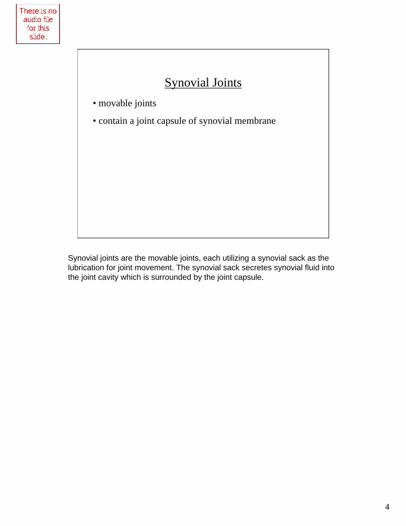
Synovial Joints• movable joints
• contain a joint capsule of synovial membrane
Synovial joints are the movable joints, each utilizing a synovial sack as the lubrication for joint movement. The synovial sack secretes synovial fluid into the joint cavity which is surrounded by the joint capsule.
5
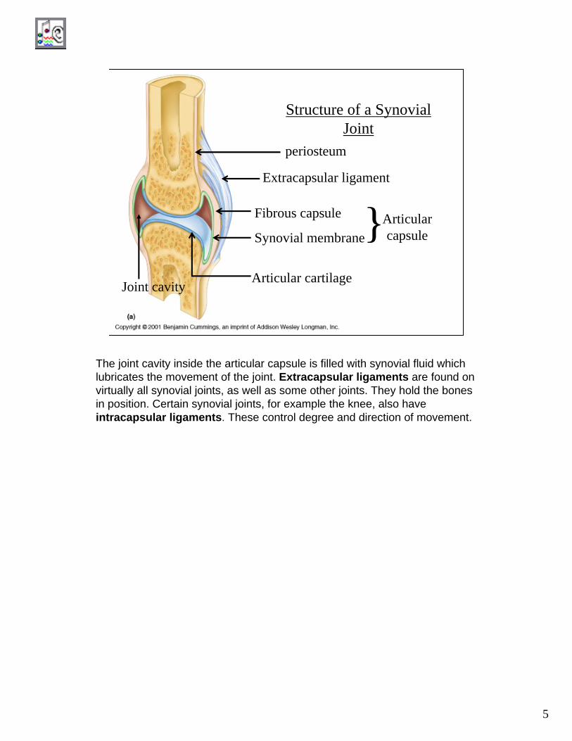
Structure of a SynovialJoint
periosteum
Extracapsular ligament
Fibrous capsule
Synovial membrane
Articular cartilage
Articularcapsule}
Joint cavity
The joint cavity inside the articular capsule is filled with synovial fluid which lubricates the movement of the joint. Extracapsular ligaments are found on virtually all synovial joints, as well as some other joints. They hold the bones in position. Certain synovial joints, for example the knee, also have intracapsular ligaments. These control degree and direction of movement.
6
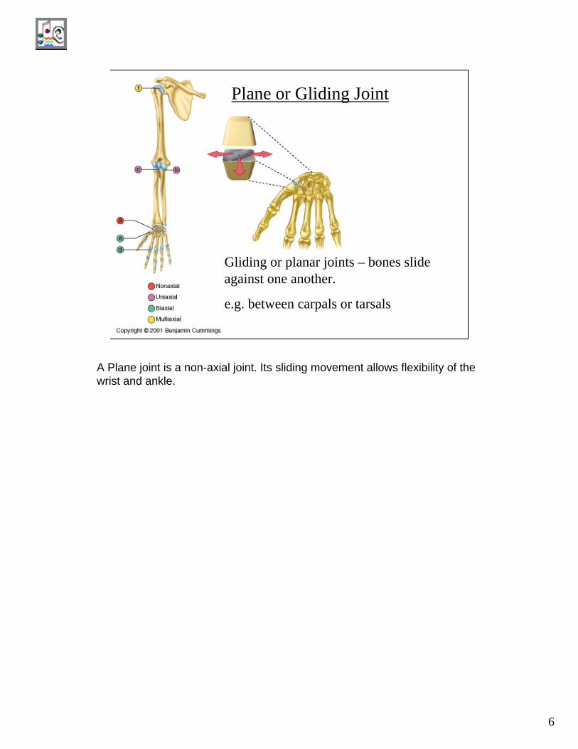
Plane or Gliding Joint
Gliding or planar joints – bones slide against one another.
e.g. between carpals or tarsals
A Plane joint is a non-axial joint. Its sliding movement allows flexibility of the wrist and ankle.

7
Gliding Movement is Non-axial
When the bones of the wrist move against one another, the don’t move in a particular axis, they slide or glide. Therefore these joints are called non-axialjoints.
8
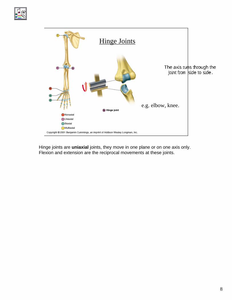
e.g. elbow, knee.
Hinge Joints
Hinge joints are uniaxial joints, they move in one plane or on one axis only. Flexion and extension are the reciprocal movements at these joints.
9
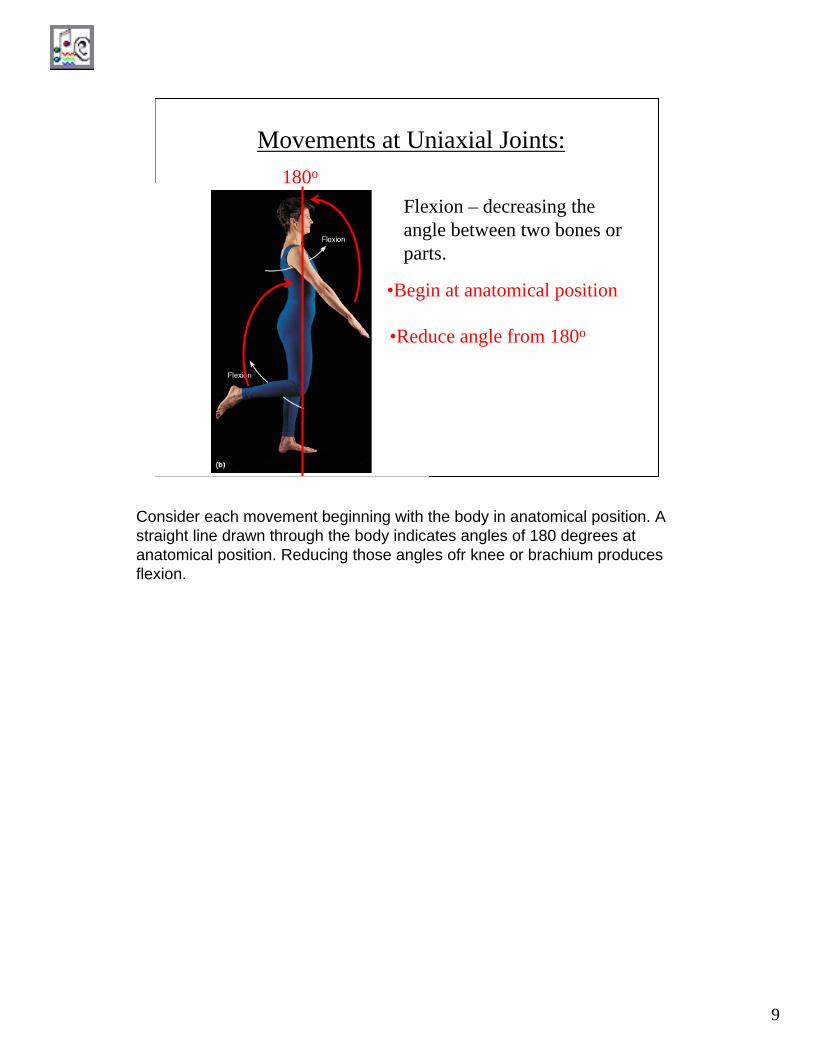
Movements at Uniaxial Joints:
Flexion – decreasing the angle between two bones or parts.
180o
•Begin at anatomical position
•Reduce angle from 180o
Consider each movement beginning with the body in anatomical position. A straight line drawn through the body indicates angles of 180 degrees at anatomical position. Reducing those angles ofr knee or brachium produces flexion.
10
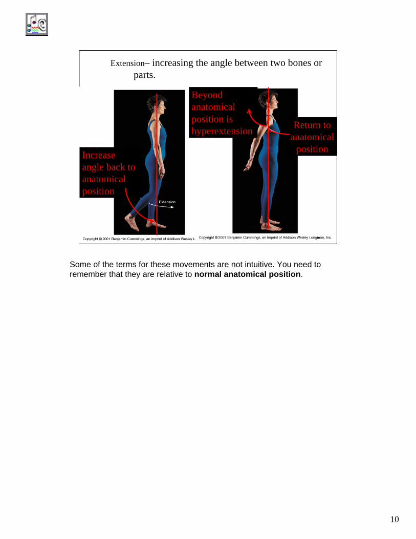
Extension– increasing the angle between two bones or parts.
Increase angle back to anatomical position
Return to anatomical
position
Beyond anatomical position is hyperextension
Some of the terms for these movements are not intuitive. You need to remember that they are relative to normal anatomical position.
11
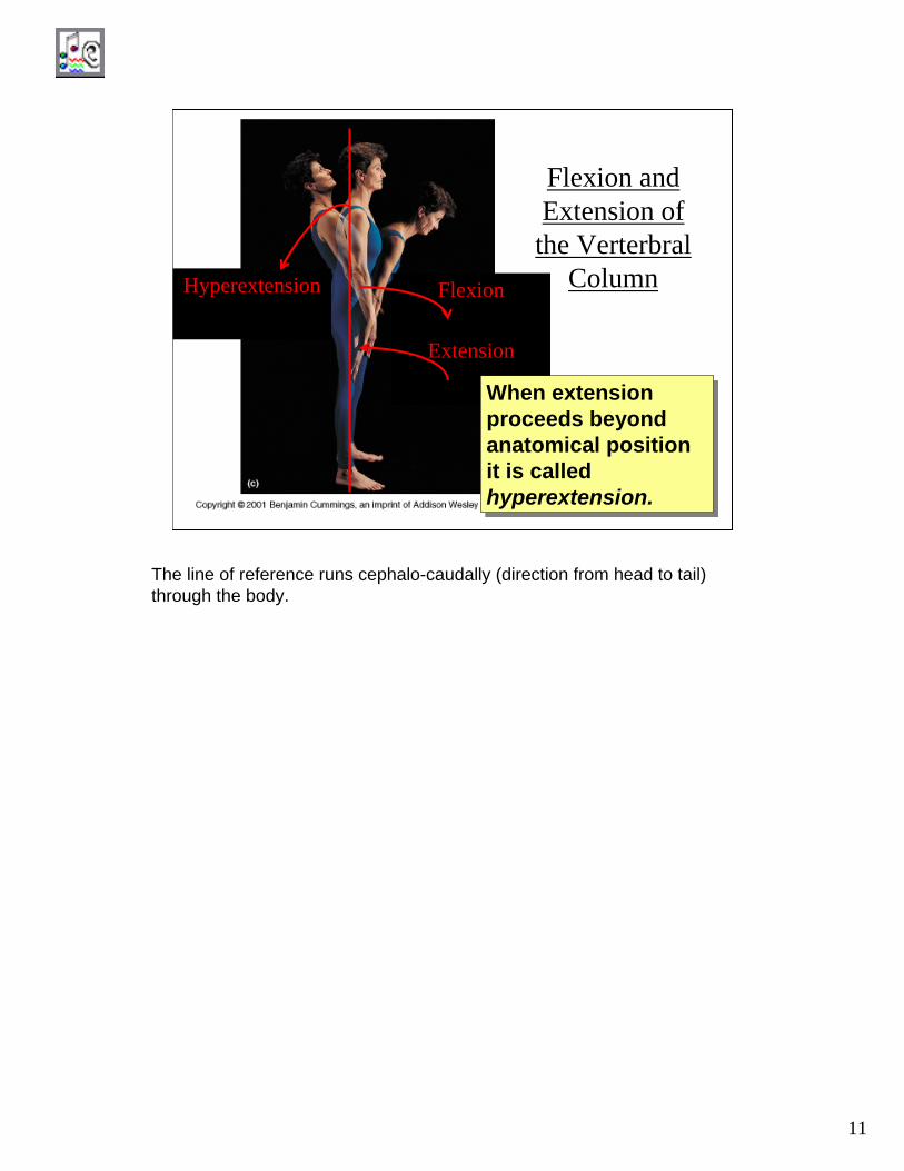
Flexion and Extension of
the VerterbralColumnFlexion
Extension
Hyperextension
When extension proceeds beyond anatomical position it is called hyperextension.
When extension proceeds beyond anatomical position it is called hyperextension.
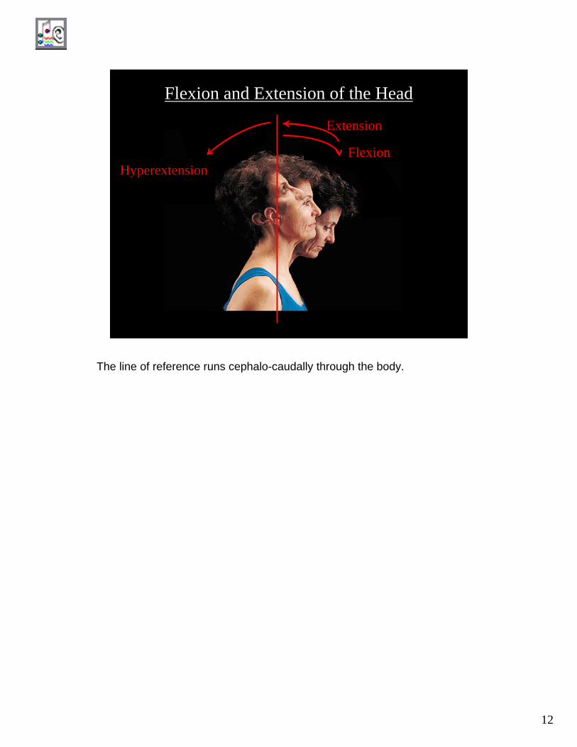
The line of reference runs cephalo-caudally (direction from head to tail) through the body.
12
Flexion and Extension of the Head
FlexionExtension
HyperextensionFlexion
The line of reference runs cephalo-caudally through the body.
13
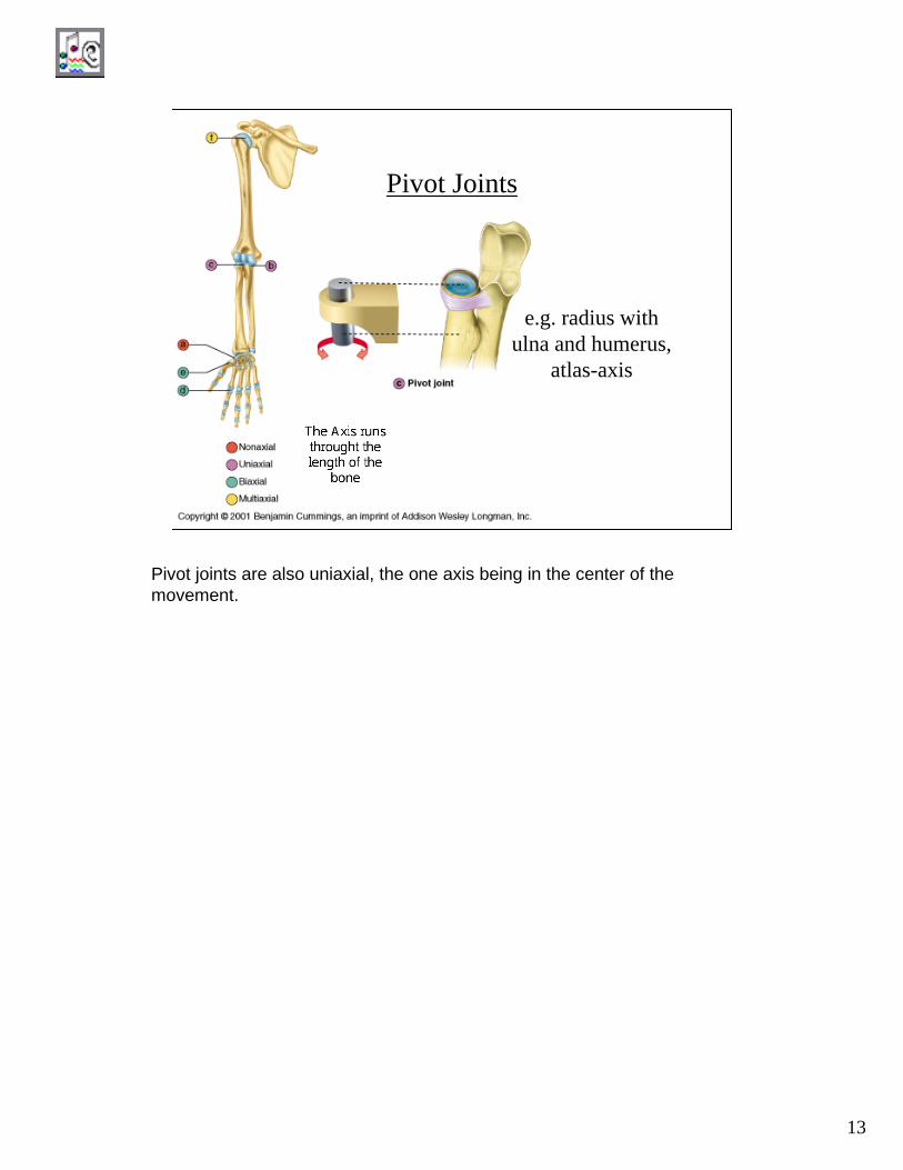
Pivot Joints
e.g. radius with ulna and humerus,
atlas-axis
Pivot joints are also uniaxial, the one axis being in the center of the movement.
14
Rotation of the Head and
Thigh
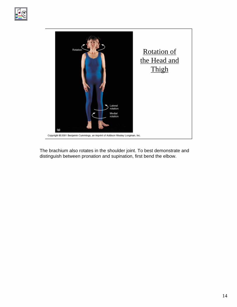
The brachium also rotates in the shoulder joint. To best demonstrate and distinguish between pronation and supination, first bend the elbow.

15
Rotation of the Radius
Supination – to move palm up
Pronation – to move palm down
Despite popular usage, supination and pronation refer only to the hand.
16
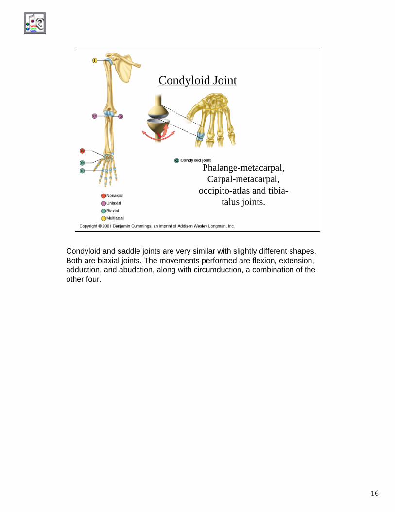
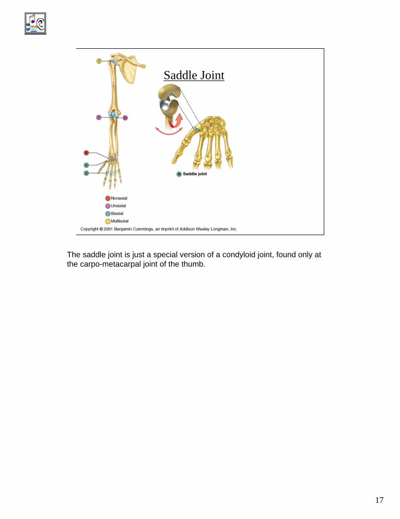
Condyloid Joint
Phalange-metacarpal, Carpal-metacarpal,
occipito-atlas and tibia-talus joints.
Condyloid and saddle joints are very similar with slightly different shapes. Both are biaxial joints. The movements performed are flexion, extension, adduction, and abudction, along with circumduction, a combination of the other four.
17
Saddle Joint
The saddle joint is just a special version of a condyloid joint, found only at the carpo-metacarpal joint of the thumb.
18
Additional Movements at Biaxial
Joints
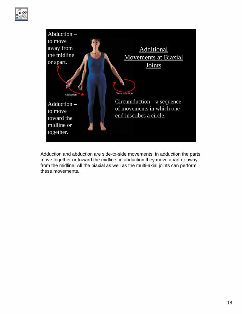
Abduction –to move away from the midline or apart.
Adduction –to move toward the midline or together.
Circumduction – a sequence of movements in which one end inscribes a circle.
Adduction and abduction are side-to-side movements: in adduction the parts move together or toward the midline, in abduction they move apart or away from the midline. All the biaxial as well as the multi-axial joints can perform these movements.
19
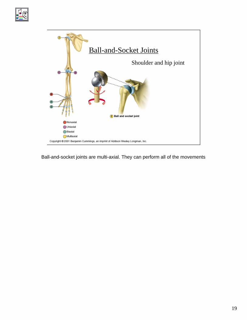
Ball-and-Socket JointsShoulder and hip joint
Ball-and-socket joints are multi-axial. They can perform all of the movements
20
Movements of the Foot
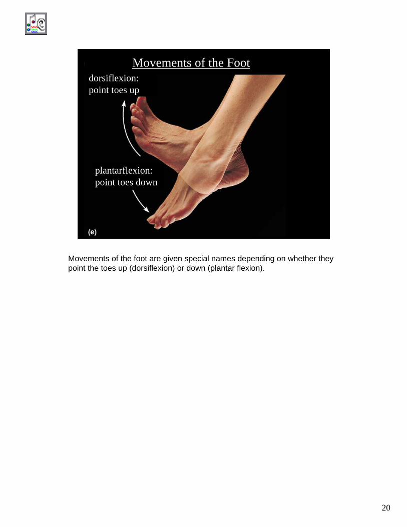
plantarflexion: point toes down
dorsiflexion: point toes up
Movements of the foot are given special names depending on whether they point the toes up (dorsiflexion) or down (plantar flexion).
21
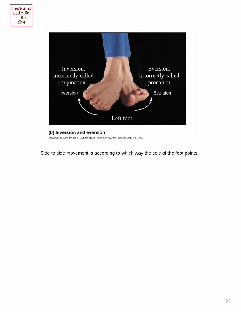
Inversion and Eversion
Left foot
Inversion, incorrectly called
supination
Eversion, incorrectly called
pronation
Side to side movement is according to which way the sole of the foot points.
22
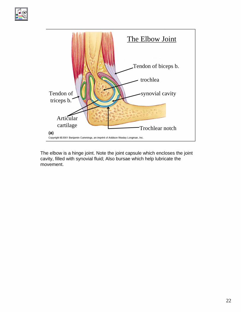
The Elbow Joint
trochlea
Trochlear notch
synovial cavity
Tendon of biceps b.
Tendon of triceps b.
Articularcartilage
The elbow is a hinge joint. Note the joint capsule which encloses the joint cavity, filled with synovial fluid; Also bursae which help lubricate the movement.

23
Annular ligament
Radial (lateral) collateral ligament
Articularcapsule
The radial head fits neatly in the collar produced by the annular ligamentand turns within this collar. Other primary ligaments are the radial (lateral) collateral ligament and the ulnar (medial) collateral ligament. These help to prevent lateral displacement of the bones at the joints.

24
Annular ligament
Ulnar (medial) collateral ligament
The annular ligament attaches to the lateral collateral ligament and to the ulna, so that the head of the radius is free to rotate.

25
Elbow radiograph, lateral view
Trochlear notch
Trochlea
Soft tissues such as the articular capsule and articular cartilage are not radio opaque, so they appear dark in the x-ray.

26
Elbow radiograph, dorsal view
Olecranon process
Head of the radius
The articular cartilage, not being radio-opaque, shows up clearly as a dark space between the bones.
27
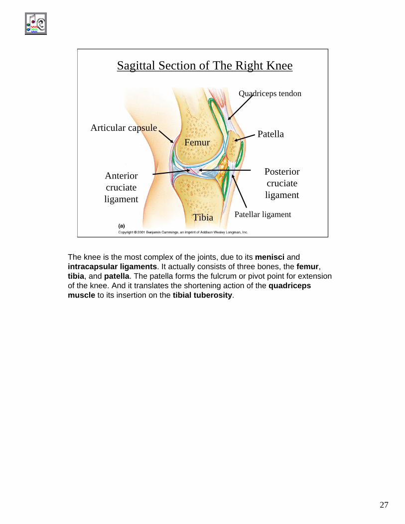
Sagittal Section of The Right Knee
Articular capsule
Anterior cruciateligament
Posterior cruciateligament
Tibia
Femur Patella
Patellar ligament
Quadriceps tendon
The knee is the most complex of the joints, due to its menisci and intracapsular ligaments. It actually consists of three bones, the femur, tibia, and patella. The patella forms the fulcrum or pivot point for extension of the knee. And it translates the shortening action of the quadriceps muscle to its insertion on the tibial tuberosity.
28
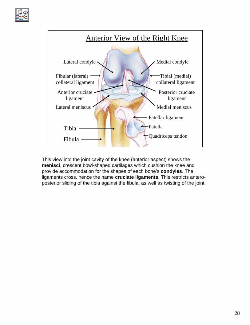
Anterior View of the Right Knee
Tibia
Fibula
Patellar ligament
Patella
Quadriceps tendon
Medial condyleLateral condyle
Tibial (medial) collateral ligament
Fibular (lateral) collateral ligament
Posterior cruciateligament
Anterior cruciateligament
Lateral meniscus Medial meniscus
This view into the joint cavity of the knee (anterior aspect) shows the menisci, crescent bowl-shaped cartilages which cushion the knee and provide accommodation for the shapes of each bone’s condyles. The ligaments cross, hence the name cruciate ligaments. This restricts antero-posterior sliding of the tibia against the fibula, as well as twisting of the joint.
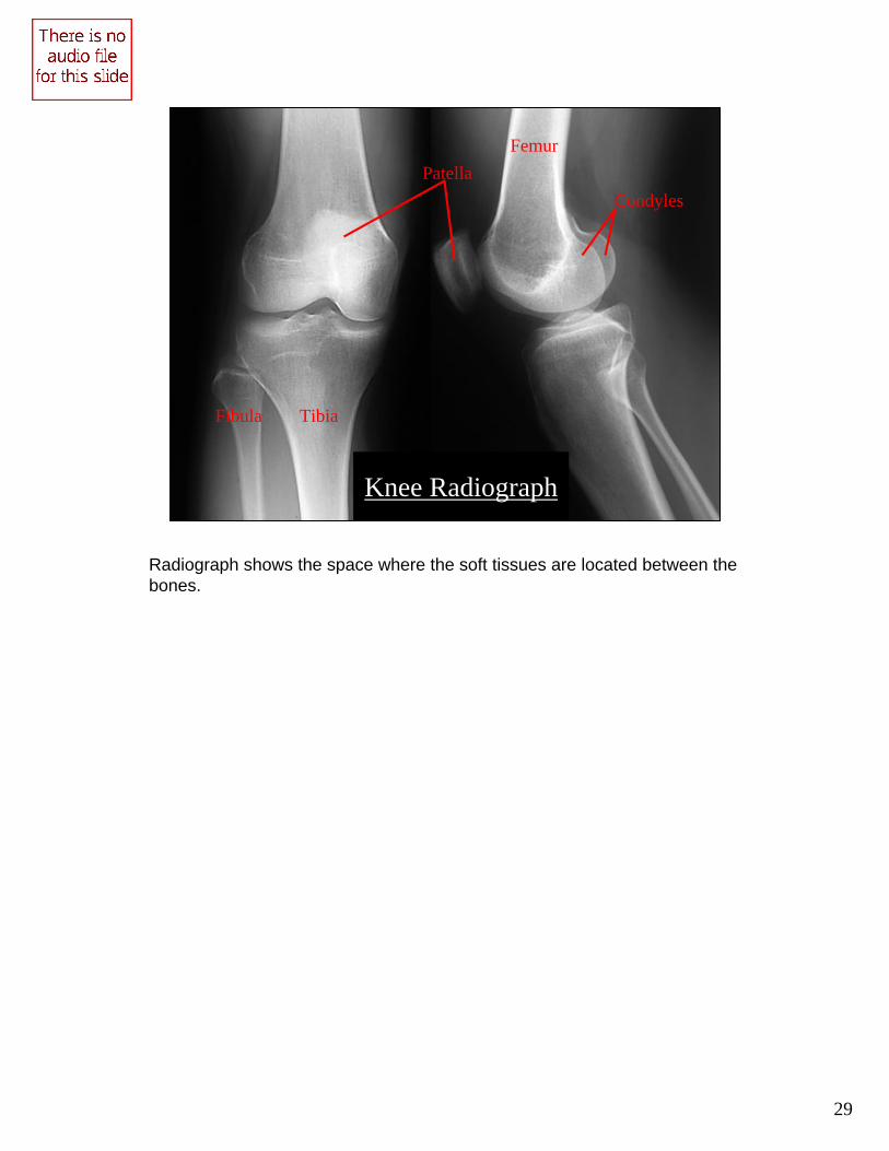
29
Patella Femur
Tibia Fibula
Condyles
Knee Radiograph
Radiograph shows the space where the soft tissues are located between the bones.
30
Lab Protocol
1. After studying the lab exercise and this PDF, complete the Review Sheet which accompanies the lab exercise.
2. Use ADAM to study the joints as per directions in the lab manual.
3. Take the quiz on the arthrology.