Embed Size (px)
Citation preview
INSTRUCTOR GUIDE
Human Anatomy & Physiology Laboratory ManualMAIN VERSION, Eighth Edition Update
CAT VERSION, Ninth Edition Update
FETAL PIG VERSION, Ninth Edition Update
San Francisco • Boston • New YorkCape Town • Hong Kong • London • Madrid • Mexico City
Montreal • Munich • Paris • Singapore • Sydney • Tokyo • Toronto
ELAINE N. MARIEB, R.N., Ph.D
Holyoke Community College
SUSAN T. BAXLEY, M.A.
Troy University, Montgomery Campus
NANCY G. KINCAID, Ph.D
Troy University, Montgomery Campus
PhysioEx™ Exercises authored by
Peter Z. Zao, North Idaho College
Timothy Stabler, Indiana University Northwest
Lori Smith, American River College
Greta Peterson, Middlesex Community College
Andrew Lokuta, University of Wisconsin—Madison
Editor-in-Chief: Serina BeauparlantProject Editor: Sabrina LarsonPhysioEx Project Editor: Erik FortierEditorial Assistant: Nicole GrazianoManaging Editor: Wendy EarlProduction Editor: Leslie AustinComposition: Cecelia G. MoralesCover Design: Riezebos Holzbaur Design GroupSenior Manufacturing Buyer: Stacey WeinbergerMarketing Manager: Gordon Lee
Copyright © 2009 Pearson Education, Inc., publishing as Pearson Benjamin Cummings,1301 Sansome St., San Francisco, CA 94111. All rights reserved. Manufactured in the UnitedStates of America. This publication is protected by Copyright and permission should beobtained from the publisher prior to any prohibited reproduction, storage in a retrieval system,or transmission in any form or by any means, electronic, mechanical, photocopying, record-ing, or likewise. To obtain permission(s) to use material from this work, please submit a writ-ten request to Pearson Education, Inc., Permissions Department, 1900 E. Lake Ave.,Glenview, IL 60025. For information regarding permissions, call (847) 486-2635.
Many of the designations used by manufacturers and sellers to distinguish their products areclaimed as trademarks. Where those designations appear in this book, and the publisher wasaware of a trademark claim, the designations have been printed in initial caps or all caps.
Pearson Benjamin Cummings™ is a trademark, in the U.S. and/or other countries, of PearsonEducation, Inc. or its affiliates.
ISBN: 0-321-54154-5
ISBN: 978-0-321-54154-3
1 2 3 4 5 6 7 8 9 10—TCS—12 11 10 09 08
www.aw-bc.com

Contents
Preface vi
Human Anatomy and Physiology Laboratory Safety Procedures viii
Trends in Instrumentation x
Part One: ExercisesExercise 1 The Language of Anatomy 1
Exercise 2 Organ Systems Overview 7
Exercise 3 The Microscope 11
Exercise 4 The Cell: Anatomy and Division 19
Exercise 5A The Cell: Transport Mechanisms and Permeability–Wet Lab 25
Exercise 6A Classification of Tissues 35
Exercise 7 The Integumentary System 43
Exercise 8 Classification of Covering and Lining Membranes 49
Exercise 9 Overview of the Skeleton: Classification and Structure of Bonesand Cartilages 53
Exercise 10 The Axial Skeleton 59
Exercise 11 The Appendicular Skeleton 67
Exercise 12 The Fetal Skeleton 77
Exercise 13 Articulations and Body Movements 81
Exercise 14 Microscopic Anatomy and Organization of Skeletal Muscle 87
Exercise 15 Gross Anatomy of the Muscular System 93
Exercise 16A Skeletal Muscle Physiology: Frogs and Human Subjects 103
Exercise 17 Histology of Nervous Tissue 113
Exercise 18A Neurophysiology of Nerve Impulses: Wet Lab 119
Exercise 19 Gross Anatomy of the Brain and Cranial Nerves 125
Exercise 20 Electroencephalography 133
Exercise 21 Spinal Cord, Spinal Nerves, and the Autonomic Nervous System 137
Exercise 22 Human Reflex Physiology 145
Exercise 23 General Sensation 153
Exercise 24 Special Senses: Vision 157
Exercise 25 Special Senses: Hearing and Equilibrium 167
Exercise 26 Special Senses: Olfaction and Taste 173
Exercise 27 Functional Anatomy of the Endocrine Glands 177
iii
Exercise 28A Role of Thyroid Hormone, Pituitary Hormone, Insulin, and Epinephrine: Wet Lab 183
Exercise 29A Blood 189
Exercise 30 Anatomy of the Heart 199
Exercise 31 Conduction System of the Heart and Electrocardiography 205
Exercise 32 Anatomy of Blood Vessels 209
Exercise 33A Human Cardiovascular Physiology: Blood Pressure and Pulse Determinations 217
Exercise 34A Frog Cardiovascular Physiology: Wet Lab 227
Exercise 35A The Lymphatic System and Immune Response 235
Exercise 36 Anatomy of the Respiratory System 241
Exercise 37A Respiratory System Physiology 247
Exercise 38 Anatomy of the Digestive System 257
Exercise 39A Chemical and Physical Processes of Digestion: Wet Lab 265
Exercise 40 Anatomy of the Urinary System 273
Exercise 41A Urinalysis 279
Exercise 42 Anatomy of the Reproductive System 285
Exercise 43 Physiology of Reproduction: Gametogenesis and the Female Cycles 291
Exercise 44 Survey of Embryonic Development 297
Exercise 45 Principles of Heredity 303
Exercise 46 Surface Anatomy Roundup 311
Part Two: Cat Dissection ExercisesDissection Exercise 1: Dissection and Identification of Cat Muscles 315
Dissection Exercise 2: Dissection of Cat Spinal Nerves 319
Dissection Exercise 3: Identification of Selected Endocrine Organs of the Cat 321
Dissection Exercise 4: Dissection of the Blood Vessels of the Cat 323
Dissection Exercise 5: The Main Lymphatic Ducts of the Cat 325
Dissection Exercise 6: Dissection of the Respiratory System of the Cat 327
Dissection Exercise 7: Dissection of the Digestive System of the Cat 329
Dissection Exercise 8: Dissection of the Urinary System of the Cat 331
Dissection Exercise 9: Dissection of the Reproductive System of the Cat 333
Part Three: Fetal Pig Dissection ExercisesDissection Exercise 1: Dissection and Identification of Fetal Pig Muscles 335
Dissection Exercise 2: Dissection of the Spinal Cord and Spinal Nerves of the Fetal Pig 339
iv
Dissection Exercise 3: Identification of Selected Endocrine Organs of the Fetal Pig 341
Dissection Exercise 4: Dissection of the Blood Vessels and Main Lymphatic Ducts of the Fetal Pig 343
Dissection Exercise 5: Dissection of the Respiratory System of the Fetal Pig 347
Dissection Exercise 6: Dissection of the Digestive System of the Fetal Pig 349
Dissection Exercise 7: Dissection of the Urinary System of the Fetal Pig 351
Dissection Exercise 8: Dissection of the Reproductive System of the Fetal Pig 353
Part Four: PhysioEx ExercisesExercise 5B Cell Transport Mechanisms and Permeability:
Computer Simulation 355
Exercise 16B Skeletal Muscle Physiology 363
Exercise 18B Neurophysiology of Nerve Impulses: Computer Simulation 371
Exercise 28B Endocrine System Physiology: Computer Simulation 377
Exercise 29B Blood Analysis: Computer Simulation 385
Exercise 33B Cardiovascular Dynamics: Computer Simulation 393
Exercise 34B Frog Cardiovascular Physiology: Computer Simulation 401
Exercise 37B Respiratory System Mechanics: Computer Simulation 409
Exercise 39B Chemical and Physical Processes of Digestion:Computer Simulation 417
Exercise 41B Renal System Physiology: Computer Simulation 425
Exercise 47 Acid-Base Balance: Computer Simulation 433
PhysioEx Frequently Asked Questions 439
AppendicesAppendix A List of Laboratory Materials 441
Appendix B List of Supply Houses 453
Appendix C Solution Preparation 455
Appendix D Multimedia Resources 463
Appendix E Guide to Multimedia Resource Distributors 473
v
PREFACE
Organization of this Instructor GuideThe Instructor Guide for Human Anatomy & Physiology Laboratory Manuals, MainVersion, Eighth Edition Update, and Cat and Fetal Pig Versions, Ninth Edition Updates byElaine N. Marieb and Susan J. Mitchell continues to feature a wealth of information for theanatomy and physiology laboratory instructor.
Each exercise in this manual includes detailed directions for setting up the laboratory,comments on the exercise (including common problems encountered), some additional oralternative activities, and answers to the questions that appear in the text of the lab manual.(Answers to questions regarding student observations and data have not been included.)
Answers to the Review Sheets that are offered in the laboratory manual have been inte-grated to follow each exercise. In some cases several acceptable answers have been provid-ed. Answers to the dissection review questions are located in this guide with the dissectionexercises.
Directions for use of the kymograph have been removed from the laboratory manual butappear in Exercise 16 in the Instructor Guide. Several complete laboratory exercises incor-porating PowerLab®, iWorx®, and Intelitool® computer data acquisition and compilation sys-tems can be downloaded from the Instructor Resource section of the new myA&P™ web-site for the Human Anatomy & Physiology Laboratory Manuals, and may be duplicated forstudent use.
The time allotment at the beginning of each exercise, indicated by the hour glassicon, is an estimate of the amount of in-lab time it will take to complete the exercise,unless noted otherwise. If you are using multimedia, add the running time to thetime allotted for a given exercise.
Suggested multimedia resources, indicated by the computer icon, are listed for eachexercise. Format options include VHS, CD-ROM, and DVD. In addition, the addressof the website for the Interactive Physiology® Modules (also available on CD-ROM)is provided. The resources are also listed by system in Multimedia Resources inAppendix D of the guide. Information includes title, format, running time, and dis-tributor. The key to format abbreviations is on the Multimedia Resources page.Street and web addresses of the distributors are listed in Appendix E.
Each exercise includes directions for preparing needed solutions, indicated by thetest tube icon. A complete list of solution preparation instructions may be found inAppendix C of the guide.
The A.D.A.M.® icon indicates links to A.D.A.M. Interactive Anatomy® in the appen-dix of the Lab Manuals.
vi
Trends in Instrumentation includes information about laboratory techniques and equipment,including information on PowerLab, iWorx, and Intelitool. There are some suggestionsabout additional investigations using techniques and equipment not described in the labora-tory manual.
The list of laboratory materials that appears in Appendix A is intended as a conveniencewhen ordering. Amounts listed assume a laboratory class of 24 students working in groupsof four. Information about several supply houses appears in Appendix B. Note: Theinformation provided is not an exhaustive list of suppliers.
Laboratory Safety Always establish safety procedures for the laboratory. Students should be given a list ofsafety procedures at the beginning of each semester and should be asked to locate exits andsafety equipment. Suggested procedures may be found on pp. viii–ix, along with a studentacknowledgment form. These pages may be copied and given to the students. Signed stu-dent acknowledgment forms should be collected by the instructor once the safety proce-dures have been read and explained and the safety equipment has been located.
Special precautions must be taken for laboratories using body fluids. Students shoulduse only their own fluids or those provided by the instructor. In many cases, suitable alter-natives have been suggested. All reusable glass and plasticware should be soaked in 10%bleach solution for 2 hours and then washed with laboratory detergent and autoclaved ifpossible. Disposable items should be placed in an autoclave bag for 15 minutes at 121°Cand 15 pounds of pressure to ensure sterility. After autoclaving, items may be discarded inany disposal facility.
Disposal of dissection materials and preservatives should be arranged according to stateregulations. Be advised that regulations vary from state to state. Contact your state Depart-ment of Health or Environmental Protection Agency or their counterparts for advice. Keepin mind that many dissection specimens can be orderd in formaldehyde-free preservatives;however, even formaldehyde-free specimens may not be accepted by local landfill organi-zations.
AcknowledgmentsThanks to the team at Benjamin Cummings: Serina Beauparlant, Editor-in-Chief; SabrinaLarson, Project Editor; Stacey Weinberger, Senior Manufacturing Buyer; and Gordon Lee,Marketing Manager. Many thanks also to Wendy Earl, Managing Editor, and Leslie Austin,Production Editor.
Susan Baxley & Nancy Kincaid
vii
Human Anatomy and Physiology Laboratory Safety Procedures
1. Upon entering the laboratory, locate exits, fire extinguisher, fire blanket, chemical shower,eye wash station, first aid kit, broken glass containers, and cleanup materials for spills.
2. Do not eat, drink, smoke, handle contact lenses, store food, or apply cosmetics or lipbalm in the laboratory. Restrain long hair, loose clothing, and dangling jewelry.
3. Students who are pregnant, taking immunosuppressive drugs, or who have any othermedical condition (e.g., diabetes, immunological defect) that might necessitate specialprecautions in the laboratory must inform the instructor immediately.
4. Wearing contact lenses in the laboratory is inadvisable because they do not provide eyeprotection and may trap material on the surface of the eye. If possible, wear regular eye-glasses instead.
5. Use safety glasses in all experiments involving liquids, aerosols, vapors, and gases.
6. Decontaminate work surfaces at the beginning and end of every laboratory period, usinga commercially prepared disinfectant or 10% bleach solution. After labs involving dis-section of preserved material, use hot soapy water or disinfectant.
7. Keep liquids away from the edge of the lab bench to help avoid spills. Liquids should be kept away from the edge of lab benches. Clean up spills of viable materials using dis-infectant or 10% bleach solution.
8. Properly label glassware and slides.
9. Use mechanical pipetting devices; mouth pipetting is prohibited.
10. Wear disposable gloves when handling blood and other body fluids, mucous membranes,or nonintact skin, and/or when touching items or surfaces soiled with blood or other bodyfluids. Change gloves between procedures. Wash hands immediately after removinggloves. (Note: Cover open cuts or scrapes with a sterile bandage before donning gloves.)
11. Place glassware and plasticware contaminated by blood and other body fluids in a dis-posable autoclave bag for decontamination by autoclaving or place them directly into a10% bleach solution before reuse or disposal. Place disposable materials such as gloves,mouthpieces, swabs, and toothpicks that come into contact with body fluids into a dis-posable autoclave bag, and decontaminate before disposal.
12. To help prevent contamination by needle stick injuries, use only disposable needles andlancets. Do not bend needles and lancets. Needles and lancets should be placed prompt-ly in a labeled puncture-resistant leakproof container and decontaminated, preferably byautoclaving.
13. Do not leave heat sources unattended.
14. Report all spills or accidents, no matter how minor, to the instructor.
15. Never work alone in the laboratory.
16. Remove protective clothing and wash hands before leaving the laboratory.
viii

Laboratory Safety Acknowledgment SheetI hereby certify that I have read the safety recommendations provided for the laboratory and have located all of the safety equipment listed in Safety Procedure Number 1 of theseprocedures.
Student’s Name
Course Date
Instructor’s Name
Adapted from:
Biosafety in Microbiological and Biomedical Laboratories. 1988. U.S. Government Printing Office, Washington, D.C.20402.
Centers for Disease Control. 1989. “Guidelines for Prevention of Transmission of Human Immunodeficiency Virus and theHepatitis B Virus to Health-Care and Public-Safety Workers.” MMWR: 38 (S6).
—. 1987. “Recommendations for Prevention of HIV Transmission in Health-Care Settings.” MMWR: 36 (2s).
Johnson, Ted, and Christine Case. 2007. Laboratory Experiments in Microbiology, Eighth Edition. San Francisco, CA:Benjamin Cummings Publishing Co.
School Science Laboratories: A Guide to Some Hazardous Substances. 1984. U.S. Consumer Product Safety Commission.Washington, D.C. 20207.
U.S. Department of Health and Human Services Centers for Disease Control and Prevention and National Institutes forHealth, Fourth Edition. May 1999. U.S. Government Printing Office. Washington, D.C.http://www.cdc.gov.od/ohs/manual/labsfty.htm.
ix
Trends in Instrumentation
Robert Anthony and Alan Wade, Triton CollegePeter Zao, North Idaho College
This section is designed for instructors interested in incorporating additional laboratory tech-nologies and instrumentation into their anatomy and physiology courses. The following tech-niques will introduce students to some standard approaches and instrumentation currently usedin clinical and research facilities. Although these techniques are used in various biology andchemistry laboratory courses, many students in basic anatomy and physiology are not routinelyintroduced to these skills. Rather than detailing specific laboratory procedures, this discussionwill provide insight into some of the options for bringing technology into the introductory anat-omy and physiology laboratory.
One of the standard methods available to medical technicians and researchers is computerizeddata acquisition. Currently available computer packages can measure and analyze variousaspects of cardiac, reflex, muscle, and respiratory physiology. Other standard methods includechromatography, spectrophotometry, and electrophoresis. Applications of available computerdata acquisition systems and clinical technologies for use in an anatomy and physiology labora-tory are listed on the following pages. Included in each application are relevant exercises in thelaboratory manual and a brief description of each possible application. A list of companiesoffering appropriate products is included at the end of this section.
Computerized Data AcquisitionComputerized equipment is commonly used to monitor patients in today’s allied healthareas. We have found that students appreciate the brief exposure to computers in our labsand begin to realize that a computer is not an intimidating machine, but a tool that allowsthem to perform specific tasks. Incorporating computer-based exercises into the lab alsogenerates increased interest because most students realize that they will be using computersin their chosen professions.
Analog-to-digital converters can be used to create customized physiological data collec-tion systems. Easy to use computer data acquisition systems include PowerLab, BIOPAC®,Intelitool, iWorx, and Vernier® systems. The packages are designed for use in college-levelcourses and require minimal computer experience.
Directions for BIOPAC are included in the lab manual. Exercises using PowerLab, iWorx,and Intelitool can be downloaded from the Instructor Resource section of the myA&P com-panion website for the laboratory manuals at www.myaandp.com. The Vernier system can beeasily adapted to sections of Exercises 31 and 31A.
General Tips for Computer Data Acquisition Systems Use in the LaboratoryThe following ideas are general guidelines designed as an introduction to the operation ofcomputer acquisition systems. Each system contains the software, equipment, and basicinstructions needed to conduct the experiments on a computer.
x
Starting the Laboratory• Prepare the laboratory for a computer-assisted data acquisition exercise by connecting
the transducers and cables to the computer.
• Run through each exercise yourself so that you have a good idea of how much time isrequired to complete the activities in the given lab time period.
• You may wish to start the program so that the main menu is visible as the students sitdown to work. If computer novices are left to start and prepare the system by themselves,their initial frustration may waste valuable lab time and detract from the experience.
• Once the program menu is up, students should be able to follow the exercise proceduresin exercises without difficulty.
• It may be helpful to have an introductory lab designed to introduce the students to thegeneral operation of the system.
Exercises Based on the PowerLab systemLaboratory Exercises with PowerLab instructions are available for download from theInstructor Resource section of myA&P for the following laboratory exercises:
Exercise 16A Skeletal Muscle Physiology: Frog and Human Subjects
Exercise 22 Human Reflex Physiology
Exercise 31 Conduction System of the Heart and Electrocardiography
Exercise 33A Human Cardiovascular Physiology: Blood Pressure and PulseDeterminations
Exercise 34A Frog Cardiovascular Physiology: Wet Lab
Exercise 37A Respiratory System Physiology
Comments and tips specific to each exercise are included in the instructions.
Exercises Based on iWorxLaboratory Exercises with iWorx instructions are available for download from the InstructorResource section of myA&P for the following laboratory exercises:
Exercise 16A Electromyography in a Human Subject Using iWorx
Exercise 20 Electroencephalography Using iWorx
Exercise 22 Measuring Reaction Time Using iWorx
Exercise 31 Electrocardiography Using iWorx
Exercise 33A Measuring Pulse Using iWorx
Exercise 34A Recording Baseline Frog Heart Activity
Exercise 37A Measuring Respiratory Variations
xi
Exercises Based on Intelitool SystemsLaboratory exercises with Intelitool instructions are available for download from theInstructor Resource section of myA&P for the following laboratory exercises:
Exercise 16i Muscle Physiology
Exercise 22i Human Reflex Physiology
Exercise 31i Conduction System of the Heart and Electrocardiography
Exercise 38i Respiratory System Physiology
Comments and tips specific to each exercise are included on a separate Tips for Instructorspage preceding each exercise.
Exercises in Cell Physiology and Clinical ChemistryModern cell physiology lab exercises frequently involve biochemical analysis of cellularcomponents and products. A number of techniques can be used to detect and quantify theconstituents of cells and body fluids. Some of the more commonly used clinical andresearch techniques include chromatography, spectrophotometry, and electrophoresis.1
Chromatography
Exercise 4: The Cell: Anatomy and Division Introduce molecular separation techniques whendiscussing the cell (or macromolecules).
Exercise 29: Blood Separate protein and lipid components during blood analysis.
Application
Chromatographic techniques have a number of applications in cell physiology and chem-istry. Chromatography is used for separation and identification of components in mixturescontaining amino acids, nucleic acids, sugars, vitamins, steroids, antibiotics, and other drugs.
The major forms of chromatography for the college physiology laboratory include thin-layer, paper, column, gas-liquid, and high-performance liquid chromatography. Descriptionsof these procedures and their clinical applications can be found in a number of clinicalmethod manuals.2
Gas and high-performance liquid chromatography offer the greatest sensitivity andquantitative ability, but the high initial investment usually makes these systems prohibitiveunless they are already in place.
Thin-layer and paper chromatography are economical, and they can be performed with aminimum of equipment. Both methods can be used as qualitative or semiquantitativescreening techniques to detect the presence of both endogenous and exogenous compounds.3
xii
1. Due to the hazards associated with the laboratory use of human body fluids, it may be advisable to avoid using stu-dent-drawn blood samples for analysis. There are a wide variety of commercially available blood components, bothnormal and abnormal, as well as blood component standards.
2. A. J. Pesce and L. A. Kaplan. 1987. Methods in Clinical Chemistry. C.V. Mosby Co.; M. L. Bishop, J. L. Duben-VonLaufen, E. P. Fody. 1985. Clinical Chemistry—Principles, Procedures, Correlations. J.B. Lippincott Co.
3. J. C. Touchstone and M. F. Dobbins. 1983. The Practice of Thin-Layer Chromatography. John Wiley and Sons.
An example of a clinically significant screening test is the determination by thin-layerchromatography of abnormal levels of certain amino acids that are associated with geneticdiseases affecting metabolism. The disorders phenylketonuria, alkaptonuria, and homo-cystinuria result in abnormal levels of phenylalanine, homogentisic acid, and methionine,respectively, in the urine and blood. The sample and standards are applied to a thin-layerplate coated with cellulose acetate, or a silica gel, or to a Whatman #4 chromatographypaper, and run in a butanol/acetic acid/water solvent. For visualization and identification ofamino acids, an indicator such as ninhydrin may be used. The color intensity for the appro-priate amino acids can be compared to normal values.
Spectrophotometry
Exercise 29A: Blood Analyze protein or lipid composition, or enzyme hydrolysis.
Exercise 41A: Urinalysis Analyze various substances present in urine.
Exercise 39A: Chemical and Physical Processes of Digestion Quantitative spectrophotometricanalysis of enzyme hydrolysis.
Application
Spectrophotometry is a common procedure used in clinical and research settings for deter-mining concentrations of substances in solution, based on the amount of radiant energytransmitted through or absorbed by a substance in solution. Spectrophotometric measure-ments include total protein, total lipid, cholesterol, lipoprotein, and hemoglobin.
Spectrophotometry can also be used as a quantitative measure of enzymatic hydrolysisusing commercially available colorigenic substrates. Most determinations in spectrophotom-etry utilize wavelengths in visible or ultraviolet ranges. For a more detailed description ofthe theory of spectrophotometry and use of the equipment, refer to a biochemistry or clini-cal methods manual.
Diagnostic kits (for specific diseases) include:
1. Bilirubin (liver disease)
2. Total cholesterol and HDL cholesterol (atherosclerosis)
3. Creatine kinase (striated muscle damage)
4. Hemoglobin (anemia)
5. Creatinine (kidney disease)
Electrophoresis
Exercise 29A: Blood Analyze protein and lipid components of blood.
Exercise 45: Principles of Heredity DNA fingerprinting systems, comparison of adult andsickle cell hemoglobin.
Application
Electrophoretic techniques, which demonstrate the migration and separation of chargedsolutes in an electrical field, have many important applications in cell and molecular biolo-gy. The most commonly used techniques involve zone electrophoresis, in which migration
xiii
occurs within a semisolid support medium. In a majority of these procedures, agarose, poly-acrylamide, or sodium dodecyl sulfate gels are used as the support medium. Sample migra-tion can be horizontal or vertical, depending on the type of apparatus. Directions for agarosegel separation of hemoglobin can be found in Exercise 45 of the laboratory manual.
An increasing number of supply companies are recognizing the importance of studies inmolecular biology and their impact on the study of cell physiology and human disease. Thecompanies are becoming involved with biotechnology education by offering lab systemsthat are designed to introduce the methods of molecular biology and biotechnology to stu-dents at the pre-college and college levels. These systems are often in kit form and facilitatehands-on experience with a variety of important procedures. Some of the experimental sys-tems available are:
1. Molecular weight determination (proteins)
2. Separation and identification of serum proteins
3. Cardiac risk assessment—analysis of lipoproteins
4. DNA fingerprinting—restriction fragmentation patterns
Sources of Equipment and Reagents
Supplies for the biochemical techniques described in the above section can be obtainedfrom the supply houses listed in Appendix B. The list is by no means complete but includescompanies that are familiar to most educators. The Intelitool products are best obtaineddirectly from the company rather than through another vendor, as delivery times are muchquicker.
xiv
If time is a problem, most of this exercise can be done as an out-of-class assignment.
Time Allotment: (in lab): 1/2 hour.
Refer to the lab manual for links to A.D.A.M. Interactive Anatomy.
Advance Preparation1. Set out human torso models and have articulated skeletons available.
2. Obtain three preserved kidneys (sheep kidneys work well). Cut one in transverse sec-tion, one in longitudinal section (usually a sagittal section), and leave one uncut. Labelthe kidneys and put them in a demonstration area. You may wish to add a fourth kidneyto demonstrate a frontal section.
3. The day before the lab, prepare gelatin or Jell-O® using slightly less water than is calledfor and cook the spaghetti until it is al dente. Pour the gelatin into several small moldsand drop several spaghetti strands into each mold. Refrigerate until lab time.
4. Set out gelatin spaghetti molds and scalpel.
Comments and Pitfalls1. Students will probably have the most trouble understanding proximal and distal, often
confusing these terms with superior and inferior. They also find the terms anterior/ven-tral and posterior/dorsal confusing since these terms refer to the same directions inhumans, but different directions in four-legged animals. Other than that there should befew problems.
Answers to QuestionsActivity 2: Practicing Using Correct Anatomical Terminology (p. 4)
The wrist is proximal to the hand.
The trachea (windpipe) is anterior or ventral to the spine.
The brain is superior or cephalad to the spinal cord.
1
1The Language of Anatomy
E X E R C I S E

The kidneys are inferior or caudal to the liver.
The nose is medial to the cheekbones.
The thumb is lateral to the ring finger.
The thorax is superior/cephalad to the abdomen.
The skin is superficial to the skeleton.
Activity 4: Identifying Organs in the Abdominopelvic Cavity (p. 7)
Name two organs found in the left upper quadrant: stomach, spleen, large intestine
Name two organs found in the right lower quadrant: small intestine, large intestine, appendix
What organ is divided into identical halves by the median plane line? urinary bladder
2 Exercise 1
3
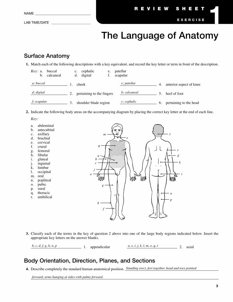
Surface Anatomy1. Match each of the following descriptions with a key equivalent, and record the key letter or term in front of the description.
Key: a. buccal c. cephalic e. patellarb. calcaneal d. digital f. scapular
1. cheek 4. anterior aspect of knee
2. pertaining to the fingers 5. heel of foot
3. shoulder blade region 6. pertaining to the head
2. Indicate the following body areas on the accompanying diagram by placing the correct key letter at the end of each line.
Key:
a. abdominalb. antecubitalc. axillaryd. brachiale. cervicalf. cruralg. femoralh. fibulari. glutealj. inguinalk. lumbarl. occipitalm. oraln. poplitealo. pubicp. suralq. thoracicr. umbilical
3. Classify each of the terms in the key of question 2 above into one of the large body regions indicated below. Insert theappropriate key letters on the answer blanks.
1. appendicular 2. axial
Body Orientation, Direction, Planes, and Sections4. Describe completely the standard human anatomical position. _________________________________________________
a; buccal
d; digital
f; scapular
e; patellar
b; calcaneal
c; cephalic
m
q
abr
jo
g
h
f
b, c, d, f, g, h, n, p a, e, i, j, k, l, m, o, q, r
Standing erect, feet together, head and toes pointed
forward, arms hanging at sides with palms forward.
el
c
d
k
i
n
p
NAME ____________________________________
LAB TIME/DATE _______________________
The Language of Anatomy
E X E R C I S E
R E V I E W S H E E T 1
4 Review Sheet 1
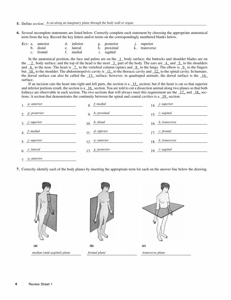
5. Define section. ______________________________________________________________________________________
6. Several incomplete statements are listed below. Correctly complete each statement by choosing the appropriate anatomicalterm from the key. Record the key letters and/or terms on the correspondingly numbered blanks below.
Key: a. anterior d. inferior g. posterior j. superiorb. distal e. lateral h. proximal k. transversec. frontal f. medial i. sagittal
In the anatomical position, the face and palms are on the _1_ body surface; the buttocks and shoulder blades are onthe __2_ body surface; and the top of the head is the most _3_ part of the body. The ears are _4_ and _5_ to the shouldersand _6_ to the nose. The heart is _7_ to the vertebral column (spine) and _8_ to the lungs. The elbow is _9_ to the fingersbut _10_ to the shoulder. The abdominopelvic cavity is _11_ to the thoracic cavity and _12_ to the spinal cavity. In humans,the dorsal surface can also be called the _13_ surface; however, in quadruped animals, the dorsal surface is the _14_surface.
If an incision cuts the heart into right and left parts, the section is a _15_ section; but if the heart is cut so that superiorand inferior portions result, the section is a _16_ section. You are told to cut a dissection animal along two planes so that bothkidneys are observable in each section. The two sections that will always meet this requirement are the _17_ and _18_ sec-tions. A section that demonstrates the continuity between the spinal and cranial cavities is a _19_ section.
1. 8. 14.
2. 9. 15.
3. 10. 16.
4. 11. 17.
5. 12. 18.
6. 13. 19.
7.
7. Correctly identify each of the body planes by inserting the appropriate term for each on the answer line below the drawing.
(a) (b) (c)
A cut along an imaginary plane through the body wall or organ.
a; anterior
g; posterior
j; superior
f; medial
j; superior
e; lateral
a; anterior
f; medial
h; proximal
b; distal
d; inferior
a; anterior
g; posterior
j; superior
i; sagittal
k; transverse
c; frontal
k; transverse
i; sagittal
median (mid-sagittal) plane frontal plane transverse plane
5Review Sheet 1
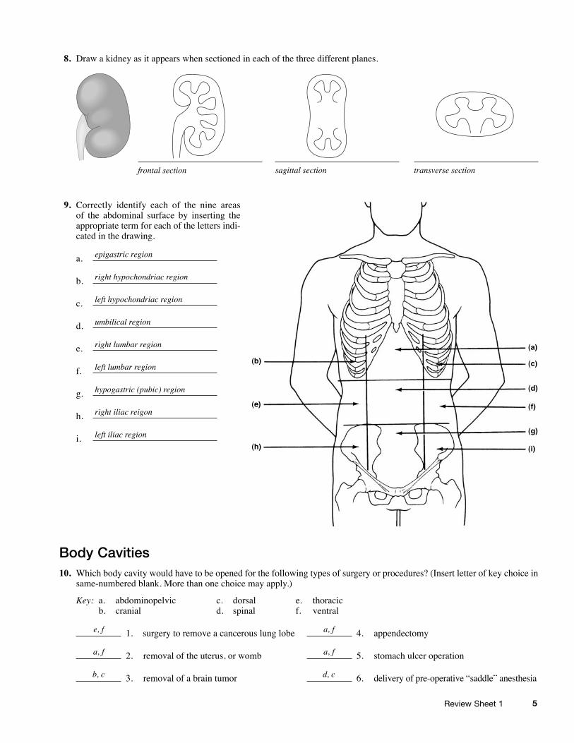
8. Draw a kidney as it appears when sectioned in each of the three different planes.
9. Correctly identify each of the nine areasof the abdominal surface by inserting theappropriate term for each of the letters indi-cated in the drawing.
a.
b.
c.
d.
e.
f.
g.
h.
i.
Body Cavities10. Which body cavity would have to be opened for the following types of surgery or procedures? (Insert letter of key choice in
same-numbered blank. More than one choice may apply.)
Key: a. abdominopelvic c. dorsal e. thoracicb. cranial d. spinal f. ventral
1. surgery to remove a cancerous lung lobe 4. appendectomy
2. removal of the uterus, or womb 5. stomach ulcer operation
3. removal of a brain tumor 6. delivery of pre-operative “saddle” anesthesia
frontal section sagittal section transverse section
(a)
(b)
(e)
(h)
(c)
(d)
(f)
(g)
(i)
epigastric region
right hypochondriac region
left hypochondriac region
umbilical region
right lumbar region
left lumbar region
hypogastric (pubic) region
right iliac reigon
left iliac region
e, f
a, f
b, c
a, f
a, f
d, c
6 Review Sheet 1
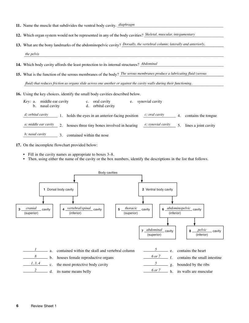
11. Name the muscle that subdivides the ventral body cavity. ____________________________________________________
12. Which organ system would not be represented in any of the body cavities? _______________________________________
13. What are the bony landmarks of the abdominopelvic cavity? __________________________________________________
___________________________________________________________________________________________________
14. Which body cavity affords the least protection to its internal structures? _________________________________________
15. What is the function of the serous membranes of the body? ___________________________________________________
__________________________________________________________________________________________________
16. Using the key choices, identify the small body cavities described below.
Key: a. middle ear cavity c. oral cavity e. synovial cavityb. nasal cavity d. orbital cavity
_________________ 1. holds the eyes in an anterior-facing position ________________ 4. contains the tongue
_________________ 2. houses three tiny bones involved in hearing ________________ 5. lines a joint cavity
_________________ 3. contained within the nose
17. On the incomplete flowchart provided below:
• Fill in the cavity names as appropriate to boxes 3–8.• Then, using either the name of the cavity or the box numbers, identify the descriptions in the list that follows.
a. contained within the skull and vertebral column e. contains the heart
b. houses female reproductive organs f. contains the small intestine
c. the most protective body cavity g. bounded by the ribs
d. its name means belly h. its walls are muscular
cavity�
1� Dorsal body cavity�
3� cavity� �(superior)�
2� Ventral body cavity�
Body ca�vities�
4� �(inferior)�
5� cavity� �(superior)�
6� cavity� �(inferior)�
7� cavity� �(superior)�
8� cavity� �(inferior)�
Skeletal, muscular, integumentary
the pelvis
Abdominal
The serous membranes produce a lubricating fluid (serous
Dorsally, the vertebral column; laterally and anteriorly,
fluid) that reduces friction as organs slide across one another or against the cavity walls during their functioning.
c; oral cavity
e; synovial cavity
d; orbital cavity
a; middle ear cavity
b; nasal cavity
1
8
1, 3, 4
2
5
6 or 7
5
6 or 7
cranial thoracic
abdominal pelvic
abdominopelvicvertebral/spinal
diaphragm
7
2Organ Systems Overview
E X E R C I S E
Time Allotment: 11/2 hours (rat dissection: 1 hour; if performing reproductive sys-tem dissection: 1/2 hour each for male and female); dissectible human torso model:1/2 hour).
Multimedia Resources: See Appendix D for a list of multimedia offerings.
Homeostasis (FHS, 20 minutes, VHS, DVD)Homeostasis: The Body in Balance (HRM, IM, 26 minutes, VHS, DVD)Organ Systems Working Together (WNS, 14 minutes, VHS)The Incredible Human Machine (CBS, 60 minutes, VHS)
Solutions:Bleach Solution, 10%Measure out 100 milliliters of household bleach. Add water to a final volume of 1 liter.
Advance Preparation1. Make arrangements for appropriate storage and disposal of dissection materials. Check
with the Department of Health or the Department of Environmental Protection, or theircounterparts, for state regulations.
2. Designate a disposal container for organic debris, set up a dishwashing area with hotsoapy water and sponges, and provide lab disinfectant such as Wavicide-01 (Carolina) orbleach solution for washing down the lab benches.
3. Set out safety glasses and disposable gloves for dissection of freshly killed animals (toprotect students from parasites) and for dissection of preserved animals.
4. Decide on the number of students in each dissecting group (a maximum of four is sug-gested, two is probably best). Each dissecting group should have a dissecting pan, dis-secting pins, scissors, blunt probe, forceps, twine, and a preserved or freshly killed rat.
5. Preserved rats are more convenient to use unless small mammal facilites are available. Iflive rats are used, they may be killed a half-hour or so prior to the lab by administeringan overdose of ether or chloroform. To do this, remove each rat from its cage and hold itfirmly by the skin at the back of its neck. Put the rat in a container with cotton soaked inether or chloroform. Seal the jar tightly and wait until the rat ceases to breathe.
6. Set out dissectible human torso models and a dissected human cadaver if available.